comment
reviews
reports
deposited research
interactions
information
refereed research
Minireview
Evolution of the initiating enzymes of the complement system
Masaru Nonaka and Seita Miyazawa
Address: Department of Biological Sciences, Graduate School of Science, University of Tokyo, Hongo, Tokyo 113-0033, Japan.
Correspondence: Masaru Nonaka. E-mail: [email protected]
Abstract
Analysis of the human MASP-1/3 gene, which encodes two proteases of the lectin-triggered complement cascade, has revealed alternatively used serine-protease-encoding regions for the gene’s two protein products. Phylogenetic studies indicate that one arose by retrotransposition early in vertebrate evolution, supporting the idea that the lectin branch of the complement cascade arose earlier than the ‘classical’ pathway.
Published: 14 December 2001
GenomeBiology2001, 3(1):reviews1001.1–1001.5
The electronic version of this article is the complete one and can be found online at http://genomebiology.com/2001/3/1/reviews/1001 © BioMed Central Ltd (Print ISSN 1465-6906; Online ISSN 1465-6914)
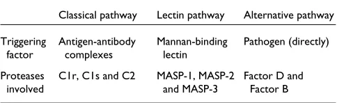
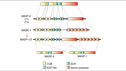
The mammalian complement system, as one of the major constituents of innate immunity, plays a pivotal role in defending the body against pathogenic microorganisms. It has more than 30 components, organized into three major pathways: the classical pathway, triggered by antibodies; the lectin pathway, triggered by engagement of mannan-binding protein; and the alternative pathway, triggered directly by pathogens (see Table 1). Of the 30 complement components, about one-fourth are serine proteases that are important for the activation or regulation of the whole system. Among these, C1r and C1s of the classical pathway and the MASPs (mannan-binding-protein-associated serine proteases) of the lectin pathway are crucial for starting the proteolytic activation cascades of their respective pathways. These components constitute a serine-protease family of the chymotrypsin type but characterized by a unique modular structure (Figure 1) [1-3].
Initial structural analysis of the C1sgene revealed a surpris-ing exon-intron organization, in which the serine-protease domain is encoded by a single uninterrupted exon, although there are several introns throughout the remainder of the gene (Figure 1) [4]. Intronless genes or pseudogenes, thought to be generated by retrotransposition, are not rare, but it was curious to imagine that retrotransposition could have led to the removal of introns from only one part of the gene. Later, the C1rand MASP-2genes were shown to have essentially the same exon-intron organization as the C1s
gene; surprisingly, though, MASP-1proved to have introns within its serine-protease-encoding region (Figure 1) [5].
The recent identification of MASP-3 [3] provided an expla-nation of how this situation might have arisen. A new intron-less serine-protease-encoding region was found in the
MASP-1 gene, upstream of the MASP-1
[image:1.609.312.554.667.741.2]encoding region. Furthermore, the two serine-protease-encoding regions were differentially selected by alternative splicing to generate MASP-1 and MASP-3, which differ only in their serine protease domains (Figure 1); the gene encod-ing them was therefore renamed MASP-1/3. In addition to the absence of introns, the upstream protease domain has characteristics that distinguish it from most of the chy-motrypsin-like serine-protease family: it lacks the disulfide bond that is believed to stabilize the structure around the active-site histidine (the histidine loop), and its active-site
Table 1
A summary of the three branches of the complement activation pathway
Classical pathway Lectin pathway Alternative pathway Triggering Antigen-antibody Mannan-binding Pathogen (directly)
factor complexes lectin
Proteases C1r, C1s and C2 MASP-1, MASP-2 Factor D and
2 GenomeBiology Vol 3 No 1 Nonaka and Miyazawa
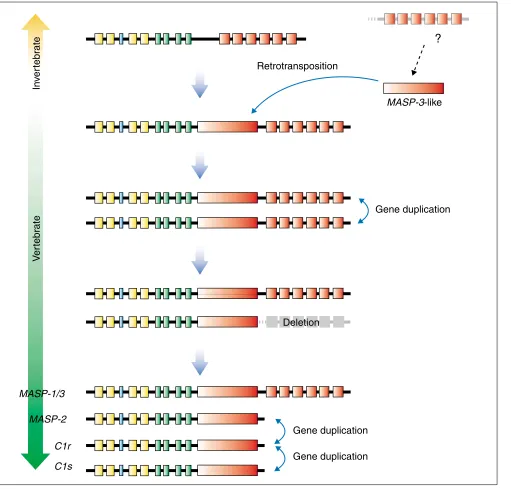
serine is encoded by the unusual AGY codon, instead of the more common TCN codon. These characteristics are all con-served in MASP-2, C1r, and C1s - we refer to this type of domain as MASP-3-like - whereas MASP-1, which uses the downstream serine-protease-encoding region of the MASP-1/3 gene, has a more typical serine-protease domain. These data provide us with a hypothesis to explain the evolutionary history of this gene family, as depicted in Figure 2. The ancestral gene, viewed here as MASP-1-type gene, had a clas-sical serine-protease-encoding region interrupted by several introns (Figure 2, top). Then, the second MASP-3-like serine-protease-encoding region was inserted by retrotrans-position upstream of the original serine-protease-encoding region, generating a MASP-1/3-type gene. After gene dupli-cation, one of the duplicates lost the downstream serine-pro-tease-encoding region, resulting in a gene of the MASP-2
type. Finally, further gene duplications generated the C1r
and C1sgenes (Figure 2, bottom), resulting in the four genes found in the human.
The order of appearance of the MASP-2, C1r and C1sgenes during evolution is not obvious from their gene structure alone, but there are other indications as to which came first.
The phylogenetic relationships among these genes are such that C1rand C1sare closest to each other, and the genomic organization is such that onlyC1rand C1sare closely linked in a tail-to-tail orientation [6], supporting a scenario in whichC1rand C1s were the last to be generated, with the final evolutionary step being an in situ duplication. This sce-nario gives further support to the generally accepted idea that the lectin pathway, in which the MASPs operate, pre-dated the classical pathway, which relies on C1r and C1s.
[image:2.609.61.554.87.368.2]So, our next question is when and how these steps actually occurred during the evolution of deuterostomes (the branch of animals that includes echinoderms, hemichordates and chordates). To date, we do not have enough data to answer this question definitively. Nevertheless, accumulating obser-vations from our group and others about the complement system of lower vertebrates and invertebrates seem to provide some indications. The two MASP genes identified from an ascidian, Halocynthia roretzi, are MASP-1-like [7] and lack the intronless serine-protease-encoding region (Y. Endo, M.N. and T. Fujita, unpublished observations), while the lamprey MASP gene seems to be MASP-1/3-like [8]. Retrotransposition of the MASP-3-like serine-protease
Figure 1
The domain structure and gene organization of the MASP/C1r/C1s serine protease family. The first line shows the domain structure of an archetypical member of the family; the central three lines are representations of the exon-intron organization of the genes encoding the family members. The MASP-1 gene was recently found to contain another serine-protease-encoding region within it, encoding MASP-3, and was therefore renamed MASP-1/3. The bottom line shows the alternative splicing pattern of MASP-1 and MASP-3. Corresponding domains and the exons encoding them are shown in the same color. Abbreviations: CUB, Clr/Cls, sea urchin Uegf, bone morphogenetic protein domain; EGF-like, similar to epidermal growth factor; SCR, short consensus repeat.
CUB
EGF-like
SCR
Serine protease
C1s
C1r
MASP-2
MASP-1
MASP-1
MASP-3
comment
reviews
reports
deposited research
interactions
information
refereed research
domain therefore most probably occurred in the main line of vertebrate evolution after the divergence of urochordates (ascidians), but before the divergence of cyclostomes (jawless vertebrates such as the lamprey and hagfish). Fur-thermore, the serine-protease domain of the Halocynthia
roretzi complement factor B gene (encoding a protease of
the alternative pathway; see Table 1) is MASP-3-like (X. Ji,
M. Sasaki and M.N., unpublished observations), except that it is interrupted by introns. All genes that have a MASP-3-like serine-protease domain may therefore prove to be on a single evolutionary lineage, as of all the known human chymotrypsin-family protease genes, only the C1r, C1s,
MASP-1/3and MASP-2 genes encode a MASP-3-like domain
[image:3.609.56.567.86.575.2][9]. Thus, it is possible that ascidian factor B was the donor
Figure 2
A proposal for the evolution of theMASP-1/3, MASP-2, C1rand C1sgenes. Colors are as in Figure 1. The top line represents the ascidian gene and the bottom lines the human genes. The intermediate lines illustrate hypothetical intermediate species. Although two MASPgenes have been reported from ascidians, they are considered to be products of a recent gene duplication in the ascidian lineage, not corresponding to the gene duplications in the vertebrate lineage.
In
v
er
tebr
ate
V
er
tebr
ate
C1s
C1r
MASP-2
MASP-1/3
Deletion
Gene duplication
Gene duplication
Gene duplication
Retrotransposition
MASP-3-like
gene, which was the source of the MASP-3-like domain acquired byMASP-1/3 through retrotransposition. The pres-ence of the classical complement pathway in bony and carti-laginous fish has been demonstrated at the functional level [10,11], and cDNA clones forC1s/C1rhave been reported in bony fish [12]. Thus, all the steps illustrated for the verte-brate pathway in Figure 2 seem to have been completed before the emergence of jawed vertebrates.
The primitive complement system
As discussed here, the complement system seems to have a more ancient origin in evolution than adaptive immunity; the latter seems to have been established only from jawed vertebrates onwards. The central component of the comple-ment system, the C3 protein on which the three activation pathways discussed here converge, has been identified in jawless vertebrates, the lamprey [13] and hagfish [14], as well as in deuterostome invertebrates, amphioxus (a cephalochordate) [15], ascidian (urochordate) [16] and sea urchin (echinoderm) [17]. Although C3- or complement-like molecules of insects show functional similarity with deuterostome C3, there is no shared derived structural char-acter between them, indicating that the deuterostome C3 and insect C3-like molecules derived independently from their common ancestor, =2-macroglobulin. Thus, the authentic complement system seems to have been estab-lished in the deuterostome lineage; the classical pathway of complement activation was then acquired in the jawed verte-brate lineage, at the time adaptive immunity arose.
In contrast to the classical pathway, which uses immunoglobulins - the central components of adaptive immunity - as recognition molecules, the primitive comple-ment system that emerged before adaptive immunity must have depended on innate recognition molecules. In this context, it is noteworthy that the human lectin pathway uses lectins - mannan-binding protein (MBP) [18] and ficolin [19] - as the molecules that recognize pathogen and trigger the pathway. These molecules share a collagen-like amino-terminal region believed to be involved in MASP binding, although their carboxy-terminal recognition domains are different - the C-type lectin domain and the fibrinogen domain, respectively. For ascidians, ficolin [20], MBP-like lectin [21] and a novel lectin termed GBL [22] have been described as the possible recognition molecules of the prim-itive complement system. No functional information is available for the first two of these, however, and GBL lacks the collagen domain, the common structure shared by MBP and the classical pathway factor C1q of vertebrates that pro-vides the binding site for MASP, C1r and C1s. We believe that further analysis of the complement system of ascidians, in which the complexity of linkage to adaptive immunity can be eliminated, will bring us important insights into the as-yet mysterious mechanisms of the lectin pathway in mammals.
In addition to C3, MASPs and lectins, factor B has also been identified from ascidian (X. Ji, M. Sasaki and M.N., unpub-lished observations) and also from sea urchin [23]. Factor B is a key component of the mammalian alternative pathway, where it acts as a catalytic subunit of the alternative path-ways C3 convertase enzyme. Thus, the lectin and alternative pathways, recognized as independent pathways in the mam-malian complement system, might in fact represent two parts of a single ancient pathway. Perhaps the lectin pathway has a recognition function and the alternative pathway an amplification function in the primitive complement system. Further analyses of the activation mechanisms of the lectin pathway in both mammals and more primitive animals will reveal how the complement system arose and how it has been built upon during evolution, in the constant war between hosts and pathogens.
References
1. Volanakis JE, Arlaud GJ: Complement enzymes. In The Human Complement System in Health and Disease. Edited by Volanakis JE, Frank MM. Marcel Dekker; 1998: 49-81.
2. Thiel S, Vorup-Jensen T, Stover CM, Schwaeble W, Laursen SB, Poulsen K, Willis AC, Eggleton P, Hansen S, Holmskov U, et al.: A second serine protease associated with mannan-binding lectin that activates complement.Nature 1997, 386:506-510. 3. Dahl MR, Thiel S, Matsushita M, Fujita T, Willis AC, Christensen T,
Vorup-Jensen T, Jensenius JC: MASP-3 and its association with distinct complexes of the mannan- binding lectin comple-ment activation pathway.Immunity 2001, 15:127-135.
4. Tosi M, Duponchel C, Meo T, Couture-Tosi E: Complement genes C1r and C1s feature an intronless serine protease domain closely related to haptoglobin. J Mol Biol 1989,
208:709-714.
5. Endo Y, Sato T, Matsushita M, Fujita T: Exon structure of the gene encoding the human mannose-binding protein-associ-ated serine protease light chain: comparison with comple-ment C1r and C1s genes. Int Immunol 1996, 8:1355-1358. 6. Tosi M, Duponchel C, Meo T, Julier C: Complete cDNA
sequence of human complement Cls and close physical linkage of the homologous genes Cls and Clr. Biochemistry 1987,26:8516-8524.
7. Ji X, Azumi K, Sasaki M, Nonaka M: Ancient origin of the com-plement lectin pathway revealed by molecular cloning of mannan binding protein-associated serine protease from a urochordate, the Japanese ascidian, Halocynthia roretzi. Proc Natl Acad Sci USA 1997, 94:6340-6345.
8. Endo Y, Takahashi M, Nakao M, Saiga H, Sekine H, Matsushita M, Nonaka M, Fujita T: Two lineages of mannose-binding lectin-associated serine protease (MASP) in vertebrates. J Immunol 1998, 161:4924-4930.
9. MEROPS database[http://merops.iapc.bbsrc.ac.uk/]
10. Nonaka M, Yamaguchi N, Natsuume-Sakai S, Takahashi M: The complement system of rainbow trout (Salmo gairdneri). I. Identification of the serum lytic system homologous to mammalian complement.J Immunol1981,126:1489-1494. 11. Jensen JA, Festa E, Smith DS, Cayer M: The complement system
of the nurse shark: hemolytic and comparative characteris-tics. Science1981, 214:566-569.
12. Nakao M, Osaka K, Kato Y, Fujiki K, Yano T:Molecular cloning of the complement (C1r/C1s/MASP2-like serine proteases from the common carp (Cyprinus carpio). Immunogenetics2001,
52:255-263.
13. Nonaka M, Fujii T, Kaidoh T, Natsuume-Sakai S, Yamaguchi N, Taka-hashi M: Purification of a lamprey complement protein homologous to the third component of the mammalian complement system. J Immunol 1984, 133:3242-3249.
14. Ishiguro H, Kobayashi K, Suzuki M, Titani K, Tomonaga S, Kurosawa Y: Isolation of a hagfish gene that encodes a complement component.EMBO J 1992, 11:829-837.
15. Suzuki MM, Satoh N, Nonaka M: C6-like and C3-like molecules from the cephalochordate, amphioxus, suggest a cytolytic complement system in invertebrates. J Mol Evol, in press. 16. Nonaka M, Azumi K, Ji X, Namikawa-Yamada C, Sasaki M, Saiga H,
Dodds AW, Sekine H, Homma MK, Matsushita M, et al.: Opsonic complement component C3 in the solitary ascidian, Halo-cynthia roretzi.J Immunol 1999, 162:387-391.
17. Al-Sharif WZ, Sunyer JO, Lambris JD, Smith LC: Sea urchin coelo-mocytes specifically express a homologue of the comple-ment component C3.J Immunol 1998, 160:2983-2997.
18. Matsushita M, Fujita T: Activation of the classical complement pathway by mannose-binding protein in association with a novel C1s-like serine protease.J Exp Med 1992,176:1497-1502. 19. Matsushita M, Endo Y, Fujita T: Cutting edge:
complement-acti-vating complex of ficolin and mannose-binding lectin-associ-ated serine protease.J Immunol 2000, 164:2281-2284.
20. Kenjo A, Takahashi M, Matsushita M, Endo Y, Nakata M, Mizuochi T, Fujita T: Cloning and characterization of novel ficolins from the solitary ascidian, Halocynthia roretzi. J Biol Chem 2001,
276:19959-19965.
21. Nair SV, Pearce S, Green PL, Mahajan D, Newton RA, Raftos DA: A collectin-like protein from tunicates. Comp Biochem Physiol B Biochem Mol Biol 2000, 125:279-289.
22. Sekine H, Kenjo A, Azumi K, Ohi G, Takahashi M, Kasukawa R, Ichikawa N, Nakata M, Mizuochi T, Matsushita M, et al.: An ancient lectin-dependent complement system in an ascidian: novel lectin isolated from the plasma of the solitary ascidian,
Halocynthia roretzi.J Immunol 2001, 167:4504-4510.
23. Smith LC, Shih CS, Dachenhausen SG: Coelomocytes express SpBf, a homologue of factor B, the second component in the sea urchin complement system.J Immunol 1998, 161: 6784-6793.
comment
reviews
reports
deposited research
interactions
information