BIROn - Birkbeck Institutional Research Online
Pereira, R. and Gendron, T. and Sanghera, C. and Greenwood, H.E. and
Newcombe, J. and McCormick, P.N. and Sander, K. and Topf, Maya and
Arstad, E. and Witney, T.H. (2019) Mapping Aldehyde Dehydrogenase 1A1
activity using an [18F]Substrate-based approach. Chemistry: A European
Journal , ISSN 0947-6539. (In Press)
Downloaded from:
Usage Guidelines:
Please refer to usage guidelines at
or alternatively
&
Biochemistry
Mapping Aldehyde Dehydrogenase 1A1 Activity using an
[
18
F]Substrate-Based Approach
Raul Pereira,
[a]Thibault Gendron,
[b]Chandan Sanghera,
[a]Hannah E. Greenwood,
[a]Joseph Newcombe,
[b, c]Patrick N. McCormick,
[a]Kerstin Sander,
[b]Maya Topf,
[c]Erik rstad,
[b]and Timothy H. Witney*
[a]Abstract: Aldehyde dehydrogenases (ALDHs) catalyze the oxidation of aldehydes to carboxylic acids. Elevated ALDH expression in human cancers is linked to metastases and poor overall survival. Despite ALDH being a poor prognostic factor, the non-invasive assessment of ALDH activity in vivo has not been possible due to a lack of sensitive and transla-tional imaging agents. Presented in this report are the syn-thesis and biological evaluation of ALDH1A1-selective chemi-cal probes composed of an aromatic aldehyde derived from N,N-diethylamino benzaldehyde (DEAB) linked to a fluorinat-ed pyridine ring either via an amide or amine linkage. Of the
focused library of compounds evaluated, N -ethyl-6-(fluoro)-N-(4-formylbenzyl)nicotinamide4 bwas found to have excel-lent affinity and isozyme selectivity for ALDH1A1 in vitro. Following18F-fluorination, [18F]4 bwas taken up by colorectal
tumor cells and trapped through the conversion to its18
F-la-beled carboxylate product under the action of ALDH. In vivo positron emission tomography revealed high uptake of [18F]4 b in the lungs and liver, with radioactivity cleared
through the urinary tract. Oxidation of [18F]4 b, however, was
observed in vivo, which may limit the tissue penetration of this first-in-class radiotracer.
Introduction
Aldehyde dehydrogenases (ALDHs) are a family of enzymes that catalyze the NAD(P)+-dependent oxidation of a wide
vari-ety of aldehydes to their corresponding carboxylic acids.[1]
There are currently 20 known functional human ALDHs[2] that
mediate the metabolism of aldehydes generated during oxida-tive stress,[3]amino acid and biogenic amine metabolism,[4]
reti-noic acid biosynthesis,[5] and ethanol metabolism.[3a] In
addi-tion, ALDHs control the detoxification of exogenous reactive aldehydes and therapeutic drugs such as cyclophosphamide.[6]
Aberrant expression of ALDH is associated with many diseases, including cancer, with increased expression and activity of ALDH shown to be a predictor of metastatic potential and poor overall survival.[7]In particular, the ALDH1A1 isozyme is a
well-characterized marker of cancer stem cells, which are known for their tumor-initiating properties and resistance to conventional therapy.[8] Studies have shown resistance to
che-motherapy and poor prognosis is associated with high ALDH1A1 activity in breast,[9] ovarian,[10] prostate,[11] colon[12]
and lung[13]cancer. As a consequence, ALDH1A1 has been
se-lected as a target for anti-cancer therapy, with ALDH inhibitors shown to reverse chemoresistance in a range of preclinical tumor models.[14]
Given the causal link between ALDH expression and cancer drug resistance, the non-invasive identification of ALDH-ex-pressing tumors is of great clinical importance. The measure-ment of chemoresistance through ALDH imaging could poten-tially enable the clinician to select the most suitable therapeu-tic intervention for the individual patient (e.g. chemotherapy versus immunotherapy) with the possibility to improve out-comes and reduce unnecessary treatment. Currently, the in vitro assessment of ALDH activity has been restricted to fluo-rescence-based assays.[15]Despite these commercially available
imaging agents being widely-adopted for the isolation of
[a]Dr. R. Pereira, C. Sanghera, H. E. Greenwood, Dr. P. N. McCormick, Dr. T. H. Witney
Centre for Advanced Biomedical Imaging University College London
Paul O’Gorman Building, 72 Huntley Street London WC1E 6DD (UK),
and
Current address: Department of Imaging Chemistry & Biology King’s College London, St. Thomas’ Hospital
London SE1 7EH (UK) E-mail: [email protected]
[b]Dr. T. Gendron, J. Newcombe, Dr. K. Sander, Prof. Dr. E. rstad Department of Chemistry
University College London 20 Gordon Street London WC1H 0AJ (UK)
[c] J. Newcombe, Prof. Dr. M. Topf Department of Biological Sciences Birkbeck, University of London Malet Street
London WC1E 7HX (UK)
Supporting information and the ORCID identification number(s) for the au-thor(s) of this article can be found under:
https://doi.org/10.1002/chem.201805473.
ALDH-positive cells in cell culture, the poor tissue penetration of the fluorescent signal currently limits their in vivo utility. In order to circumvent these inherent limitations, we propose the use of positron emission tomography (PET) as an alternative to fluorescence-based assays.[16] Previous attempts to develop
ALDH1A1-specific radiotracers have so far failed due to the poor cellular retention of the carboxylate product, presumed to be a consequence of its high hydrophobicity.[17] Here, we
report the synthesis and biological evaluation of18F-fluorinated
aldehyde-based probes for the non-invasive detection of ALDH1A1 activity in tumor cell models.
Results and Discussion
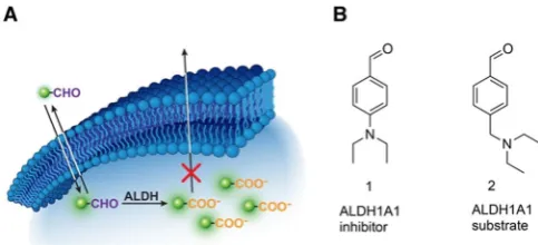
ALDH1A1 chemical probes were designed to have a) an alde-hyde that can serve as a substrate for ALDH1A1; b) contain a (radio)fluorine atom that would allow for detection via gamma counting/PET imaging; c) a suitable hydrophobic-hydrophilic balance which would allow for passive diffusion in and out of cells, and importantly; d) subsequent trapping of the in situ generated carboxylic acid product within the cytosol as a result of the acquired negative charge (Figure 1 A). We took a
substrate-based approach for the imaging of ALDH1A1 to pro-vide a functional readout of enzymatic activity. Substrate-based radiotracers provide an advantage over radiolabeled in-hibitors which only report on enzyme expression. Moreover, multiple substrate molecules can be turned over by a single enzyme, thereby increasing the sensitivity of detection when compared to radiolabeled inhibitors.
The starting point for our small molecule probe develop-ment wasN,N-diethylaminobenzaldehyde (DEAB)1, which is a well-known inhibitor of ALDH1A1 (Ki values of 10–40 nm).[18]
Given the advantages of substrate-based radiotracers, our ini-tial goal was to convert1 in to a substrate whilst maintaining suitable ALDH1A1 selectivity over other commonly expressed ALDH isozymes.1is thought to form a stalled acyl-enzyme in-termediate as a result of the delocalization of the electron lone pair of the para-substituted amine into the aromatic ring.[18a,c]
To convert 1 from an inhibitor to a substrate, we uncoupled the amine nitrogen from the aromatic p-system through the introduction of a methylene linker to give2(Figure 1 B), which
was rapidly converted to the carboxylate product under the action of ALDH1A1 (Figure S1). In order to assess the enzyme kinetics of the compounds in this study, we examined the effect of substrate concentration on the initial enzyme velocity using recombinant human ALDH1A1, ALDH2 and ALDH3A1 en-zymes, which are commonly expressed in human cancer. ALDH2 is expressed in the mitochondrial matrix and plays a critical role in alcohol metabolism,[19] whereas ALDH3A1 is
lo-calized in both the nucleus and cytosol and functions to detox-ify aldehydes formed during UV-induced lipid peroxidation.[20]
Furthermore, these three isoforms exhibit three different rate-limiting steps: ALDH1A1’s being the cofactor dissociation,[21]
the deacylation-step for ALDH2,[22] and hydride transfer for
ALDH3A1.[23]
We made a single-point modification to2, with the addition of a fluorine to the aromatic ring—an essential requirement for 18F-radiofluorine-based radiotracers—to give compounds
3 aand3 b. The lithium/bromine exchange on11 followed by quenching with DMF afforded aldehyde 12. Stirring 12 with NaBH(OAc)3 and diethylamine in DCE afforded the crude
re-ductive amination product which was directly treated with aqueous hydrochloric acid in THF to furnish 3 a (Scheme 1 A). Reduction of nitrile13with diisobutylaluminium hydride clean-ly produced aldehyde 14 which was thereafter stirred with excess diethylamine in THF to yield aldehyde3 b(Scheme 1 B). Amide16was prepared by reacting the acid chloride of 5-fluo-ronicotinic acid 15with amine B (Scheme 1 C). The acid-cata-lyzed cleavage of the acetal furnished compound4 a. LiAlH4
re-duction of the amide bond in 16 followed by the acid-cata-lyzed acetal cleavage afforded amine5(Scheme 1 C). In a simi-lar manner, the acid chloride of 6-fluronicotinic acid16was re-acted with amineAto yield amide18which following an acid-catalyzed acetal cleavage furnished aldehyde4 b. The sulfona-mide6was accessed by reacting the commercially available 5-fluoropyridine-3-sulfonyl chloride 19 with amine A following an acid-catalyzed acetal cleavage (Scheme 1 E).
The benzylic amine 2 showed a higher affinity (lower KM)
and catalytic efficiency (Vmax/KM) for ALDH1A1 over both
ALDH2 and ALDH3A1 (Table 1, entry 1 and Figure 2 B, respec-tively), indicating that the isozyme selectivity of DEAB was maintained. Interestingly, 3 a and 3 b exhibited lower affinity for ALDH1A1 than the non-fluorinated analogue 2, with a KM
of 0.280.12 mm, 0.260.08 mm and 0.160.03 mm,
respec-tively (Table 1, entries 1–3).
[image:3.595.47.289.347.457.2]Given that fluorination proximal to the aldehyde moiety de-creased affinity for ALDH1A1, we next explored compounds with fluorine atoms that were remote from the aldehyde. Com-pounds 4 aand4 b exhibited a five-fold increase in ALDH1A1 affinity over 2 (Table 1, entries 1, 4 and 5). Furthermore, the enzyme efficiency for4 awas 7-fold higher for ALDH1A1 than ALDH2, with the enzyme efficiency for ALDH1A1 over 20-fold higher with respect to ALDH3A1; that is, the linking of the pyri-dine via an amide bond resulted in improved selectivity for ALDH1A1 (Figure 2 B; Table 1, entry 4). The position of the fluo-rine on the pyridine ring crucially did not play a key role in substrate kinetics, as seen with amide 4 b which exhibited analogous behavior to 4 a, albeit with marginally decreased
Figure 1.A) Schematic illustrating ALDH-mediated trapping of18
F-labeled al-dehydes by conversion to their corresponding acid. B) Chemical structures of DEAB (1), an ALDH1A1 inhibitor, and2, an ALDH1A1 substrate.
Chem. Eur. J.2019,25, 1 – 8 www.chemeurj.org 2 2019The Authors. Published by Wiley-VCH Verlag GmbH & Co. KGaA, Weinheim
&&
ALDH1A1 enzyme selectivity (Table 1, entry 5). In the absence of the amide linkage, the tertiary amine 5, whilst exhibiting a similar ALDH1A1 binding profile as compounds 4 a and 4 b, was readily oxidized by ALDH2 when compared to the other compounds tested (Figure 2 B; Table 1, entry 6). Replacing the amide of compound 4 a with a sulfonamide gave us com-pound 6 with diminished ALDH1A1 enzyme efficiency when compared to4 a (Figure 2 B; Table 1, entries 5 and 7). In sum-mary, we have shown4 a,4 band5to be excellent substrates
for ALDH1A1, with4 aand4 bto have good ALDH1A1 selectiv-ity over the other isoforms tested (Figure 2 B).
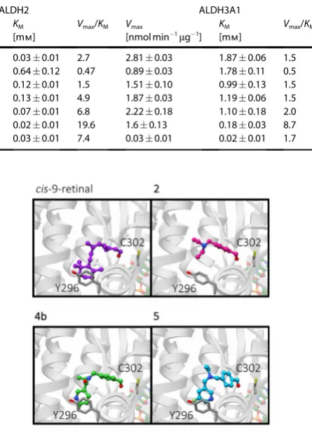
To understand why compounds 4 a and 4 b exhibited en-hanced selectivity for ALDH1A1 over the other isozymes tested, we carried out an in silico docking study wherein com-pounds 2–6, and the natural ALDH1A1 substrates 9-cis-retinal, 13-cis-retinal, and all-trans-retinal, were docked into protein structures for ALDH1A1, ALDH2, and ALDH3A1 (Protein data bank ID: 4WB9, 1O01 and 4L2O respectively). In comparison to
Scheme 1.Synthetic route to3 a-b,4 a-b,5,6,7,8and9. All yields are isolated yields. See Supporting Information for further details and for the synthesis of
ALDH2 and ALDH3A1, ALDH1A1 has the largest access tunnel to the active site residue (Figure S3), which suggests that bulki-er and more rigid substrates would be prefbulki-erentially turned over by ALDH1A1. Tunnel topography may also explain why reducing the amide in compound 4 a to a flexible tertiary amine in compound5, resulted in increased affinity for ALDH2 and ALDH3A1. Compounds4 aand4 bdisplayed similar bind-ing modes as2to ALDH1A1 in our in silico model, as shown in Figure 3. The benzaldehyde portions of compounds4 a,4 b,5, and6occupied a similar site to that of compound2; however, the pyridyl-ring was predicted to make additional contacts, presumably viap-stacking between the pyridine and the tyro-sine-Y296 of ALDH1A1 (Figure 3). This putative p-stacking in-teraction may explain the increase in binding affinity for com-pounds 4 a,4 b,5, and6when compared to compounds that lack the pyridine functional group. The low binding affinity of compounds4 a,4 b,5, and6 for ALDH2 (Figure 2 and Table 1) might be ascribed to the equivalent phenylalanine residue within the active site, which being less electron-rich than tyro-sine, results in a lowered p-stacking efficiency.[24]In ALDH3A1
the active site tyrosine is replaced by a methionine, which is incapable of p-stacking. Consequently, no pattern in binding affinity for ALDH3A1 was observed for substrates with the pyr-idyl substituent.
Compound4 bwas further considered as a potential radio-chemical probe to assess cellular ALDH activity as it not only had excellent enzyme efficiencies and selectivity for ALDH1A1, but also potential radiochemical accessibility. Radiolabeling via
nucleophilic substitution with [18F]fluoride allows for
substitu-tion at the 2-posisubstitu-tion on a pyridine ring, whilst the 3-posisubstitu-tion 4 a exhibits poor reactivity.[25] Consequently, the 18
F-radiola-beled analogue of4 b,N-ethyl-6-(fluoro-18F)-N
-(4-formylbenzyl)-nicotinamide was chosen as our lead candidate radiotracer. To access [18F]4 b, a nucleophilic aromatic substitution (S
NAr) was
performed on the 6-chloronicotinamide precursor 7, which was prepared from the acid chloride of 6-chloronicotinic acid 20 and amine A (Scheme 1 F). Stirring 7 with [18F]KF/K
222 in
DMSO at 1508C for 25 min followed by an acid-catalyzed acetal cleavage step furnished [18F]4 b in 351 % (n=3)
decay-corrected radiochemical yield (RCY) after reverse phase semi-preparative HPLC purification following manual radiola-beling (Scheme 2). Automation of this procedure improved the RCY to 431 % (decay-corrected ;n=3). Starting the synthesis with 1.0 GBq of [18F]fluoride, the radiotracer was obtained
with a radiochemical purity of 99 % (see Supporting Informa-tion) and a molar activity of up to 4.4 GBqmmol 1 (manual)
and up to 7.2 GBqmmol 1(automated).
[image:5.595.48.340.86.322.2]Furthermore, reference compounds for cellular metabolite analysis, carboxylic acid8and alcohol9, were prepared by the
Table 1.Kinetic properties of human ALDH1A1, ALDH2 and ALDH3A1 towards oxidation of aldehydes in this study.
Entry Com- ALDH1A1 ALDH2 ALDH3A1
pound Vmax
[nmol min1m
g 1
]
KM
[mm]
Vmax/KM Vmax
[nmol min1m
g 1
]
KM
[mm]
Vmax/KM Vmax
[nmol min1m
g1
]
KM
[mm]
Vmax/KM
1 2 1.220.09 0.160.03 7.9 0.090.01 0.030.01 2.7 2.810.03 1.870.06 1.5
2 3 a 1.100.18 0.280.12 3.9 0.300.02 0.640.12 0.47 0.890.03 1.780.11 0.5
3 3 b 1.420.17 0.260.08 5.5 0.190.01 0.120.01 1.5 1.510.10 0.990.13 1.5
4 4 a 1.080.07 0.030.01 33.9 0.670.01 0.130.01 4.9 1.870.03 1.190.06 1.5
5 4 b 0.980.02 0.030.01 28.7 0.530.01 0.070.01 6.8 2.220.18 1.100.18 2.0
6 5 1.360.08 0.040.01 33.3 0.320.02 0.020.01 19.6 1.60.13 0.180.03 8.7
7 6 1.52.68 0.100.24 14.6 0.270.01 0.030.01 7.4 0.030.01 0.020.01 1.7
Figure 2.Structure-activity relationship. A) Chemical structures for the com-pounds in this study. B) Enzyme efficiency of human recombinant ALDH1A1, ALDH2 and ALDH3A1 for compounds2–6. The “enzyme efficiency” (Vmax/KM)
[image:5.595.319.542.87.396.2]provides a measure of how efficiently an enzyme converts a substrate into product. Note: see Scheme 1 and Supporting Information for chemical syn-thesis of compounds2–6and Figure S2 for Michaelis Menten plots used to derive enzyme kinetic data.
Figure 3.In silico modeling. Predicted binding modes of ALDH1A1 sub-strates from in silico docking studies. Predicted binding modes in ALDH1A1 of 9-cis-retinal (purple),2(magenta),4 b(green) and5(cyan), shown in ball and stick representation. Catalytic residue C302, and Y296 which was identi-fied inp-stacking interactions with the substrates are shown as dark grey sticks. Ribbons are shown in grey and faded for clarity, the cofactor NAD(H) is shown in pastel orange, green, red and blue ball and stick representation.
Chem. Eur. J.2019,25, 1 – 8 www.chemeurj.org 4 2019The Authors. Published by Wiley-VCH Verlag GmbH & Co. KGaA, Weinheim
&&
[image:5.595.339.514.206.391.2]KMnO4mediated oxidation of4 b, and the NaBH4mediated re-duction of aldehdye 4 b, respectively (Scheme 1 G). With a tracer candidate in hand we assessed whether [18F]4 b could
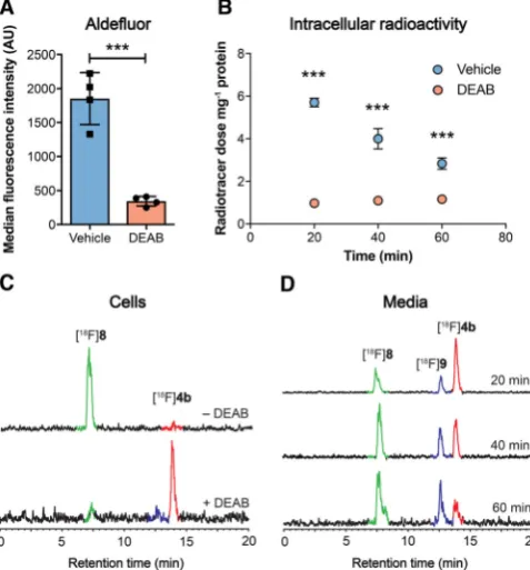
provide a readout of ALDH activity in tumor cells grown in cul-ture. We used the HCT116 KrasG13D/ mutant (HCT116 mut)
human colorectal cancer cell line as a model of aggressive, therapy-resistant cancer. HCT116 mut lines displayed high ALDH activity, which was specifically inhibited through the in-cubation of cells with the ALDH inhibitor DEAB (30mm;
Fig-ure 4 A). Incubation of HCT116 mut cells with [18F]4 b resulted
in rapid cell uptake and intracellular retention of radioactivity, reaching 5.70.2 % radiotracer dose mg 1 protein at 20 min
(n=3). Treatment of cells with DEAB resulted in an 83 % reduc-tion in cell-associated radioactivity to 1.00.1 % radiotracer dose mg 1 protein (n=3;P<0.0001), indicating ALDH-specific
intracellular trapping of either [18F]4 b or its products
(Fig-ure 4 B). To confirm the identity of the intracellular radioactive species present, we performed radio-HPLC analysis of the re-sulting cell lysates. 20 min after the addition of the radiotracer, near-complete conversion of the [18F]4 baldehyde to the
corre-sponding carboxylic acid [18F]8 was observed (Figure 4 C; see
Scheme 1 G for structures of8 and9), confirmed through co-injection with non-radioactive [19F]8 (See Supporting
Informa-tion for details). IncubaInforma-tion of cells with DEAB resulted in a substantial reduction in the production of [18F]8, with >90 %
of radioactivity present as the parent compound (Figure 4 C), suggesting that in the absence of ALDH1A1 activity the alde-hyde does not undergo intracellular oxidation and therefore is not retained.
Whilst the levels of [18F]4 bin DEAB-treated cells did not
sig-nificantly change over the 60 min time course of the experi-ment (P>0.05; Figure 4 B, red circles), the amount of intracellu-lar radioactivity decreased in a time-dependent fashion in vehi-cle (DMSO)-treated cells following addition of [18F]4 b
(Fig-ure 4 B, blue circles). Despite washout of radioactivity from ve-hicle-treated cells, cell-associated radioactivity at 60 minutes remained 2.4-fold higher than cells treated with DEAB, at 2.8 0.3 % radiotracer dose mg 1 and 1.20.1 % radiotracer
dose mg 1 protein, respectively (P=0.0005; n=3). Analysis of
media samples by radio-HPLC following cell incubation showed a progressive increase in the levels of [18F]8 and the
subsequent appearance of the alcohol [18F]9 (see Scheme 1 G
for structures), indicating imperfect intracellular trapping of [18F]8 following its production by ALDH (Figure 4 D).
Appear-ance of [18F]8in the media therefore accounts for the
time-de-pendent reduction in cell-associated radioactivity observed in vehicle-treated cells following incubation with [18F]4 b,
poten-tially as the result of efflux pump-mediated excretion of the carboxylate. Media incubation of [18F]4 bat 378C for 60 min in
the absence of cells did not result in the production of [18F]8
(Supporting Information; chromatogram S3), confirming that the conversion to the carboxylate was cell-mediated. Together, these data show [18F]4 bto be specific and sensitive marker of
ALDH activity in tumor cells.
Given that [18F]4 b can measure ALDH activity in HCT116
mut cells with high sensitivity and specificity, we next explored [18F]4 b’s in vivo biodistribution. Dynamic microPET imaging
following intravenous injection of [18F]4 b into healthy balb/c
mice (Figure 5) revealed rapid and extensive uptake in the lungs, known to express high levels of ALDH1A1. Liver uptake peaked at 5 min post injection (p.i.) at 20.02.6 % injected dose (ID) g 1tissue (n=3 mice). [18F]4 b clearance was initially
via the kidneys and bladder to afford high contrast images with low uptake in background tissue (2.20.3 %ID g 1 in the
[image:6.595.306.545.67.324.2]muscle at 5 min p.i.;n=3 mice). However, hepatobiliary excre-tion was evident by 30 min p.i., as shown by radiotracer
Figure 4.Detection of ALDH activity in HCT116 mut cells. A) ALDH activity in vehicle (DMSO) and DEAB-treated cells (30mm, 45 min), as measured by
ALDH-mediated trapping of Aldefluor and detection by flow cytometry. Data are meansSD (n=4). B) Intracellular radioactivity levels in HCT116 mut cells treated with either vehicle or DEAB (30mm, 45 min) following
incuba-tion with [18
F]4 b. Data are meansSD (n=3). C) Radio-HPLC chromato-grams from cell lysates following 20 min incubation of [18
F]4 b(red peak) with (bottom), or without DEAB treatment (top). The green peak corre-sponds to the carboxylate [18
F]8. D) Composition of radioactive metabolites in the media following 20, 40 and 60 min incubation of [18F]4 b(red) with
HCT116 mut cells. The presence of [18
F]8(green peak) along with the alcohol [18
F]9(blue peak) were identified via co-injection with [19
F]4 b, [19
F]8, and [19F]9and their detection at 254 nm (see Supporting Information for further
information). ***,P<0.001 in vehicle vs. DEAB-treated cells.
Scheme 2.Radiofluorination of [18F]4 b. See Supporting Information for
uptake in the gallbladder and gastrointestinal (GI) tract, indi-cating possible metabolism of the parent compound at this time point. [18F]4 b was further cleared from all other organs
other than gallbladder and GI over the remainder of the imag-ing time course. For time activity curves for organs with high [18F]4 buptake, see Figure S4.
The combined renal and hepatobiliary excretion observed at 30 min p.i., prompted us to assess the in vivo stability of [18F]4 b. Radio-HPLC analysis of plasma taken by terminal
ex-sanguination from anesthetized mice following tail vein intra-venous (i.v.) injection of [18F]4 brevealed complete conversion
to [18F]8by 2 min (Supporting Information; chromatogram S7).
The appearance of a second, unknown peak of similar reten-tion time to [18F]8 (Supporting Information chromatogram S7)
was evident by 5 min, increasing to 40 % total radioactivity in the blood by 60 min, which may account for the mixed routes of excretion observed at later imaging time points. Im-portantly, the rapid metabolism of the free aldehyde may limit the in vivo tissue penetration of [18F]4 b.
Conclusions
In conclusion, we have developed a focused library of ALDH substrates based on the well-known inhibitor DEAB. The addi-tion of a fluoronicotinamide dramatically increased ALDH1A1 isoform specificity, thought to result from p-stacking interac-tions with a tyrosine residue proximal to the active site. N -Ethyl-6-(fluoro)-N-(4-formylbenzyl)nicotinamide 4 b was taken forward for radiolabeling and evaluation in both tumor cells and in mice. [18F]4 bwas rapidly taken up by ALDH-expressing
colorectal cancer cells in culture and intracellularly trapped through ALDH-specific conversion to the corresponding car-boxylic acid. In vivo, high radiotracer uptake in the lung and liver were observed, combined with rapid clearance from back-ground tissues and excretion via the urinary tract. Rapid oxida-tion of this lead compound in vivo however highlights a po-tential limitation of aldehyde-based radiotracers for ALDH
imaging. Future strategies will focus on the development of second generation ALDH1A1 radiotracers with improved in vivo stability to image drug resistance in animal models of cancer.
Experimental Section
See Supporting Information for detailed synthetic, radiochemical methods, enzyme assays, cellular and studies in mice.
Acknowledgements
This study was funded through a Wellcome Trust and Royal So-ciety Sir Henry Dale Fellowship (107610/Z/15/Z), the CRUK & EPSRC Comprehensive Cancer Imaging Centre at KCL & UCL (C1519/A16463). UCL radiochemistry is funded in-part by the Department of Health’s NIHR Biomedical Research Centres funding Scheme. We would like to thank Dr. Adam Shuhendler for his thoughtful comments during the preparation of this manuscript.
Conflict of interest
The authors declare no conflict of interest.
Keywords: [18F]fluorination · aldehyde dehydrogenase ·
cancer·radiochemistry·radiolabeling
[1] S. Singh, C. Brocker, V. Koppaka, Y. Chen, B. C. Jackson, A. Matsumoto, D. C. Thompson, V. Vasiliou,Free Radical Biol. Med.2013,56, 89 – 101. [2] W. J. Black, D. Stagos, S. A. Marchitti, D. W. Nebert, K. F. Tipton, A.
Bair-och, V. Vasiliou,Pharmacogenet. Genomics2009,19, 893 – 902. [3] a) V. Vasiliou, A. Pappa, D. R. Petersen,Chem.-Biol. Interact.2000,129, 1 –
19; b) D. P. Hartley, J. A. Ruth, D. R. Petersen, Arch. Biochem. Biophys.
1995,316, 197 – 205.
[4] W. Ambroziak, R. Pietruszko,J. Biol. Chem.1991,266, 13011 – 13018. [5] G. Duester, F. A. Mic, A. Molotkov,Chem.-Biol. Interact.2003,143 – 144,
201 – 210.
[6] N. E. Sldek,J. Biochem. Mol. Toxicol.2003,17, 7 – 23.
[7] P. Marcato, C. A. Dean, C. A. Giacomantonio, P. W. K. Lee,Cell Cycle2011,
10, 1378 – 1384.
[8] a) K. Nakahata, S. Uehara, S. Nishikawa, M. Kawatsu, M. Zenitani, T. Oue, H. Okuyama,PLoS One2015,10, e0125454; b) D. Raha, T. R. Wilson, J. Peng, D. Peterson, P. Yue, M. Evangelista, C. Wilson, M. Merchant, J. Set-tleman,Cancer Res.2014,74, 3579 – 3590; c) S. Liu, C. Liu, X. Min, Y. Ji, N. Wang, D. Liu, J. Cai, K. Li,PLoS One2013,8, e81050.
[9] Y. Liu, D.l. Lv, J. Duan, S.l. Xu, J. Zhang, X. Yang, X. Zhang, Y. Cu, X. Bian, S. Yu,BMC Cancer2014,14, 444.
[10] C. N. Landen, B. Goodman, A. A. Katre, A. D. Steg, A. M. Nick, R. L. Stone, L. D. Miller, P. V. Mejia, N. B. Jennings, D. M. Gershenson, R. C. Bast, R. L. Coleman, G. Lopez-Berestein, A. K. Sood, Mol. Cancer Ther. 2010, 9, 3186 – 3199.
[11] T. Li, Y. Su, Y. Mei, Q. Leng, B. Leng, Z. Liu, S. A. Stass, F. Jiang, Lab. Invest.2010,90, 234.
[12] C. Kahlert, E. Gaitzsch, G. Steinert, C. Mogler, E. Herpel, M. Hoffmeister, L. Jansen, A. Benner, H. Brenner, J. Chang-Claude, N. Rahbari, T. Schmidt, F. Klupp, N. Grabe, B. Lahrmann, M. Koch, N. Halama, M. Bchler, J. Weitz,Ann. Surg. Oncol.2012,19, 4193 – 4201.
[13] C.-P. Huang, M.-F. Tsai, T.-H. Chang, W.-C. Tang, S.-Y. Chen, H.-H. Lai, T.-Y. Lin, J. C.-H. Yang, P.-C. Yang, J.-Y. Shih, S.-B. Lin,Cancer Lett.2013,328, 144 – 151.
[image:7.595.52.287.64.229.2][14] L. N. Abdullah, E. K.-H. Chow,Clin. Transl. Med.2013,2, 3.
Figure 5.PET/CT maximum intensity projections of [18F]4 bin a healthy balb/
c mouse. Representative time course images are shown to illustrate [18
F]4 b
pharmacokinetics. Gray scale is CT image, and color scale is PET image. Uptake in key organs are illustrated. GI, gastrointestinal tract. Images are representative of three separate mice.
Chem. Eur. J.2019,25, 1 – 8 www.chemeurj.org 6 2019The Authors. Published by Wiley-VCH Verlag GmbH & Co. KGaA, Weinheim
&&
[15] a) O. Christ, K. Lucke, S. Imren, K. Leung, M. Hamilton, A. Eaves, C. Smith, C. Eaves,Haematologica2007,92, 1165 – 1172; b) R. W. Storms, A. P. Trujillo, J. B. Springer, L. Shah, O. M. Colvin, S. M. Ludeman, C. Smith,Proc. Natl. Acad. Sci. USA 1999, 96, 9118 – 9123; c) I. Minn, H. Wang, R. C. Mease, Y. Byun, X. Yang, J. Wang, S. D. Leach, M. G. Pomper,
Nat. Commun.2014,5, 3662 ; d) D. Ucar, C. R. Cogle, J. R. Zucali, B. Ost-mark, E. W. Scott, R. Zori, B. A. Gray, J. S. Moreb,Chem.-Biol. Interact.
2009,178, 48 – 55; e) S. Maity, C. M. Sadlowski, J.-M. G. Lin, C.-H. Chen, L.-H. Peng, E.-S. Lee, G. K. Vegesna, C. Lee, S.-H. Kim, D. Mochly-Rosen, S. Kumar, N. Murthy,Chem. Sci.2017, 8, 7143 – 7151; f) C. Anorma, J. Hedhli, T. E. Bearrood, N. W. Pino, S. H. Gardner, H. Inaba, P. Zhang, Y. Li, D. Feng, S. E. Dibrell, K. A. Kilian, L. W. Dobrucki, T. M. Fan, J. Chan,ACS Cent. Sci.2018,4, 1045 – 1055.
[16] a) K. Sander, E. Galante, T. Gendron, E. Yiannaki, N. Patel, T. L. Kalber, A. Badar, M. Robson, S. P. Johnson, F. Bauer, S. Mairinger, J. Stanek, T. Wanek, C. Kuntner, T. Kottke, L. Weizel, D. Dickens, K. Erlandsson, B. F. Hutton, M. F. Lythgoe, H. Stark, O. Langer, M. Koepp, E. rstad,J. Med. Chem.2015, 58, 6058 – 6080; b) T. H. Witney, M. L. James, B. Shen, E. Chang, C. Pohling, N. Arksey, A. Hoehne, A. Shuhendler, J.-H. Park, D. Bodapati, J. Weber, G. Gowrishankar, J. Rao, F. T. Chin, S. S. Gambhir,Sci. Transl. Med.2015,7, 310ra169; c) G. J. Kelloff, J. M. Hoffman, B. Johnson, H. I. Scher, B. A. Siegel, E. Y. Cheng, B. D. Cheson, J. O’Shaughnessy, K. Z. Guyton, D. A. Mankoff, L. Shankar, S. M. Larson, C. C. Sigman, R. L. Schil-sky, D. C. Sullivan,Clin. Cancer Res.2005,11, 2785 – 2808.
[17] G. Vaidyanathan, H. Song, D. Affleck, D. L. McDougald, R. W. Storms, M. R. Zalutsky, B. B. Chin,Nucl. Med. Biol.2009,36, 919 – 929.
[18] a) V. Koppaka, D. C. Thompson, Y. Chen, M. Ellermann, K. C. Nicolaou, R. O. Juvonen, D. Petersen, R. A. Deitrich, T. D. Hurley, V. Vasiliou, Phar-macol. Rev.2012,64, 520 – 539; b) M. Luo, K. S. Gates, M. T. Henzl, J. J. Tanner,ACS Chem. Biol.2015,10, 693 – 697; c) C. A. Morgan, B. Parajuli, C. D. Buchman, K. Dria, T. D. Hurley,Chem.-Biol. Interact.2015,234, 18 – 28.
[19] B. Yoval-Snchez, J. S. Rodrguez-Zavala, Chem. Res. Toxicol. 2012, 25, 722 – 729.
[20] T. Estey, M. Cantore, P. A. Weston, J. F. Carpenter, J. M. Petrash, V. Vasi-liou,J. Biol. Chem.2007,282, 4382 – 4392.
[21] L. F. Blackwell, R. L. Motion, A. K. H. MacGibbon, M. J. Hardman, P. D. Buckley,Biochem. J.1987,242, 803 – 808.
[22] H. Weiner, J. H. Hu, C. G. Sanny,J. Biol. Chem.1976,251, 3853 – 3855. [23] C. J. Mann, H. Weiner,Protein Sci.1999,8, 1922 – 1929.
[24] C. A. Hunter, K. R. Lawson, J. Perkins, C. J. Urch, J. Chem. Soc. Perkin Trans. 22001, 651 – 669.
[25] M. Karramkam, F. Hinnen, F. Vaufrey, F. Doll,J. Labelled Compd. Radio-pharm.2003,46, 979 – 992.
FULL PAPER
&
BiochemistryR. Pereira, T. Gendron, C. Sanghera, H. E. Greenwood, J. Newcombe, P. N. McCormick, K. Sander, M. Topf, E. rstad, T. H. Witney*
&&–&&
Mapping Aldehyde Dehydrogenase 1A1 Activity using an [18
F]Substrate-Based Approach
Presented in this report are the syn-thesis and biological evaluationof ALDH1A1-selective chemical probes composed of an aromatic aldehyde de-rived fromN,N-diethylamino benzalde-hyde (DEAB) linked to a fluorinated pyri-dine ring either via an amide or amine linkage.
Chem. Eur. J.2019,25, 1 – 8 www.chemeurj.org 8 2019The Authors. Published by Wiley-VCH Verlag GmbH & Co. KGaA, Weinheim
&&
![Figure 5. PET/CT maximum intensity projections of [18F]4b in a healthy balb/c mouse. Representative time course images are shown to illustrate [18F]4bpharmacokinetics](https://thumb-us.123doks.com/thumbv2/123dok_us/8850059.934533/7.595.52.287.64.229/figure-maximum-intensity-projections-healthy-representative-illustrate-bpharmacokinetics.webp)
