Simple and Rapid Determination of Diuretics by
Luminescent Method
Iuna Tsyrulneva, Olga Zaporozhets
Chemistry Department, Taras Shevchenko National University of Kyiv, Kyiv, Ukraine. Email: [email protected]
Received August 2nd, 2013; revised August 6th, 2013; accepted September 18th, 2013
Copyright © 2013 Iuna Tsyrulneva, Olga Zaporozhets. This is an open access article distributed under the Creative Commons Attri-bution License, which permits unrestricted use, distriAttri-bution, and reproduction in any medium, provided the original work is properly cited.
ABSTRACT
Diuretics are drugs widely used in treatment of heart failure and hypertension and as doping agents in sports. Wrong prescription and excessive abuse can lead to negative side effects. Despite the effectiveness of methods usually used for the determination of diuretics (gas or liquid chromatography, capillary electrophoresis), they do not always provide necessary sensitivity. Moreover, sample preparation increases time of analysis. A rapid and sensitive luminescent method for determination of 5 diuretics (amiloride, bendroflumethiazide, bumetanide, furosemide, triamterene) in aqueous solutions and amiloride and triamterene in human urine is described. Intrinsic luminescent properties of proto-lytic forms of diuretics were studied in order to provide highly sensitive analysis. Investigation of interfering influence of diuretics was carried out to provide selective determination of triamterene, bumetanide and furosemide in aqueous mixtures of diuretics. Influence of urine at luminescent properties of diuretics was studied. The possibility of determina-tion of triamterene and amiloride in human urine as individual substances and in mixture was proved. Simple and rapid technique for their determination in human urine was elaborated. The techniques elaborated for determination of triam-terene in presence of other diuretics and furosemide in presence of commensurate amount of bumetanide allow enhanc-ing specifity of analysis. Sufficient selectivity and sensitivity were reached in determination of amiloride and triam-terene in human urine. The reduction of time of analysis due to avoiding sample preparation merits the techniques pro-posed.
Keywords: Furosemide; Bumetanide; Amiloride; Triamterene; Thiazides; Separation; Luminescence; Human Urine
1. Introduction
Diuretics are drugs that increase the amount of urine pro- duced and enhance the excretion of electrolytes and water with urine as a result of disruption of ion transport in the kidney [1]. They are widely used in clinical prac- tice in the treatment of hypertension and in different kinds of edema as well as for the correction of acid-base balance, in the treatment of intoxication and some in- juries. Despite their effectiveness, diuretics may cause se- rious problems in case of wrong administration and excessive abuse [2]. Thus, it is of primary importance to control the intake of diuretics in order to avoid negative side effects.
Diuretics are prohibited for samples taken out of and in competition according to World Anti-Doping Agency (WADA) [3]. In sports, athletes misuse diuretics for several reasons: to reduce body weight in order to qualify for a lower weight, to reduce the urinary concentration of
other prohibited substances to avoid a positive doping result and to overcome fluid retention caused by the use of anabolic steroids. Diuretics need to be detected at or a lower level than the minimum required performance limit (MRPL) −250 ng·mL−1 [4]. The factors mentioned de- mand to carry out rapid, multicomponent, accurate and selective analysis for the determination of pharmaceu- ticals in urine.
The widely used methods for the determination of diuretics in urine are liquid and gas chromatography with mass-spectroscopic detection. These methods provide ne- cessary selectivity and expressivity [5-13]. The main disadvantage of the methods is insufficient selectivity towards the urine components. Necessary selectivity is achieved by preliminary liquid-liquid or solid phase extraction. However, it leads to an increase in analysis time.
molecules is luminescent method. Isopotential fluori- metry and fluorimetry in combination with partialleast squares multivariative calibration for simultaneous de- termination of amiloride and triamterene in human urine are described in [14,15]. Although these methods are characterized by high specifity and rapidity, high dilution of urine and low recoveries of diuretics lead to low sensitivity. Besides, the interfering influence of other diuretics was not studied.
Sensitive method of determination of furosemide based on its luminescent properties was proposed by Ioannou et al. [16], but the selectivity of method was not reached. Determination of amiloride and furosemide in urine involves separation of the substances on nylon membrane [17]. The method of the luminescent deter- mination of triamterene and its metabolite in urine de- mands preliminary separation on octadecyl (C18) discs that consist of glass fibber embedded with C18 bonded silica, providing a hydrophobic surface for retaining non- polar compounds, and the method of determination of triamterene in pharmaceuticals—separation on MP1 (a mixed phase of nonpolar and strong cation which in- volves both reversed phase and cation exchange) [18]. Literature data concerning luminescent determination of other diuretics have not been found yet.
The luminescent properties of some representatives of different classes of diuretics (loop, thiazide and potas- sium sparing) in aqueous solutions and in human urine with the aim to develop method of determination of furo- semide, bumetanide, chlorthiazide, hydrochlorothiazide, bendroflumethiazide, triamterene and amiloride were studied in present work.
2. Materials and Methods
2.1. InstrumentsLuminescence spectrometer LS55 (Perkin Elmer, USA) equipped with a xenon impulse lamp source and 1.0 cm cell, spectrophotometer UV-VIS Unico UV-2800, pH- meter with glass electrode, centrifuge OPN-3Y4.2 (Rus- sia), evaporator in dry nitrogen Liebisch (Germany), ana- lytical balance KERN ABS (Germany), pipettes of adjust- able volume Eppendorf (Germany) 2 - 20 µL, 20 - 200 µL, 100 - 1000 µL.
2.2. Chemicals and Reagents
Stock standard solutions were prepared for 7 diuretic drugs (amiloride, bendroflumethiazide, bumetanide, chlor- thiazide, furosemide, hydrochlorothiazide, triamterene) (Merck, Germany) by dissolving 1.0 mg of the diuretic in 10 mL of methanol (Sigma, USA, qualification “HPLC grade”) to obtain concentration of 0.1 mg·mL−1. The methanol stock solutions of diuretics were stored in a tightly closed container in a cool and dry place. Working solutions were obtained by taking an appropriate volume
of standard solution, evaporating the methanol in a ni- trogen stream and diluting it with water. Standard aque- ous solution was freshly prepared daily.
The pH was regulated by adding of fixed volume of acid or alkali. Hydrochloric acid and sodium hydroxide (both Merck, Germany) were prepared by dilution of the initial concentrated solution.
Urine samples were collected from 5 volunteers who did not consume banned substances. Urine samples were stored in polypropylene bottles at a temperature of −20˚C. Before conducting the experiment samples were defreez- ed and centrifuged for 10 min at 3000 rpm. Urine sam- ples were diluted 10 times and the pH value was meas- ured. Spiked urine samples were prepared by adding an aliquot of diuretic and appropriate volume of hydroch- loric acid to negative urine samples after sample prepara- tion to reach pH 2.0 or 4.0.
3. Results and Discussion
[image:2.595.310.535.394.739.2]The diuretics selected for investigation can be classified as strong acids (bumetanide and furosemide), weak acids (thiazide derivatives) and basic compounds (triamterene and amiloride) (Table 1). Considering this fact and that
Table 1. The characteristics of selected diuretics [1].
рКа Name Structure
рКа1 рКа2
Furosemide S N H Cl O O O NH2 O H O 3.9 - Bumetanide S O O NH2 O OH NH
O 1.4 3.7
Bendroflumethiazide NH N H S F F F S NH2 O O O O 8.5 -
Chlorthiazide N S N H Cl S O O
O O NH2
6.7 9.5 Hydrochlorthiazide S N H Cl S O O
O O NH2
N
H 7.9 9.2
Amiloride N
N Cl N H2
NH2 O
N H
NH2
NH
8.7 -
Triamterene N
pH of urine of healthy person is 5.5 - 7.0, it was nece- ssary to study luminescent properties of their different protolytic forms.
It is seen that at pH of urine of healthy person fu- rosemide and bumetanide exist as anions in solutions: furosemide as monoanionic and bumetanide as dianionic. The spectra of excitation and emission of molecular and anionic forms of furosemide and bumetanide are shown in Figures 1 and 2. It is seen that luminescent properties of protolytic forms of both diuretics differ appreciably. The intrinsic luminescence of furosemide at рН > 5 significantly decreases due to the destruction of rigidity of molecule which can be explained by breach of inter- molecular hydrogen bonds with carboxylic groups of other furosemide molecules [17].
[image:3.595.60.286.300.470.2]In contrast to furosemide the highest intensity of lu- minescence is peculiar to dianionic form of bumetanide,
[image:3.595.60.284.523.699.2]Figure 1. Excitation and emission spectra of molecular (1, 2) and monoanionic (1', 2') forms of furosemide. pH = 2.0 (1, 2), pH = 6.0 (1', 2'), ex = 270 nm, em = 410 nm.
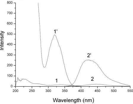
Figure 2. Excitation and emission spectra of monoanionic (1, 2) and dianionic (1', 2') forms of bumetanide. pH = 2.0 (1, 2), pH = 6.0 (1', 2'), ex = 325 nm, em = 420 nm.
which can be explained by simultaneous deprotonation of the benzoic acid and of the anilinium ion with con- sequent formation of stable structure [19]. Dianionic form of bumetanide exhibits intensive excitation band at 325 nm with emission at 420 nm. So, at pH of urine bumetanide is characterized with high luminescence intensity while furosemide demonstrates low intensity that is the evidence of possibility to determine amount of furosemide.
In acidic medium (pH 2) furosemide demonstrates strong intrinsic luminescence with maxima ex/em 270/
410 nm in contrast to bumetanide. These properties are served as the basis for the determination of furosemide in presence of commensurate amount of bumetanide after acidifying urine sample till pH 2.
Bendroflumethiazide and hydrochlorothiazide at pH of urine exist in molecular form, while in case of chloro- thiazide at pH 5.5 - 7.0 molecular and monoanionic forms coexist in solution. The spectra of excitation and emission of bendroflumethiazide, hydrochlorothiazide and chlorothiazide are shown in Figures 3 and 4. The highest luminescence intensity of bendroflumethiazide is peculiar to its molecular form (ex/em 270/400 nm)
which exists at pH 5.5 - 7.0. Anionic form is charac- terized by negligible emission. Luminescent properties of protolytic forms of hydrochlorothiazide and chloro- thiazide do not differ much. Maxima of excitation and emission for protolytic forms of both diuretics coincide which can be explained by their identical molecular structures. Luminescent intensity of chlorothiazide is 5 times more than of hydrochlorothiazide. However, consi- derable overlapping of excitation and emission spectra makes their simultaneous determination impossible. At the same time, determination of bendroflumethiazide in presence of commensurate amount of hydrochlorothia- zide is possible due to low luminescent intensity of the latter.
[image:3.595.316.531.540.696.2]At pH of urine amiloride is present in solution in protonated form, and triamterene in protonated and mo- lecular forms. Molecular forms of amiloride and triam- terene are characterized with intensive luminescence
(Figures 5 and 6). Protonated forms which predominate at pH of urine have less intensive luminescence. At pH 4.0 insignificant bathochromic shift for excitation (Δλex =
6 nm) and emission maxima (Δλem = 4 nm) is observed.
This fact can be explained by protonation of amino- groupes [18].
[image:4.595.312.535.86.254.2]Determination of amiloride in mixture with triam- terene is impossible due to high luminescent intensity of the latter. However, triamterene can be determined in presence of any amount of amiloride. Elaborated method of determination of triamterene as individual substance in aqueous solution without pre-concentration demonstrates
Figure 4. Excitation and emission spectra of molecular (1, 1') and anionic (2, 2') forms of hydrochlorothiazide and of mo-lecular (3, 3') and anionic (4, 4') forms of chlorothiazide. pH = 6.0 (1, 3), pH = 10.0 (2, 4), ex= 295 nm, em= 405 nm.
Figure 5. Excitation and emission spectra of protonated (1, 2) and molecular (1', 2') forms of amiloride. pH = 6.0 (1, 2), pH = 10.0 (1', 2'), ex= 285 nm, em= 420 nm.
Figure 6. Excitation and emission spectra of protonated (1, 2) and molecular (1', 2') forms of triamterene. pH = 6.0 (1, 2), pH = 10.0 (1', 2'), ex= 360 nm, em= 440 nm.
the results competitive with that obtained in [18]. The same LOD and linearity range were achieved without solid-phase extraction which decreased time of analysis and simplified the method of determination.
[image:4.595.60.285.293.464.2]The excitation and emission intensities as function of diuretic concentration under optimal conditions were studied. Limit of Detection was calculated as relation of threefold standard deviation of noise signal to coefficient of instrumental sensitivity. The results are shown in
Table 2.
3.1. Interfering Influence
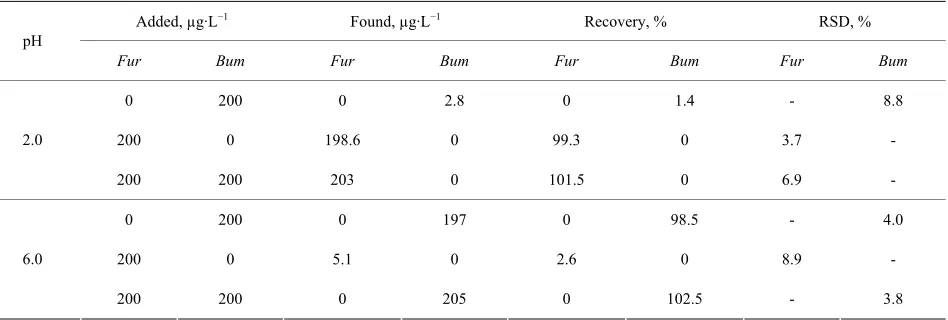
The highest sensitivity is inherent for the methods of determination of furosemide and triamterene as it can be seen in Table 2. At pH of urine all diuretics except furosemide show luminescent properties. Furosemide at pH > 5 does not interfere with determination of other diuretics but all diuretics except bumetanide interfere with its determination. Furosemide and bumetanide can be determined in one sample by adjusting pH. So, at pH 2.0 bumetanide molecule does not emit light which allows determination of furosemide. On the contrary, at pH 6.0 bumetanide is characterized with high lumine- scence intensity, and furosemide molecule does not emit light. The possibility of determination of one diuretic in presence of other in commensurate amount with suffi- cient sensitivity was proved. Results are shown in Table 3.
[image:4.595.60.285.527.697.2]Table 2. Some analytical figures of merit of determination of diuretics by intrinsic luminescence in aqueous solutions. R2 =
0.998 - 0.999.
I = (a ± Δa) + (b ± Δb) C, µg·mL−1 Diuretic pH emmax (exmax), nm
I a ± Δa b ± Δb
LOD,
µg·L−1 Linearity range, µg·L−1
Іex 9 ± 4 1988 ± 19 6.0
Furosemide 2.0 410 (270)
Іem 14 ± 6 1980 ± 26 9.0
10 - 2000
Іex 3 ± 4 392 ± 4 30
Bumetanide 6.0 420 (325)
Іem 1 ± 2 395 ± 2 20
50 - 5000
Іex 16 ± 5 988 ± 13 20
Bendroflumethiazide 6.0 400 (270)
Іem 17 ± 6 992 ± 15 20
10 - 5000
Іex 12 ± 7 489 ± 7 40
Amiloride 6.0 420 (285)
Іem 16 ± 6 413 ± 6 40
50 - 5000
Іex 1 ± 1 3805 ± 26 0.8
Triamterene 4.0 440 (360)
Іem 2 ± 1 3802 ± 3 0.8
[image:5.595.65.539.379.539.2]1 - 1000
Table 3. Determination of furosemide (Fur) and bumetanide (Bum) in aqueous solutions.
Added, µg·L−1 Found, µg·L−1 Recovery, % RSD, %
pH
Fur Bum Fur Bum Fur Bum Fur Bum
0 200 0 2.8 0 1.4 - 8.8
200 0 198.6 0 99.3 0 3.7 - 2.0
200 200 203 0 101.5 0 6.9 -
0 200 0 197 0 98.5 - 4.0
200 0 5.1 0 2.6 0 8.9 -
6.0
200 200 0 205 0 102.5 - 3.8
3.2. Urine Analysis
Determination of diuretics in urine is complicated due to the influence of matrix which contains a variety of organic substances. Most of these substances exhibit high absorbance in the ultraviolet region [20] and have strong background luminescence that interferes with the direct determination of diuretics. As a result, the urine must be diluted and the fluorescence intensity should be meas- ured at the maximum of the highest excitation wave- length where the urine exhibits low absorbance.
Dilution in 10 times and measuring of intensity signal versus blank urine was proved to be effective in order to avoid an interfering influence of matrix and distortion of the spectra. As it was established earlier the highest luminescence intensity was inherent to molecular forms
of triamterene and amiloride (pH > 8.0 and 9.5, res- pectively), but alkalization of urine till pH 9.5 - 10.0 leads to appearance of turbidity. Thus, to avoid addi- tional step in sample preparation including filtration determination of these diuretics was realized at pH 4.0 at which both diuretics exist in protonated forms.
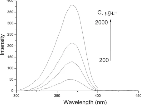
As it can be seen from Figure 7 triamterene in urine is characterized by high intensive excitation and emission with maxima at 370 and 440 nm, respectively.
20 - 500 µg·L−1.
Amiloride does not exhibit luminescence in urine (Fi- gure 8) which can be explained by binding with urine components such as derivatives of kynurenine, xanthure- nic and folic acids [20]. As individual substance it can be determined by excitation spectra with LOD 30 µg·L−1 and linearity range 200 - 2000 µg·L−1. The presence of triamterene makes determination of amiloride by excita- tion spectra impossible since their maxima coincide. Overlapping leads to the increase of total signal intensity. However, determination of amiloride in presence of any amount of triamterene is still manageable by spectro- photometry, by intrinsic absorbance at 360 nm (Figure 9), in area where other diuretics in urine do not absorb. The linearity range is 50 - 1000 µg·L−1 with LOD 10 µg·L−1 obtained. Determination of triamterene in pre- sence of any amount of amiloride is possible by its emis- sion spectrum at wavelength 440 nm.
[image:6.595.309.537.84.249.2]High recoveries of amiloride and triamterene from
Figure 7. Excitation and emission spectra of triamterene in urine. C = 20 - 500 µg·L−1. pH = 4.0.
ex = 360 nm, em = 440
nm.
Figure 8. Excitation spectrum of amiloride in urine С = 200 - 2000 µg·L−1. рН = 4.0.
[image:6.595.60.286.320.486.2]ex = 285 nm, em = 420 nm.
Figure 9. Spectrum of absorbance of amiloride in urine. C = 50 - 1000 µg·L−1. pH = 4.0. = 360 nm.
human urine and low relative standard deviations (Table 4) are the evidence of accuracy and reproducibility of the method given. Retaining of necessary specifity on reduction the analysis time compared with that proposed in [18] owing to avoiding solid-phase extraction is im- portant achievement of our work.
Bumetanide, furosemide and bendroflumethiazide were proved to exhibit high luminescence under optimal con- ditions in aqueous solutions. The influence of complex urine matrix leads to disappearance of luminescent pro- perties of these diuretics at optimal conditions. Lumi- nescence quenching probably can be explained by their binding with components of urine, supposedly with ami- noacids [20]. So, determination of bumetanide, furo- semide and bendroflumethiazide assume preliminary sample preparation of urine.
4. Conclusions
Intrinsic luminescent properties of different protolytic forms of furosemide, bumetanide, bendroflumethiazide, amiloride and triamterene are taken as a principle of sensitive determination of the diuretics in aqueous so- lutions. The simplicity and rapidity of method make it a useful tool in determination.
Techniques elaborated for determination of triam- terene in presence of other diuretics and furosemide in presence of commensurate amount of bumetanide allow enhancing specifity of analysis. Other studied diuretics can not be determined in presence of triamterene which requires its preliminary separation.
[image:6.595.59.285.540.708.2]Table 4. Analytical recoveries of amiloride and triamterene from human urine.
Amiloride Triamterene
Added,
µg·L−1 Found*a, µg·L−1 Recovery, % RSD, % Found *b
,
µg·L−1 Recovery,% RSD,% Added, µg·L−1 Found *
,
µg·L−1 Recovery, % RSD, %
225 227 100.9 8.7 228 101.3 5.7 22.5 22.7 100.9 6.2
400 397 99.2 6.2 402 100.5 6.4 90 88.6 98.4 5.9
1000 998 99.8 6.4 1000 100.0 6.5 250 252 100.8 5.8
1500 1508 100.5 5.3 1510 100.6 5.5 400 396 99.0 7.6
*Average of 3 measurements. aQuantity found by luminescent method. bQuantity found by spectrophometry.
which demands preliminary sample preparation of urine. Triamterene in urine can be determined in presence of studied diuretics by emission intensity with high sen- sitivity. Simple method of amiloride determination in human urine was proposed. It does not include long sam- ple preparation but provides high specifity of analysis with sufficient sensitivity. The reduction of time of ana- lysis due to avoiding sample preparation merits the tech- niques proposed.
REFERENCES
[1] R. Ventura and J. Segura, “Detection of Diuretic Agents in Doping Control,” Journal of Chromatography B: Bio-medical Sciences and Applications, Vol. 687, No. 1, 1996, pp. 127-144.
http://dx.doi.org/10.1016/S0378-4347(96)00279-4 [2] A. Morganti, “Should a Diuretic Always Be the First
Choice in Patients with Essential Hypertension? The Case for No,” Journal of the American Society of Nephrology, Vol. 16, No. 3, 2005, pp. 70-73.
http://dx.doi.org/10.1681/ASN.2004110964
[3] WADA, “The World Anti-Doping Code—The 2009 Pro-hibited List: International Standard,” World Anti-Doping Agency, Montreal, 2009.
[4] WADA, “Minimum Required Performance Limits for Detection of Prohibited Substances (Technical Document TD2009MRPL),” World Anti-Doping Agency, Montreal, 2009.
[5] C. Brunelli, C. Bicchi, A. Di Stilo, A. Salomone and M. Vincenti, “High-Speed Gas Chromatography in Doping Control: Fast-GC and Fast-GC/MS Determination of β- Adrenoceptor Ligands and Diuretics,” Journal of Separa-tion Science, Vol. 29, No. 18, 2006, pp. 2765-2771. http://dx.doi.org/10.1002/jssc.200500387
[6] R. Ventura, M. Roig, N. Monfort, P. Saez, R. Berges and J. Segura, “High-Throughput and Sensitive Screening by Ultra-Performance Liquid Chromatography Tandem Mass Spectrometry of Diuretics and Other Doping Agents,”
European Journal of Mass Spectrometry, Vol. 14, No. 3, 2008, pp. 191-200.http://dx.doi.org/10.1255/ejms.920 [7] M. Mazzarino, X. de la Torre and F. Botrè, “A Screening
Method for the Simultaneous Detection of Glucocorti-coids, Diuretics, Stimulants, Anti-Estrogens, Beta-Adre-
nergic Drugs and Anabolic Steroids in Human Urine by LC-ESI-MS/MS,” Analytical and Bioanalytical Chemis-try,Vol. 392, No. 4, 2008, pp. 681-698.
http://dx.doi.org/10.1007/s00216-008-2292-5
[8] O. J. Pozo, P. Van Eenoo, K. Deventer and F. T. Delbeke, “Development and Validation of a Qualitative Screening Method for the Detection of Exogenous Anabolic Ster-oids in Urine by Liquid Chromatography-Tandem Mass Spectrometry,” Analytical and Bioanalytical Chemistry, Vol. 389, No. 4, 2007, pp. 1209-1224.
http://dx.doi.org/10.1007/s00216-007-1530-6
[9] K. Deventer, O. J. Pozo, P. Van Eenoo and F. T. Delbeke, “Qualitative Detection of Diuretics and Acidic Metabo-lites of Other Doping Agents in Human Urine by High-Performance Liquid Chromatography-Tandem Mass Spectrometry. Comparison between Liquid-Liquid Ex-traction and Direct Injection,” Journal of Chromatogra-phy A,Vol. 1216, No. 31, 2009, pp. 5819-5827.
http://dx.doi.org/10.1016/j.chroma.2009.06.003
[10] V. Morra, P. Davit and P. Capra, “Fast Gas Chromatogra- phic/Mass Spectrometric Determination of Diuretics and Masking Agents in Human Urine. Development and Vali- dation of a Productive Screening Protocol for Antidoping Analysis,” Journal of Chromatography A,Vol. 1135, No. 2, 2006, pp. 219-229.
http://dx.doi.org/10.1016/j.chroma.2006.09.034
[11] L. Amendola, C. Colamonici, M. Mazzarino and F. Botrè, “Rapid Determination of Diuretics in Human Urine by Gas Chromatography-Mass Spectrometry Following Mi- crowave Assisted Derivatization,” Analytica Chimica Acta, Vol. 475, No. 1-2, 2003, pp. 125-136.
http://dx.doi.org/10.1016/S0003-2670(02)01223-0 [12] Yi.-L. Tseng, M.-H. Shieh, Ch.-Ts. Lin and F.-H. Kuo,
“Detection of Diuretics in Urine during Sports Events in Taiwan,” Tzu Chi Medical Journal,Vol. 16, No. 2, 2004, pp. 69-77.
[13] O. Zaporozhets, I. Tsyrulneva and M. Ischenko, “Deter-mination of 8 Diuretics and Probenecid in Human Urine by Gas Chromatography-Mass Spectrometry: Confirma-tion Procedure,” American Journal of Analytical Chemis-try, Vol. 3, No. 4, 2012, pp. 320-327.
http://dx.doi.org/10.4236/ajac.2012.34044
2001, pp. 179-187.
http://dx.doi.org/10.1016/S0003-2670(01)01356-3 [15] J. A .M. Pulgarin, A. A. Molina and P. F. Lopez,
“Simul-taneous Direct Determination of Amiloride and Triam-terene in Urine Using Isopotential Fluorometry,” Ana-lytical Biochemistry, Vol. 292, No. 1, 2001, pp. 59-68.
http://dx.doi.org/10.1006/abio.2001.5064
[16] P. C. Ioannou, N. V. Rusakova, D. A. Andrikopoulou, K. M. Glynou and G. M. Tzompanakia, “Spectrofluorimetric Determination of Anthranilic Acid Derivatives Based on Terbium Sensitized Fluorescence,” Analyst, Vol. 123, No. 12, 1998, pp. 2839-2843.
http://dx.doi.org/10.1039/a806093b
[17] C. M. Peralta, L. P. Fernández and A. N. Masi, “Solid Phase Extraction Using Nylon Membranes with Fluores-cence Detection as a Fast and Sensitive Method for Amiloride and Furosemide Determination in Urine Sam-ples,” Microchemical Journal, Vol. 98, No. 1, 2011, pp.
39-43.http://dx.doi.org/10.1016/j.microc.2010.10.009 [18] G. A. Ibanez, G. M. Escandar, A. E. Mansilla and A. M.
de la Pena, “Determination of Triamterene in Pharmaceu-tical Formulations and of Triamterene and Its Main Me-tabolite Hydroxytriamterene Sulfate in Urine Using Solid- Phase and Aqueous Solution Luminescence,” Analytica Chimica Acta, Vol. 538, No. 1-2, 2005, pp. 77-84.
http://dx.doi.org/10.1016/j.aca.2005.02.001
[19] B. Song, A. K. Galande, K. Kodukula, W. H. Moos and S. M. Miller, “Evaluation of the PKa Values and Ionization
Sequence of Bumetanide Using 1H and 13C NMR and UV
Spectroscopy,” Drug Development Research, Vol. 72, No. 5, 2011, pp. 416-426.http://dx.doi.org/10.1002/ddr.20443 [20] M. J. P. Leiner, M. R. Hubmann and O. S. Wolfbeis,
“The Total Fluorescence of Human Urine,” Analytica Chimica Acta, Vol. 198, 1987, pp. 13-23.
![Table 1. The characteristics of selected diuretics [1].](https://thumb-us.123doks.com/thumbv2/123dok_us/7869011.738233/2.595.310.535.394.739/table-the-characteristics-of-selected-diuretics.webp)