ISSN Online: 2333-9721 ISSN Print: 2333-9705
Kirschner Wire Fixation of Neglected Lateral
Condylar Fracture of the Humerus in Children
Nguyen Ngoc Hung
Vietnam National Hospital of Pediatrics, Hanoi, Vietnam
Abstract
Background: Delayed presentation of lateral condylar fractures of the hume-rus is relatively common in the developing regions of the world. Fractures of lateral condyle of humerus in pediatric age group, the most common being distal humerus epiphyseal injury, are commonly associated with delayed presentation to terminal health care providers. Materials and Methods: Twenty-one children having fracture of lateral condyle of humerus with dura-tion of trauma more than 3 weeks were included in the prospective study. Among the 21 patients, 13 were male and 8 were female. Average time of presentation was after 3 weeks of injury. All patients had Milch type II injury. The patients were treated by open reduction and internal fixation using Kir-schner wire. The outcome assessment according to the scoring system pro-posed by Dhillon et al. was used. Result: Mean month at operation is 81.5 months (range, 64 - 112 months); 8 Right/13 Left. Reason for presentation: Pain, swelling in 12; Pain, decreased elbow motion in 7; Restriction of the el-bow flexion in 2. Time from injury to Operation with mean 6.34 weeks, 3 weeks - ≤ 6 weeks in 13 Patients, and > 6 weeks in 8 patients; Previous treat-ment: Plaster cast in 15 Patients, and No treatment in 6 Patients; Mean time of Follow-up: 66 Months; Mean Age at last Follow-up was 11.6 (range, 8.4 - 15.0). To tell result, 38.1% is in Excellent; 33.3% is in Good; 14.3% is in Fair; and 14.3% is in Poor. Comparison of accepted result from time of injury to operation was 3 - 6 weeks better than over 6 weeks with PValuate 0.001387. There were AVN in 1 case, Nonunion in 2 cases. Conclusion: Osteosynthesis by Kirschner wire without bone grafting could provide solid bony union and improve elbow function. However, this technique could not prevent an angu-lar deformity completely. Corrective osteotomy should be considered in pa-tients with valgus or varus deformities.
Subject Areas
Orthopedics, Pediatrics
How to cite this paper: Hung, N.N. (2017) Kirschner Wire Fixation of Neglected Lateral Condylar Fracture of the Humerus in Child- ren. Open Access Library Journal, 4: e3330. http://dx.doi.org/10.4236/oalib.1103330
Received: December 15, 2016 Accepted: January 17, 2017 Published: January 20, 2017
Copyright © 2017 by author and Open Access Library Inc.
This work is licensed under the Creative Commons Attribution International License (CC BY 4.0).
Keywords
Delayed Presentation, Lateral Condyle Fractures, Delayed, Surgical Management
1. Introduction
Fracture of lateral condyle of humerus in pediatric age is a common occurrence. The injury is basically an epiphyseal injury; hence, in the long run, it is inhe-rently associated with potential problem of growth arrest, premature physeal closure, range of motion restriction, angular deformity of elbow, and neural complications. Current literature advises for operative management of displaced fractures of less than 3 weeks’ duration. Orthopedic societies are having odd views regarding treatment of the same fracture of more than 3 weeks’ period. Fractures of the lateral condyle of the humerus are the second most frequently encountered fractures in children, accounting for 16.9% of those occurring in the upper limb [1]. Timely treatment with accurate reduction, Kirschner wire fixation and regular follow-up yields good outcomes [1]. If, however, the diag-nosis is delayed or reduction loses in plaster after the initial conservative treat-ment, malunion or nonunion can occur, leading to persistent pain and a de-creased range of movement (ROM) in the elbow [2].
The place of operative treatment for these patients remains controversial. In 1975, Jakob et al. [3] reported the results of open reduction in seven patients af-ter three to 12 weeks, describing a mean loss of ROM of 34˚, and concluded that open reduction was not superior to non-operative management of these patients. Opponents of operative treatment have put forward the stand that operative treatment may jeopardize the already precarious vascular supply of the displaced fragment by surgical manipulation, threatening to avascular necrosis of the fragment; moreover, the fracture surfaces are no longer conforming to each oth-er and tenuous amount of attached cuff of soft tissue is shortened and con-tracted, creating difficulty in accurate reduction. It was further reported that anatomical reduction was difficult in a maluniting fracture and the surgery itself may lead to avascular necrosis (AVN) of the fragment [3] [4] [5]. Several authors have reported surgical outcomes in delayed cases where AVN has been avoided
[5]-[11].
in-stability of joint, restriction of rotatory motion of forearm and nerve palsy. We report the results of operative management of twenty-one lateral condyle fractures of the humerus in the paediatric population that presents after more than 3 weeks of the initial trauma. Our hypothesis was that open reduction and internal fixation (ORIF) could improve the function of the elbow without in-creasing the incidence of complications such as humeral dysplasia, AVN and premature physeal closure.
2. Materials and Methods
A retrospective study was carried out to evaluate the results of surgical tech-niques performed from December 2003 to December 2013 in 25 patients with neglected lateral condylar humerus in Children. The operations were performed by single surgeon (Author).
The study had the approval of the Ethical Review Committee of our Institute and was carried out in accordance with the tenets of the Declaration of Helsinki.
The Patients underwent “Osteosynthesis in situ without bone grafting” for neglected of LCF that developed after >3 weeks after injury. According to the criteria of Flynn et al. [12] if the fracture had not united until 3 months, it was defined as a nonunion. From 2003 to 2013, 25 patients visited our institution secondary to nonunion of LCF and underwent the index procedure. One patient loss of Follow-up and three patients were excluded because the duration from Operation was <3 years, remaining 21 patients in this study. All patients with metabolic bone disease, skeletal dysplasia and lateral condyle fracture with asso-ciated supracondylar, medial condyle, and elbow dislocation were excluded from the study.
All patients were referred from other hospitals and had tenderness over the lateral aspect of the elbow and pain on elbow motion at the time of visit. None of the children had signs of ulnar neuropathy. All patients showed varying degrees of impairment on elbow motion. Most of patients had some degrees of elbow flexion contracture and pain on elbow motion, so carrying angle and elbow in-stability could not be correctly assessed.
At the time of presentation, radiographs showed an obvious fracture gap be-tween the distal humerus and lateral condyle fragments and no callus formation on fracture gap. Physis of lateral humeral condyle was open in all patients. All the fractures were of Milch type II, with various degrees of superolateral dis-placement. The amount of displacement was measured from medial and lateral metaphyseal ends of the lateral condylar fragments to origin site of the distal humerus on the anteroposterior or internal oblique radiograph. The measure-ment with the most displacemeasure-ment was regarded as the amount of medial and lat-eral displacement. The amount of displacement of latlat-eral condyle fragment was measured as 3 to 9 mm, averaging 5.2 mm medially, and 4 to 10.4 mm, averaging 6.8 mm laterally.
wire fixation and cast immobilization.
2.1. Surgical Technique
A lateral approach was made to the elbow. Dissection is through the plane be-tween the triceps and the brachioradialis. The approach was carried through the lateral fascia right down to the fracture. The fragment was often found to be dis-placed and fibrous tissue often made it difficult to assess the orientation of the fracture. Careful dissection of the fibrous tissue was made and posterior attach-ments were saved. Thorough irrigation was done to remove the fibrinous debris. Any dissection needing to be done on the lateral epicondyle and metaphysis was made anterior, to avoid the posterior blood supply and minimize the risk for avascular necrosis. The displaced fragment was reduced under direct visualiza-tion, often with the aid of a reduction clamp, “joystick” Kirschner wires, or the assistant’s manual pressure. We used 1.4-mm-diameter Kirschner wires for pa-tients younger than five years of age, 1.6-mm-diameter wires for those between five and eight years of age and 1.8-mm diameter wires for those older than eight years of age. Grafting was not added in any patient. Post operatively all patients underwent a common protocol of 3 weeks padded cramer wire splint immobili-zation followed by range of motion exercises intermittently for a further 3 weeks. At 6 weeks the Kirschner wire was removed. The patient was allowed to do range of motion exercises without splint protection at 6 weeks.
All patients were followed up for a period of one year and so far according to Dhillon et al. [4]. Radiologically avascular necrosis, malunion, nonunion and heterotropic ossification were specifically looked at. Union was assessed on the anteroposterior and lateral radiographs of the elbow. Union was said to have occurred when the fracture was obliterated by the trabeculae or the callus. Radi-ographical outcome was assessed for lateral bony overgrowth, fishtail deformity, presence of osteonecrosis, and valgus or varus deformity at the latest Follow-up. For functional evaluation, range of elbow motion, and the signs of ulnar neuro-pathy were checked and presence of tenderness and pain on elbow motion and fatigue on strenuous activity such as sports were surveyed. To assess cosmetic issues, carrying angle was measured and compared with that of the contralateral side and development of bony hump on lateral side of distal humerus after sur-gery was examined. For overall outcome assessment, the scoring system pro-posed by Dhillon et al. [4] was used. This scoring system includes pain on activ-ity, range of motion, and carrying angle (see, Table 1).
2.2. Statistical Analysis
The data were analysed with Epi Info 6.04 sofware public domain statistical software for epidemiology, developed by Centers for Disease Control and Pre-vention (CDC) in Atlanta, Georgia, USA
(http://wwwn.cdc.gov/epiinfo/html/prevVersion.htm). We performed the χ2 test
Table 1. Scoring system for the outcome of fractures of the lateral humeral Condyle in children [4].
Function Carrying Angle
(Degree) Each Column Score Points Pain Range of Motion (Deg,)
None 0 - 140 Valgus 7 - 10 3
Occasional >15 - 125 Valgus < 20 Varus< 0 2
After heavy work (or activities) >30 - 110 Valgus 20 - 30 Varus 0 - 15 1
With normal activity
(morto or sensory loss) >30 - 110 Valgus > 30 Varus > 15 0 Functional grading (points): Excellent 6, Good 5. Fair 4, Poor < 4. Overall grading (points): Excellent 9, Good 7-8. Fair 5-6, Poor < 4.
3. Results
Total of 21 patients were included in our study (Table 2). There were 13 Male and 8 Female. 81.5 moths (range, 64-112 months). Time from injury to Opera-tion 3 weeks - ≤ 6 weeks with 13 Patients (Case 3, Figure 1), and >6 weeks - 12 weeks with 8 patients. Union was achieved in 19 of them (90.5%) patients at av-erage 9 weeks (range, 7 to 11 weeks) (Case 9, Figure 2). The average duration of follow-up was 66 months (range, 38 to 89 months). One patient had evidences of osteonecrosis on the latest follow-up radiographs. In 5 patients, variable degrees of lateral bony hump were shown on anteroposterior radiographs. It originated from the superolaterally displaced metaphyseal fragment of fractured lateral humeral condyle, which was fixed in situ. Although lateral bony hump was shown on radiographs, it was hardly detectable on gross clinical examination and no patients complained about mild bony protrusion on lateral side of elbow. In one patient (case 14), bony hump had remodeled completely on the latest fol-low-up radiographs, and the size of bony hump had decreased with time in the remaining patients.
Cubitus valgus was seen in 4 patients (range, 12 to 18) and cubitusvarus in 5 patients (range, 10 to 8 degrees) by the criteria of scoring system proposed by Dhillon et al. [4] which defined the ideal carrying angle as 7 to 10 degrees of valgus. However, in 7 patients who had angular deformities, the difference of carrying angle between the affected side and contralateral side was within 5 de-grees at the latest follow-up. The parents and patients were satisfied with the alignment of upper extremity in 11 patients. In the remaining 3 patients, cubi-tusvarus deformity of 21 degrees or more than contralateral side developed in 1 patient. One cubitus valgus deformity (case 12) with a carrying angle of 18 de-grees was not found to be substantially improved at the latest follow-up, and both the patient and the parents refused to undertake the corrective osteotomy. The other one patient with cubitus valgus deformity (Case 7, Figure 3) did not want corrective osteotomy.
(a) (b)
[image:6.595.268.486.66.218.2]Figure 1. (Case 3). Initial Injury 5 Weeks; (a) Pre-Operation; (b) Post-Operative 5 weeks.
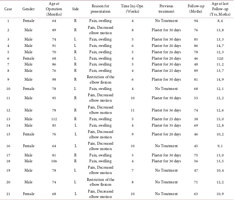
Table 2. General information of the patient.
Case Gender Operation Age at
(Months) Side
Reason for
presentation Time Inj-Ope. (Weeks) treatment Previous Follow-up (Moths)
Age at last Follow-up (Yrs, Moths)
1 Female 64 R Pain, swelling 4 No Treatment 94 8, 4
2 Male 89 R Pain, Decreased elbow motion 8 Plaster for 30 days 76 13, 8
3 Male 74 L Pain, swelling 5 Plaster for 30 days 85 13, 3 4 Male 91 L Pain, swelling 6 Plaster for 20 days 86 14, 7 5 Male 70 R Pain, swelling 5 Plaster for 26 days 78 12, 3 6 Female 68 L Pain, swelling 4 Plaster for 20 days 46 12,0 7 Male 86 R Pain, swelling 5 Plaster for 30 days 48 11, 2 8 Male 76 R Pain, swelling 4 Plaster for 25 days 89 13, 7 9 Male 98 R Restriction of the elbow flexion 6 Plaster for 30 days 81 14, 9
10 Female 78 L Pain, swelling 4 No Treatment 68 12, 1
11 Male 95 R Pain, Decreased elbow motion 10 Plaster for 30 days 53 12, 2
12 Male 78 R Pain, Decreased elbow motion 11 Plaster for 36 days 74 12, 6 13 Male 112 R Pain, swelling 5 Plaster for 25 days 38 15, 0 14 Male 85 L Pain, swelling 4 Plaster for 35 days 69 12, 8 15 Female 76 L Pain, Decreased elbow motion 9 Plaster for 20 days 46 10, 2
16 Female 64 L Pain, Decreased elbow motion 10 No Treatment 45 9, 1 17 Male 81 R Pain, swelling 5 Plaster for 30 days 75 13, 0 18 Male 106 R Pain, swelling 4 Plaster for 30 days 56 13, 5 19 Male 78 L Pain, Decreased elbow motion 7 No Treatment 47 10, 4
20 Male 74 L Restriction of the elbow flexion 8 No Treatment 71 12, 2
21 Female 68 L Pain, Decreased elbow motion 10 No Treatment 63 10, 9
[image:6.595.58.540.269.678.2](a) (b)
Figure 2. (Case 9). Initially 6 Weeks. (a) Pre-Operation; (b) Union Postoperative 6 weeks.
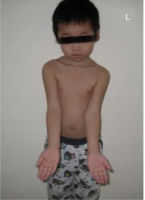
Figure 3. (Case 7). Patients have cubitus valgus.
ties, and there were no signs or symptoms suggestive of ulnar nerve dysfunction. Range of motion was remarkably improved in most of the patients. The average flexion contracture of the elbow joint decreased from 25 degrees (range, 0 to 52) before surgery to 4.6 degrees (range, 0 to 14 degrees), and the range of elbow flexion increased from 118 degrees (range, 90 to 135 degrees) to 136 degrees (range, 125 to 140 degrees). Pronation and supination were full in all patients with initial injury under 6 weeks (Figure 4). Overall outcome according to the scoring system proposed by Dhillon et al. [4] in time from injury to operation 3-6 weeks was excellent in 8 patients (61.5%), good in 5 patients (39.5%); in time from injury to operation 6-12 weeks was, good in 2 (25%) patients, fair in 3 pa-tient (37.5%), and poor in 3 papa-tient (37.5) (see, Table 3).
4. Discussion
[image:7.595.271.487.62.226.2] [image:7.595.302.445.261.458.2](a) (b)
[image:8.595.261.487.65.254.2]Figure 4. (Case 7). Function of the elbow with full Extension and Flexion.
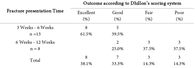
Table 3. Compare latest results of operation according to Fracture presentation time.
Fracture presentation Time
Outcome according to Dhillon’s scoring system Excellent
(%) Good (%) Fair (%) Poor (%) 3 Weeks - 6 Weeks
n =13 61.5% 8 39.5% 5 6 Weeks - 12 Weeks
n = 8 25.0% 2 37.5% 3 37.5% 3
Total 38.1% 8 33.3% 7 14.3% 3 14.3% 3
Accepted Result (Excellent and Good) was all Patients 100% in time of injury to Operation 3 weeks - 6 weeks and 25% in time 6 weeks - 12 weeks. To tall result, 38.1% in Excellent, 33.3% in Good, 14.3% in Fair, and 14.3% in Poor. Comparison of accepted result in time of injury to Operation 3 - 6 weeks better than 6 weeks with P Valuate 0.001387. There were AVN in 1 case, Nonunion in 2 cases.
eral studies demonstrated a significant improvement in postoperative outcome and advocated some surgical techniques in order to preserve vascularity of the lateral condyle fragment and create intraoperative acceptable reduction [3] [4] [14]. Nonetheless, the aforementioned techniques did not provide a clear refer-ence for deformity correction in the sagittal plane and studies had also not proven the correlation between the sagittal plane malalignment and the post-operative functional outcome.
In developing countries, patients with fractures of the lateral condyle of the humerus usually present late [14] [15]. Sometimes the diagnosis is missed due to incorrect interpretation of the radiograph, as the fracture fragment is partially cartilaginous; the radiographs are also often of poor quality. A prospective co-hort study showed that internal oblique radiographs are more sensitive than a plain anteroposterior (AP) view for diagnosing displaced or minimally displaced fractures [16] [17]. Recently, a 20˚ tilt AP radiograph has been suggested to demonstrate fragment dislocation more precisely than a standard radiograph
[image:8.595.210.540.309.425.2]available in the rural and suburban areas in most developing countries. The di-agnosis of minimally displaced fractures is therefore often missed in the early stages, being made late or only after more displacement has occurred.
Fractures of the lateral condyle of the humerus are amongst the commonest injuries encountered by the orthopaedic surgeon. Henry Milch differentiated between the two fractures patterns. A fracture exiting the trochleocapitellar groove as type 1 and the fracture exiting the trochlea as type 2 [19] and Milch type I fracture is salter type IV epiphyseal injury and Milch type II is salter type II injury. Most trauma of the degree of displacement depends on the preserva-tion of the articular hinge. If the hinge is intact the condylar fragment shows only a lateral tilt. If the fracture is complete the fracture can be rotated com-pletely up to almost 180 degrees.
The undisplacedmilch type I fractures are usually treated with plaster immo-bilization. There is inherent risk of late displacement and nonunion of fracture if the injury is not regularly followed with frequent radiographic check up. We be-lieve that delayed union is a relevant sequela of any fracture as this can result in prolonged casting which can be frustrating for the patient, increases costs and resource utilization, and increases risk of elbow stiffness.
The late displacement of fracture warrants immediate open reduction and in-ternal fixation of fracture. Toh et al. [20] observed the long term functional re-sults of nonunion of fracture lateral condyle in children and concluded that the Milch type I fracture contributes more functional deficit owing to loss of normal radiocapitellar relationship owing to separation of a part of the capitulum. Thus, what appears initially as a stable tiny fragment, if displaced, could meet with more functional deficit than potentially unstable Milch type II fracture.
5. Operation
The surgical technique should not be too aggressive to disturb the condylar vas-cularization. In order to control the intra-articular reduction, it may be neces-sary to cut some parts of the capsule and the synovial [18] [19]. Jakob et al. felt that surgical intervention in cases with delayed presentation did not improve results in comparison to patients with no treatment at all [3]. They pointed out the difficulty caused by the early callus formation. There is a general agreement that surgical intervention in old established non unions should be avoided as os-teosynthesis may reduce the range of motion of the elbow or the bone may not unite, so operative treatment for such patients has not been popular [20] [21] [22].
Osteosynthesis in situ permits malposition of lateral condyle fragment result-ing in incongruent articular surface of distal humerus. Some author mentioned that the fragment should be reduced on anatomic or functional position for good prognosis [4] (Case 7, Figure 4). Anatomic or functional reduction needs to mobilize the fragment and have a relative high risk of osteonecrosis than os-teosynthesis in situ [3] [4].
synthesis [4] [5] [23]. However, Inoue et al. [24] mentioned that bone grafting carried the risk of decreased elbow motion by blocking the distal humeral fossa or inadvertent distal migration of the fracture fragment resulting in violation of the radiocapitellar articulation. Previous studies reported favorable results of their treatment method for nonunion of LCF without bone grafting [25] [26] [27]. In the current study, bone union could be obtained completely by open curettage and firm fixation without bone grafting. Therefore, we think that bone grafting is not compulsory to achieve union in osteosynthesis in situ and the more important issue may be firm fixation and curettage of fibrous tissue. We didn’t perform bone grafting for all patients in this study.
A number of surgical techniques have been described to treat nonunion of LCF. These procedures have been generally undertaken with an open technique including pinning, bone grafting, ulnar nerve anterior transposition, and correc-tive osteotomy of the humerus for cubitus valgus deformity [20]. However, de-bate has existed whether to secure the fragment in situ, neglecting the physeal and joint deformity, or to reduce the fragment anatomically. Repositioning of the fragment anatomically to restore the articular surface often requires exten-sive soft tissue dissection, which can cause osteonecrosis of the lateral humeral condyle or decreased range of elbow motion after surgery [23] [28].
The current study demonstrates significant biomechanical advantages of screw fixation as compared with Kirschner wire fixation of lateral condyle frac-tures using a synthetic bone model. Displaced fracfrac-tures of the lateral condyle in pediatric patients have traditionally been treated with open reduction and inter-nal fixation using Kirschner wires (Case 3, Figure 1). Potential exists for loo-sening of the pins with loss of fixation, and if the pins are not buried, supple-mental casting is required for the duration of fixation. Screw fixation has been advocated as an alternative technique with potential advantages of providing compression at the fracture site and allowing continued fixation after supple-mental casting is no longer felt necessary by the treating physician. Theoretical-ly, this could result in improved healing, decreased duration of casting, and fast-er return of range of motion. Potential disadvantages include need for subse-quent removal and the theoretical possibility of growth disturbance (although this has never been reported with screw fixation of a lateral condyle fracture). Although some clinical studies have compared the fixation methods, no previous biomechanical studies have provided a biomechanical comparison [29].
Two studies have compared clinical use of Kirschner wire and screw fixation for pediatric lateral condyle fractures. Li et al. [31] compared 30 lateral condyle fractures repaired with Kirschner wires to 32 fractures repaired with 3.5 mm cannulated screws with an average follow-up of 39.4 months [2]. No statistically significant difference in clinical outcome was noted; however, 5 infections oc-curred in the Kirschner wire group compared with none in the screw group. In addition, 11 patients in the Kirschner wire group and 4 patients in the screw group developed an obvious lateral prominence. Nine fractures repaired with Kirschner wires and 2 repaired with screws experienced a 10 degree loss of elbow extension. Gilbert et al. [32] in a series of displaced lateral condyle fractures re-quiring open reduction compared 41 patients treated with screw fixation and 43 patients treated with Kirschner wire fixation. They found decreased time to un-ion, improved range of motun-ion, and fewer complications in the screw fixation group [3].
The present study compares the optimal 2 Kirschner wire configuration de-termined by Bloom and colleagues [30] with lag screw fixation and shows that lag screws provide increased biomechanical stability at the fracture site. Testing in tension was performed to simulate the force exerted by the extensor muscu-lature, a commonly cited possible mechanism for displacement and poor healing of lateral condyle fractures. In tension, screw fixation was biomechanically supe-rior to Kirschner wire fixation both in terms of stiffness and maximum force required to achieve displacement, as expected. Testing in compression was per-formed to simulate the mechanism of failure described by the radial head “pushing off” the fracture fragment. Compression testing showed smaller in-creases in maximum force with screw fixation, and no statistically significant difference in stiffness. The more similar biomechanical performance of the 2 fix-ation methods in compression is consistent with expected characteristics [29].
Contrary to all above literature, of late, some authors reported satisfactory re-sults of osteosynthesis in old fractures and established nonunion of lateral hum-eral condyle in children, and advocated the fractures to be entertained with re-duction and fixation with bone grafting before physeal closure. Smith [33]
treated established nonunion of lateral humeral condyle with cubitus valgus de-formity using ilizarov apparatus and found 53.5% excellent result, 39.3% good result, and 2% fair result with mean postoperative humerusulna angle as 6˚. Roye et al.[11] treated 4 cases of established nonunion of fracture lateral hum-eral condyle and found satisfactory results, and advocated that established non-union of fracture can be safely treated with osteosynthesis. Agarwal et al. [23]
reduction and internal fixation with bone grafting was performed and found ex-cellent results in 8 patients and good results in 7 patients. Moorhead [37] ob-served nonunion of lateral condyle of humerus for 17 years and reported good functions of elbow Contrary to fearing that the dicey situation of over release and over pulling might compromise the vascular supply of fragment, Gaur et al.
[35] advocated a technique of making multiple incisions in the common exten-sor aponeurosis for easy reduction of fragment. There was one patient with AVN in this study (Case 15, Figure 5).
Ali and Tahir [36] fixed 18 fractures lateral condyle 3 to 12 weeks old with Kirschner wires with average duration of follow up of 15 months and noted ex-cellent results in five patients, good in three, fair in six and poor in four patients using Modified Aggarwal et al. Criteria. They concluded that open reduction and internal fixation is an effective treatment in fractures of lateral condyle of hu-merus in children presenting between 3 to 12 weeks after injury. In another local study fifty children with neglected fracture lateral condyle were fixed with Kir-schner wires and outcome was evaluated in terms of pain relief, range of motion, and union at fracture. Follow-up at the end of two months revealed excellent results in 68% patients, good in 14% patients, fair in 10% patients and poor in 8% patients. Similarly in another study 22 patients with fracture lateral humeral condyle presented up to 12 weeks after injury were fixed with Kirschner wires and the results were assessed by Modified Aggarwal et al. criteria after follow-up for 1 year. Excellent results were achieved in 12 patients, good in three, fair in three and poor in three patients and they concluded that open reduction and in-ternal fixation is an effective treatment in all cases of displaced fractures of the lateral condyle of humerus presenting up to 12 weeks post injury on the basis of low surgical complication and high union rate. All the participants of our study had lateral condyle fracture of 3 to 8 weeks old. Sarafand and Khare [6] in a se-ries analyzed their results in 16 patients with lateral condylar humeral fractures 5 - 12 weeks old using criteria defined by Agarwal et al. They observed excellent
[image:12.595.306.440.520.719.2]to good results in 6, fair in 6 and poor results in 4 patients. They concluded that open reduction and internal fixation is recommended in all cases of displaced fractures of the lateral condyle of the humerus presenting at up to 12 weeks post injury. However, the results become poorer with increase in duration after injury and the grade of displacement.
In our study, excellent results were achieved in 38.1%, 33.3% in Good, 14.3% in Fair, and 14.3% in Poor. Comparison of accepted result in time of injury to Operation 3 - 6 weeks better than 6 weeks with P Valuate 0.001387 (see, Table 3).
There was high rate of union and satisfactory elbow function in late present-ing lateral condyle fractures in children followpresent-ing osteosynthesis attempt. The study showed poor correlation between patient’s age, duration of late presenta-tion or Milch type I or II and final elbow funcpresenta-tion as determined by LES. Our study noted only one patient with post-operative AVN of lateral condyle (Figure 5). Watt enbarger [8] fixed 11 children with fracture of more than 3 weeks with K-wires and observed that there were no cases of AVN even though four of their cases had displacement of more than 10 mm. Three patients had occasional pain. The risk of AVN with late open reduction of lateral condyle at more than 3 weeks is reduced if no tissue is stripped off the fracture fragment posteriorly.
Even children without anatomical reduction had functional arms with little or no pain. Weiss [37] reported that 3.8% of patients developed skin infection around Kirschner wires while Chao Li [38] reported this to be 16.7%. In our study, skin infection around the Kirschner wires occurred in 9 (36%) patients, while no patients developed skin infection over the cancellous screws. Thus it indicated that more infection in patients occurred with percutaneous Kirschner wires than screws. Therefore, it is suggested that skin care should be done to prevent skin infection. Oral antibiotics and wound care should be given in treatment for infection.
some of the cases. Almost all of our patients had yet to gain their pubertal growth spurt, and obviously longer studies need to be taken to find the behavior of the lateral condylar epiphysis and alteration in function with remodeling. It is highly desirable to keep these patients under longer follow up.
Whilst there is a consensus about the management of fractures treated early, fixation of delayed fractures is surrounded by confusion. A late presentation leads to difficulty in management due to displacement of the fragment as a result of the pull of the common extensors, incongruous reduction of articular surfac-es, injury/early closure of the epiphyseal growth plate, and possible damage to vascular supply.
Lagrange and Rigault showed that the blood supply to the lateral condyle en-ters by its soft tissue attachments, particularly posteriorly at the origin of the long extensor muscles, and disruption of this will destroy the vessels and render the condyle ischaemic [40].
Jacob et al.[3] contended that the results of open reduction and internal fixa-tion > 3 weeks after the fracture did not show better results than those of no treatment at all and may result in AVN of fragment.
Soft tissue dissection during surgery can be a factor affecting healing of these fractures. As the blood supply to the lateral condyle enters through the posterior cortex [41] it is recommended to avoid posterior soft-tissue dissection during surgery to prevent avascular necrosis.
Achieving anatomical reduction is often not possible because of remodeling of the fragment, new bone formation, and sclerosis and smoothening of the frac-ture line. For these various reasons, in long standing untreated nonunion, reduc-tion of the fracture has been a concern. With higher grades of displacement, it sometimes becomes impossible to bring the fragment into normal position without stripping the soft tissue attachments on the displaced fragment. As ex-tensive soft tissue dissection may lead to avascular necrosis of the fragment, many recommend that these fractures should be left alone [3] [42].
Complications of nonunion viz, cubitus valgus deformity, lateral instability of elbow, weakness of limb, radiocapitular malformation, tardy ulnar nerve palsy, etc., Possibly, it avoids the future need of osteotomy and anterior transposition of ulnar nerve (Case 12, Figure 6).
Dhillon et al.[4], Chao et al. [38], Zionts et al. [43] Speed and Macey [44] re-porting uniformly bad results which included cubitusvarus and valgus deformi-ties, osteonecrosis, nonunion and malunion, and loss of motion. They recom-mended that patients presenting late be left alone and any sequelae evaluated at a late stage. Fractures that are operated upon after a delay are also complicated by the presence of fibrosis, and callus formation. Preoperative stiffness that is found in these cases is likely to affect the post operative result [4].
(a) (b)
Figure 6. (Case 12). Initial injury 10 weeks; (a) Pre-Operation; (b) Post-Operative 14 weeks.
this study, there were no premature growth arrest, and fishtail deformity and none of the patients had ulnar neuropathy at latest follow-up. Because all frac-tures were classified as Milch type II, we could achieve osteosynthesis between the metaphyseal fragments with epiphysis and distal humerus without physeal damage. This results means that premature growth arrest of lateral condyle could be avoidable with osteosynthesis in situ, especially in nonunion of Milch type II fracture.
At the latest follow-up, meaningful angular deformitieswere found in 4 pa-tients. This result was probably because osteosynthesis in situ could not correct a preexisting angular deformity at the time of surgery. However, this presumption could not be substantiated because we could not evaluate initial angular defor-mities of these patients exactly due to flexion contracture. In addition, we as-sumed that these deformities resulted from surgical technique itself. If the frac-ture fragment did not fit exactly to the distal humerus and a fracfrac-ture gap still remained even after tightening the Kirschner wire, cubitusvarus deformity will develop. If the Kirschner wire was too tightened, cubitus valgus deformity would occur. Therefore, we agreed with the opinion of some authors that if the patient has an angular deformity at presentation or latest follow-up, concomitant or staged corrective osteotomy should be considered [26].
[image:15.595.231.519.66.281.2]hump seldom causes cosmetic problems, it should be kept in mind that the children and parents might have concerns about gross appearance especially in eminent cases [45].
It has been observed that nonunion and growth arrest more commonly result from minimally displaced fractures than from markedly displaced and rotated fractures, probably because severe fractures are treated more adequately with surgery [14]. A late presentation leads to difficulty in management due to dis-placement of the fragment as a result of the pull of the common extensors, in-congruous reduction of articular surfaces, injury/early closure of the epiphyseal growth plate, and possible damage to vascular supply because of stripping of soft tissue attachments (Case 15, Figure 5). For these reasons, when the patient presents at 3 - 8 weeks, the controversy is with regard to whether to treat these fractures by nonoperative or operative methods. If these fractures are treated nonoperatively, the various possible complications are nonunion, malunion, de-formity at the site, instability of the elbow joint, stiffness, cubitus valgus/varus, and tardy ulnar nerve palsy (Case 7, Figure 3). In addition, precarious blood supply to the fractured fragment due to excessive stripping of the soft tissues, may result in avascular necrosis of the fragment [3] [46]. So the majority favor management of established nonunion by no treatment as the functional prob-lems are not very severe [3]. It is easier to treat cubitus valgus/varus at a later date by corrective osteotomy or to treat tardy ulnar nerve palsy by ulnar nerve transposition rather than to attempt a difficult reduction. Despite the inherent risk associated with the surgery, there are reports in the literature of successful outcomes of open reduction and internal fixation of these established nonunion cases [24] [47]. The current controversy regarding the management of fractures of the lateral condyle of the humerus presenting between 3 to 8 weeks excited us to evaluate our results of open reduction and internal fixation of such fractures.
6. Conclusions
Osteosynthesis by Kirschner wire without bone grafting could provide solid bony union and improve elbow function. We show that this technique is a safe and effective procedure for growing children with relatively minimal displaced and fresh nonunion of LCF. However, this technique could not prevent an an-gular deformity completely. Corrective osteotomy should be considered in pa-tients with valgus or varus deformities.
present in these patients, it may be meaningful to observe the progression until maturity.
References
[1] Beaty, J.H. and Kasser, J.R. (2006) Rockwood and Wilkins’ Fracture in Children. 6th Edition, Lippincott-Raven, Philadelphia.
[2] Agarwal, A., Qureshi, N.A., Gupta, N., Verma, I. and Pandey, D.K. (2012) Man-agement of Neglected Lateral Condyle Fractures of Humerus in Children: A Re-trospective Study. Indian Journal of Orthopaedics, 46, 698-704.
https://doi.org/10.4103/0019-5413.104221
[3] Jakob, R., Fowles, J.V., Rang, M. and Kassab, M.T. (1975) Observations Concerning Fractures of the Lateral Humeral Condyle in Children. Journal of Bone & Joint Surgery, 57, 430-436.
[4] Dhillon, K.S., Sengupta, S. and Singh, B.J. (1988) Delayed Management of Fracture of the Lateral Humeral Condyle in Children. Acta Orthopaedica Scandinavica, 59, 419-424. https://doi.org/10.3109/17453678809149395
[5] Flynn, J.C. (1989) Nonunion of Slightly Displaced Fractures of the Lateral Humeral Condyle in Children: An Update. Journal of Pediatric Orthopaedics, 9, 691-696. https://doi.org/10.1097/01241398-198911000-00012
[6] Saraf, S.K. and Khare, G.N. (2011) Late Presentation of Fractures of the Lateral Condyle of the Humerus in Children. Indian Journal of Orthopaedics, 45, 39-44. https://doi.org/10.4103/0019-5413.67119
[7] Yang, W.E., Shih, C.H., Lee, Z.L., Chang, C.H. and Chen, W.J. (2008) Anatomic Reduction of Old Displaced Lateral Condylar Fractures of the Humerus in Children via a Posterior Approach with Olecranon Osteotomy. Journal of Trauma-Injury In-fection & Critical Care, 64, 1281-1289.
https://doi.org/10.1097/TA.0b013e318069117b
[8] Wattenbarger, J.M., Gerardi, J. and Johnston, C.E. (2002) Late Open Reduction In-ternal Fixation of Lateral Condyle Fractures. Journal of Pediatric Orthopaedics, 22, 394-398. https://doi.org/10.1097/01241398-200205000-00026
[9] Bauer, A.S., Bae, D.S., Brustowicz, K.A. and Waters, P.M. (2013) Intra-Articular Corrective Osteotomy of Humeral Lateral Condyle Malunions in Children: Early Clinical and Radiographic Results. Journal of Pediatric Orthopaedics, 33, 20-25. https://doi.org/10.1097/BPO.0b013e318279c4cd
[10] Badelon, O., Bensahel, H., Mazda, K. and Vie, P. (1988) Lateral Humeral Condylar Fractures in Children: A Report of 47 Cases. Journal of Pediatric Orthopaedics, 8, 31-34. https://doi.org/10.1097/01241398-198801000-00008
[11] Roye, D.P., Bini, S.A. and Infosino, A. (1991) Late Surgical Treatment of Lateral Condylar Fractures in Children. Journal of Pediatric Orthopaedics, 11, 195-199. https://doi.org/10.1097/01241398-199103000-00011
[12] Flynn, J.C., Richards, J.F. and Saltzman, R.I. (1976) Prevention and Treatment of Non-Union of Slightly Displaced Fractures of the Lateral Humeral Condyle in Children. An End-Result Study. Journal of Bone & Joint Surgery, 57, 1087-1092. https://doi.org/10.2106/00004623-197557080-00009
[13] Flynn, J.C. and Richards, J.F. (1971) Non-Union of Minimally Displaced Fractures of the Lateral Condyle of the Humerus in Children. Journal of Bone & Joint Sur-gery, 53, 1096-1101. https://doi.org/10.2106/00004623-197153060-00004
[14] Vrisha, M. (2009) Neglected Lateral Condyle Injuries. Proceedings of the CME at
24-27 November 2009, 5-6.
[15] Song, K.S., Kang, C.H., Min, B.W., Bae, K.C. and Cho, C.H. (2007) Internal Oblique Radiographs for Diagnosis of Nondisplaced or Minimally Displaced Lateral Condy-lar Fractures of the Humerus in Children. Journal of Bone & Joint Surgery, 89, 58- 63.
[16] Imada, H., Tanaka, R., Itoh, Y. and Kishi, K. (2010) Twenty-Degree-Tilt Radiogra-phy for Evaluation of Lateral Humeral Condylar Fracture in Children. Skeletal Ra-diology, 39, 267-272. https://doi.org/10.1007/s00256-009-0708-8
[17] Zhang, J.D. and Chen, H. (2008) Ultrasonography for Non-Displaced and Mini-Displaced Humeral Lateral Condyle Fractures in Children. Chinese Journal of Traumatology, 11, 297-300. https://doi.org/10.1016/S1008-1275(08)60060-7 [18] Horn, B.D., Herman, M.J., Crisci, K., Pizzutillo, P.D. and MacEwen, G.D. (2002)
Fractures of the Lateral Humeral Condyle: Role of Cartilage Hinge in Fracture Sta-bility. Journal of Pediatric Orthopaedics, 22, 8-11.
https://doi.org/10.1097/01241398-200201000-00003
[19] Milch, H. (1964) Fractures and Fracture Dislocations of the Humeral Condyles.
Journal of Trauma-Injury Infection & Critical Care, 4, 592-607. https://doi.org/10.1097/00005373-196409000-00004
[20] Toh, S., Tsubo, K., Nishikawa, S., Inoue, S., Nakamura, R. and Harata, S. (2002) Long-Standing Nonunion of Fracture of the Lateral Condyle Humerus. Journal of Bone & Joint Surgery, 84, 593-598.
https://doi.org/10.2106/00004623-200204000-00013
[21] Thomas, D.P., Howard, A.W., Cole, W.G. and Hedden, D.M. (2001) Three Weeks of Kirschner Wire Fixation for Displaced Lateral Condylar Fractures of the Hume-rus in Children. Journal of Pediatric Orthopaedics, 21, 565-569.
https://doi.org/10.1097/01241398-200109000-00002
[22] Wilkins, K.E. (1984) Fractures and Dislocations of the Elbow Region. In: Rock-wood, C.A., Wilkins, K.E. and King, R.E., Eds., Fractures in Children, JB Lippincott, Philadelphia, 447-457.
[23] Shimada, K., Masada, K., Tada, K. and Yamamoto, T. (1997) Osteosynthesis for the Treatment of Non-Union of the Lateral Humeral Condyle in Children. Journal of Bone & Joint Surgery, 79, 234-240.
https://doi.org/10.2106/00004623-199702000-00011
[24] Inoue, G. and Tamura, Y. (1993) Osteosynthesis for Longstanding Nonunion of the Lateral Humeral Condyle. Archives of Orthopaedic and Trauma Surgery, 112, 236- 238. https://doi.org/10.1007/BF00451882
[25] Morris, S., McKenna, J., Cassidy, N. and Stephens, M. (2000) A New Technique for Treatment of a Non-Union of a Lateral Humeral Condyle. Injury, 31, 557-559. https://doi.org/10.1016/S0020-1383(00)00033-4
[26] Tien, Y.C., Chen, J.C., Fu, Y.C., Chih, T.T., Huang, P.J. and Wang, G.J. (2005) Su-pracondylar Dome Osteotomy for Cubitus Valgus Deformity Associated with a Lateral Condylar Nonunion in Children. Journal of Bone & Joint Surgery, 87, 1456- 1463.
[27] Knight, D.M., Alves, C., Alman, B. and Howard, A. (2014) Percutaneous Screw Fix-ation Promotes Healing of Lateral Condyle Nonunion in Children. Journal of Pe-diatric Orthopaedics, 34, 155-160. https://doi.org/10.1097/BPO.0000000000000077 [28] Papandrea, R. and Waters, P.M. (2000) Posttraumatic Reconstruction of the Elbow
[29] Ryne, S., Joseph, M.S., Alan, W.E. and Shawn, R.G. (2015) Biomechanical Analysis of Screws versus K-Wires for Lateral Humeral Condyle Fractures. Journal of Pedia-tric Orthopaedics, 35, e93-e97.
[30] Bloom, T., Chen, L.Y. and Sabharwal, S. (2011) Biomechanical Analysis of Lateral Humeral Condyle Fracture Pinning. Journal of Pediatric Orthopaedics, 31, 130-137. https://doi.org/10.1097/BPO.0b013e3182074c5b
[31] Li, W.C. and Xu, R.J. (2012) Comparison of Kirschner Wires and AO Cannulated Screw Internal Fixation for Displaced Lateral Humeral Condyle Fracture in Child-ren. International Orthopaedics, 36, 1261-1266.
https://doi.org/10.1007/s00264-011-1452-y
[32] Gilbert, S.R., MacLennan, P.A., Schlitz, R.S. and Estes, A.R. (2015) Open Reduction with Screw vs. Pin Fixation of Pediatric Lateral Condyle Fractures. Journal of Pe-diatric Orthopaedics B, 25, 148-152.
https://doi.org/10.1097/BPB.0000000000000238
[33] Smith, F.M. (1971) An Eighty Four Year Follow-Up on a Patient with Ununited Fracture of the Lateral Condyle of Humerus. A Case Report. Journal of Bone & Joint Surgery, 55, 378-380. https://doi.org/10.2106/00004623-197355020-00015 [34] Moorehead, E.L. (1919) Old Untreated Fracture of External Condyle of Humerus-
Factors Influencing Choice of Treatment. Surgical Clinics, 3, 987-989.
[35] Gaur, S.C., Varma, A.N. and Swarup, A. (1993) A New Surgical Technique for Old Ununited Lateral Condyle Fractures of the Humerus in Children. Journal of Trau-ma-Injury Infection & Critical Care, 34, 68-69.
https://doi.org/10.1097/00005373-199301000-00012
[36] Ali, Z., Tahir, A. and Ali, N. (2012) Treatment of Old Fractures of Lateral Condyle of Humerus in Children. Pakistan Pediatric Journal, 36, 152-157.
[37] Weiss, J.M., Graves, S. and Yang, S. (2009) A New Classification System Predictive of Complications in Surgically Treated Pediatric Humeral Lateral Condyle Frac-tures. Journal of Pediatric Orthopaedics, 29, 602-605.
https://doi.org/10.1097/BPO.0b013e3181b2842c
[38] Chao, L.W. and Jiang, X.R. (2012) Comparison of Kirschner Wires and AO Cannu-lated Screw Internal Fixation for Displaced Lateral Humeral Condyle Fracture in Children. International Orthopaedics, 36, 1261-1266.
https://doi.org/10.1007/s00264-011-1452-y
[39] Mahmood, K., Shah, F.A., Atiq, G., et al. (2014) Outcome of Open Reduction and Internal Fixation of Fracture Lateral Condyle of Humerus in Children Presented late. Pakistan Journal of Surgery, 30, 263-267.
[40] Lagrange, J. and Rignault, P. (1990) Fractures du condyle externe. Revue de Chfrur-gie Orthop, 48, 415-416.
[41] Haraldsson, S. (1959) On Osteochondrosis Deformas Juvenilis Capitulihumeri In-cluding Investigation of Intra-Osseous Vasculature in Distal Humerus. Acta Or-thopaedica Scandinavica, 38, 1-232.
[42] Rohl, L. (1952) On Fractures through the Radial Condyle of the Humerus in Chi-dren. Acta Chirurgica Scandinavica, 104, 74-80.
[43] Zionts, L.E. and Stolz, M.R. (1984) Late Fracture of the Lateral Humeral Condyle.
Orthopedics, 7, 541-545.
[44] Speed, J.S. and Macey, H.B. (1933) Fractures of the Lateral Humeral Condyle in Children. Journal of Bone & Joint Surgery, 15, 903-919.
Orthopae-dics B, 10, 123-130.
[46] Fontanetta, P., Mackenzie, D.A. and Rosman, M. (1978) Missed, Maluniting, and Malunited Fractures of the Lateral Humeral Condyle in Children. Journal of Trau-ma-Injury Infection & Critical Care, 18, 329-335.
https://doi.org/10.1097/00005373-197805000-00006
[47] Mazurek, T. and Skorupski, M. (2006) Nonunion of the Lateral Humeral Condyle- Operative Treatment, Case Report. Chirurgia Narzadow Ruchu I Ortopedia Polska, 71, 227-229.
Submit or recommend next manuscript to OALib Journal and we will pro-vide best service for you:
Publication frequency: Monthly
9 subject areas of science, technology and medicine
Fair and rigorous peer-review system Fast publication process
Article promotion in various social networking sites (LinkedIn, Facebook, Twitter, etc.)
Maximum dissemination of your research work
Submit Your Paper Online: Click Here to Submit
![Table 1. Scoring system for the outcome of fractures of the lateral humeral Condyle in children [4]](https://thumb-us.123doks.com/thumbv2/123dok_us/7786830.723990/5.595.211.539.102.228/table-scoring-outcome-fractures-lateral-humeral-condyle-children.webp)