ISSN Online: 2164-3199 ISSN Print: 2164-3180
Diagnostic and Prognostic Significance of
Histidine-Rich Glycoprotein in Acute
Lymphoblastic Leukemia
Manal Basyouni Ahmed1,2*, Ebtehal Almogbel3, Iman Khirry2, Sara Hassan4, Tarek Salem5, Aliaa Saeed6
1Medical Biochemistry Department, Faculty of Medicine, Qassim University, Al-Mulida, KSA 2Medical Biochemistry Department, Faculty of Medicine, Ain Shams University, Cairo, Egypt 3Family Medicine, Faculty of Medicine, Qassim University, Al-Mulida, KSA
4Clinical Pathology, Faculty of Medicine, Ain Shams University, Cairo, Egypt 5Clinical Pathology, Faculty of Medicine, Qassim University, Al-Mulida, KSA
6Internal Medicine (Hematology Department), Faculty of Medicine, Ain Shams University, Cairo, Egypt
Abstract
Background: Histidine-rich glycoprotein (HRG), a multifunctional plasma protein, has a regulatory role in homeostasis, angiogenesis, and immunity; which in turn could greatly affect tumor control and metastasis. Objectives: To assess the possible role of HRG in acute lymphoblastic leukemia (ALL) tumorgenesis and follow-up. Design and Methods: HRG was quantitatively measured in serum by ELISA and its expression was assessed by real-time PCR (qPCR) in 35 patients with ALL and compared to same 25 ALL patients after induction therapy and 30 age and sex matched healthy control subjects. Results: HRG-serum protein (at cutoff value 63.55 pg/ml) and HRG-RNA (at cutoff value 0.955) were positive in all ALL patients before therapy, but in on-ly 76% after therapy for HRG-protein and 60% for HRG-RNA and they could not be detected in the control group; P < 0.001. Additionally, the serum HRG level showed a significant positive correlation with its expression level, bone marrow blast percentage, peripheral blood blast count, P < 0.01. Also its se-rum and expression levels were positively related to the poor risk Philadelphia chromosome; P < 0.01. Conclusions: HRG (protein and RNA) might be con-sidered as a novel diagnostic and prognostic marker in ALL. HRG-serum protein level, detected by simple methodology of ELISA, has more significant advantages than its expression level, motivating its application in large clinical studies as a potential marker.
Keywords
Acute Lymphoblastic Leukemia, Histidine-Rich Glycoprotein and Serum
How to cite this paper: Ahmed, M.B., Almogbel, E., Khirry, I., Hassan, S., Salem, T. and Saeed, A. (2017) Diagnostic and Prognostic Significance of Histidine-Rich Glycoprotein in Acute Lymphoblastic Leu-kemia. Open Journal of Blood Diseases, 7, 16-28.
https://doi.org/10.4236/ojbd.2017.71002
Received: November 7, 2016 Accepted: December 27, 2016 Published: December 30, 2016
Copyright © 2017 by authors and Scientific Research Publishing Inc. This work is licensed under the Creative Commons Attribution International License (CC BY 4.0).
Markers
1. Introduction
Acute lymphoblastic leukemia (ALL) is a highly heterogeneous disease compris-ing many entities for which distinct treatment strategies are pursued. Treatment of ALL remains one of the most challenging adult malignancies, especially with respect to therapy [1]. The inherent heterogeneity of ALL requires an accurate assessment of risk to aid treatment decisions. In the past, the classic prognostic factors were age, presenting white blood cell (WBC) counts, cytogenetic abnor-malities and upfront response to induction therapy. One of the strongest adverse prognostic features is the presence of the Philadelphia chromosome t (9; 22) [2]. Although more than 80% of adult patients with Philadelphia chromosome (Ph)-negative ALL achieve complete remission (CR) with conventional induc-tion therapy, their 5-year survival is only 30% - 40%. Leukemia relapse is the most common cause of treatment failure in ALL [3].
Histidine-rich glycoprotein (HRG) is, a ~75-kDa single polypeptide chain protein, synthesized by the liver and secreted from activated platelets [4]. It is a multidomain protein displaying two cystatin-like regions of the N-terminus and a histidine-rich region (HRR) flanked by proline-rich regions (PRR) closer to the C-terminus [5]. HRG could interact with many ligands, including heparin, phospholipids, plasminogen, fibrinogen, immunoglobulin G, C1q, heme, and Zn2+ [6]. Through these interactions, HRG could function as an adaptor
mole-cule and thereby modulates numerous important biologic processes, such as immunity, angiogenesis, cell adhesion, cell proliferation, and remodeling of the extra cellular matrix (ECM). Many of these functions are involved in tumor progression and antitumor response [7].
Some studies reported the proangiogenic effect of HRG through its high binding affinity to thrombospondin and interfering with TSP-CD36-mediated antiangiogenic signaling which inhibits angiogenesis induced by basic fibroblast growth factor [8]. While other studies suggested the antiangiogenic activity of HRG as it could inhibit endothelial cell adhesion and migration, block angioge-nesis and induce apoptosis in endothelial cells [9]. Kärrlander and his colleagues [10] found that the quality of the vasculature is impaired by increasing expres-sion of HRG in mouse malignant glioma cells. Meanwhile, Rolny et al. [11] re-ported that angiogenesis was improved in HRG-transduced tumors, including increased vessel perfusion and percentage of priest covered vessels. Moreover, the presence of HRG in the stroma of most tumor biopsies, indicates that its ef-fects are likely dependent on their concentration in the tumor and type of tumor [12].
and evaluate its diagnostic and prognostic value in ALL patients.
2. Patients and Methods
2.1. Subjects
The current case-control study was conducted on 65 adult subjects (age ≥18 years) including 35 ALL patients, and 30 healthy control subjects. All patients were recruited from the Hematology and Clinical Oncology Unit, Internal Med-icine Department, Faculty of MedMed-icine, Ain Shams University, Cairo, Egypt in the period from July 2014 to March 2015. An informed consent was taken from all subjects participating in the present study according to declaration of Helsin-ki and was approved by the Research Ethics Committee of Ain Shams Universi-ty, Cairo, Egypt.
Leukemia was diagnosed and classified according to the criteria of the French- American-British (FAB) Cooperative Group [13].
2.1.1. The Subjects Enrolled in This Study Were Divided into the Following Groups
Group-I (Malignant Group): Included 35 adult patients with newly diag-nosed acute lymphoblastic leukemia (23 males and 12 females) with mean age of 38.8 ± 8.1 years, classified based on FAB classification into 23 patients having pre-B-ALL (8 with +ve Philadelphia chromosome), 5 patients as hav-ing B-ALL and 7 patients as havhav-ing T-ALL (4 with +ve Philadelphia chro-mosome).
Group-II (Follow-Up Group): Included 25 patients with ALL, they are the same individuals of group I after receiving induction chemotherapy protocol. Of them, unfortunately 10 have succumbed their illness during induction. The remaining 25 patients were segregated into chemotherapy responsive and chemorefractory patients in accordance with the complete remission criteria that will be detailed below.
Group-III (Control Group): Included 30 healthy controls subjects (21 males and 9 females) with mean age of 40.5 ± 3.6 years with a complete normal demographic data.
2.1.2. Inclusion Criteria
Adults (age ≥ 18 years). Newly diagnosed ALL patients.
Exclusion criteria: Age < 18 years.
Relapsed or refractory ALL patients who have received prior chemotherapy protocols.
2.1.3. Plan of Treatment in ALL Patients
Even courses (2, 4, 6, 8) included high-dose methotrexate and cytarabine: 200 mg/m2 methotrexate. Addition of tyrosine kinase inhibitor namely
imati-nibmesylate; at a dose of 400 mg a day was done when the patient was proved to be pheladelphia positive ALL [14].
2.1.4. Definition of Response
Response assessment was done at day 21 of course 1, if patient did not achieve complete remission, then he/she proceeded to course 2 and the response was as-sessed at day 21 of course. Response criteria were defined as no evidence of leu-kemic blasts in the BM (<5%), complete resolution of extramedullary manifesta-tions, and recovery of peripheral cell counts [15].
Follow up period: Patients were followed up from the beginning of induction with course 1 to the end of course 2.
2.2. Methods
2.2.1. Sample Collection
Blood samples (5 - 10 ml) were drawn from all subjects before any therapeutic intervention and after 3 weeks of completed induction therapy. Five-milliliters blood were collected into tubes without anticoagulant for serum samples; another 5 ml blood were collected into EDTA-anticoagulated tubes for RNA ex-traction and PCR protocol. Serum and RNA samples were separated and then stored at −80˚C until subsequent processing and measurements.
2.2.2. Assay Procedures
HRG concentration was measured using enzyme-linked immunosorbent assay (ELISA) kit (Catalog No: E2267h; Wuhan EIAab Science Co., Ltd, China). The assay employs the quantitative sandwich enzyme immunoassay technique ac-cording to the steps described by the manufacturer.
2.2.3. RNA Isolation and Real-Time Polymerase Chain Reaction (qPCR)
Aliquots of plasma from peripheral blood were processed using RNA extraction kits supplied by Ambion (nirvana TmParis TmKit). Extraction was carried on
un-der complete sterile conditions in a level II Biosafety cabinet (Lobonco), steps were carried out according to the manufacture’s instructions. Ethanol was added to the samples and they were passed through a filter cartridge containing a glass, fiber filter immobilizes the RNA. The filter was then washed few times and fi-nally the RNA was eluted with a low ionic strength solution. The RNA purity and concentration were determined by spectrophotometric measurement of ab-sorbance at 260 and 280 nm.
c-DNA, H2O as required) was wanted. An initial denaturation at 95˚C for 10
minutes, then 40 cycles were done. Each cycle consisted of denaturation at 94˚C for 15 seconds, annealing at 60˚C for 25 seconds and elongation at 72˚C for 20 seconds. The following primer sequences were used for Histidine rich glyco-protein (HRG) (forward, 5'-GATCATCATCATCCCCACAAG-3'; reverse, 5'-GGGTCACAAGGTCCATAGTC-3', GenBank: NM_000412.2). B-actin (for-ward, 5'-AGCGGG AAA TCG TGC GTG-3'; reverse, 5'-CAG GGT ACA TGG TGC C-3') which was used as an endogenous reference. Bio-Rad software was used to calculate threshold cycle (Ct) values for the target gene and for the ref-erence gene (B-actin). The expression values of the tumor samples are presented as a fold expression in relation to the control sample; the actual values were cal-culated using the 2−ΔΔCt equation, where ΔΔCt = [Ct Target – Ct B-actin] (tumor
sample) − [Ct Target – Ct B-actin] (control sample).
2.2.4. Statistical Analysis
The analysis was done using the Statistical Package for the Social Sciences (SPSS software version 19, SPSS Inc., Chicago, IL). Statistical comparisons were made using parametric test, ANOVA (followed by Post Hoc test) or nonparametric Mann-Whitney U (to compare two groups) and Kruskal-Wallis tests (to com-pare three groups). Chi-square test was used to comcom-pare quantitative parameters between groups. Correlation between different variables was performed by Pearson’s correlation coefficient. Statistical significance was set at a value of p < 0.05. The best cutoff value that maximizes sensitivity and specificity and diffe-rentiates acute lymphoblastic patients from controls was calculated by using the Receiver Operating Characteristic (ROC) curve, which was constructed by cal-culating the true positive fraction (sensitivity percent) and false positive fraction (100-specificity) of markers at several cut-off points. Positive predictive value was calculated as percent of truly positive patients while negative predictive val-ue was calculated as percent of truly subjects that don’t have the disease.
3. Results
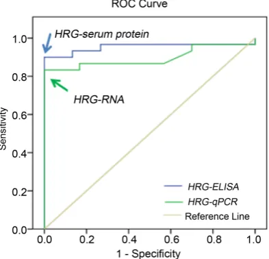
pg/ml with 90.0% sensitivity and 100.0% specificity, and for HRG-RNA was 0.955 with 83.3% sensitivity and 100% specificity (p < 0.01), Figure 2 and Table 2. Moreover, HRG positivity rates detected by both methods was 100% in ALL
Table 1. Clinicopathological parameters of ALL patients.
Parameters Mean ± SD Median Range
Age (years) 38.3 ± 8.3 35.0 17.0 - 65.0
TLC (1000/mm3) 8.3 ± 5.7 6.7 4.4 - 20.0
HB % 8.6 ± 2.3 7.0 6.0 - 14.7
Platelet count (1000/mm3) 69.8 ± 66 45.0 6.0 - 21.1
LDH (IU/L) 714.7 ± 428.2 961.5 312.0 - 1561.0
BM blast cells (%) 78.7 ± 21.8 88.0 32.0 - 98.0
Peripheral blast cells (%) 87.7 ± 29.3 58.0 23.0 - 95.0
Figure 1. Serum HRG mean level detected by ELISA method (a) and qPCR (b) in ALL
[image:6.595.206.541.149.431.2]patients before and after therapy and control groups. (p1 = after versus before therapy, p2 = normal group versus ALL before therapy and p3 = normal group versus ALL after thera-py **P < 0.01 is highly significant. P > 0.05 is non-significant).
Figure 2. ROC curves for serum HRG-serum protein and HRG-RNA to discriminate
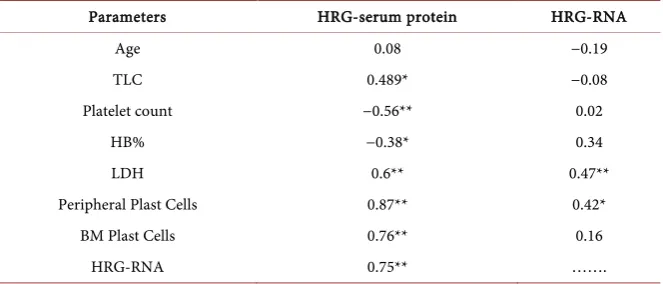
[image:6.595.276.473.501.690.2]patients before therapy and 0.0% in the normal group, while in follow-up group it was 76.0% for in HRG-serum protein and 60.0% for HRG-RNA (P < 0.001), Figure 3. No significant differences between ALL subtypes and HRG, however, the HRG protein level was significantly higher in AAL patients with +ve Phila-delphia chromosome (P < 0.01) and both HRG protein and RNA protein were significantly high in ALL patients who died during the course of treatment compared to those had complete remission (P < 0.01), Table 3. Furthermore, HRG protein showed a significant negative correlation with platelets (P < 0.01), HB% (P < 0.05) and significant positive correlations with its expression level, LDH, BM blast cells and peripheral blast cells (P < 0.01), while HRG-RNA showed significant positive correlations only with LDH (<0.05) and peripheral blast cells (P < 0.01), Table 4.
4. Discussion
[image:7.595.135.538.392.695.2]Histidine-rich glycoprotein (HRG) is a multifunctional plasma protein with two cystatin-like domains and a wide spectrum of targets and functions [16]. Exten-sive research studied the potential role of HRG in carcinogenesis, however, its effect on overall survival remains to be clearly determined. In the current study,
Table 2. Diagnostic performance of serum HRG-serum protein and HRG-RNA from
pa-tients with acute lymphoblastic leukemia and normal control group.
Variable Cut-off points Sensitivity Specificity NPV PPV AUC HRG-serum protein 63.55 pg/ml 90.0% 100% 83.3% 100% 0.953
[image:7.595.139.537.394.694.2]HRG-RNA 0.955 83.3% 100% 75.0% 100% 0.896
Figure 3. Positivity rate of patients of serum HRG-serum protein (a) and HRG-RNA (b) above the cutoff
Table 3. Relation between mean rank of HRG serum level and RNA expression with cli-nicopathological factors in the ALL patients.
Clinicopathological factors HRG serum level HRG-RNA Expression
Type of ALL; Pre-B-ALL (23) B-ALL (5) T-ALL (7) 19.24 18.5 15.2 χ2: 1.06
p: 0.5
17.74 20.5 17.55 χ2: 0.2
p: 0.8 Chromosomal abnormalities;
Normal (23) +ve Philadelphia chromosome
(12)
13.83 26.0 χ2: 11.1 *p: 0.001
16.28 21.29 χ2: 1.89
p: 0.16
ALL patients Complete remission (25)
Dead patients (10)
13.48 29.30 χ2: 17.0 *p: 0.001
13.16 30.1 χ2: 19.5 *p: 0.001
P* Significant difference between types by applying non parametric Mann-Whitney U (to compare Chro-mosomal abnormalities) and Kruskal-Wallis tests (to compare Types of ALL).
Table 4. Pearson correlation between HRG-ELISA and HRG-qPCR and other parameters.
Parameters HRG-serum protein HRG-RNA
Age 0.08 −0.19
TLC 0.489* −0.08
Platelet count −0.56** 0.02
HB% −0.38* 0.34
LDH 0.6** 0.47**
Peripheral Plast Cells 0.87** 0.42*
BM Plast Cells 0.76** 0.16
HRG-RNA 0.75** …….
*P value < 0.05 is significant, **P value < 0.01 is highly significant.
[image:8.595.208.540.335.477.2]demon-strated that HRG expression was increased by glioma cells in both subcutaneous and orthotopic brain tumor models resulted in an increase in tumor size and an-giogenesis, possibly through interfering with the antiangiogenic activity of vas-culostatin. On the other hand, HRG has been found to be reduced in the serum of alpha fetoprotein-negative hepatitis B virus-related hepatocellular carcinoma [19] and down regulated in endometrial carcinoma [20]. Moreover, Wu et al. [21] reported that fucosylated HRG levels were significantly higher in patients with stage III ovarian cancer compared to normal and benign donors but was not significantly higher in patients with stage I/II disease. HRG’s antitumor ac-tivity has been ascribed due to its immune modulator functions and its effects on tumor vessels. The contradiction of the previous reports about pro- and antian-giogenic properties of HRG might be owing to its multi-domain structure and the activities of its proteolytically-released fragments, notably the histi-dine-proline rich region [22] and different experimental systems used [23]. Rol-ny et al. [11] assumed that HRG affects other cell types, such as macrophages, known to regulate angiogenesis and HRG might regulate tumor angiogenesis in-directly through tumor-associated macrophages.
HRG level was still high in follow-up ALL patients despite their complete re-mission (blast cells were around 5% and the rest of cells were normal), that had a statistically significant difference between HRG-serum protein levels after ther-apy and normal subjects. We previously explained it probably by persistence of residual tumor cells leading to relapse if no further additional consolidation chemotherapy was administered. This may highlight the molecular biolo-gy-based methods with a greater prognostic significance than conventional cri-teria for the detection of remission [24] [25]. Nagafuji et al. [26] reported that chemotherapy should be continued as follow up therapy in ALL even with good prognosis in order to prevent leukemia relapse. However, patients with less fa-vorable prognosis should be treated more aggressively. Although allogeneic he-matopoietic stem cell transplantation (HSCT) for patients with ALL in complete remision is much more intensive than multi-agent combined chemotherapy, it is associated with increased morbidity and mortality when compared with such chemotherapy. Minimal residual disease (MRD) status has been proven to be a strong prognostic factor for adult patients with Ph-negative ALL.
patients with pancreatic cancer. However, they explained this weak positivity of HRG as a prognostic marker due to its anti-angiogenic properties as the patients in trial phase were receiving anti-VEGF monoclonal antibody. Moreover, Zhang et al. [28] suggested the negative regulatory role HRG on hepatocellular carci-noma cell line through regulating cell proliferation via the Erk1/2 signaling pathway. Mantovani and Sica [29] revealed the antitumor activity of HRG as it is not only increased tumor infiltration by antigen-presenting DCs, cytolytic NK cells, and cytotoxic T-lymphocytes but also enhances their antigen presentation and tumor cell lysis potential, immune changes known to inhibit tumor growth. The possible, increasing levels of HRG in malignant cases might be due to the recognition of ‘‘malignant danger’’, in line with its presumed role as a ‘‘pattern recognition molecule’’ [16]. We could not find any literature evaluating serum HRG protein or its expression levels in ALL.
5. Conclusion
All these findings indicated that HRG might be a novel diagnostic biomarker in ALL patients, with high sensitivity and specificity. The more significant positive correlations of HRG-serum protein over HRG-RNA and its easy method of ap-plication, motivate its application in large clinical studies as a potential prognos-tic marker.
Acknowledgements
Our thanks are due to all patients and healthy subjects involved in this study and to Clinical Hematology Unit, Faculty of Medicine, Ain Sham University for pro-viding us the samples. This study was supported by grant No; 3380, 2015 from the Deanship of scientific research, Qassim University, Saudi Arabia.
Conflict of Interest
The authors declare that they have no competing interests.
References
[1] Faderl, S., O’Brien, S., Pui, C., Stock, W., Wetzler, M., Hoelzer, D., et al. (2010) Adult Acute Lymphoblastic Leukemia Concepts and Strategies. Cancer, 1165-1176.
https://doi.org/10.1002/cncr.24862
[2] Zhao, Y., Huang, H. and Wei, G. (2013) Novel Agents and Biomarkers for Acute Lymphoid Leukemia. Journal of Hematology & Oncology, 6, 40.
https://doi.org/10.1186/1756-8722-6-40
[3] Faderl, S., O’Brien, S., Pui, C.H., Stock, W., Wetzler, M., Hoelzer, D., et al. (2010) Adult Acute Lymphoblastic Leukemia: Concepts and Strategies. Cancer, 116, 1165- 1176. https://doi.org/10.1002/cncr.24862
[4] Jones, A.L., Hulett, M.D. and Parish, C.R. (2005) Histidine-Rich Glycoprotein: A Novel Adaptor Protein in Plasma That Modulates the Immune, Vascular and Coa-gulation Systems.Immunology & Cell Biology, 83, 106-118.
https://doi.org/10.1111/j.1440-1711.2005.01320.x
Con-formation of Histidine-Proline-Rich Glycoprotein. Biochemistry, 35, 1925-1934.
https://doi.org/10.1021/bi952061t
[6] Tugues, S., Roche, F., Noguer, O., Orlova, A., Bhoi, S., Padhan, N., et al. (2014) His-tidine-Rich Glycoprotein Uptake and Turnover Is Mediated by Mononuclear Pha-gocytes. PLoS ONE, 9, e107483. https://doi.org/10.1371/journal.pone.0107483
[7] Johnson, L., Goubran, H. and Kotb, R. (2014) Histidine Rich Glycoprotein and Cancer: A Multi-faceted Relationship. Anticancer Research, 34, 593-603.
[8] Simantov, R., Febbraio, M. and Silverstein, R.L. (2005) Theantiangiogenic Effect of Thrombospondin-2 Is Mediated by CD36 and Modulated by Histidine-Rich Glyco-protein. Matrix Biology, 24, 27-34. https://doi.org/10.1016/j.matbio.2004.11.005
[9] Juarez, J.C., Guan, X., Shipulina, N.V., Plunkett, M.L. Parry, G.C., Elliot Shaw, D.E.,
et al. (2002) Histidine-Proline-Rich Glycoprotein Has Potent Antiangiogenic
Activ-ity Mediated through the Histidine-Proline-Rich Domain.Cancer Research, 62, 5344- 5350.
[10] Kärrlander, M., Lindberg, N., Olofsson, T., Kastemar, M., Olsson, A.K. and Uhr-bom, L. (2009) Histidine-Rich Glycoprotein Can Prevent Development of Mouse Experimental Glioblastoma. PLoS ONE, 4, e8536.
https://doi.org/10.1371/journal.pone.0008536
[11] Rolny, C., Mazzone, M., Tugues, S., Laoui, D., Johansson, I., Coulon, C., et al. (2011) HRG Inhibits Tumor Growth and Metastasis by Inducing Macrophage Pola-rization and Vessel Normalization through Down-Regulation of PlGF. Cancer Cell, 19, 31-44. https://doi.org/10.1016/j.ccr.2010.11.009
[12] Thulin, A., Ringvall, M., Dimberg, A., Karehed, K., Vaisanen, T., Vaisanen, M.R., et al. (2009) Activated Platelets Provide a Functional Microenvironment for the Anti-angiogenic Fragment of Histidine-Rich Glycoprotein. Molecular Cancer Research, 7, 1792-1802. https://doi.org/10.1158/1541-7786.MCR-09-0094
[13] Bennettt, J.M., Catovsky, D. and Daniel, M.T. (1976) Proposals for the Classification of the Acute Leukaemias. French-American-British (FAB) Co-Operative Group.
British Journal of Haematology, 3, 451-458.
https://doi.org/10.1111/j.1365-2141.1976.tb03563.x
[14] Thomas, D.A., Faderl, S., Cortes, J., O’Brien, S., Giles, F.J., Kornblau, S.M., et al. (2004) Treatment of Philadelphia Chromosome-Positive Acute Lymphocytic Leu-kemia with Hyper-CVAD and Imatinibmesylate. Blood, 103, 4396-4407.
https://doi.org/10.1182/blood-2003-08-2958
[15] Gokbuget, N., Kneba, M., Raff, T., Trautmann, H., Bartram, C.R., Arnold, R., et al.
(2012) Adult Patients with Acute Lymphoblastic Leukemia and Molecular Failure Display a Poor Prognosis and Are Candidates for Stem Cell Transplantation and Targeted Therapies. Blood, 120, 1868-1876.
[16] Poon, I.K., Hulett, M.D. and Parish, C.R. (2010) Histidine-Rich Glycoprotein Is a Novel Plasma Pattern Recognition Molecule That Recruits IgG to Facilitate Necrot-ic Cell Clearance via Fcgammari on Phagocytes. Blood, 115, 2473-2482.
https://doi.org/10.1182/blood-2009-07-234013
[17] Matboli, M., Eissa, E. and Said, H. (2014) Evaluation of Histidine-Rich Glycoprotein Tissue RNA and Serum Protein as Novel Markers for Breast Cancer. Medical
On-cology, 31, 897. https://doi.org/10.1007/s12032-014-0897-4
[18] Klenotic, P.A., Huang, P. and Palomo, J. (2010) Histidine-Rich Glycoprotein Mod-ulates the Anti-Angiogenic Effects of Vasculostatin. American Journal of Pathology, 176, 2039-2050. https://doi.org/10.2353/ajpath.2010.090782
Carcinoma Using iTRAQ-MALDI-MS/MS. Neoplasma, 61, 17-26.
https://doi.org/10.4149/neo_2014_001
[20] Wang, Y.S., Cao, R., Jin, H., Huang, Y.P., Zhang, X.Y., Congm, Q., et al. (2011) Al-tered Protein Expression in Serum from Endometrial Hyperplasia and Carcinoma Patients. Journal of Hematology & Oncology, 4, 15.
https://doi.org/10.1186/1756-8722-4-15
[21] Wu, J., Xie, X., Liu, Y., He, J., Benitez, R., Buckanovich, R.J., et al. (2012) Identifica-tion and ConfirmaIdentifica-tion of Differentially Expressed Fucosylated Glycoproteins in the Serum of Ovarian Cancer Patients Using a Lectin Array and LC-MS/MS. Journal of
Proteome Research, 11, 4541-4552. https://doi.org/10.1021/pr300330z
[22] Lee, C., Bongcam-Rudloff, E., Sollner, C., Jahnen-Dechent, W. and Claesson-Welsh, L. (2009) Type 3 Cystatins, Fetuins, Kininogen and Histidine-Rich Glycoprotein.
Frontiers in Bioscience, 14, 2911-2922. https://doi.org/10.2741/3422
[23] Poon, I.K., Patel, K.K., Davis, D.S., Parish, C.R. and Hulett, M.D. (2011) Histi-dine-Rich Glycoprotein: The Swiss Army Knife of Mammalian Plasma. Blood, 117, 2093-2101. https://doi.org/10.1182/blood-2010-09-303842
[24] Ahmed, M.B., Shehata, H.H., Moussa, M. and Ibrahim, T.M. (2012) Prognostic Sig-nificance of Survivin and Tumor Necrosis Factor-Alpha in Adult Acute Lymphob-lastic Leukemia. Clinical Biochemistry, 45, 112-126.
https://doi.org/10.1016/j.clinbiochem.2011.08.1147
[25] Alkhouly, N., Shehata, I., Ahmed, M.B., Shehata, H., Hassan, S. and Ibrahim, T. (2013) HLA-G Expression in Acute Lymphoblastic Leukemia: A Significant Prog-nostic Tumor Biomarker. Medical Oncology, 30, 460.
https://doi.org/10.1007/s12032-013-0460-8
[26] Nagafuji, K., Miyamoto, T., Eto, T., Kamimura, O., Taniguchi, S., Okamura, T., et al. (2013) Monitoring of Minimal Residual Disease (MRD) Is Useful to Predict Prognosis of Adult Patients with Ph-Negative ALL: Results of a Prospective Study (ALL MRD2002 Study). Journal of Hematology & Oncology, 6, 14.
https://doi.org/10.1186/1756-8722-6-14
[27] Roberts, A.S., Campa, M.J., Gottlin, E.B., Jiang, C., Owzar, K., Kindler, H.L., et al. (2012) Identification of Potential Prognostic Biomarkers in Patients with Untreated, Advanced Pancreatic Cancer from a Phase 3 Trial (Cancer and Leukemia Group B 80303). Cancer, 118, 571-578. https://doi.org/10.1002/cncr.26270
[28] Zhang, Q., Jiang, K., Li, Y., Gao, D., Sun, L., Zhang, S., et al. (2015) Histidine-Rich Glycoprotein Function in Hepatocellular Carcinoma Depends on Its N-Glycosyla- tion Status, and It Regulates Cell Proliferation by Inhibiting Erk1/2 Phosphoryla-tion. Oncotarget, 6, 30222-30231.
[29] Mantovani, A. and Sica, A. (2010) Macrophages, Innate Immunity and Cancer: Balance, Tolerance, and Diversity. Current Opinion in Immunology, 22, 231-237.
Abbreviations
ALL Acute lymphoblastic leukemia AUC Area under ROC curve BM Bone marrow
ELISA Enzyme-linked immunosorbent assay HRG Histidine-rich glycoprotein
LD Lactate dehydrogenase NPV Negative predictive value PPV Positive predictive value
qPCR Real-time polymerase chain reaction ROC curve Receiver operating characteristic curve. TLC Total leukocyte count
Submit or recommend next manuscript to SCIRP and we will provide best service for you:
Accepting pre-submission inquiries through Email, Facebook, LinkedIn, Twitter, etc. A wide selection of journals (inclusive of 9 subjects, more than 200 journals)
Providing 24-hour high-quality service User-friendly online submission system Fair and swift peer-review system
Efficient typesetting and proofreading procedure
Display of the result of downloads and visits, as well as the number of cited articles Maximum dissemination of your research work
Submit your manuscript at: http://papersubmission.scirp.org/