R E S E A R C H A R T I C L E
Open Access
The Sp1 transcription factor is essential for the
expression of gliostatin/thymidine phosphorylase
in rheumatoid fibroblast-like synoviocytes
Kenji Ikuta
1, Yuko Waguri-Nagaya
1*, Kae Kikuchi
1, Takaya Yamagami
1, Masahiro Nozaki
1, Mineyoshi Aoyama
2,
Kiyofumi Asai
2and Takanobu Otsuka
1Abstract
Introduction:Gliostatin/thymidine phosphorylase (GLS/TP) has angiogenic and arthritogenic activities, and aberrant GLS production has been observed in the active synovial membranes of rheumatoid arthritis (RA) patients. The human GLS gene promoter contains at least seven consensus binding sites for the DNA binding protein Sp1. Here we examined whether Sp1 is necessary for GLS production in RA. We also studied the effects of the Sp1 inhibitor mithramycin on GLS production in RA fibroblast-like synoviocytes (FLSs).
Methods:FLSs from RA patients were treated with specific inhibitors. The gene and protein expression of GLS were studied using the quantitative reverse-transcription polymerase chain reaction (qRT-PCR) and an enzyme immunoassay. Intracellular signalling pathway activation was determined by western blotting analysis, a luciferase assay, a chromatin immunoprecipitation (ChIP) assay and a small interfering RNA (siRNA) transfection.
Results:The luciferase and ChIP assays showed that Sp1 binding sites in the GLS promoter were essential for GLS messenger RNA (mRNA) expression. GLS production was suppressed in FLSs by siRNA against Sp1 transfection. Mithramycin decreased GLS promoter activity, mRNA and protein expression in FLSs. Tumour necrosis factor-a (TNF-a) significantly increased GLS expression in RA FLSs; this effect was reduced by pre-treatment with cycloheximide and mithramycin.
Conclusions:Pretreatment of mithramycin and Sp1 silencing resulted in a significant suppression of GLS production in TNF-a-stimulated FLSs compared to controls. GLS gene expression enhanced by TNF-a was partly mediated through Sp1. As physiological concentrations of mithramycin can regulate GLS production in RA, mithramycin is a promising candidate for anti-rheumatic therapy.
Introduction
Many components of the immune system contribute to the development and progression of rheumatoid arthritis (RA), and angiogenesis is considered a critical step in its initiation and progression. Levels of inflammatory cyto-kines such as tumor necrosis factor-alpha (TNF-a) and interleukin-1-beta (IL-1b) and IL-6 [1-3] are increased in arthritic joints, whereas high levels of angiogenic fac-tors such as vascular endothelial growth factor (VEGF) [4,5] and gliostatin (GLS) [6,7] have been reported in
synovial fluid and sera from patients with RA. For this reason, the past decade has seen the development of biological treatments for RA, such as TNF-ainhibitors.
GLS is thought to be similar to thymidine phosphory-lase (TP) and platelet-derived endothelial cell growth fac-tor (PD-ECGF) [8] and induces angiogenesis through the proliferation and chemotactic migration of endothelial cells [9-11]. It also inhibits the growth of glial cells [12], promotes glial cell differentiation, and has neurotrophic effects on cortical neurons [13]. GLS production by cul-tured RA fibroblast-like synoviocytes (FLSs) was shown to be enhanced by TNF-a[14], IL-1b[15], and GLS itself [16]. GLS was also found to induce VEGF expression in FLSs [14], and we reported that direct injection of GLS * Correspondence: waguri@med.nagoya-cu.ac.jp
1Department of Orthopedic Surgery, Nagoya City University Graduate School
of Medical Sciences, 1 Kawasumi, Mizuho-cho, MuzuhoMizuho-ku, Nagoya, 467-8601, Japan
Full list of author information is available at the end of the article
into rabbit knees led to pronounced RA-like synovitis [17]. Inhibition of GLS is therefore regarded as an impor-tant approach in reducing damage to RA tissues.
Analysis of the TP promoter region revealed several potential zinc finger-type transcription factor Sp1-binding sites, including those in numerous housekeeping and inducible genes [18], in the interferon-stimulated response element (ISRE), and in the gamma-activated sequence [19]. In the present study, we used a luciferase assay and a small interfering RNA (siRNA) against Sp1 to show that the Sp1 box is essential for GLS production. We also used the inhibitor mithramycin to demonstrate the effect of Sp1-binding inhibition on GLS expression. Mithramycin is an aureolic acid anti-neoplastic antibiotic used for treating cancer-related hypercalcemia, leukemia [20], and testicular cancer [21] and prevents Sp1 from binding to its cognate site in DNA by modifying CG sequences [22]. Here, we show that mithramycin potently suppresses GLS induction through the transduction of Sp1 in RA FLSs.
Materials and methods
Preparation of human fibroblast-like synoviocytes
This study was approved by the ethics committee of Nagoya City University (Nagoya City, Japan). Before parti-cipation, informed consent was obtained from all subjects in accordance with the Declaration of Helsinki. FLSs were cultured from the synovial tissues of 10 patients who had RA, who were undergoing total knee arthroplasty, and who met the American Rheumatism Association 1987 revised criteria for the classification of RA [23], as pre-viously described [24-26]. The clinical characteristics of these patients are shown in Table 1. FLSs were maintained
in Dulbecco’s modified Eagle’s medium supplemented
with penicillin (100 units per mL), streptomycin (100μg/
mL), and 10% fetal bovine serum at 37°C in a 5% CO2
atmosphere. The cultures were found to be completely free of lymphoid and monocytic cells. Cells were allowed to adhere overnight, and then non-adherent cells were removed and adherent FLSs were split at a 1:3 ratio when they reached 70% to 80% confluency. FLSs were used between passages 3 and 9, during which time they were a homogeneous population of cells.
Luciferase assay
The 1,243-base pair (bp) GLS/TP promoter fragment was a kind gift from Mayumi Ono (Department of Pharmaceu-tical Oncology, Kyushu University, Fukuoka, Japan) [19]. The fragment was cloned into luciferase gene Basic Vector 2 (NIPPON GENE CO., LTD, Tokyo, Japan) and digested
withXhoI andHindIII. This reporter construct was
desig-nated pTP-Luc1. To prepare other deletion constructs, pTP-Luc1 was digested with different restriction enzymes, includingXhoI,StuI,SacI, andKpnI. Both 5’overhangs and 3’overhangs of the digested product were blunted by T4 DNA polymerase (Takara Bio Inc., Otsu, Japan) and self-ligated by T4 DNA ligase (Takara Bio Inc.) to produce pGLS/TP I, pGLS/TP II, and pGLS/TP III (Figure 1).
FLSs were seeded into a 48-well culture plate (5 × 10³ cells per well) and incubated at 37°C in a humidified
atmo-sphere of 5% CO2/air for 1 week. They were then
co-transfected with luciferase reporter vector (400 ng/well) and pRL-SV40 internal control construct (10 ng/well) (Promega Corporation, Madison, WI, USA) by using
Lipo-fectamine™ LTX (0.5 μL/well) and Plus™ Reagent
(0.5μL/well) (Invitrogen Corporation, Life Technologies Corporation, Carlsbad, CA, USA). The medium was replaced after 12 hours, and FLSs were treated with 100 or 300 nM mithramycin (Sigma-Aldrich, St. Louis, MO, USA) for 24 hours. Cells were harvested by using passive lysis buffer (65μL/well), and the luciferase activity was measured by using a Dual-Luciferase Assay System (Pro-mega Corporation) and normalized to the internal control.
Chromatin immunoprecipitation assays
Chromatin immunoprecipitation (ChIP) assays were
per-formed by using ChIP-IT™Express kits (Active Motif,
[image:2.595.57.290.509.705.2]Carlsbad, CA, USA) in accordance with a reported proto-col [27] with minor modifications. In brief, 1.0 × 10⁶FLSs cells from 60-mm dishes were treated with or without 300 nM mithramycin for 24 hours and then harvested. Cells were cross-linked by formaldehyde (1% final concen-tration) and incubated at room temperature for 10 min-utes. Cells were then washed with ice-cold phosphate-buffered saline (PBS) containing protease inhibitors, and the fixation reaction was stopped by adding 10 mL of Gly-cine Stop-Fix Solution (Active Motif). Samples were lysed for 30 minutes in lysis buffer on ice, and the chromatin
Table 1 Clinical characteristics of patients
Characteristic n = 10
Gender, female/male 7/3
Age in years, range (mean) 48-74 (65.0)
Disease duration in years, range (mean) 1-18 (10.6)
CRP in mg/dL, range (mean) 0.05-3.34 (0.66)
ESR in mm/hour, range (mean) 1-83 (20.8)
Rheumatoid factor, positive/negative 9/1
Anti-CCP antibody in U/mL, range (mean) 0.6-100 (37.8)
MMP-3 in ng/mL, range (mean) 38.9-525.7 (222)
Steinbrocker stage, I/II/III/IV 0/5/3/2
Patients using DMARDs and biologics
Methotrexate 7
Infliximab 1
Etanercept 2
FK506 2
Patients using oral steroids 5
was sheared by sonicating 20 times for 40 seconds each at maximum power with an ultrasonic processor, including 30 seconds of cooling on ice between each pulse (Misonix, Inc., Farmingdale, NY, USA). Cross-linked and released chromatin fractions were immunoprecipitated with mag-netic beads, Sp1 antibodies, and non-specific IgG (rabbit) on a rolling shaker overnight at 4°C. Cross-linking of the immunoprecipitates containing fragmented DNA was che-mically reversed, and the polymerase chain reaction (PCR) was performed with MESA GREEN qPCR MasterMix Plus for SYBR Assay I Low ROX (Eurogentec, San Diego, CA, USA). The PCR primers used for amplifying promoters containing the Sp1-binding site furthest upstream were 5’
-AACTGTGGGCCTTCCCACTC-3’and 5’-TGCTGAGGT
CCTCGAAGAAAC-3’, which produced a 227-bp
frag-ment (Figure 2a).
Western blot analysis for Sp1
[image:3.595.57.541.89.249.2]FLSs were incubated in 60-mm dishes with 1 ng/mL TNF-a(R&D Systems, Minneapolis, MN, USA) in the presence or absence of mithramycin. To prepare nuclear extracts, cells were rinsed in ice-cold PBS and lysed in 10 mM Tris-HCl (pH 7.5), 1 mM ethylenediaminetetraacetic acid (EDTA), 0.5% Nonidet P-40, and a protease inhibitor cocktail (Sigma-Aldrich) for 10 minutes at 4°C. Cell lysates were centrifuged at 20,000gfor 10 minutes at 4°C to sepa-rate the cytoplasmic fraction (supernatant). Insoluble materials were dissolved in sodium dodecyl sulphate sam-ple buffer to collect the nuclear extracts. After measuring
Figure 1Effects of mithramycin on gliostatin/thymidine phosphorylase (GLS/TP) promoter activity based on deletion constructs. Closed ovals indicate Sp1-binding sites. Open ovals indicate interferon-stimulated response element (ISRE) and gamma-activated sequence (GAS) sequences. Fibroblast-like synoviocytes were transiently transfected with deletion constructs and then incubated with mithramycin (100 nM, shaded column, or 300 nM, closed column) for 24 hours. Control cells were incubated without mithramycin (open column). Data were normalized by measuring the luminescent reaction of the internal control. Results are presented as mean ± standard error of the mean of five determinations. Statistical significance was calculated by using the Mann-WhitneyUtest: compared with samples without mithramycin *P< 0.05, **P< 0.01; compared with samples with pTP-Luc1 without mithramycin†P< 0.01.
[image:3.595.305.539.382.593.2]the protein concentration, samples (20μg of total protein/ lane) were loaded and separated by electrophoresis on a 7.5% polyacrylamide gel (Bio-Rad Laboratories, Hercules, CA, USA) and then transferred to a polyvinylidene difluor-ide membrane (Immobilin-P; EMD Millipore, Billerica, MA, USA). The blots were blocked for 60 minutes with 5% non-fat dry milk in Tris-buffered saline containing 0.1% Tween 20 (TBS-T) and then incubated with an anti-Sp1 antibody (1:1,000; Cell Signaling Technology, Inc., Danvers, MA, USA), anti-lamin C (1:1,000; ImmuQuest Ltd, Seamer, North Yorkshire, UK), and anti-a-tubulin antibody (1:1,000; Cell Signaling Technology, Inc.) as load-ing controls in TBS-T for 2 hours at room temperature. After three washes with TBS-T, membranes were incu-bated with appropriate horseradish peroxidase-conjugated secondary antibodies in TBS-T (1:3,000; GE Healthcare, Little Chalfont, Buckinghamshire, UK) for 1 hour at room temperature and washed three times with TBS-T. Protein bands were detected by using the ECL Plus Western Blotting Detection System (GE Healthcare) and then quantified with a densitometric scanner by using ImageJ software [28].
Reverse transcription-polymerase chain reaction assay
Expression of the GLS gene was assessed by using reverse transcription-PCR (RT-PCR). Total RNA was isolated by using TRIzol reagent (Invitrogen Corporation), and RT was carried out by using random primers and Ready-To-Go You-Prime First-Strand Beads (GE Healthcare). The resultant cDNA was subjected to real-time PCR by using a 7500 Fast Real-time PCR System (Life Technologies Cor-poration) with MESA GREEN qPCR MasterMix Plus for SYBR Assay I Low ROX (Eurogentec) and primers. The PCR conditions were an initial denaturation at 95°C for 10 minutes, followed by 40 cycles at 95°C for 5 seconds and 60°C for 1 minute. The relative quantification value of
GLS was normalized to an endogenous control,b-actin,
after confirming that the GLS andb-actin cDNAs were
amplified with the same efficiency. The primers were as
follows: GLS, 5’-ACAGGAGGCACCTTGGATAA-3’and
5’-CCGAACTTAACGTCCACCAC-3’(272 bp), andb
-actin, 5’-GACCTGACTGACTACCTCAT-3’ and 5’
-TCGTCATACTCCTGCTTGCT-3’(542 bp).
RNA interference
Sp1 Stealth siRNAs and negative control siRNA were pur-chased from Invitrogen Corporation. The following Sp1
siRNA oligos were used: sense sequence 5’
-GACAGGU-CAGUUGGCAGACUCUACA-3’and anti-sense sequence
5’-UGUAGAGUCUGCCAACUGACCUGUC-3’. FLSs
were transfected with the indicated combinations of siRNA against Sp1 or negative control siRNA at a final concentration of 20 nM by using Lipofectamine
RNAiMAX transfection reagent (Invitrogen Corporation) in accordance with the recommendations of the manufac-turer. FLSs were harvested for Western blotting 48 hours after transfection. FLSs were incubated for 24 hours after
transfection and further incubated with 1 ng/mL TNF-a
for 24 hours and harvested for RT-PCR.
Enzyme immunoassay for gliostatin
GLS was measured by using an enzyme immunoassay system, as described by Hirano and colleagues [29]. Polyclonal antibodies on the solid phase were obtained
by immunizing New Zealand albino rabbits with 40 μg
of purified natural human GLS, and the monoclonal antibody was used with the b-galactosidase-labeled sec-ondary antibody. The detection limit of this assay was 150 pg/mL, and no significant cross-reactivity or inter-ference was observed. The GLS concentration in cul-tured FLSs was normalized to the protein content, as measured by using a Pierce BCA protein assay kit (Thermo Fisher Scientific, Waltham, MA, USA).
Immunocytochemistry
Confluent FLSs in chambered slides coated with BD Matrigel Matrix (BD Biosciences, Franklin Lakes, NJ, USA) were fixed for 30 minutes in 3% paraformaldehyde, permeabilized with 0.2% Triton X-100 for 5 minutes, washed with PBS, and blocked for 60 minutes at room temperature with blocking solution comprising 3% bovine serum albumin and 0.1% glycine in PBS. After washing, cells were labeled overnight at 4°C with the primary anti-body (1:1,000 anti-TP/GLS antianti-body; a gift from Chugai Pharmaceutical Co., Ltd., Tokyo, Japan). Cells were washed and labeled with an Alexa Flour 594-labeled (red) goat anti-mouse IgG (1:1,000; Molecular Probes Inc., now part of Invitrogen Corporation) secondary antibody. After washing, sections were mounted on glass slides containing ProLong Gold Antifade with 4’ ,6-diamidino-2-phenylin-dole (DAPI) (Invitrogen Corporation). Stained cells were visualized by using an AX70 fluorescence microscope (Olympus, Tokyo, Japan). Total red intensity of immunos-taining in nine random fields was quantified by ImageJ. The numbers of cells were counted in the fields. Data were presented as mean of intensity per cell.
Statistical analysis
Results
Mithramycin inhibited gliostatin gene expression
We prepared three deletion constructs of the GLS/TP promoter and fused them to the reporter luciferase plas-mid (Figure 1) to investigate whether mithramycin inhibited GLS/TP promoter activity. The promoter activity was expressed relative to the internal control. Treatment with mithramycin inhibited the promoter activity in RA FLSs transfected with pTP-Luc1 (55.5%, 100 nM mithramycin; 59.2%, 300 nM mithramycin), whereas promoter activity was measured at 41.8% in RA FLSs transfected with pGLS/TP I, relative to those transfected with pTP-Luc1. The observed inhibition was accelerated following treatment with mithramycin (38.2%, 100 nM mithramycin; 35.1%, 300 nM mithramy-cin) in RA FLSs transfected with pGLS/TP I. The pro-moter activity of pGLS/TP II and pGLS/TP III was largely inhibited with or without mithramycin.
Analysis of the gliostatin/thymidine phosphorylase promoter by using a chromatin immunoprecipitation assay
We performed a ChIP assay to confirm Sp1 binding to the promoter and examined the inhibitory effect of mithramycin on GLS gene expression. Sp1 was shown to bind to the furthest upstream Sp1-binding site of the GLS/TP promoter fragment (1,243 bp). Treatment with mithramycin suppressed binding at this site (Figure 2b). No amplification band was observed by using the non-specific rabbit IgG, indicating the non-specificity of the DNA immunoprecipitation and ChIP assays.
Nuclear accumulation of Sp1 in response to TNF-aand mithramycin
To examine whether the nuclear localization of Sp1
changed following treatment with TNF-aor
mithramy-cin, we prepared nuclear extracts from FLSs treated with or without 300 nM mithramycin for 30 minutes, further
incubated with 1 ng/mL TNF-afor 24 hours, and then
subjected to immunoblotting with anti-Sp1, anti-lamin C, and anti-a-tubulin antibodies. TNF-atreatment led to a remarkable accumulation of Sp1 in nuclear extracts, which was inhibited by treatment with mithramycin (Fig-ure 3). The nuclear extracts were stained by the anti-lamin C antibody as a nuclear anti-lamin marker but not by the anti-a-tubulin antibody as a cytoplasmic marker.
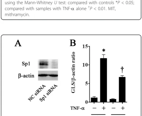
Inhibition of gliostatin expression by RNA interference
[image:5.595.305.539.86.282.2]To investigate the direct effect of Sp1 on GLS expression, we employed an siRNA against Sp1 for as an approach to inhibit GLS expression. The efficiency and specificity of siRNA gene knockdown of Sp1 were determined by Wes-tern blotting for Sp1 expression. Sp1 siRNA suppressed the Sp1 protein expression (Figure 4a). Sp1 siRNA
Figure 3Effect of tumor necrosis factor-alpha (TNF-a) and mithramycin on Sp1 protein expression in fibroblast-like synoviocyte nuclei. Fibroblast-like synoviocytes were cultured to confluence in the presence or absence of 300 nM mithramycin for 30 minutes and then further incubated with or without TNF-a(1 ng/mL) for 24 hours. Nuclear extracts were processed for
immunoblotting with an anti-Sp1 antibody. Anti-lamin C and anti-a -tubulin immunoblotting were included to assess the purities of nuclear and cytoplasmic fractions, respectively. Results are presented as mean ± standard error of the mean of five determinations. Statistical significance compared with controls was calculated by using the Mann-WhitneyUtest: compared with controls *P< 0.05; compared with samples with TNF-aalone†P< 0.01. MIT, mithramycin.
[image:5.595.303.540.400.594.2]reduced TNF-a-induced GLS gene expression by 56.7% at 48 hours post-transfection (Figure 4b).
Effects of mithramycin on gliostatin production stimulated by TNF-ain cultured fibroblast-like synoviocytes
FLSs were treated in the presence or absence of 10 to 300 nM mithramycin for 30 minutes and then further
incubated with 1 ng/mL TNF-afor 24 hours. GLS mRNA
(Figure 5a) and protein (Figure 5b) levels were significantly
induced by treatment with TNF-aalone (11.3-fold and
2.1-fold compared with the control, respectively), and
these inductions were suppressed by mithramycin treat-ment in a dose-dependent manner. GLS was not induced by treatment with mithramycin alone. We confirmed the non-toxic concentration at least 48 hours after incubation
with 10 to 300 nM mithramycin and 1 ng/mL TNF-aby
using WST-8 assays (Cell Counting Kit-8; Dojindo Laboratories, Kumamoto, Japan) (data not shown). GLS
mRNA expression increased in response to TNF-a(1 ng/
[image:6.595.57.539.252.642.2]mL) and reached a maximum at 24 hours (21.9-fold com-pared with the level at 0 hours) (Figure 5c). GLS protein expression increased in response to TNF-aand continued to rise until at least 48 hours after treatment (5.6-fold
compared with the level at 0 hours) (Figure 5d). These increases in GLS mRNA and protein were significantly suppressed by 300 nM mithramycin treatment. To
con-firm whether TNF-adirectly regulates GLS expression,
FLSs were pre-treated with the protein synthesis inhibitor,
cycloheximide. TNF-a-induced GLS mRNA production
significantly decreased in a dose-dependent manner with treatment of cycloheximide (Additional file 1).
FLSs were cultured to confluence in the presence or absence of 300 nM mithramycin for 30 minutes, followed by further incubation with or without 1 ng/mL TNF-afor 24 hours. We observed no morphological change in FLSs during immunocytochemical staining. FLSs were immu-nostained with GLS antibody (red). Staining was weakly diffuse in the cytoplasm of FLSs that had not undergone treatment (Figure 6a). Treatment with mithramycin alone had no effect on GLS staining. The expression of GLS pro-tein was significantly induced by treatment with TNF-a alone (1.5-fold compared with the control). This induction was significantly suppressed by mithramycin treatment
(0.6-fold compared with the sample induced by TNF-a
alone) (Figure 6b).
Discussion
TNF-ahas been identified as a crucial cytokine in the pathogenesis of RA [30,31] and is involved in the activa-tion of the nuclear factor-kappa-B (NF-B) transcription factor [32,33]. In the present study, no NF-B-binding sites were identified within the upstream GLS/TP promo-ter examined. It is therefore unlikely that TNF-adirectly modulates transcriptional GLS regulation through bind-ing the GLS/TP promoter. To determine the principal elements in the GLS/TP promoter which are relevant to GLS expression, we constructed a series of deletion mutant plasmids. Treatment with mithramycin inhibited GLS/TP promoter activity, confirming the potential for GLS production through Sp1 binding in FLSs.
Goto and colleagues [19] reported that
interferon-gamma (IFN-g) induced the expression of TP through
ISRE and gamma-activated sequence in human macro-phages. In the present study, the promoter activity was not
regulated by IFN-gin FLSs (data not shown); however,
[image:7.595.57.539.371.635.2]this discrepancy could be explained by differences in the cellular properties of macrophages and FLSs. In addition, luciferase and ChIP assays showed that the Sp1-binding
sites played an important role in regulating the promoter activity of the GLS gene and mithramycin exactly behaved as an Sp1 inhibitor. Other reports suggested that activated Sp1 is transported into the nucleus [34,35]; this was sup-ported by our analysis of FLS nuclear fractions by using Western blotting, which revealed a remarkable accumula-tion of Sp1 in the nuclei following TNF-atreatment that was inhibited by treatment with mithramycin. We further investigated whether Sp1 directly regulated expression of GLS. We used RNA interference to detect the effects of Sp1 depletion on GLS expression at the mRNA levels.
We confirmed that TNF-a-induced GLS mRNA and
protein production were inhibited by mithramycin in a dose-dependent manner. Our immunocytochemical stu-dies revealed that GLS was stained weakly and diffusely in FLS cytoplasm that had not undergone treatment. The intensity of GLS staining was increased by TNF-a treat-ment and suppressed by mithramycin at concentrations of 100 to 300 nM. These concentrations did not affect cell viability, as measured by a WST-8 assay, and anin vivostudy demonstrated that leukemia patients receiving
a 2-hour continuous infusion of 25μg/kg mithramycin
did not exceed plasma levels of 300 to 350 nM [36]. The protein synthesis inhibitor cycloheximide
signifi-cantly decreased TNF-a-induced GLS mRNA production
in a dose-dependent manner. GLS gene transcription might requirede novoprotein synthesis in FLSs stimulated by TNF-a, although the key protein involved remains to be identified.
In the pathogenesis of RA, we reported that GLS increased the expression of VEGF mRNA and protein in FLSs [14]. As angiogenesis is necessary for the perpetua-tion of inflammaperpetua-tion, inhibiperpetua-tion of angiogenic factors such as GLS and VEGF could provide a means to suppress the inflammatory cascade in RA synovitis [37]. We also reported that GLS induced the expression of matrix metal-loproteinase (MMP)-1 and MMP-3 and GLS itself in FLSs [16]. The MMP family of proteins, including MMP-1, MMP-3, and MMP-13, plays a crucial role in excessive cartilage degradation in RA [38,39]. Other reports indi-cated that the gene expression levels of VEGF in fibro-blasts [40,41] and of MMPs in articular chondrocytes [42] were closely related to that of Sp1. It was suggested, based on these findings, that an Sp1 inhibitor could be effective in reducing RA disease activity.
Conclusions
The present study indicates that expression of the GLS gene is mediated, in part, through the transcription fac-tor Sp1. Our data suggest that the beneficial effects of mithramycin in RA might be at least partly due to anti-angiogenic and anti-arthritogenic activity involving the downregulation of GLS. Mithramycin is therefore a pro-mising candidate anti-rheumatic drug.
Additional material
Additional file 1: Effect of cycloheximide on TNF-ainduced GLS mRNA production in FLSs. Confluent FLSs in 6-well plate were incubated in the presence or absence of 10-100 (μg/ml cycloheximide for 30 min, followed by further incubation with TNF-a(1 ng/ml) for 24 h. The GLS mRNA levels are expressed as a RT-PCR product ratio (GLS/b -actin). Results are presented as mean ± SEM of four determinations. Statistical significance compared with controls was calculated using the Mann-Whitney U-test: compared to controls *P< 0.01; compared to samples with TNF-aalone†P< 0.01.
Abbreviations
bp: base pair; ChIP: chromatin immunoprecipitation; FLS: fibroblast-like synoviocyte; GLS: gliostatin; IFN-γ: interferon-gamma; IL: interleukin; ISRE: interferon-stimulated response element; MMP: matrix metalloproteinase; NF-κB: nuclear factor-kappa-B; PBS: phosphate-buffered saline; PCR: polymerase chain reaction; RA: rheumatoid arthritis; RT: reverse transcription; RT-PCR: reverse transcription-polymerase chain reaction; siRNA: small interfering RNA; TBS-T: Tris-buffered saline containing 0.1% Tween 20; TNF-α: tumor necrosis factor-alpha; TP: thymidine phosphorylase; VEGF: vascular endothelial growth factor.
Acknowledgements
This research was supported by a Grant-in-Aid for Scientific Research (C) (23592225) from the Japan Society for the Promotion of Science. We thank Mieko Suzuki for her excellent technical assistance. We thank Mayumi Ono for the gift of the GLS/TP promoter fragment.
Author details
1Department of Orthopedic Surgery, Nagoya City University Graduate School
of Medical Sciences, 1 Kawasumi, Mizuho-cho, MuzuhoMizuho-ku, Nagoya, 467-8601, Japan.2Department of Molecular Neurobiology, Nagoya City
University Graduate School of Medical Sciences, Mizuho-Ku, 1 Kawasumi, Mizuho-cho, MuzuhoMizuho-ku, Nagoya, 467-8601, Japan.
Authors’contributions
KI and KK performed all the experiments, data analysis, and drafting of the manuscript. TY and MN participated in the interpretation of the data and performed the statistical analysis. MA and KA conceived of the study, participated in its design and coordination, and helped to draft the manuscript. YW-N conceived this study, provided financial support, designed experiments, interpreted the data, and drafted the manuscript. TO carried out administrative and financial support and helped to draft the manuscript. All authors read and approved the final manuscript.
Competing interests
The authors declare that they have no competing interests.
Received: 16 November 2011 Revised: 1 April 2012 Accepted: 25 April 2012 Published: 25 April 2012
References
1. Arend WP, Dayer JM:Cytokines and cytokine inhibitors or antagonists in rheumatoid arthritis.Arthritis Rheum1990,33:305-315.
2. Arend WP, Dayer JM:Inhibition of the production and effects of interleukin-1 and tumor necrosis factorαin rheumatoid arthritis.Arthritis Rheum1995,38:151-160.
3. Choy EHS, Panayi GS:Cytokine pathways and joint inflammation in rheumatoid arthritis.N Engl J Med2001,344:907-916.
4. Harada M, Mitsuyama K, Yoshida H, Sakisaka S, Taniguchi E, Kawaguchi T, Ariyoshi M, Saiki T, Sakamoto M, Nagata K, Sata M, Matsuo K, Tanikawa K:
Vascular endothelial growth factor in patients with rheumatoid arthritis. Scand J Rheumatol1998,27:377-380.
6. Asai K, Hirano T, Matsukawa K, Kusada J, Takeuchi M, Otsuka T, Matsui N, Kato T:High concentration of immunoreactive gliostatin/platelet-derived endothelial cell growth factor in synovial fluid and serum of rheumatoid arthritis.Clin Chim Acta1993,218:1-4.
7. Takeuchi M, Otsuka T, Matsui N, Asai K, Hirano T, Moriyama A, Isobe I, Eksioglu YZ, Matsukawa K, Kato T, Tada T:Aberrant production of gliostatin/platelet-derived endothelial cell growth factor in rheumatoid arthritis.Arthritis Rheum1994,37:662-672.
8. Asai K, Nakanishi K, Isobe I, Eksioglu YZ, Hirano A, Hama K, Miyamoto T, Kato T:Neurotrophic action of gliostatin on cortical neurons: identity of gliostatin and platelet-derived endothelial cell growth factor.J Biol Chem
1992,267:20311-20316.
9. Furukawa T, Yoshimura A, Sumizawa T, Haraguchi M, Akiyama S, Fukui K, Ishizawa M, Yamada Y:Angiogenic factor.Nature1992,356:668. 10. Moghaddam A, Zhang H-T, Fan T-PF, Hu DE, Lees VC, Turley H, Fox SB,
Gatter KC, Harris AL, Bicknell R:Thymidine phosphorylase is angiogenic and promotes tumor growth.Proc Natl Acad Sci USA1995,92:998-1002. 11. Miyadera K, Sumizawa T, Haraguchi M, Yoshida H, Konstanty W, Yamada Y,
Akiyama S:Role of thymidine phosphorylase activity in the angiogenic effect of platelet-derived endothelial cell growth factor/thymidine phosphorylase.Cancer Res1995,55:1687-1690.
12. Asai K, Hirano T, Kaneko S, Moriyama A, Nakanishi K, Isobe I, Eksioglu YZ, Kato T:A novel glial growth inhibitory factor, gliostatin, derived from neurofibroma.J Neurochem1992,59:307-317.
13. Ueki T, Nakanishi K, Asai K, Okouchi Y, Isobe I, Eksioglu YZ, Kato T, Kohno K:
Neurotrophic action of gliostatin on cocultured neuron with glial cells. Brain Res1993,622:299-302.
14. Tanikawa T, Waguri-Nagaya Y, Kusabe T, Aoyama M, Asai K, Otsuka T:
Gliostatin/thymidine phosphorylase-regulated vascular endothelial growth-factor production in human fibroblast-like synoviocytes. Rheumatol Int2007,27:553-559.
15. Waguri Y, Otsuka T, Sugimura I, Matsui N, Asai K, Moriyama A, Kato T:
Gliostatin/platelet-derived endothelial cell growth factor as a clinical marker of rheumatoid arthritis and its regulation in fibroblast like synoviocytes.Br J Rheumatol1997,36:315-321.
16. Muro H, Waguri-Nagaya Y, Mukofujiwara Y, Iwahashi T, Otsuka T, Matsui N, Moriyama A, Asai K, Kato T:Autocrine induction of gliostatin/platelet-derived endothelial cell growth factor (GLS/PD-ECGF) and GLS-induced expression of matrix metalloproteinases in rheumatoid arthritis synoviocytes.Rheumatol1999,38:1195-1202.
17. Waguri-Nagaya Y, Otsuka T, Sugimura I, Matsui N, Asai K, Nakajima K, Tada T, Akiyama S, Kato T:Synovial inflammation and hyperplasia induced by gliostatin/platelet-derived endothelial cell growth factor in rabbit knees.Rheumatol Int2000,20:13-19.
18. Black AR, Black JD, Azizkhan-Clifford J:Sp1 and kruppel-like factor family of transcription factors in cell growth regulation and cancer.J Cell Physiol
2001,188:143-160.
19. Goto H, Kohno K, Sone S, Akiyama S, Kuwano M, Ono M:Interferon gamma-dependent induction of thymidine phosphorylase/platelet-derived endothelial growth factor through gamma-activated sequence-like element in human macrophages.Cancer Res2001,61:469-473. 20. Zojer N, Keck AV, Pecherstorfer M:Comparative tolerability of drug
therapies for hypercalcaemia of malignancy.Drug Saf1999,21:389-406. 21. Kennedy BJ, Torkelson JL:Long-term follow-up of stage III testicular
carcinoma treated with mithramycin (plicamycin).Med Pediatr Oncol
1995,24:327-328.
22. Blume SW, Snyder RC, Ray R, Thomas S, Koller CA, Miller DM:Mithramycin inhibits SP1 binding and selectively inhibits transcriptional activity of the dihydrofolate reductase genein vitroandin vivo.J Clin Invest1991,
88:1613-1621.
23. Arnett FC, Edworthy SM, Bloch DA, McShane DJ, Fries JF, Cooper NS, Healey LA, Kaplan SR, Liang MH, Luthra HS, Medsger TA, Mitchell DM, Neustadt DH, Pinals RS, Schaller JG, Sharp JT, Wilder RL, Hunder GG:The American Rheumatism association 1987 revised criteria for the classification of rheumatoid arthritis.Arthritis Rheum1988,31:315-324. 24. Kusabe T, Waguri-Nagaya Y, Tanikawa T, Aoyama M, Fukuoka M,
Kobayashi M, Otsuka T, Asai K:The inhibitory effect of disease-modifying anti-rheumatic drugs and steroids on gliostatin/platelet-derived endothelial cell growth factor production in human fibroblast-like synoviocytes.Rheumatol Int2005,25:625-630.
25. Yamagami T, Waguri-Nagaya Y, Ikuta K, Aoyama M, Asai K, Otsuka T:FK506 inhibition of gliostatin/thymidine phosphorylase production induced by tumor necrosis factor-αin rheumatoid fibroblast-like synoviocytes. Rheumatol Int2011,31:903-909.
26. Nagahara M, Waguri-Nagaya Y, Yamagami T, Aoyama M, Tada T, Inoue K, Asai K, Otsuka T:TNF-αinduced aquaporin 9 in synoviocytes from patients with OA and RA.Rheumatol (Oxford)2010,49:898-906. 27. Tsuchiya A, Imai K, Asamitsu K, Waguri-Nagaya Y, Otsuka T, Okamoto T:
Inhibition of inflammatory cytokine production from rheumatoid synovial fibroblasts by a novel kappaB kinase inhibitor.J Pharmacol Exp Ther2010,333:236-243.
28. ImageJ, Image Processing and Analysis in Java.[http://rsbweb.nih.gov/ij/]. 29. Hirano T, Asai K, Matsukawa K, Kato T, Takeuchi M, Yonezawa M, Otsuka T,
Matsui N:Establishment of enzyme immunoassay system for gliostatin/ platelet-derived endothelial cell growth factor (PD-ECGF).Biochim Biophys Acta1993,1176:299-304.
30. McInnes IB, Schett G:Cytokines in the pathogenesis of rheumatoid arthritis.Nat Rev Immunol2007,7:429-442.
31. Firestein GS:Evolving concepts of rheumatoid arthritis.Nature2003,
423:356-361.
32. Feldmann M:Pathogenesis of arthritis: recent research progress.Nat Immunol2001,2:771-773.
33. Okamoto T:The epigenetic alteration of synovial cell gene expression in rheumatoid arthritis and the roles of nuclear factor kappaB and Notch signaling pathways.Mod Rheumatol2005,15:79-86.
34. Ito T, Kitamura H, Uwatoko C, Azumano M, Itoh K, Kuwahara J:Interaction of Sp1 zinc finger with transport factor in the nuclear localization of transcription factor Sp1.Biochem Biophys Res Commun2010,403:161-166. 35. Kuwahara J, Watanabe Y, Kayasuga T, Ito K:Zn finger and nuclear
localization of transcription factor Sp1.Nucleic Acids Symp Ser2000,
44:265-266.
36. Fang K, Koller CA, Brown N, Covington W, Lin JR, Ho DH:Determination of plicamycin in plasma by radioimmunoassay.Ther Drug Monit1992,
14:255-260.
37. Colville-Nash PR, Scott DL:Angiogenesis and rheumatoid arthritis: pathogenicand therapeutic implications.Ann Rheum Dis1992,51:919-925. 38. Kobayashi A, Naito S, Enomoto H, Shiomoi T, Kimura T, Obata K, Inoue K,
Okada Y:Serum levels of matrix metalloproteinase 3 (stromelysin 1) for monitoring synovitis in rheumatoid arthritis.Arch Pathol Lab Med2007,
131:563-570.
39. Lindy O, Konttinen YT, Sorsa T, Ding Y, Santavirta S, Ceponis A, López-Otín C:Matrix metalloproteinase 13 (collagenase 3) in human rheumatoid synovium.Arthritis Rheum1997,40:1391-1399.
40. Milanini J, Vinals F, Pouyssegur J, Pages G:p42/p44 MAP kinase module plays a key role in the transcriptional regulation of the vascular endothelial growth factor gene in fibroblasts.J Biol Chem1998,
273:18165-18172.
41. Milanini-Monqiat J, Pouyssequr J, Pages G:Identification of two Sp1 phosphorylation sites for p42/p44 mitogen-activated protein kinases: their implication in vascular endothelial growth factor gene transcription.J Biol Chem2002,277:20631-20639.
42. Liacini A, Sylvester J, Li WQ, Zafarullah M:Mithramycin downregulates proinflammatory cytokine-induced matrix metalloproteinase gene expression in articular chondrocytes.Arthritis Res Ther2005,7:R777-783.
doi:10.1186/ar3811