dysfunction in mice fed a high-cholesterol diet
Naoki Terasaka, … , Ira J. Goldberg, Alan R. Tall
J Clin Invest. 2008;
118(11)
:3701-3713.
https://doi.org/10.1172/JCI35470
.
Plasma HDL levels are inversely related to the incidence of atherosclerotic disease. Some
of the atheroprotective effects of HDL are likely mediated via preservation of EC function.
Whether the beneficial effects of HDL on ECs depend on its involvement in cholesterol
efflux via the ATP-binding cassette transporters ABCA1 and ABCG1, which promote efflux
of cholesterol and oxysterols from macrophages, has not been investigated. To address
this, we assessed endothelial function in Abca1
–/–, Abcg1
–/–, and Abca1
–/–Abcg1
–/–mice
fed either a high-cholesterol diet (HCD) or a Western diet (WTD). Non-atherosclerotic
arteries from WTD-fed Abcg1
–/–and Abca1
–/–Abcg1
–/–mice exhibited a marked decrease
in endothelium-dependent vasorelaxation, while Abca1
–/–mice had a milder defect. In
addition, eNOS activity was reduced in aortic homogenates generated from Abcg1
–/–mice
fed either a HCD or a WTD, and this correlated with decreased levels of the active dimeric
form of eNOS. More detailed analysis indicated that ABCG1 was expressed primarily in
ECs, and that these cells accumulated the oxysterol 7-ketocholesterol (7-KC) when
Abcg1
–/–mice were fed a WTD. Consistent with these data, ABCG1 had a major role in
promoting efflux of cholesterol and 7-KC in cultured human aortic ECs (HAECs).
Furthermore, HDL treatment of HAECs prevented 7-KC–induced ROS production and
active eNOS dimer disruption in an ABCG1-dependent manner. Our data suggest that
ABCG1 and HDL maintain EC function in […]
Research Article
Cardiology
Find the latest version:
ABCG1 and HDL protect against
endothelial dysfunction in mice
fed a high-cholesterol diet
Naoki Terasaka,1 Shuiqing Yu,2 Laurent Yvan-Charvet,1 Nan Wang,1 Nino Mzhavia,2 Read Langlois,1
Tamara Pagler,1 Rong Li,1 Carrie L. Welch,1 Ira J. Goldberg,2,3 and Alan R. Tall1
1Division of Molecular Medicine, 2Division of Cardiology, and 3Division of Preventive Medicine and Nutrition, Department of Medicine, Columbia University College of Physicians and Surgeons, New York, New York, USA.
Plasma HDL levels are inversely related to the incidence of atherosclerotic disease. Some of the
atheroprotec-tive effects of HDL are likely mediated via preservation of EC function. Whether the beneficial effects of HDL
on ECs depend on its involvement in cholesterol efflux via the ATP-binding cassette transporters ABCA1 and
ABCG1, which promote efflux of cholesterol and oxysterols from macrophages, has not been investigated.
To address this, we assessed endothelial function in Abca1
–/–, Abcg1
–/–, and Abca1
–/–Abcg1
–/–mice fed either a
high-cholesterol diet (HCD) or a Western diet (WTD). Non-atherosclerotic arteries from WTD-fed Abcg1
–/–and Abca1
–/–Abcg1
–/–mice exhibited a marked decrease in endothelium-dependent vasorelaxation, while Abca1
–/–mice had a milder defect. In addition, eNOS activity was reduced in aortic homogenates generated from Abcg1
–/–mice fed either a HCD or a WTD, and this correlated with decreased levels of the active dimeric form of eNOS.
More detailed analysis indicated that ABCG1 was expressed primarily in ECs, and that these cells accumulated
the oxysterol 7-ketocholesterol (7-KC) when Abcg1
–/–mice were fed a WTD. Consistent with these data, ABCG1
had a major role in promoting efflux of cholesterol and 7-KC in cultured human aortic ECs (HAECs).
Further-more, HDL treatment of HAECs prevented 7-KC–induced ROS production and active eNOS dimer disruption
in an ABCG1-dependent manner. Our data suggest that ABCG1 and HDL maintain EC function in HCD-fed
mice by promoting efflux of cholesterol and 7-oxysterols and preserving active eNOS dimer levels.
Introduction
Endothelial dysfunction is a key feature of early atherosclerotic lesions in both humans and animal models (1–3). It is characterized by decreased eNOS activity and NO bioavailability and increased expression of cell adhesion molecules such as VCAM-1 and ICAM-1, promoting atherosclerotic lesion formation, impaired blood flow, and thrombus formation. In animal models, increased dietary cho-lesterol plays a central role in inducing endothelial dysfunction (4–6). Dietary oxysterols, particularly 7-oxysterols, appear to have a key role in inducing decreased NO-induced vascular relaxation (7, 8). 7-Ketocholesterol (7-KC) is detected at high levels in human atherosclerotic plaques and in the plasma of patients with a high cardiovascular risk, and is abundant in oxidized LDL (9–11). In addition, oxysterols may be present in the diet and incorporated into plasma lipoproteins (12, 13). Dietary sources of oxysterols are cholesterol-rich foods (dairy, egg, meat products), especially those products that are heated in air during processing or are stored for long periods (14, 15). Thus, many foods in the Western diet (WTD) contain cholesterol oxidation products.
Plasma HDL levels are inversely related to the incidence of athero-thrombotic disease (16, 17). A part of the atheroprotective effect of HDL may be related to its role in preserving endothelial
function (18, 19). The beneficial effects of HDL on ECs may include stimulation of proliferation, cell survival, migration, and NO synthesis as well as inhibition of the expression of VCAM-1 and ICAM-1 (20–23). HDL may have a specific role in reversing decreased eNOS activity in human ECs treated with oxidized LDL (24) or in reversing the decrease in eNOS-dependent vascular relax-ation induced by high-cholesterol diets (HCDs) (4). The ability of HDL to cause relaxation of vascular rings has been reported to be impaired in scavenger receptor B-I–deficient (SR-BI–deficient) mice, and SR-BI expression in cultured cells enables an increase in eNOS activity in response to HDL through a mechanism that depends on the cholesterol efflux properties of HDL (25). While ATP-binding cassette transporters ABCA1 and ABCG1 have a major role in inducing cellular cholesterol efflux (26–28) and are known to be expressed in ECs (29), to our knowledge their role in preserving endothelial function has not been explored. ABCA1 mediates cholesterol efflux to lipid-poor apoA-I but only mod-estly increases cholesterol efflux to HDL (28, 30, 31). In contrast, ABCG1 promotes macrophage cholesterol efflux to HDL but not to lipid-poor apoA-I (28, 32–34). ABCG1 was recently shown to have a specific role not shared by ABCA1 in promoting efflux of 7-oxysterols from macrophages and transfected cells to HDL (28, 35). To better understand the adverse effects of dietary cholesterol and 7-oxysterols on endothelial function (7, 8), the present study was undertaken to test the hypothesis that ABCG1 and/or ABCA1, by promoting efflux of sterols and oxysterols from ECs, plays a key role in preserving eNOS activity in animals fed HCDs. Our stud-ies show a major role for ABCG1 in defending endothelial eNOS activity in mice fed HCDs regulated to the efflux of cholesterol and 7-oxysterols and the preservation of eNOS dimer.
Nonstandard abbreviations used: CM-H2
DCFDA, 6-carboxy-2,7-dichlorodihydro-fluorescein diacetate, diacetoxymethyl-ester; GSH, glutathione; HAEC, human aortic EC; HCD, high-cholesterol diet; 7-KC, 7-ketocholesterol; l-NAME, NG-nitro-l
-argi-nine methyl ester; NAC, N -acetylcysteine; SNP, sodium nitroprusside; SR-BI, scaven-ger receptor B-I; WTD, Western diet.
Conflict of interest: A.R. Tall has received consulting fees from Pfizer, Merck, Astra-Zeneca, and Roche; lecture fees from Merck; and grant support from Merck and Pfizer.
Results
Impact of ABC transporter deficiency on endothelium-dependent vasore-laxation. Abcg1–/– and control mice were placed on a HCD (1.25%
cholesterol, 7.5% cocoa butter, and 0.5% sodium cholate). After 11 weeks, both groups developed a similar moderate hypercho-lesterolemia (control, 331 ± 34 mg/dl; Abcg1–/–, 321 ± 46 mg/dl).
To test vascular function in these mice, femoral arteries were pre- constricted with phenylephrine, and relaxant responses to endo-thelium-dependent ACh and endothelium-independent sodium nitroprusside (SNP) vasodilating agents were measured. Arte-rial vasorelaxation in response to ACh was markedly attenuated in Abcg1–/– mice (Figure 1A). The ACh dose-response curve was
shifted to the right in Abcg1–/– mice, and the maximum relaxation
response was significantly reduced compared with arteries from control mice (P < 0.01; Figure 1B). In contrast, there was no sig-nificant difference in relaxation in response to SNP (Figure 1C). There was also no significant difference in ACh-induced or SNP-induced arterial relaxation in WT and Abcg1–/– mice fed the chow
diet (Figure 1, D and E). We also assessed ACh-induced vascular relaxation in WTD-fed mice with single or combined deficiencies of ABCA1 and ABCG1 (Figure 1, F and G). This revealed a similar severe defect in vascular relaxation in Abcg1–/– and Abca1–/–Abcg1–/–
mice (EC50, 79.6 ± 13.0 vs. 88.3 ± 14.7 nM), while the response of
Abca1–/– mice (EC50, 42.1 ± 11.8 nM) was intermediate between
these groups and that of the controls (17.4 ± 2.7 nM) (Figure 1F). There was no difference in relaxation in response to SNP in any of the groups (Figure 1G). These findings suggest that while both transporters may be involved in preserving vascular relaxation responses, ABCG1 has a more prominent role than ABCA1.
Table 1 summarizes the effect of diet on vascular relaxation parameters in WT and Abcg1–/– mice. In the control group, the
response to ACh was similar on the chow or WTD (Table 1) but impaired in response to the HCD (Table 1). There was a progres-sive, more marked impairment with increasing dietary cholesterol content in the Abcg1–/– mice (Table 1). The EC50 value for the
response to ACh was approximately 4-fold greater than control in
Abcg1–/– mice on the WTD, and 10-fold greater on the HCD, while
there was no difference in response to the chow diet (Table 1). The data indicate that ABCG1 plays a progressively more important role in maintaining endothelium-dependent vasorelaxation as dietary cholesterol content is increased.
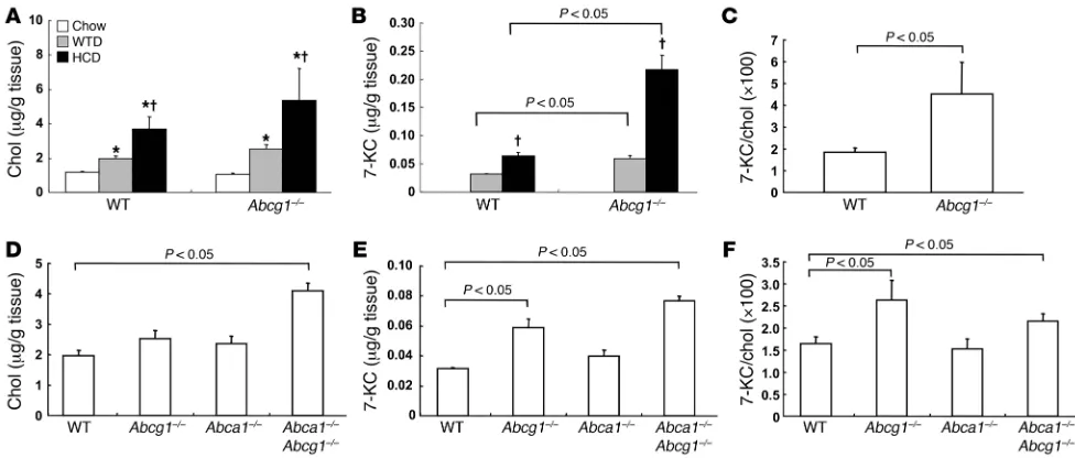
Cholesterol and 7-KC accumulation in aorta: effects of diet and genotype. In view of the specific role of ABCG1 in efflux of 7-oxysterols from cells (35), we next measured the content of cholesterol and 7-KC in
Table 1
EC50 values of vasorelaxation induced by ACh in femoral arteries
from control and Abcg1–/– mice a chow diet, WTD, or HCD
Diet (cholesterol, %) EC50 (nM)
Control Abcg1–/–
Chow (0.025) 27.7 ± 7.5 18.5 ± 3.4 WTD (0.25) 17.4 ± 2.7 79.6 ± 13.0A,B HCD (1.25) 72.4 ± 8.5A 696.2 ± 64.3A,B
[image:3.585.52.543.83.282.2]The results are represented as mean ± SEM. AP < 0.05 vs. chow diet. BP < 0.05 vs. control. n = 4–5 in each group.
Figure 1
Response to vasoconstrictive agents in the femoral arteries from control (WT), Abcg1–/–, Abca1–/–, and Abca1–/–Abcg1–/– mice. (A–C) WT and
Abcg1–/– mice (n = 4 per group) were put on a HCD (1.25% cholesterol, 7.5% cocoa butter and 0.5% sodium cholate) for 11 weeks. (A) Original trace recordings showing vessel tension increase after addition of 3 μM phenylephrine (PE) and relaxation in response to different concentra-tions of ACh. ACh-induced vasorelaxation occurred at the indicated concentraconcentra-tions in control and Abcg1–/– mice. (B) Vasorelaxation in response to ACh was markedly attenuated in Abcg1–/– mice. (C) There was no significant difference in relaxation in response to SNP. (D and E) WT and
non-atherosclerotic thoracic and abdominal aortas excluding the proximal aorta. Total cholesterol content was increased by the HCDs in a dietary cholesterol concentration–dependent manner (Figure 2A). However, there was no significant difference in cholesterol content between the control and Abcg1–/– mice (Figure 2A). 7-KC
was not detectable in aortas of chow-fed mice but accumulated in response to the HCD (Figure 2B) and WTD (Figure 2, B and E). Accumulation of 7-KC was more prominent in Abcg1–/–
mice (Fig-ure 2, B and E). The ratio of 7-KC to cholesterol was significantly higher in Abcg1–/–
mice than in controls in response to HCD (Fig- ure 2C) and WTD feeding (Figure 2F). We also compared aor-tic cholesterol and 7-KC contents in Abcg1–/– mice with those in
Abca1–/– and Abca1–/–Abcg1–/– mice in response to the WTD. There
was no significant difference in cholesterol content between the control and Abcg1–/– or Abca1–/– mice (Figure 2D), while in Abca1–/–
Abcg1–/– mice cholesterol content was significantly higher than
in the controls (Figure 2D). In contrast, 7-KC was significantly increased in Abcg1–/– and Abca1–/–Abcg1–/– mice compared with
controls, although no difference was found between Abcg1–/– and
Abca1–/–Abcg1–/– (Figure 2E). The ratio of 7-KC to cholesterol was
also significantly increased in Abcg1–/– and Abca1–/–Abcg1–/– mice
(Figure 2F). Thus, we conclude that deficiency of both ABCA1 and ABCG1 results in increased cholesterol accumulation compared with accumulation associated with a single deficiency of the trans-porters, while accumulation of 7-KC specifically reflects deficiency of ABCG1. The latter finding parallels the impairment of vasodila- tory responses and suggests that the impaired ACh-induced vas-cular relaxation in Abcg1–/– mice could be brought about by aortic
accumulation of 7-oxysterols.
eNOS protein expression and dimerization in aorta. Previous studies have shown that the formation of eNOS homodimers is necessary for eNOS activity (36, 37). In response to the HCD, eNOS dimer levels were dramatically reduced in Abcg1–/– mice (Figure 3, A and
B). Total eNOS and phospho-eNOS levels were also moderately
decreased in Abcg1–/–
mice (Figure 3, C and D), but the ratio of phos-pho-eNOS to eNOS did not change. On the WTD, Abca1–/–, Abcg1–/–,
and Abca1–/–Abcg1–/– mice exhibited decreased eNOS dimer levels in
aortas (Figure 3, E and F). This reduction was most prominent in
Abcg1–/– and Abca1–/–Abcg1–/– mice (Figure 3, E and F). There was no
difference in eNOS or phospho-eNOS levels between the groups (Figure 3, G and H). In aortas of chow-fed mice, there was no sig-nificant difference in eNOS dimer levels, eNOS, or phospho-eNOS levels between the control and Abcg1–/– mice (Figure 3, I–L). PECAM
levels were not changed in any groups or diets (Figure 3, C, G, and K), indicating an intact endothelium. These data suggest that endothelial dysfunction induced by ABCG1 deficiency in response to the HCD resulted from the reduction of eNOS dimer levels.
ABCG1 expression and accumulation of 7-KC in aorta. To further evaluate the role of ABCG1 in endothelium-dependent vasorelax-ation, we investigated ABCG1 expression in non-atherosclerotic aorta of Abcg1–/– mice that harbor a lacZ cassette insertion at the
Abcg1 locus. Blue nuclear lacZ expression was detected specifically in ECs (Figure 4A, arrowheads), but not in other cells indicated by nuclear fast red staining (Figure 4A, arrows). We also carried out PECAM staining in aorta of WTD-fed Abcg1–/–
mice, which indi- cated an intact endothelium (Figure 4B). As expected, these seg-ments of abdominal and thoracic aorta did not show any evidence of atherosclerosis or macrophage accumulation (data not shown). We also measured NOS activity using aortic lysates in WTD-fed WT and Abcg1–/– mice. The NOS activity in Abcg1–/–
mice was sig-nificantly decreased (Figure 4C). These data are consistent with the reduction of eNOS dimer levels in aorta (Figure 3, E and F). We also isolated ECs from aorta in WTD-fed WT and Abcg1–/– mice
[image:4.585.47.535.81.289.2]using an affinity column with anti-PECAM antibody. After isola-tion of ECs, PECAM, eNOS, and Abcg1 mRNA levels were increased by 15- to 20-fold compared with the non-endothelial fraction (data not shown). There was no significant difference in cholesterol con-tent between WT and Abcg1–/– mice (Figure 4D). 7-KC levels (Figure
Figure 2
4E) and the 7-KC/cholesterol ratio (Figure 4F) were significantly increased in the ECs isolated from Abcg1–/– mice, but not in the
non-EC fraction. These findings suggest that lack of ABCG1 in ECs leads to 7-KC accumulation and reduced eNOS dimer levels and that decreased eNOS activity is responsible for impaired vas-cular relaxation in mice fed HCDs.
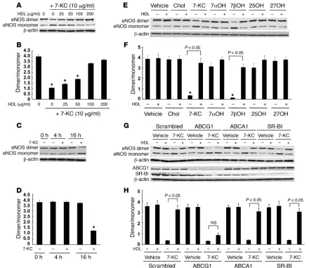
Effects of HDL and 7-KC on eNOS dimer and NOS activity . To fur-ther investigate the role of HDL and ABCG1 in promoting efflux of 7-oxysterols and preserving eNOS dimer levels and activity, we carried out experiments using human aortic ECs (HAECs), which are known to have a high level of ABCG1 (29). We first tested the effects of different concentrations of 7-KC (5–40 μg/ml) and HDL (100 μg/ml). 7-KC (5–40 μg/ml) significantly reduced eNOS dimer levels (Figure 5, A and B). Treatment of cells with HDL (100 μg/ ml) following exposure to 7-KC prevented disruption of eNOS dimer levels by 7-KC (Figure 5, A and B). Only treatment with a high concentration of 7-KC (40 μ g/ml) reduced eNOS and phos-pho-eNOS levels (Figure 5, A and C), and this did not change the ratio of phospho-eNOS to eNOS. The 7-KC concentration of 40
μg/ml also reduced eNOS mRNA (Supplemental Figure 1A; sup-plemental material available online with this article; doi:10.1172/
JCI35470DS1) and induced apoptosis (Supplemental Figure 1B), but this was not observed at lower concentrations. Notably the concentration range of 5–10 μg/ml led to 7-KC levels that were comparable with those in isolated ECs from Abcg1–/– mice (see
below). HDL treatment completely preserved eNOS dimer levels up to a concentration of 7-KC of 20 μg/ml (Figure 5, A and C). Increasing doses of 7-KC also progressively impaired eNOS activ-ity, and this effect was reversed by HDL (Figure 5D). These data demonstrate a strong correlation between decreased eNOS dimer levels and NOS activity in response to increasing doses of 7-KC and show that both effects are reversed by HDL. 7-KC did not affect inflammatory gene expression such as Il6 or Mcp1 (Supplemental Figure 2). Insig1 and LDL receptor mRNA levels, which are regulated by SREBP-2, were reduced by 7-KC (Supplemental Figure 2). The reduction of these mRNAs most likely reflected intracellular accu-mulation of 7-KC.
[image:5.585.80.504.81.439.2]Cholesterol and 7-KC mass efflux. Next, we measured cholesterol and 7-KC mass efflux to different acceptors in HAECs. HAECs were loaded with cholesterol (5 μg/ml) and 7-KC (5 μg/ml) for 24 h. Before starting efflux, intracellular cholesterol and 7-KC contents were measured to determine the baseline contents for the control
Figure 3
Western blot for eNOS protein of mouse aorta. (A–D) Aortas from HCD-fed WT and Abcg1–/– mice. (E–H) Aortas from WTD-fed WT, Abca1–/–,
Abcg1–/–, and Abca1–/–Abcg1–/– mice. (I–L) Aortas from chow-fed WT and Abcg1–/– mice. (A, E, and I) Western blot for eNOS dimer levels. (B,
dishes. Intracellular 7-KC contents were around 10 μg/mg protein and corresponded to the content in the isolated ECs from aortas in WTD-fed Abcg1–/– mice (Figure 4E).
HDL2 (25–100 μg/ml) and HDL3 (25–100 μg/ml) stimulated
both cholesterol and 7-KC mass efflux, whereas apoA-I did not (Figure 6A). We examined the effect of suppression of ABCG1, ABCA1, and SR-BI by siRNA transfection on HDL-mediated cho-lesterol and 7-KC mass efflux. Western blotting showed effective suppression of ABCG1 and SR-BI; however, ABCA1 was not readily detected in these non–liver X receptor agonist–treated cells (Figure 6B, inset). Suppression of ABCG1 significantly reduced both cho-lesterol and 7-KC mass efflux (Figure 6B). Neither suppression of ABCA1 nor SR-BI affected cholesterol or 7-KC mass efflux (Figure 6B). These data indicate that the ABCG1-mediated sterol efflux pathway is predominant in HAECs.
Effects of ABCG1 and HDL on eNOS dimer levels. We examined the effects of different concentrations of HDL on eNOS dimer disrup-tion by 7-KC. HDL treatment protected the disruption of eNOS dimer levels in a concentration-dependent manner with concen-trations between 25 and 100 μ g/ml (Figure 7, A and B). The reduc-tion of eNOS by 7-KC required a relatively long incubation time (>4 h) (Figure 7, C and D). We have previously reported a specific role of ABCG1 in the efflux of 7-oxysterols (35). To further evalu-ate the role of ABCG1 on eNOS dimer levels, we tested the effects of different oxysterols and cholesterol (each 10 μg/ml) in similar experiments. 7β-Hydroxycholesterol as well as 7-KC significantly decreased eNOS dimer levels (Figure 7, E and F). HDL treatment prevented 7β -hydroxycholesterol–induced eNOS dimer disrup-tion. Cholesterol, 7α-hydroxycholesterol, 25-hydroxycholesterol, or 27-hydroxycholesterol did not affect eNOS dimer levels (Figure
7, E and F). This pattern of predominant effects of 7-oxysterols on eNOS dimer levels parallels the specific role of ABCG1 in promot-ing efflux of these oxysterols compared with other sterols (35).
To assess the specific role of ABCG1 in 7-KC–induced disruption of eNOS dimer levels, we knocked down expression of ABCG1 by siRNA. In ABCG1 siRNA-transfected HAECs, the protective effect of HDL was abolished (Figure 7, G and H). In contrast, suppres-sion of neither ABCA1 nor SR-BI affected the ability of HDL to protect against disruption of eNOS dimer levels by 7-KC (Figure 7, G and H). These experiments show a specific requirement for ABCG1 in the ability of HDL to promote 7-KC efflux and to pro-tect ECs from eNOS dimer disruption induced by 7-KC.
Effects of ABCG1 and HDL on ROS production. Previous studies have shown that disruption of eNOS dimer levels can be mediated by peroxynitrite (ONOO–), which is generated from superoxide (O2–)
and NO (38). To investigate the hypothesis that HDL and ABCG1 reverse the effects of 7-KC on ROS production, we used the cell- permeable reagent 6-carboxy-2,7-dichlorodihydrofluorescein diac-etate, diacetoxymethyl-ester (CM-H2DCFDA) (39). In HAECs, 7-KC
[image:6.585.46.540.84.326.2](5–40 μg/ml) induced ROS formation in a dose-dependent manner (Figure 8, A and B). The ROS production by 7-KC required more than 4 h incubation (Figure 8C). The concentration dependence and the time-course response paralleled those of eNOS dimer dis-ruption induced by 7-KC (Figure 7, A–D). 7β-Hydroxycholesterol (10 μg/ml) also significantly increased ROS (P < 0.01), whereas cholesterol, 7α-hydroxycholesterol, 25-hydroxycholesterol, or 27-hydroxycholesterol did not (data not shown). Incubation with HDL (100 μg/ml) significantly reduced ROS production by 7-KC (10 μg/ml) (Figure 8, D and E). This HDL protection was virtually abolished by ABCG1 knockdown (Figure 8, D and E).
Figure 4
Similar protective effects of HDL and ABCG1 on ROS production by 7-KC were observed in primary mouse aortic ECs isolated in a manner similar to the studies described in Figure 4, D–F (see also Supplemental Figure 3). We also measured NOS activity in the similar experiments. HDL preserved the reduction of NOS activ-ity by 7-KC, whereas the HDL protection was abolished by ABCG1 siRNA transfection (Figure 8F). These experiments suggest HDL preserves eNOS dimer levels and activity by promoting efflux of 7-KC via ABCG1 and thus reducing ROS formation.
Effects of antioxidants and NG-nitro-l-arginine methyl ester on eNOS
dimer levels . To confirm that 7-KC–induced eNOS dimer disrup-tion is mediated by ROS, we also evaluated the effects of 2 potent antioxidants, glutathione (GSH) and N-acetylcysteine (NAC). Both GSH and NAC showed protective effects on eNOS dimer disrup-tion (Figure 9, A and B) and ROS production (Figure 9, C and D). To determine the site of 7-KC–induced intracellular ROS produc-tion, HAECs were also incubated with carbonyl cyanide m -chloro- phenylhydrazone (CCCP), an uncoupler of oxidative phosphoryla-tion that abolishes the mitochondrial membrane proton gradient. CCCP did block the ability of 7-KC to induce ROS, suggesting that
mitochondria were the source of ROS production by 7-KC (Sup-plemental Figure 4). By contrast, knockdown of NADPH oxidases Nox1, Nox2, and Nox4, did not affect 7-KC–induced ROS produc-tion (data not shown).
To further analyze the mechanism of eNOS dimer disruption by 7-KC, we investigated the effect of NOS inhibitor NG-nitro-l
-argi-nine methyl ester (l-NAME). l-NAME treatment prevented eNOS
dimer disruption by 7-KC in a dose-dependent manner (Figure 9, E and F). We also investigated the effect of 7-KC on protein tyrosine nitrosylation, since nitrotyrosine formation is considered an indi-cator for ONOO– production. Treatment with 7-KC significantly
increased the detection of nitrotyrosine-positive protein (Figure 9G). In addition, either the presence of l
-NAME or HDL signifi-cantly reduced the level of nitrotyrosine formation (Figure 9G). These data strongly suggest that 7-KC induces formation of O2–,
which reacts with eNOS-generated NO to form ONOO–, which in
turn leads to eNOS oxidation.
Effect of apoA-I transgene expression in endothelial function . To fur-ther evaluate the role of HDL in endothelial function, we also investigated the effect of apoA-I transgene expression on endothe-lium-dependent vasorelaxation in HCD-fed Ldlr+/– mice. ApoA-I
transgene expression significantly improved endothelium-depen-dent vasorelaxation (EC50: Ldlr+/–, 84.4 ± 11.6 vs. Ldlr+/–apoA-I Tg,
23.4 ± 6.5 nM; P < 0.05) (Figure 10A). There was no difference between the groups in the response to SNP (Figure 10B). ApoA-I transgene expression also significantly increased eNOS dimer levels (Figure 10, C and D) and NOS activity (Figure 10F). There was no difference between the groups in eNOS and phospho-eNOS levels (Figure 10, C and E). We also measured cholesterol and 7-KC con-tents in the aortas. In Ldlr+/–apoA-I Tg mice, both cholesterol (Figure
10G) and 7-KC (Figure 10H) contents were significantly decreased, but the magnitude of 7-KC reduction was more pronounced. These data suggest that increased HDL levels resulting from apoA-I trans- gene expression promote efflux of 7-KC from the aorta, contribut-ing to preservation of eNOS dimer levels and activity.
Discussion
[image:7.585.49.282.77.492.2]One of the most important athero-protective functions of HDL is thought to be the stimulation of macrophage cholesterol efflux, and recent studies have highlighted the key roles of ABCA1 and ABCG1 in reversing macrophage foam cell formation (40) and ath-erosclerosis (41, 42). HDL has also been shown to exert a variety of beneficial actions that are independent of macrophage cholesterol efflux. For example, HDL inhibits LDL oxidation, smooth muscle cell migration, and platelet aggregation and reverses endothelial dysfunction (20–22). Our studies revealed a non-redundant role of ABCG1 and a lesser role of ABCA1 in preserving endothelial eNOS activity in mice fed HCDs, and our results suggest that this may be a major mechanism underlying the ability of HDL to defend endothelial NO activity in response to such diets. The ability of
Figure 5
Effects of HDL on eNOS dimer, eNOS, and phospho-eNOS levels, and NOS activity in HAECs. HAECs were incubated with 7-KC (5–40
ABCG1 to preserve endothelial function appears to be at least partly related to its role in promoting efflux of 7-oxysterols such as 7-KC to HDL.
HDL has consistently been shown to increase eNOS-dependent NO activity in cultured ECs (24, 25) and aortic rings (43) and in human forearm blood flow studies (44, 45). In humans, HDL lev-els are correlated with flow-mediated vasodilation responses of the brachial artery (18, 19) and with decreased coronary vasoconstrictor responses (44). Importantly, infusion of recombinant phospholip-id/apoA-I particles into Tangier disease heterozygotes with isolated low HDL levels reversed defective forearm blood flow measurements (46). Deckert et al. (7, 8) showed that 7-oxysterols can produce decreased eNOS activity in rabbits and HUVECs. In apoE–/– mice
fed a chow diet, arterial eNOS activity was preserved but became impaired when mice were challenged with a HCD (4). Importantly, apoA-I transgene expression reversed the decrease in eNOS activity induced by the HCD (4). The current study in HCD-fed Ldlr+/– mice
reproduced preservation of endothelium-dependent aortic relax-ation by apoA-I transgene overexpression (Figure 10), and we show that this effect is associated with preservation of eNOS dimer levels and eNOS activity and reduced aortic 7-KC levels.
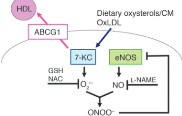
Our studies extend these important earlier observations (4, 7, 8) and suggest that the underlying mechanism by which increased or basal HDL levels protect the endothelium involves efflux of dietary sterols, especially 7-oxysterols from ECs to HDL, mediated principally by ABCG1 (Figure 11). It is most likely that dietary oxysterols are normally incorporated into chylomicrons, cleared by the liver, converted into bile acids, and excreted (13). However, when there is delayed clearance of chylomicron remnants, as
occurs in apoE–/– mice or in humans with increased coronary heart
disease risk (47), the vascular endothelium has increased exposure to dietary oxysterols, and ABCG1 and HDL likely have a key role in excluding or promoting efflux of 7-oxysterols from ECs. Indeed, we found that ABCG1 was expressed specifically in endothelium in non-atherosclerotic mouse aorta (Figure 4A) and 7-KC also accu-mulated in ECs isolated from the aorta in Abcg1–/– mice (Figure
4E). Even though HDL may have a variety of different antioxidant properties in different settings (48, 49), the ability of HDL to pro-mote efflux of 7-KC, reduce ROS production, and preserve eNOS dimer levels and activity were all dependent on ABCG1 expression, indicating that the underlying mechanism involves ABCG1-medi-ated oxysterol efflux. Notably, while 7-KC was readily detected in non-lesioned arteries from mice fed HCDs, it was not measurable in arteries from mice fed chow diets (Figure 2), making it unlikely that 7-KC was artifactually formed during sample processing. Moreover, 7-KC was specifically increased as a result of ABCG1 deficiency (Figure 2), consistent with the role of ABCG1 and not ABCA1 in promoting efflux of this oxysterol to HDL (35).
In the present study in HAECs, disruption of eNOS dimer levels was induced by a 7-KC concentration of 5 μg/ml, which might be equivalent to levels found in human plasma after a fat-rich meal (50). Intracellular 7-KC content in HAECs treated with the relevant concentrations of 7-KC (5–10 μg/ml) were around 10 μ g/mg pro-tein, approximating the concentration found in isolated ECs from aortas in WTD-fed Abcg1–/– mice (Figure 4), in which eNOS dimer
levels were reduced (Figure 3). Thus, these 7-KC concentrations are likely sufficient to induce endothelial dysfunction. The current findings agree with the notion that endothelial dysfunction is a key feature of early atherosclerosis (1) and also occurs transiently in the postprandial state (51).
Our parallel studies in mice and in HAECs suggest that ABCG1 mediates the efflux of 7-oxysterols from ECs to HDL, resulting in decreased ROS formation and preservation of the active dimeric form of eNOS (Figure 9). O2–
is known to inactivate NO and gener-ate ONOO– (37, 38). ONOO– can disrupt eNOS dimers through
oxidation and displacement of the zinc metal ion (37, 52). Our stud-ies also demonstrate that both l-NAME and antioxidants reversed
the disruption of eNOS dimer levels by 7-KC (Figure 9). These data strongly suggest that 7-KC induced O2– and ONOO– production
through interaction with NO, resulting in eNOS oxidation (Figure 11). There is considerable evidence that increased ROS can inhibit eNOS dimer formation and produce endothelial dysfunction in vivo, for example in diet-induced diabetic mice (53, 54) or in apop-tosis signal–regulating kinase-1–deficient mice (55).
A number of different mechanisms have been proposed to account for the ability of HDL to preserve or increase arterial eNOS activity. HDL appears to be moderately effective in inducing
eNOS-dependent vascular relaxation when directly added to aor-Figure 6
Effects of ABCG1 and HDL on sterol efflux in HAECs. (A) HAECs
[image:8.585.48.280.78.411.2]tic rings isolated from rats or mice (43, 56). However, the effect is very rapid (within a few minutes) and is saturated at very low con-centrations of HDL (10 μg/ml), far below that normally bathing the endothelium (43). The response to added HDL is defective in vascular rings isolated from chow diet–fed SR-BI–/– mice (43), and
from mice lacking the lysophospholipid S1P3 receptor (56). The
direct effect of HDL on induction of eNOS activity has also been attributed to minor components such as lysophospholipids (56) or estrogen (57), but the concentrations of these components may not be sufficiently high to be physiologically relevant (25). While SR-BI may not have a major role in mediating net cellular choles-terol efflux to HDL in vivo, it is likely that ABCA1 and ABCG1 do mediate net efflux (27, 42, 58). Finally, our study has not assessed
the role of ABC transporters in efflux to HDL of oxidized phos- pholipids, which are also likely to be important in endothelial dys- function (59–61). Further studies are required to assess the rela-tive roles of these different potential mechanisms in HDL-induced eNOS activity in vivo. Our study conclusively demonstrates the essential role of the ABC transporters, and especially ABCG1, in this process and delineates one mechanism involving efflux of 7-oxysterols and preservation of eNOS dimer levels.
[image:9.585.80.520.81.463.2]Therapies that increase HDL levels, such as niacin and cholesteryl ester transfer protein inhibitors, probably activate the ABCG1-cho-lesterol/oxysterol efflux pathway not only in macrophages (26–28, 35) but also in ECs, likely with beneficial effects on endothelial function. Importantly, niacin therapy has been shown to improve
Figure 7
NO-mediated vascular relaxation in humans (62). Our studies sug-gest that the underlying mechanism may involve increased efflux of cholesterol and 7-oxysterols via the ABCA1 and ABCG1 pathway.
Methods
Materials. The ROS-sensitive fluorescent probe CM-H2
DCFDA and nucle-ar fast red were from Invitrogen. Anti-eNOS and anti–phospho-eNOS (S1177) antibodies were obtained from BD Transduction Laboratories. Anti-ABCG1, anti-PECAM, and anti-nitrotyrosine antibodies were pur-chased from Abcam. SR-BI antibody was from Santa Cruz Biotechnology Inc. Anti–β-actin antibody, X-gal (5-bromo-4-chloro-3-indolyl β-d -galac-topyranoside), lipoprotein-deficient serum, NAC, GSH, phenylephrine,
ACh, SNP, cholesterol, 7-KC, 7β -hydroxycholesterol, and 25-hydroxycho-lesterol were purchased from Sigma-Aldrich. 27-Hydroxycholesterol was obtained from Steraloids. l-NAME was purchased from Cayman Chemical. Human apoA-I was obtained from Biodesign International. HDL (density 1.063–1.210 g/ml), HDL2 (density 1.063–1.125 g/ml), and HDL3 (density
1.125–1.210 g/ml) were isolated by preparative ultracentrifugation from normolipidemic human plasma and stored in PBS.
[image:10.585.72.510.80.499.2]Mouse studies. Abcg1–/–, Abca1–/–, and Abca1–/–Abcg1–/– mice have been previ- ously described (41). We performed studies with a chow diet (0.025% cho-lesterol), a HCD (1.25% cholesterol, 7.5% cocoa butter, and 0.5% sodium cholate; catalog no. TD88051; Harlan Teklad) and a WTD (21% milk fat, 0.2% cholesterol; catalog no. TD88137; Harlan Teklad).
Figure 8
C57BL/6 Ldlr–/– mice and C57BL/6 apoA-I Tg mice (63) were obtained from the Jackson Laboratory and crossed to generate Ldlr+/–apoA-I Tg mice. Next, these animals were crossed with DBA/1LacJ mice (The Jackson Laboratory) to obtain the genetically uniform F1 generation. F1 hybrid C57BL/6 × DBA Ldlr+/–apoA-I Tg mice were put on the HCD.
Animals had ad libitum access to both food and water. Animal protocols were approved by the Institutional Animal Care and Use Committee of Columbia University.
Tissue collection. Mice were anesthetized with an intraperitoneal injection of ketamine. The chest and peritoneal cavity were opened and the circulatory system was perfused via the left ventricle with PBS. Aortas were removed and processed for all assays. For vascular studies, the left superficial femoral artery was removed and immediately placed in ice-cold physiologic salt solution.
Vascular function studies. Femoral arteries with intact endothelium and
similar dimensions were mounted on a small vessel wire myograph (Dan-ish MyoTechnology) as described previously (53). Vessels were bathed in physiologic salt solution at 37°C and aerated continuously with 5% CO2/95% O2 to achieve pH 7.4. The startup protocol and evaluation of
vessel viability was conducted as described previously (53). Concentra-tion response curves were performed for ACh (endothelium dependent) and SNP (endothelium-independent NO-releasing agent). Wall tension was expressed as mN/mm of artery length. Sensitivity to the agonist was expressed as the negative log of EC50 (–log EC50). Sensitivity was calculated
from each concentration response curve by fitting the Hill equation using Prism (GraphPad Software).
[image:11.585.61.521.80.456.2]Isolation of ECs from aorta. Mice aortas were perfused with PBS and digested in RPMI 1640 medium containing collagenase D (2 mg/ml; Roche Applied Science) at 37°C for 45 min. The digest was sequentially fil-tered through 100-μm, 70-μm, and 40-μm cell strainers and was washed with PBS. The cells were incubated with anti-PECAM biotin-conjugated Figure 9
antibody (Millipore) at 4°C for 15 min and were washed with PBS. Next, the cells were labeled with streptavidin microbeads (Miltenyi Biotec) and aortic ECs were separated by MACS column (Miltenyi Biotec) according to the manufacturer’s instructions. Isolated aortic ECs were used for sterol mass measurement.
LacZ expression and PECAM immunostaining . The tissues were snap-frozen in OCT and stored at –80°C. Frozen sections 10 μm long were prepared. To determine β-galactosidase activity, the glass slides were incubated for 16 h in the presence of X-gal. The slides were counter-stained with nuclear fast red. PECAM immunostaining was carried out as previously described (64).
Cell culture. HAECs and the culture medium EMG-2 were purchased from Lonza. The cells were grown in EMG-2 at 37°C in humidified 5% CO2 and
used for experiments between passages 3 and 5. All siRNAs were purchased from Invitrogen or Santa Cruz Biotechnology Inc. HAECs were transfected with siRNA using Lipofectamine RNAiMAX reagent (Invitrogen) accord-ing to the manufacturer’s protocol. Forty-eight hours after transfection, HAECs were treated with 7-KC in the presence or absence of HDL.
Sterol mass analysis. The lipid fractions of abdominal aortas, isolated ECs or non-ECs from aorta, and HAECs were extracted using hexane/isopropa-nol (3:2 vol/vol) in presence of stigmasterol added as the internal standard. Total cholesterol and 7-KC were determined after saponification by gas-liquid chromatography (26, 35).
Sterol mass efflux assay . HAECs were incubated in EGM-2 plus 5% lipopro-tein-deficient serum with cholesterol (5 μg/ml) and 7-KC (5 μg/ml) for 24 h. The next day, cells were washed with PBS and then incubated in EGM-2 plus 5% lipoprotein-deficient serum alone or supplemented with human apoA-I or HDL for 16 h. After the efflux period, media and cells were col-lected separately and lipids were extracted with hexane/isopropanol (3:2 vol/vol) with stigmastanol as the internal standard. Sterol mass of media and cells was determined using gas chromatography. Percentages of sterol mass efflux were calculated by the ratio of sterol mass in the medium to total (medium plus cellular) sterol mass.
NOS activity assay. The NO synthesizing activity was determined by quantifying the rate of the conversion of [3H]l-arginine to [3H]l
-citrul-line with kits obtained from Calbiochem-Novabiochem according to the manufacturer’s instructions (52).
[image:12.585.87.499.81.347.2]Western blotting. Protein was resolved on 4%–20% SDS-PAGE reducing gels (Bio-Rad). Protein was transferred to PVDF membranes and probed Figure 10
Effect of apoA-I transgene expression in endothelial function in HCD-fed Ldlr+/– mice. Ldlr+/– and Ldlr+/–apoA-I Tg mice (n = 6 per group) were put on a HCD for 6 weeks. (A) ACh-induced vasorelaxation. (B) SNP-induced vasorelaxation. (C) Western blot for eNOS dimer and monomer, eNOS, and phospho-eNOS in aorta. (D) Quantification of the eNOS dimer/monomer ratio. (E) Quantification of eNOS and phospho-eNOS. (F) Aortic NOS activity. (G) Cholesterol and (H) 7-KC contents in aorta. The results are represented as mean ± SEM. *P < 0.05 versus control.
Figure 11
[image:12.585.326.511.623.741.2]with primary antibodies overnight. For detection of eNOS dimer levels, we performed low-temperature SDS-PAGE (4°C) (52, 53). Mice aorta lysates and HAEC lysates were heated to 55°C and room temperature, respec-tively, for 30 min in the presence of SDS and 2.5% β-mercaptoethanol.
Quantification of intracellular ROS. The generation of intracellular ROS was estimated by incubating CM-H2DCFDA (1 μM) with cells 30 min before
determination, as described previously (39).
Statistics. Statistical analysis was performed using the Student’s t test. Bonferroni post-hoc tests were utilized. Results are represented as means ± SEM. Acknowledgments This work was supported by grants from the NIH (HL 54591). Received for publication February 27, 2008, and accepted in revised form September 10, 2008. Address correspondence to: Naoki Terasaka, Division of Molecular Medicine, Department of Medicine, Columbia University, PS 8-401, West 168th St., New York, New York 10032, USA. Phone: (212) 305-5789; Fax: (201) 305-5052; E-mail: nt2188@columbia.edu. 1. Zeiher, A.M., Drexler, H., Wollschlager, H., and Just, H. 1991. Modulation of coronary vasomotor tone in humans. Progressive endothelial dysfunction with different early stages of coronary atheroscle-rosis. Circulation. 83:391–401.
2. Bossaller, C., et al. 1987. Impaired muscarinic endo-thelium-dependent relaxation and cyclic guanosine 5ʹ-monophosphate formation in atherosclerotic human coronary artery and rabbit aorta. J. Clin. Invest. 79:170–174.
3. Forstermann, U., Mtigge, A., Alheid, U., Haverich, A., and Frdlich, J.C. 1988. Selective attenuation of endo-thelium-mediated vasodilation in atherosclerotic human coronary arteries. Circ. Res. 62:185–190.
4. Deckert, V., et al. 1999. Impairment of endothelium-dependent arterial relaxation by high-fat feeding in ApoE-deficient mice. Circulation. 100:1230–1235. 5. Minor, R.L., Jr., Myers, P.R., Guerra, R., Jr., Bates,
J.N., and Harrison, D.G. 1990. Diet-induced ath-erosclerosis increases the release of nitrogen oxides from rabbit aorta. J. Clin. Invest. 86:2109–2116. 6. Shimokawa, H., and Vanhoutte, P.M. 1989. Impaired
endothelium-dependent relaxation to aggregating platelets and related vasoactive substances in por-cine coronary arteries in hypercholesterolemia and atherosclerosis. Circ. Res. 64:900–914.
7. Deckert, V., et al. 1997. Inhibitors of arterial relax-ation among components of human oxidized low-density lipoproteins. Cholesterol derivatives oxidized in position 7 are potent inhibitors of endothelium-dependent relaxation. Circulation.
95:723–731.
8. Deckert, V., Katan, M. 1998. Inhibition by choles-terol oxides of NO release from human vascular endothelial cells. Arterioscler. Thromb. Vasc. Biol.
18:1054–1060.
9. Brown, A.J., and Jessup, W. 1999. Oxysterols and atherosclerosis. Atherosclerosis. 142:1–28. 10. Myoishi, M., et al. 2007. Increased endoplasmic
reticulum stress in atherosclerotic plaques associ-ated with acute coronary syndrome. Circulation.
116:1226–1233.
11. Zhou, Q., Wasowicz, E., Handler, B., Fleischer, L., and Kummerow, F.A. 2000. An excess concentra- tion of oxysterols in the plasma is cytotoxic to cul-tured endothelialcells. Atherosclerosis. 149:191–197. 12. Jacobson, M.S. 1987. Cholesterol oxides in Indian
ghee: possible cause of unexplained high risk of atherosclerosis in Indian immigrant populations. Lancet. 2:656–658.
13. Vine, D.F., Mamo, J.C.L., Beilin, L.J., More, T.A., and Croft, K.D. 1998. Dietary oxysterols are incorporat-ed in plasma triglyceride-rich lipoproteins, increase their susceptibility to oxidation and increase aortic cholesterol concentration of rabbits. J. Lipid Res.
39:1995–2004.
14. Sander, B.D., Smith, D.E., Addis, P.B., and Park, S.W. 1989. Effects of prolonged and adverse stor-age conditions on levels of cholesterol oxidation products in dairy products. J. Food Sci. 54:874–879. 15. van de Bovenkamp, P., Kosmeijer-Schuil, T.G., and
Katan, M.B. 1988. Quantification of oxysterols in Dutch foods: egg products and mixed diets. Lipids.
23:1079–1085.
16. Gordon, D.J., and Rifkind, B.M. 1989. High-density lipoprotein-the clinical implications of recent stud-ies. N. Engl. J. Med. 321:1311–1316.
17. Assmann, G., and Gotto, A.M., Jr. 2004. HDL cho-lesterol and protective factors in atherosclerosis. Circulation. 109(Suppl. 1):III-8–III-14.
18. Li, X.P., et al. 2000. Protective effect of high density lipoprotein on endothelium-dependent vasodilata-tion. Int. J. Cardiol. 73:231–236.
19. Kuvin, J.T., et al. 2003. Relation between high-den- sity lipoprotein cholesterol and peripheral vasomo-tor function. Am. J. Cardiol. 92:275–279.
20. O’Connell, B.J., and Genest, J., Jr. 2001. High-den-sity lipoproteins and endothelial function. Circula-tion. 104:1978–1983.
21. Rohrer, L., Hersberger, M., and von Eckardstein, A. 2004. High density lipoproteins in the intersection of diabetes mellitus, inflammation and cardiovas-cular disease. Curr. Opin. Lipidol. 15:269–278. 22. Mineo, C., Deguchi, H., Griffin, J.H., and Shaul,
P.W. 2006. Endothelial and antithrombotic actions of HDL. Circ. Res. 98:1352–1364.
23. Collins, T., and Cybulsky, M.I. 2001. NF-kappaB: pivotal mediator or innocent bystander in athero-genesis? J. Clin. Invest. 107:255–264.
24. Uittenbogaard, A., Shaul, P.W., Yuhanna, I.S., Blair, A., and Smart, E.J. 2000. High density lipoprotein prevents oxidized low density lipoprotein-induced inhibition of endothelial nitric-oxide synthase localization and activation in caveolae. J. Biol. Chem.
275:11278–11283.
25. Mineo, C., Deguchi, H., Griffin, J.H., and Shaul, P.W. 2006. Endothelial and antithrombotic actions of HDL. Circ. Res. 98:1352–1364.
26. Matsuura, F., Wang, N., Chen, W., Jiang, X.C., and Tall, A.R. 2006. HDL from CETP-deficient subjects shows enhanced ability to promote cholesterol efflux from macrophages in an apoE- and ABCG1-dependent pathway. J. Clin. Invest. 116:1435–1442.
27. Yvan-Charvet, L., et al. 2007. Inhibition of choles-teryl ester transfer protein by torcetrapib modestly increases macrophage cholesterol efflux to HDL. Arterioscler. Thromb. Vasc. Biol. 27:1132–1138. 28. Tall, A.R., Yvan-Charvet, L., Terasaka, N., Pagler, T.,
and Wang, N. 2008. HDL, ABC transporters, and cholesterol efflux: implications for the treatment of atherosclerosis. Cell Metab. 7:365–375.
29. O’Connell, B.J., Denis, M., and Genest, J. 2004. Cel-lular physiology of cholesterol efflux in vascular endothelial cells. Circulation. 110:2881–2888. 30. Wang, N., Silver, D.L., Costet, P., and Tall, A.R.
2000. Specific binding of ApoA-I, enhanced cho-lesterol efflux, and altered plasma membrane morphology in cells expressing ABC1. J. Biol. Chem.
275:33053–33058.
31. Oram, J.F., Lawn, R.M., Garvin, M.R., and Wade, D.P. 2000. ABCA1 is the cAMP-inducible apolipoprotein receptor that mediates choles-terol secretion from macrophages. J. Biol. Chem.
275:34508–34511.
32. Wang, N., Lan, D., Chen, W., Matsuura, F., and Tall, A.R. 2004. ATP-binding cassette transporters G1 and G4 mediate cellular cholesterol efflux to high-density lipoproteins. Proc. Natl. Acad. Sci. U. S. A.
101:9774–9779.
33. Kennedy, M.A., et al. 2005. ABCG1 has a critical role in mediating cholesterol efflux to HDL and preventing cellular lipid accumulation. Cell Metab.
1:121–131.
34. Vaughan, A.M., and Oram, J.F. 2005. ABCG1 redis-tributes cell cholesterol to domains removable by high density lipoprotein but not by lipid-depleted apolipoproteins. J. Biol. Chem. 275:34508–34511. 35. Terasaka, N., Wang, N., Yvan-Charvet, L., and Tall,
A.R. 2007. High-density lipoprotein protects mac- rophages from oxidized low-density lipoprotein- induced apoptosis by promoting efflux of 7-keto-cholesterol via ABCG1. Proc. Natl. Acad. Sci. U. S. A.
104:15093–15098.
36. Rodriguez-Crespo, I., Moenne-Loccoz, P., Loehr, T.M., and Ortiz de Montellano, P.R. 1997. Endothelial nitric oxide synthase: modulations of the distal heme site produced by progressive N-ter-minal deletions. Biochemistry. 36:8530–8538. 37. Forstermann, U., and Munzel, T. 2006. Endothelial
nitric oxide synthase in vascular disease: from mar-vel to menace. Circulation. 113:1708–1714. 38. Xu, J., Xie, Z., Reece, R., Pimental, D., and Zou, M.H.
2006. Uncoupling of endothelial nitric oxidase synthase by hypochlorous acid: role of NAD(P)H oxidase-derived superoxide and peroxynitrite. Arte-rioscler. Thromb. Vasc. Biol. 26:2688–2695. 39. Robbesyn, F., et al. 2003. HDL counterbalance
the proinflammatory effect of oxidized LDL by inhibiting intracellular reactive oxygen species rise, proteasome activation, and subsequent NF-kap-paB activation in smooth muscle cells. FASEB J.
17:743–745.
40. Wang, X., et al. 2007. Macrophage ABCA1 and ABCG1, but not SR-BI, promote macrophage reverse cholesterol transport in vivo. J. Clin. Invest.
117:2216–2224.
41. Yvan-Charvet, L., et al. 2007. Combined deficiency of ABCA1 and ABCG1 promotes foam cell accu-mulation and accelerates atherosclerosis in mice. J. Clin. Invest. 117:3900–3908.
42. Rader, D.J. 2006. Molecular regulation of HDL metabolism and function: implications for novel therapies. J. Clin. Invest. 116:3090–3100.
43. Yuhanna, I.S., et al. 2001. High-density lipopro-tein binding to scavenger receptor-BI activates endothelial nitric oxide synthase. Nat. Med.
7:853–857. 44. Zeiher, A.M., Schachlinger, V., Hohnloser, S.H., Saurbier, B., and Just, H. 1994. Coronary athero-sclerotic wall thickening and vascular reactivity in humans. Elevated high-density lipoprotein levels ameliorate abnormal vasoconstriction in early ath-erosclerosis. Circulation. 89:2525–2532.
45. Bisoendial, R.J., et al. 2003. Restoration of endothelial function by increasing high-density lipoprotein in subjects with isolated low high-den-sity lipoprotein. Circulation. 107:2944–2948. 46. Bisoendial, R.J., et al. 2003. Restoration of
47. Bansal, S., et al. 2007. Fasting compared with non-fasting triglycerides and risk of cardiovascular events in women. JAMA. 298:309–316.
48. Nicholls, S.J., et al. 2005. Reconstituted high-den-sity lipoproteins inhibit the acute pro-oxidant and proinflammatory vascular changes induced by a periarterial collar in normocholesterolemic rabbits. Circulation. 111:1543–1550.
49. Tall, A.R. 2008. Cholesterol efflux pathways and other potential mechanisms involved in the athero- protective effect of high density lipoproteins. J. Intern. Med. 263:256–273.
50. Emanuel, H.A., Hassel, C.A., Addis, P.B., Bergman, S.D., and Zavoral, J.H. 1991. Plasma cholesterol oxi-dation products (oxysterols) in human subjects fed a meal rich in oxysterols. J. Food Sci. 56:843–847. 51. Vogel, R.A., Corretti, M.C., and Plotnick, G.D. 1997.
Effect of a single high-fat meal on endothelial func-tion in healthy subjects. Am. J. Cardiol. 79:350–354. 52. Zou, M.H., Shi, C., and Cohen, R.A. 2002. Oxida-tion of the zinc-thiolate complex and uncoupling of endothelial nitric oxide synthase by peroxyni-trite. J. Clin. Invest. 109:817–826.
53. Molnar, J., et al. 2005. Diabetes induces endothelial
dysfunction but does not increase neointimal for-mation in high-fat diet fed C57BL/6J mice. Circ. Res. 96:1178–1184.
54. Hink, U., et al. 2001. Mechanisms underlying endothelial dysfunction in diabetes mellitus. Circ. Res. 88:E14–E22.
55. Yamashita, T., et al. 2007. Apoptosis signal-regu-lating kinase-1 is involved in vascular endothelial and cardiac remodeling caused by nitric oxide defi-ciency. Hypertension. 50:519–524.
56. Nofer, J.R., et al. 2004. HDL induces NO dependent vasorelaxation via the lysophospholipid receptor S1P3. J. Clin. Invest. 113:569–581.
57. Gong, M., et al. 2003. HDL-associated estradiol stimulates endothelial NO synthase and vasodila-tion in an SR-BI-dependent manner. J. Clin. Invest.
111:1579–1587.
58. Yvan-Charvet, L., et al. 2008. SR-BI inhibits ABCG1- stimulated net cholesterol efflux from cells to plas-ma HDL. J. Lipid Res. 49:107–114.
59. Watson, A.D., et al. 1997. Structural identification by mass spectrometry of oxidized phospholipids in minimally oxidized low density lipoprotein that
induce monocyte/endothelial interactions and evidence for their presence in vivo. J. Biol. Chem.
272:13597–13607.
60. Rikitake, Y., et al. 2000. Inhibition of endothelium- dependent arterial relaxation by oxidized phospha-tidylcholine. Atherosclerosis. 152:79–87.
61. Navab, M., et al. 2004. The oxidation hypothesis of atherogenesis: the role of oxidized phospholipids and HDL. J. Lipid Res. 45:993–1007.
62. Kuvin, J.T., et al. 2002. A novel mechanism for the beneficial vascular effects of highdensity lipopro-tein cholesterol: enhanced vasorelaxation and increased endothelial nitric oxide synthase expres-sion. Am. Heart J. 144:165–172.
63. Rubin, E.M., Ishida, B.Y., Clift, S.M., and Krauss, R.M. 1991. Expression of human apolipoprotein A-I in transgenic mice results in reduced plasma levels of murine apolipoprotein A-I and the appearance of two new high density lipoprotein size subclasses. Proc. Natl. Acad. Sci. U. S. A. 88:434–438.
64. Welch, C.L., et al. 2007. Spontaneous atherothrom-bosis and medial degradation in Apoe–/–, Npc1–/–