Biology Dissertations Department of Biology
Fall 12-14-2010
Quorum Sensing Inhibitory Activities of Various
Folk-Medicinal Plants and the Thyme-tetracycline
Effect.
Maria M. Nagy
Georgia State University
Follow this and additional works at:https://scholarworks.gsu.edu/biology_diss Part of theBiology Commons
This Dissertation is brought to you for free and open access by the Department of Biology at ScholarWorks @ Georgia State University. It has been accepted for inclusion in Biology Dissertations by an authorized administrator of ScholarWorks @ Georgia State University. For more information, please [email protected].
Recommended Citation
Nagy, Maria M., "Quorum Sensing Inhibitory Activities of Various Folk-Medicinal Plants and the Thyme-tetracycline Effect.." Dissertation, Georgia State University, 2010.
QUORUM SENSING INHIBITORY ACTIVITIES OF VARIOUS FOLK MEDICINAL
PLANTS AND THE ELUCIDATION OF THE THYME-TETRACYCLINE EFFECT
by
MARIA M. NAGY
Under the Direction of Dr. Sidney A. Crow
ABSTRACT
Pseudomonas aeruginosa is an opportunistic, nosocomial pathogen for which antibiotic resistance and biofilm development is common. Quorum sensing communication is known to be
a major controlling factor in virulence gene expression, biofilm development, antibiotic
resistance factors, and specifically MexAB-OprM multi-drug efflux pump expression in
P.aeruginosa. MexAB-OprM efflux pumps contribute to antibiotic resistance of tetracycline and other antibiotics in pseudomonads and other organisms. P.aeruginosa infections are problematic in cystic fibrosis and burn patients; it is also the number one causative agent of respiratory
infections for intensive care unit patients. Present day antibiotics are losing the battle against
these infections. In theory, quorum sensing inhibitors (QSI) reduce pathogencity of the organism;
making it less virulent, thus allowing either the host immune system to clear the infection or use
two alternative modes of treatment were explored in this study: quorum sensing inhibition by
folk-medicinal plant extracts and an example of combination drug therapy, the
―thyme-tetracycline effect‖.
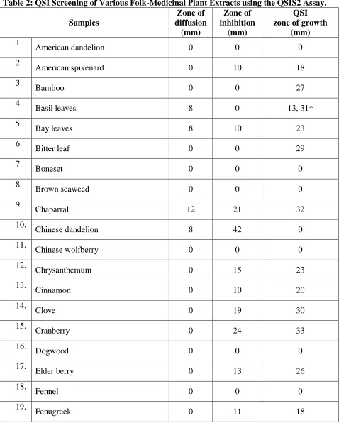
Fifty folk-medicinal plant extracts were screened for potential anti-quorum sensing
activity using two quorum sensing inhibition (QSI) reporter strains, Pseudomonas aeruginosa
QSIS2 and Chromobacterium violaceum 12725. These were used to test specifically for C4-C6 and C12 HSL quorum sensing inhibition. Of the fifty plants tested, thirty plant families were
represented. Eleven plant extracts (basil, chaparral, clove, cranberry, oregano, pomegranate,
rosemary, sage, sassafras, thyme and witch hazel) showed C4 HSL quorum sensing inhibition as
determined by both assays. Interestingly, five of the plants were from the Lamiaceae family.
Thymus vulgaris (thyme), also from the Lamiaceae family, was chosen for further assessment. Previous research has shown that thyme extract can synergistically augment tetracycline
activity against tetracycline-resistant Pseudomonas aeruginosa, creating the ―thyme-tetracycline effect.‖ Disc diffusion assay, thin layer chromatography (TLC), and TLC bioassay techniques
were used to show that thymol is the active component in the thyme extract that augments
tetracycline activity against resistant Pseudomonas. This study also showed that thymol is a potent C4 HSL quorum sensing inhibitor. The collective data suggests a potential mode of action
for the thyme-tetracycline effect: thymol appears to prevent MexAB-OprM efflux pump gene
expression. By blocking MexAB-OprM expression, tetracycline antibiotic accumulation can
occur within the cell, thus allowing cellular damage.
INDEX WORDS: Thymus vulgaris, Quorum sensing inhibition, Medicinal plants,
QUORUM SENSING INHIBITORY ACTIVITIES OF VARIOUS FOLK MEDICINAL
PLANTS AND THE ELUCIDATION OF THE THYME-TETRACYCLINE EFFECT
by
MARIA M. NAGY
A Dissertation Presented in Partial Fulfillment of Requirements for the Degree of
Doctor of Philosophy
in the College of Arts and Sciences
Georgia State University
Copyright by Maria M. Nagy
QUORUM SENSING INHIBITORY ACTIVITIES OF VARIOUS FOLK MEDICINAL PLANTS AND THE ELUCIDATION OF THE THYME-TETRACYCLINE EFFECT
by
MARIA M NAGY
Committee Chair: Dr. Sidney Crow
Committee: Dr. Keith Pascoe Dr. George Pierce Dr. Eric Gilbert
Electronic Version Approved:
Office of Graduate Studies
College of Arts and Science
Georgia State University
ACKNOWLEDGEMENTS
Thank you God!
Thank you! Dr. Crow and Dr. Pascoe. I‘d like to thank Dr. Crow for forever being the
stern disciplinarian in this process, and Dr. Pascoe for believing in me since day one. I‘d also like
to thank Dr. Gilbert and Dr. Peirce for participating on my committee.
Thanks of course to my family Mom, Dad, Kati, Peti, Paul, and everyone else that stood
by me and supported me emotionally and intellectually on this long arduous journey. Thank you
so much for never giving up on me, especially during those times I wanted to give up on myself.
I am very blessed.
Thank you!
***
I‘d also like to acknowledge others that have been critical to my success. First, the 4905
Biology classes at Georgia State University that helped pick, prepare and test many of the
medicinal plant extracts discussed in this study. Without them I wouldn‘t have the samples. I‘d
also like to thank Brandi and Jeannie for proof reading my work, and Bryan Stubblefield and
TABLE OF CONTENTS
ACKNOWLEDGEMENTS iv
LIST OF TABLES vi
LIST OF FIGURES viii
ABBREVIATIONS x
INTRODUCTION 1
MATERIALS AND METHODS 30
RESULTS: SCREENING OF FIFTY FOLK-MEDICINAL EXTRACTS
FOR QSI ACTIVITY 38
RESULTS: THE THYME-TETRACYCLINE EFFECT 61
RESULTS: QSI ACTIVITY OF THYME AND ITS MAJOR CONSITUENTS 79
DISCUSSION 88
REFERENCES 97
LIST OF TABLES
Table 1: Folklore and Published Test Results Pertaining to the Folk-Medicinal Plants
Tested. 38
Table 2: QSI Screening of Various Folk-Medicinal Extracts using the QSIS2 Assay. 49
Table 3: Glucose Concentration and pH Readings of the Folk-Medicinal Extracts. 54
Table 4: QSI Results of the Folk-Medicinal Extracts using the Chromobacterial QSI
Assay. 57
Table 5: DDA of Ethanol Extract of Thymus vulgaris Leaves. 62 Table 6: Percentage Yield of Crude Extracts Produced by Extraction of Thyme Leaves in
Various Solvents. 64
Table 7: Percent Yield from Gradient extraction of Thyme Leaves using Recycled and
Nonrecycled (Fresh) Hexane. 66
Table 8: Disc Diffusion Assay Results of the Crude Extracts made with Non-recycled
Solvent for each Extraction in Hexane, Methanol, and Ethanol of Thymus vulgaris Leaves. 67 Table 9: Percentage Yield of Crude Extracts made with Non-recycled Solvent. 67
Table 10: Disc Diffusion Assay of Hexane, Methanol, and Ethanol Resoaks using
Recycled Solvent (300-303). 69
Table 11: Percentage Yield of Samples 300-303. 69
Table 12: Percentage Yield of Crude Samples (304-311) Comparing Three Soaks Versus
One Soak. 71
Table 13: Disc Diffusion Assay of Crude Samples (304-311) Comparing Three Soaks
Versus One Soak. 71
Table 14: DDA Results of the Methanol Crude Extract and its Corresponding Fraction. 73
Table 15: DDA of Major Constituents at Low Concentrations. 77
Table 16: DDA of Thymol and Carvacrol constituents at Higher Concentrations. 78
Table 17: QSI Activity of Crude Extracts with the QSIS2 Assay. 79
Table 19: QSI Activity of Thymol at Various Concentrations Using the QSIS2 Assay. 82
Table 20: Activity of Rosmarinic Acid at Various Concentrations using the QSIS2 Assay. 83
Table 21: QSI Activity of Carvacrol at Various Concentrations with the QSIS2 Assay. 84
LIST OF FIGURES
Figure 1: Life magazine--Advertisement for penicillin (1944). 2 Figure 2: Scanning electron micrograph of Pseudomonas aeruginosa-(CDC). 4
Figure 3: Biofilms. 7
Figure 4: Quorum Sensing cascading system in Pseudomonas aeruginosa. 11 Figure 5: N-(3-oxododecanoyl) homoserine lactone (3-oxo-C12-HSL). 11
Figure 6: N-(butanoyl)-L-homoserine lactone (C4-HSL). 11
Figure 7: Tetracycline structures. 14
Figure 8: MexAB-OprM multidrug efflux pump. 16
Figure 9: QSI molecules. 21
Figure 10: Thymus vulgaris. 24
Figure 11: Baicalein. 26
Figure 12: Rosmarinic acid. 26
Figure 13: A) Thymol and B) Carvacrol. 27
Figure 14: QSI results screened with the QSIS2 assay. 51
Figure 15: QSI results screened with the QSIS2 assay. 52
Figure 16: QSI results screened with the QSIS2 assay. 52
Figure 17: QSI results screened with the QSIS2 assay 53
Figure 18: QSI results of crude samples screened with the Chromobacterial QSI
assay. 59
Figure 19: QSI results of crude samples screened with the Chromobacterial QSI
assay. 60
Figure 20: QSI results of crude samples screened with the Chromobacterial QSI
Figure 21: DDA of ethanol thyme leaf extract at a 500 mg/mL concentration. 61
Figure 22: 2D-TBA of the thyme leaf extract. 63
Figure 23: TLC comparison of thyme leaves extracted with various solvents. 64
Figure 24: TLC results of graduated extraction of thyme leaves using recycled and
non-recycled hexane. 65
Figure 25: TLC of extracts samples 270-275 (thyme leaves) extracted in hexane,
methanol, and ethanol, prepared using non-recycled solvent with each of three soaks. 67
Figure 26: TLC of thyme leaf extracts. (Sample 300-303) extracted in hexane,
methanol, and ethanol resoaks using recycled solvent with each soak. 68
Figure 27: TLC comparison of three soaks verses one soak. 70
Figure 28: TLC of the methanol crude (307) and its corresponding fractions. 72
Figure 29: TLC of four major constituents. 74
Figure 30: TLC diagram and TBA of hexane crude, thymol, and control. 75
Figure 31: This is a close up photo of the rows of the above TBA. 75
Figure 32: Disc Diffusion Assay of thymol. 78
Figure 33: QSI activity of various thyme crude extracts with the QSIS2 assay. 80
Figure 34: QSI activity of baicalein at various concentrations with the QSIS2 assay. 81
Figure 35: QSI activity of thymol at various concentrations with the QSIS2 assay. 82
Figure 36: QSI activity of Rosmarinic acid at various concentrations with the QSIS2
assay 83
Figure 37: QSI activity of carvacrol at various concentrations with the QSIS2 assay. 85
Figure 38: QSI Chromobacterial QSI assay results of the thyme extract and its
various constituents. 87
LIST OF ABBREVIATIONS
Acyl homoserine lactone AHL
Disc diffusion assay DDA
Infectious Disease Society of America IDSA
Luria-Bertani LB
Methicillin resistant Staphylococcus aureus MRSA
Mueller Hinton II MH
N-(butanoyl)--L-homoserine lactone C4 HSL
N-(3-oxododecanyoyl)-L-homoserine lactone C12 HSL
N-hexanoyl-L-homoserine lactone C6 HSL
Nutrient broth NB
Office of Technical Assessment OTA
Quorum sensing QS
Quorum-sensing inhibition QSI
QSIS2 reporter strain QSIS2
Reference factor RF
Rosmarinic acid RA
Thin-layer chromatography TLC
TLC bioassay TSA
Tetracycline TET
Tryptic soy agar TSA
INTRODUCTION
Infectious Disease
Infectious diseases are defined by the World Health Organization (WHO) as diseases
caused by microbes; these microbes may include bacteria, fungi, protozoa, and viruses (WHO,
2010). These organisms may be found in either the environment or participate in normal
commensal flora for humans, plants, or animals. When in their natural environment, these
microbes are typically kept in balance by the surrounding flora. Thus, many of these organisms
can be beneficial to their environment by providing nutrient turnover; but when certain bacteria
are introduced into a foreign niche within the human body, they may cause disease. Diseases
caused by bacterial infections can range from severe to mild and may include wound infections,
pneumonia, septicemia, endocarditis, colds, and eye and ear infections (Todar, 2008).
Prior to the discovery of penicillin antibiotics, doctors were unable to treat even relatively
simple bacterial infections such as otitis media (inner ear infection) and streptococcal infection
of the throat (Office of Technical Assessment [OTA], 1995). Doctors were instructed to keep
the infected patient clean and comfortable until the immune system could clear the infection;
more serious infections, however, were incurable (OTA, 1995). The treatment of bacterial
infections was revolutionized in 1929, when Alexander Fleming discovered penicillin. For
several years, antibiotics were thought to be the ―end-all‖ curative agent for many bacterial
infections and were considered to be the wonder drug of the era. Penicillin was used to treat
infections caused by Staphylococci, Streptococci and other Gram-positive organisms (OTA, 1995). On August 14, 1944, Schenley Laboratories unveiled an advertisement campaign in Life
magazine (Figure 1) that suggested the revolutionary usefulness of penicillin on the battle field
Figure 1: Life magazine--Advertisement for penicillin (1944).
ncmuseumofhistory.org/.../topic/16/
However, by 1945 penicillin-resistant strains of Staphylococcus aureus were being isolated in hospitals. Methicillin, a semi-synthetic version of penicillin, was introduced in 1959
in hopes of combating these resistant strains. Only one year later, the first isolates of
methicillin-resistant strains of S. aureus (MRSA) were being found in hospitals in the United States. Vancomycin was introduced in 1956 to treat MRSA (OTA, 1995). The first case of
vancomycin resistance was seen in Japan in 1996 (CDC, 2002).
In 2001, WHO officially deemed antibiotic resistance the number three public health
concern of the 21st century (Levy, 2002). In April of 2010, the ―10 x‘20‖ initiative was launched
by the Infectious Disease Society of America (IDSA) to assist in the development of 10 new
antibiotic drugs by the end of 2020. The focus of this drug development initiative is to target
specific infectious agents deemed ―ESKAPE‖ pathogens. These organisms include:
prominent causes of hospital-acquired infections today. ―ESKAPE‖ also refers to the ability of
these organisms to ―escape‖ present-day antimicrobial treatments (IDSA, 2010). Clearly,
exploration of new and alternative drug treatments for infectious diseases is vital.
One particularly troubling organism is Pseudomonas aeruginosa; it is a nosocomial, or hospital-acquired, opportunistic pathogen. It is the number one infectious agent causing
respiratory infections in people with extended hospital stays (Driscoll et al., 2007).
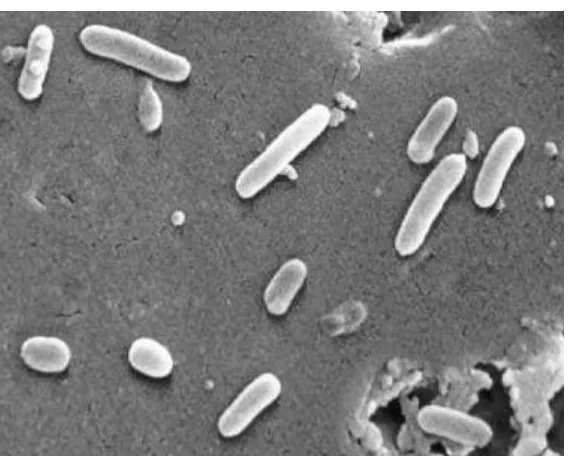
Pseudomonas aeruginosa
Pseudomonas aeruginosa (Figure 2) is a Gram-negative rod that is propelled by a single flagellum (Todar, 2008). Pseudomonas is a genus within the medically-relevant class of
Gammaproteobacteria, and belongs to the family Pseudomonadaceae. There are eight genera in
the family Pseudomonadaceae and twelve species in the genus Pseudomonas. P. aeruginosa,
native to both soil and water, is also a well-known plant pathogen (Todar, 2008). As a human
pathogen, opportunistic infections by this organism can occur when the host immune system is
previously weakened due to a prior ailment-such as AIDS, cancer, cystic fibrosis,
transplantation, or substantial burns (Driscoll et al., 2007). In these scenarios, pseudomonad‘s
have the ability to infect any tissue where the defenses of the host have been compromised
(Todar, 2008). While P. aeruginosa infections are most commonly associated with pneumonia in cystic fibrosis patients and wound infections of burn patients, it is also the causative agent of
several other illnesses including urinary tract infections, endocarditis, bactermia (Todar, 2008),
ulcerative keratitis (in contact lens wearers) and otitis externa (in diabetics) (Driscoll et al.,
site infections (Driscoll et al., 2007). Furthermore, it is the second-most common cause of
health-associated and ventilator-associated pneumonia (Driscoll et al., 2007).
[image:17.612.73.357.177.405.2]Figure 2: Scanning electron micrograph of Pseudomonas aeruginosa-(CDC).
(Todar, 2008)
Virulence
When P. aeruginosa infects the host, it expresses a myriad of virulence factors that allow it to attack the host immune system while evading treatment. An intrinsic defensive feature of
this organism is its outer phospholipid membrane which has limited permeability for most
molecules.
Pseudomonads also secrete a number of compounds that interfere with host defenses
including enzymes, exotoxins, and pyocyanin. Enzymes, such as alkaline protease, elastase, and
protein synthesis and can even cause cellular death. Pyocyanin is a blue-green pigment which is
bactericidal against competing organisms. In the lungs, pyocyanin can also disrupt ciliary action
and cause inflammation (Driscoll et al., 2007).
Another virulence factor that can be expressed in Pseudomonas is the type III secretion apparatus. Expression of the genes encoding the various parts of the secretion system is seen
predominantly in acute or invasive infections more than in chronic infections (Driscoll et al.,
2007). This mechanism is a ―contact-dependent‖ system that is triggered by the interaction of P. aeruginosa with the host cell. The secretion apparatus allows the cell to inject exoenzymes directly into the host cell. There are four known exoenzymes that are typically expressed by P. aeruginosa: Exo S, Exo T, Exo U, and Exo Y (Driscoll et al., 2007). These exoenzymes can damage the host‘s cellular machinery and even cause cell death (Driscoll et al., 2007).
In addition to the impermeability of the cellular membrane and the secretion apparatus,
Pseudomonas also has another mechanism of protection. If a damaging molecule does gain entry into the cell, then unique pumps quickly expel the compounds from the cell before any
damage can occur. These efflux pumps can remove antibiotics, dyes, detergents, solvents and
other compounds from the cell before they can cause harm (Driscoll et al., 2007). There are two
types of efflux pumps that are seen in bacteria: those which are encoded in the chromosome and
those which are plasmid borne. For example, the MexAB-OmpM efflux pumps found in P. aeruginosa are intrinsic to the genome. Plasmid-borne antibiotic resistance genes that code for efflux pumps can be located on specialty plasmids, such as tetracycline-resistant (Tc) plasmids.
These plasmids are typically transferred through conjugation between bacteria and can encode
Biofilms
A final mechanism for protection from the host immune system may be incurred through
biofilm formation. It well known that bacteria grow and develop into ―tight-knit‖ communities
on biotic and abiotic surfaces. The innate features of a biofilm can protect bacteria from
antibiotic damage. Biofilms are so efficient in preventing antibiotic damage that concentrations
of antibiotics must be increased by 100-1000--fold to be effective (Costerton et al., 1994).
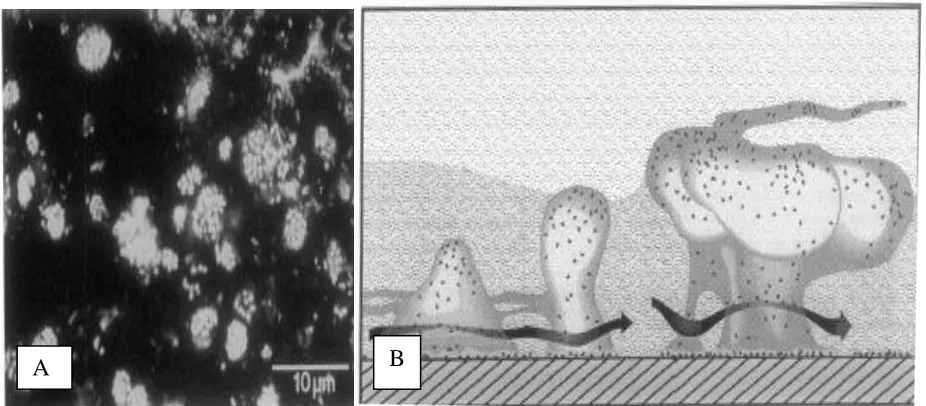
Biofilms (Figure 3-A) typically consist of microbial communities that secrete and are
encased in a thick exopolysaccharide matrix. There are special features associated with most
biofilms that allow for increased efficiency of these communities. These particular morphological
characteristics include: an attachment surface, mushroom-like clusters of bacterial cells, a thick
polysaccharide matrix encasing the bacterial communities; extensive well-hydrated channels that
allow for directed liquid flow (providing delivery and removal of nutrients and waste products);
and swarmer cells (Figure 3-B). Swarmer cells are cells that easily break off from the mushroom
Figure 3: Biofilms. A) Mixed colony biofilm from Bow River Alberta, Canada; B) Diagram of a biofilm: Notice the following features: mushroom structures, thick polysaccharide matrix, excessive channels, well hydrated channels, directed flow (allowing for delivery of nutrients and removal of waste products and swarmer cells. (Costerton et al., 1994)
Medical devices, such as internal catheters, artificial joints, and heart value replacement
units, have become commonplace in medicine. While these advances have extended the lives of
many, they have also ironically brought about subsequent problems. The number of people that
are in extended ―immuno-compromised‖ states has greatly increased. Unfortunately, medical
devices provide the perfect breeding ground for the development of opportunistic bacterial and
biofilm infections, and for the proliferation of resistant bacteria (Reid, 1999).
As of 1999, catheter and urinary stent usage occurred in over 100 million patients per
year (Reid, 1999). Sixty percent of all hospital-acquired microbial infections are caused by
biofilms (Lewis, 2001). Without the use of prophylactic antibiotic treatments, the infection rate
for urethral stents is 28% and is nearly 100% with catheter usage. For this reason, prophylactic
antibiotic treatments are administered to patients to reduce infection rates (Reid, 1999).
There are many features of a catheter that promote bacterial growth. These features
include liquid flow, nutrient access, attachment surface, and the occasional planktonic organism, all
of which are readily associated with biofilm development. Because of this, biofilm infections are
prominent in catheter-type environments. If a biofilm does develop, then it requires an antibiotic
dosage of a 100-1000 times greater concentration of antibiotics than with non-biofilm bacterial
infections. The excessive use of these antibiotic treatments increases the development of resistant
bacteria, thus reducing the overall effectiveness of available antibiotics (Costerton, 1999).
Many characteristics of biofilms contribute to the difficulty in treating these types of
infections with normal antibiotic doses. First, the exopolysaccharide matrix produces a physical
barrier, which reduces the amount of antibiotic that can enter into this microbial community.
Second, even if antibiotic treatments infiltrate the exopolysaccharide matrix, the antibiotic may
still have difficultly accessing the internal cells of the mushroom-like structures. Third, during the
formation of a biofilm, specialized virulence gene expression can occur. These virulence factors
include the expression of antibiotic denaturing enzymes, efflux pump, and increased plasmid
exchange. The development and expression of many of these virulence features is typically under
quorum sensing control (Bassler, 1999; Camara et al., 2002; Costerton, 1999; Lewis, 2001).
Quorum Sensing
Quorum sensing (QS) is a population-dependent expression of genes that influences
biofilm development, efflux pump expression, toxin production, and many other virulence factors.
Quorum sensing occurs through chemical signaling and has been observed in bacteria, fungi, and
even plants. Individual cells in an environment constantly express and expel low levels of quorum
sensing molecules. It is only when the cell population reaches a certain concentration that the
threshold gradient is achieved; these signal molecules diffuse back into the cell and bind to a
population thus responds in a joint expression or repression of a multitude of genes. It has been
shown that many organisms have variations on this quorum sensing theme (Bassler, 1999; Camara
et al., 2002; Hogan et al., 2004; Otto, 2004). Quorum sensing responses may result from
intra-species or interintra-species communication. In this way, bacteria within a given area can attack the host
as unified army (Bassler, 1999).
While the basics are similar, there are some differences with Gram-positive quorum
sensing. First Gram-positive organisms use small peptide units called pheromones as their QS
molecules. They also use transmembrane proteins and they utilize a two- component signal
transduction system in order to trigger QS-mediated gene expression. For example, in
Staphylococcus aureus QS communication is encoded on the agrACDB operon. First the Agr D
gene product produces a nonreactive oligopeptide, which later becomes cleaved and a small peptide
unit is exported from the cell by the AgrB ATP binding cassette transmembrane protein. When the
concentration of these newly expelled pheromone units reach quorum levels, they eventually bind to
a second transmembrane protein, AgrC. Next, the AgrC protein; can autophophorylate the
transcription regulator, AgrA. Lastly, the phosphorylated AgrA will bind to the P3 promoter sites
of the agrACDB operon triggering the expression of RNA III. RNA III will up regulate or down
regulate the expression of many quorum sensing mediated genes such as surface
proteins-fibronectin-binding protein, coagulase, toxin expression-enteroxin (TSS), and toxic shock syndrome
toxin (TSST-1) (Camara et al., 2002; Otto, 2004).
One of the QS motifs found in Gram-negative bacteria is the Lux I/R system. An
autoinducer synthase, Lux I, is responsible for manufacturing QS molecules called acyl homoserine
lactone (AHL) derivatives. Acyl homoserine lactones are lactone rings with a carbonyl tail (C4-C16)
When the organisms grow to sufficient density, the concentration of the AHL reaches a critical
value; these compounds diffuse back down their concentration gradient and into the cell, binding to
an autoinducer regulator compound, Lux R. This triggers the expression of quorum sensing
controlled genes. One main difference between the LuxR/I system and other QS systems is that transmembrane proteins are not used predominantly because acyl homoserine lactone units tend to
be small carbon (C4-C6) units (Bassler, 1999).
In Pseudomonas, the quorum sensing mechanism consists of two main cascading regulatory systems Las I/R and Rhl I/R (Figure 4) and varying size acyl-homoserine lactone units (Bassler, 1999). There is a low level constitutive expression of the Las I gene product, N -(3-oxododecanoyl) homoserine lactone (3-oxo-C12-HSL) (Figure 5). When the 3-oxo-C12-HSL signal
molecule binds to the Las R regulator protein, two functions are carried out. First, it triggers
modulation of QS-regulated genes including genes associated with exoenzymes- elastase, alkaline
and acid proteases, exotoxin A, secretion apparatus (Xcp) and biofilm development. Second, it also
triggers the cascading regulatory expression of Rhl I/R genes. Rhl I/R gene products control the expression of a number of secondary genes including regions that code for: elastase, lectins,
hydrogen cyanide, rhamnolipids and siderophores. Expression of these genes occurs when the gene
product of Rhl I, N-(butanoyl)-L- homoserine lactone (C4-HSL) (Figure 6) is excreted into the environment; also reaching sufficient levels, and diffusing back into the cell thus binding to the Rhl
Figure 4: Quorum Sensing cascading system in Pseudomonas aeruginosa. (Camara et al., 2002)
O NH CH
3
O O
O
Figure 5: N-(3-oxododecanoyl) homoserine lactone. (3-oxo-C12-HSL).
O NH CH3
O O
Prevention and Treatment
Complete eradication of P. aeruginosa in hospitalsis likely unattainable, since
Pseudomonas species can be found growing in every conceivable reservoir in the hospital environment. Reduction is the first line of defense when it comes to infections in hospitals.
Reducing nosocomial infection rates can be achieved thru methods such as: proper disinfecting,
aseptic techniques and monitoring of patient respirators, ventilators, and other equipment
(Driscoll et al., 2007). However if an infection does occur, antibiotic treatment is needed.
P. aeruginosa infections are typically treated with aminoglycosides antibiotics such as: gentamicin, tobramycin, monobactams, and some flouroquinolones antibiotics.
Antibiotics
In general there are five major groups of antibiotics used to treat most bacterial
infections: β-lactams, sulfonamides, streptomycin, chloramphenicol, and tetracyclines. There are
three major sources from which these well known antibiotics are derived. These are the molds
Penicillium and Cephalosporium, the Actinomycetes, such as Streptomycesspp., and the Gram-positive Bacillus spp. Penicillium and Cephalosporium molds produce the β-lactam antibiotics such as: penicillin, cephalosporin; semi-synthetic versions such as amoxicillin and ampicillin are
also available. These drugs interfere with cell wall development.
Actinomycetes, such as Streptomyces, are the source for: the aminoglycosides, macrolides, tetracyclines and chloramphenicol. These antibiotics interfere with ribosome or
protein synthesis in the target bacteria. Bacillusspp. produces polypeptide antibiotics such as polymixins, and bacitracin. Polymixin disrupts phospholipid membrane function, while
Antibiotic Resistance
Antibiotic resistance is a major problem encountered by physicians treating bacterial
infections. There are some antibiotics to which pseudomonads are naturally resistant. These
include: macrolides, β-lactams, tetracyclines and some fluoroquinolones (Driscoll et al., 2007).
Pseudomonads, as well as other Gram-negative organisms‘ posse several mechanisms which
promote antimicrobial resistance to common antibiotics. First, they have an outer phospholipid
membrane that creates a selective barrier, which limits uptake into the cell. Next, many bacteria
can express a number of proteins that can either degrade or expel the antibiotic from the cell.
For instance, efflux pumps can quickly expel a number of compounds from the cell before they
can cause injury to the cell‘s machinery. Third, many bacteria quickly develop into biofilm
communities. These biofilms are quite resistant to antibiotic treatment. Lastly, resistance by
bacteria is also easily gained by conjugation and plasmid transfer of antibiotic resistance genes
between organisms. It is through these mechanisms that many bacteria can circumvent
antibiotic treatments. For instance, Pseudomonas aeruginosa is naturally resistant to tetracycline antibiotics.
Tetracycline
The tetracycline family of antibiotics was first discovered in the 1940‘s and originally
isolated from Streptomyces. They are broad-spectrum antibiotics that are effective against both Gram-positive and Gram-negative bacteria. They are also used to treat chlamydia, rickettsia,
mycoplasms and some protozoa. Tetracycline (Figure 7-A) disables protein synthesis in a cell
2001). The family of tetracycline antibiotics includes: chlorotetracycline, oxytetracycline,
tetracycline, demethylchlorotetracycline, and minocycline.
The tetracycline scaffolding (Figure 7-B) has four 6-carbon rings designated as A, B, C
and D, with varying functional groups. 6-deoxy-6-demethyltetracycline (Figure 7-C) is the
simplest known tetracycline to show antimicrobial activity. From this scaffolding, functional
side groups may be added and removed to augment activity and reduce toxicity (Chopra and
Roberts, 2001).
OH O OH
OH O
CONH2 OH N(CH3)2 OH
CH3
A.
OH R
R
R R
O
R
OH R
O
OH
O R
OH O OHOH
H H
N(CH3)2
OH
CONH2
OH
B. C.
[image:27.612.77.537.289.547.2]In Gram-negative organisms, tetracyclines are strong chelating agents; prior to entering
the cell they bind to positively charged cations such as magnesium (Mg+). This tetracycline
complex gains entry into the cell via OmpC porin channels and accumulates in the periplasmic
space where it dissociates from the cation. Tetracycline is mildly lipophilic so it can easily
diffuse through the bi-lipid membrane and gain entry into the cytoplasm of the cell. In
Gram-positive organisms, tetracycline takes advantage of the proton motive force and it is thrust
through the cytoplasmic membrane, where it will bind another magnesium ion. The complex
then settles into the A site of the of the 30S ribosomal subunit. This binding of tetracycline
prevents normal binding of aminoacyl t-RNA to the A site of the ribosome and thus disrupting
normal protein synthesis within the cell (Chopra and Roberts, 2001).
Intrinsic Expression of Efflux Pumps
Bacteria can intrinsically express a number of multidrug resistant (MDR) efflux pumps.
These efflux pumps are made up of three protein subunits, which each function to bridge
different sections of the phospholipid bilayer of the bacterial membrane. In Gram-negative
organisms; there are two phospholipid bilayers (outer and inner membranes) that are located on
either side of the periplasmic space. The outer membrane factor (OMP) crosses the outer
membrane; the periplasmic membrane fusion protein (MFP) covers the periplasmic space.
Lastly, proteins called the Resistance–Nodulation-Division proteins (RND); transverse the inner
membrane of the cells. Nomenclatures for these complexes are written as: RND-MFP-OMP
types.
(Figure 8) is heavily involved in tetracycline resistance, it is also involved in the removal of
other substrates including: β-lactams, fluoroquinolones, chloramphenicol, novobiocin,
macrolides, ethidium bromide, crystal violet, sodium dodecanoyl sulfate, toluene, aromatic
hydrocarbons, and homoserine lactones (Poole, 2001).
Figure 8: MexAB-OprM multidrug efflux pump. OM: outer membrane, CM: cytoplasmic or inner membrane. © 2009 University of Cambridge Department of Pharmacology
Expressions of these complexes are under multi-level control. For, the MexAB-OprM
pump, Mex R is the auto-regulating negative repressor for the OprM operon.
MexAB-OprM is also under quorum sensing control.
In Pseudomonas aeruginosa, the MexAB-OprM pump, releases the C12HSL QS
molecule. After quorum levels are reached, this molecule diffuses back into the cell, after which
a second QS signal molecule, C4HSL, is expressed. It is this second signal molecule which
greatly up-regulates the expression of MexAB-OprM and other MDR efflux pumps. Maximum
transcription expression of these pumps is typically achieved during the mid-stationary growth
Mex T is the positive regulator protein of a second efflux pump system MexEF-OprM. It is this
protein which represses further transcription of the MexAB-OprM gene transcription (Maseda et al., 2004).
Plasmid Gene Expression Efflux Pumps
Tetracycline resistance genes can be found on Tetracycline-resistant (Tc) plasmids
(Chopra and Roberts, 2001). There have been up to 29 tetracycline resistance genes (tet) that have been identified and at least 3 oxytetracycline resistance genes (orp) that have been
characterized in both Gram-positive and Gram-negative organisms. P. aeruginosa is known to express four ―tet” genes: tet(A), tet(C), tet(E) and tet(G). These genes code for multi-drug resistant efflux pumps which function to expel the antibiotic from the cell. These ―tet‖ genes
typically code for two proteins: a repressor and the efflux pump complex. Expression of these
genes is partially regulated by the absence or presence of the antibiotic. For instance, in the
absence of tetracycline in the cell, a repressor protein prevents the exposure of the ―tet‖ gene
promoter binding site. At the appearance of even nanomolar concentrations of tetracycline in the
cell, the Tetracycline-Mg+ complex binds to the repressor, it to change conformation and release
the binding site. This binding of the Tetracycline-Mg+ complex thus allows for transcription of
the efflux pump genes (Chopra and Roberts, 2001).
Statistical Data about P. aeruginosa Resistance Found in Hospitals
A 2003 study (Driscoll et al., 2007) of P. aeruginosa resistance in ICU units showed that approximately 22% percent of isolates were resistant to imipenem. There was almost a 30%
there was a 15% surge (from 1% to 16%) of multi-drug resistance pseudomonad strains that are
resistant to three or more antibiotics (Driscoll et al., 2007). For these reasons, alternate
therapeutic leads are being explored.
Folk-Medicinal Leads
According to the WHO, 70-80% of the world‘s population still relies on folk-medicinal
medicine as part of their main form of medical treatments (WHO, 2008). The pharmaceutical
community has taken this knowledge to heart. Ethnopharmacology as this concept is called is the
theory of looking at folk medicinal treatments and assessing then as potential leads in drug
development. Twenty-five percent of all drugs on the market have at least one compound derived
from a plant source. If fungal and animal sources are included, the number jumps to 40%
(Houghton, 2001). There are several successful drugs that have been derived from plant or fungal
sources. For instance, Taxol isolated from the Pacific yew tree, is one of the most successful
anticancer treatments to date, with over 11 billion in revenue the first few years on the market
(Stephenson, 2004). Ephedrine, a popular bronchodilator, was derived from the folk-medicinal
plant Ma Hung, which demonstrates medicinal properties that were first recorded in China 5,000
years ago (Abourashed et al., 2003). Because of their history of medicinal properties, many
folk-medicinal plants have been screened for antibacterial (Cowen, 1999) and anti-quorum sensing
Quorum Sensing Inhibitors
Studies have found that one method of defense used by many organisms to protect
themselves from invading microbes is the production of compounds called quorum sensing
inhibitors (QSI). Bacteria, fungi and plants have all been shown to produce these compounds,
which interfere with the QS-regulated gene expression in the invading organism (Hogan et al.,
2004; Manefield et al., 2001; Persson et al., 2005; Rasmussen et al., 2005b). Acyl-homo serine
lactone analogs and other quorum sensing inhibitors (QSI) have been investigated to determine their
ability to prevent expression of quorum sensing controlled genes. QSIs may also reduce microbial
virulence by interrupting quorum communication thus preventing microbes to attack the host as a
unified army, by prevent the expression of pathogenic and virulence gene expression, and by
reducing or preventing the development of biofilm formation. Several compounds have been
identified that have the ability to interfere with QS-mediated gene expression (Manefield et al.,
2001) through competitive inhibition, thus reducing biofilm thickness (Hentzer et al., 2002).
QSI compounds produced by Fungi
Rasmussen et al. (2005a) studied 100 extracts from 50 Penicillium species and found that 33 produced QSI compounds. From these 100 extracts, penicillic acid and patulin (Figure 9–A, B)
proved to be inhibitory against P. aeruginosa QS-controlled gene expression. Further tests showed that 3-day-old biofilms which were grown on patulin-treated media were more susceptible to
tobramycin treatment compared to untreated media, as indicated by a higher degree of cell death.
Secondly, studies in a mouse model of chronic pulmonary infections showed that after 3 days of
These investigators showed that when QS communication is blocked, the host immune system is
better able to combat infection. One way that host-derived polymorphonuclear leukocytes
neutrophils (PMNs) can clear infection within a host is by production of hydrogen peroxide (H2O2).
This activity is blocked by rhamnolipids, which are P. aeruginosa QS-expressed compounds. Rasmussen‘s group (2005a) demonstrated that if the QS communication can be blocked by a
quorum sensing inhibitor, PMNs could function normally. The compound patulin interrupts
pseudomad QS-mediated gene expression; thus PMNs are able to combat biofilm infections by the
production of H2O2 during oxidative burst.
QSI Compounds Produced by an Alga
Halogenated furanones (Figure 9-C) are QSI molecules that are produced by the micro alga
Delisea pulchra. These compounds have been shown to prevent quorum sensing gene expression in various organisms. For example, Hentzer et al. (2002) showed that synthesized furanone 56, a
halogenated furanone of Delisea pulchra is able to block Las I/R quorum sensing system of P. aeruginosa and prevent expression of elastase and chitinase. Confocal microscopy demonstrated that this furanone helped reduced biofilm thickness (after 7 days) from 61 + 6 m (untreated) to 23
+ 4 m (treated), and it lowered QS-mediated gene expression, as measured using fluorescent
markers. The appearance of bioluminescence in Vibrio fischeri and swarming motility in Serratia liquefaciens are also affected (Manefield et al., 2001). The furanone
(4-bromo-5-(bromomethylene)-3-(1‘-hydroxybutyl)-2-(5H)-furanone) has been shown to inhibit antibiotic
production and many extracellular degradative enzymes (pectate lyases, cellulases and proteases) in
QSI Compounds Produced by Plants
Plant extracts have also been examined for QSI activity (Adonizio et al., 2006; Rasmussen
et al., 2005b). Because of its extensive antifungal reputation in medicinal folklore, Allium sativum L., commonly known as garlic, has been examined for this type of activity. Persson et al. (2005) reported that toluene extracts of garlic contained several compounds with varying levels of quorum
sensing inhibition against Gram-negative transcriptional regulators Lux R or Las R. Specifically,
N-heptysulfanylacetyl–L-homoserine lactone, a synthetic derivative, showed QSI activity against both Lux I/R and Las I/R QS mediated systems. Collectively, the evidence presented here suggests that QSIs may be useful in the treatment and/or prevention of biofilm infections.
Figure 9: QSI molecules. A) Penicillic acid, B) Patulin and C) Halogenated Furanone (Rasmussen et al., 2005b)
Combination Therapy
Since biofilm infections and antibiotic resistance are on the rise (Lewis, 2001), research
has begun to explore the effects of combination therapy to treat these types of infections
(Nascimento et al., 2000; Rasmussen et al., 2005b). The term ―synergistic activity‖ is often used to
describe various herbal remedies in reference to the fact that their antimicrobial activity is
dependent on several compounds working in combination with one another within the whole plant
the ―sum of its parts,‖ compared to an additive effect that is equal to the ―sum of its parts.‖ Many
studies have shown that a synergistic effect may be occurring between crude plant extracts and
antibiotic compounds against various resistant organisms (Nascimento et al., 2000; Rasmussen et
al., 2005b).
Betoni et al. (2006) looked at the effects of sub-inhibitory levels of eight plant extracts
(1/4 of MIC 90%) combined with 13 antibiotics against 32 strains of Staphylococcus aureus. Each extract was able to produce a synergistic effect (synergy was defined as values p<0.05 of the
Wilcox nonparametric test) with at least two antibiotics. The highest level of synergy occurred
with antibiotics that worked by inhibiting protein synthesis (with five extracts/drug).
Tetracycline produced a synergistic activity with all eight of the extracts tested. Finally, the
weakest and the most potent extracts, from Cymbopogon citratus-lemongrass (MIC 90% --17.84 mg/mL) and Syzygium aromaticum-clove (MIC 90% --0.36 mg/mL) both showed synergistic activity with 11 of the 13 antibiotics tested. This suggests that synergy maybe an important
aspect in antimicrobial treatment against resistant S. aureus.
Aburjai et al. (2001) surveyed nineteen methanolic extracts of Jordanian plants. Extracts
were combined with seven antibiotics to determine effectiveness against both an
antibiotic-resistant strain and a non-antibiotic-resistant strain of P. aeruginosa. Individual extracts increased activity of some antibiotics while decreasing the activity of others. For instance, the methanolic plant
extracts from Euphorbia macroclada antagonized penicillin G and nalidixic acid activity,
allowing over 100% percent bacterial growth. In contrast, tetracycline inhibition was augmented
and only 26.9 % bacterial growth was observed. The extract of Mentha piperita L. prevented gentamicin and erythromycin activity allowing over 100% growth activity compared to the
bacterial growth. Thea sinensis L. extract had the most significant effect increasing tetracycline inhibitory activity thus only allowing 13.0 % percentage growth. Of the seven antibiotics tested,
tetracycline was the most easily augmented, with five of the nineteen plants that were tested
against the resistant Pseudomonas strain.
Nascimento et al. (2000) demonstrated that a number of crude plant extracts, including
thyme, showed an increased killing effect against Pseudomonas species when combined with ineffective dosages of commercial antibiotics. For example, the plant extract of Thymus vulgaris
can inhibit Pseudomonas growth at 70 g/mL; but when combined with ineffective doses (50 g/mL) of tetracycline, the amount of thyme needed to produce growth inhibition was reduced to
10 g/mL.
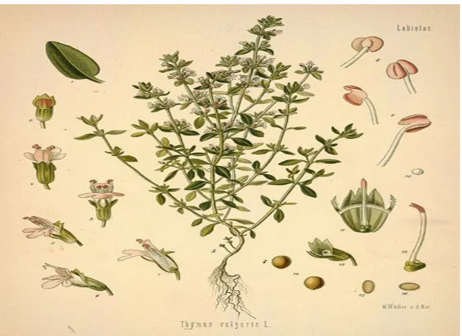
Thymus vulgaris
Thymus vulgaris, (Figure 10) is a well-known plant that has been regarded as a potent medicinal herb for thousands of years. Thyme belongs to the family Lamiaceae, and is an
aromatic perennial, that grows to 20-30 cm in height. The stems are long-slender, woody and
quadrangular, and they grow from a basal center. The leaves are typically grayish-green in color,
smooth oblong-lanceolate in shape, and with little or no petioles which connect the leaves to the
woody stems. Set in pairs, the flowers grow at the top of these slightly twisting foot-stalks in a
whorl-like pattern. The petals grow in together in a form that creates a closed-corolla tube, and
they are typically pinkish-white in color with green sepals at the base of the corolla (Grieves,
Figure 10: Thymus vulgaris. (Grieve, 1995)
The word ―thyme‖ originated from the Greek word thymus, which means brave or
courageous. During ancient times it was used a fumigant, as it's aroma was meant to inspire
courage and bravery, and to invigorate and energize those who smelled it. During the middle
ages, people believed that the scent of thyme could keep away venomous creatures (Grieve,
1995). The flower and leaves were also used to treat a number of ailments. More recently,
thyme is a well-known and potent antiseptic, and useful for oral hygiene (WHO, 1999). The
plant has often been dried and made into an infusion, sweetened with honey and used to treat
sore throats. Syrup made out of the fresh herb was used to treat whooping coughs; it has also
been dried and added to tobacco "to promote good digestion" (Grieve, 1995). Other
folk-medicinal uses include the treatment of coughs which stem from bronchitis, laryngitis, pertussis,
or tonsillitis (WHO, 1999). Present day uses include cooking, cosmetics, food preservation, and
Many of thyme's medicinal activities can be attributed to its essential oil. Essential oils
of plants are typically extracted from the plant leaves and flowers through steam distillation.
Constituents of essential oils include terpenes, monoterpenes, sequesterpines and phenols (Torras
et al., 2007). The major constituents of the essential oil thyme include: phenols, thymol and
carvacrol, which make up about 20-25% of the essential oils. The other constituents include
linaalool, p-cymol, cymeme, thymeme, pinene, apigenin, leteolin and geraniol (Grieves, 1995;
Torras et al., 2007; WHO, 1999). Fabio et al. (2007) found potent antibacterial activity of the
essential oil against seven various bacteria at a minimum concentration of 0.0002 mL/mL. Four
major constituents were chosen for further assessment due to strong scientific evidence of their
effectiveness against resistant bacteria and biofilm formations.
Four Main Constituents of Thyme
Baicalein (Figure 11) is a flavone compound that can be isolated from the methanolic
extract of Thymus vulgaris leaves; it has been shown to produce synergistic activities with tetracycline and various β-lactam antibiotics against methicillin-resistant Staphylococcus aureus
(MRSA). It appears that the mode of action for this synergistic antibiotic effect varies, dependent upon the resistant features of the organism, such as whether or not the MRSA
O
CH3 O
O H
O H
Figure 11: Baicalein.
Rosmarinic acid (Figure 12) is a caffeic acid ester that is excreted naturally by roots of
the plant sweet basil when challenged by Pseudomonas aeruginosa (Walker et al., 2004). It is also produced by several other aromatic herbs including Thymus vulgaris (Wang et al., 2004). Rosmarinic acid has been shown to inhibit planktonic growth of some Pseudomonas spp., and interferes with quorum sensing activities and biofilm development (Walker et al., 2004).
O O
OH O
OH
OH
OH O
H
Figure 12: Rosmarinic acid.
Thymol (2-isopropyl-5-methylphenol) (Figure 13.A) and Carvacrol (2
known for their antimicrobial activity against many organisms, including MRSA. These
compounds have also been shown to interfere withstaphylococcal biofilm growth (Nostro et al.,
2007).
CH3
CH3 C
H3
OH
OH
C
H3 CH3
CH3
Figure 13: A) Thymol and B) Carvacrol.
Purpose
The purpose of this dissertation was to perform preliminary investigations on two
alternative treatment avenues against Pseudomonas aeruginosa infections: quorum sensing inhibition and combination therapy. First, fifty folk medicinal plant extracts were screened for
anti- quorum sensing properties. Next, specific attention was placed on the thyme extract, a folk-
medicinal plant shown to possess anti-quorum sensing properties (Vattam et al., 2007) but also
first shown by Nascimento et al., 2000 to have synergism when combined with tetracycline
against resistant Pseudomonas aeruginosa. Furthermore, this study pursued the isolation and identification of the active component in thyme involved in the thyme-tetracycline effect, and the
elucidation of its mode of action.
Rationale
There are many clues indicating that ethanolic thyme extract may act as a quorum sensing
inhibitor, thus augmenting tetracycline activity. First, the thyme extract has been shown to
augment activity of various antibiotics, such as ampicillin, tetracycline and chloramphenicol
(Nascimento et al., 2000). Interestingly, these three antibiotics work through different modes of
action (Mims et al., 1998). This indicates a more general mode of action for the plant extract.
Aburjai et al. (2001) demonstrated that while the activity of tetracycline (typically active against
Gram-negative organisms) is augmented by various plant extracts, activity is greatly reduced
with other antibiotics such as penicillin G (typically active against Gram-positive bacteria). This
suggests that the mechanism involved may be type-specific. Next, tetracycline resistance occurs
by either expression of efflux pumps and/or ribosome protection expressed via the tet R protein
(Rosen and Mobashery, 1998). It has been shown that quorum sensing can modulate many
virulence genes including efflux pump expression (Lewis, 2001). Thus, control of quorum
sensing activity by an inhibitor would present a logical explanation of these observations.
More evidence suggesting that the thyme extract may include a QSI comes to light upon
examination of various green tea studies. Yam et al. (1997) studied the activity of Camellia sinensis, green tea, against various bacteria. They discovered that ‗cup of tea‘ concentrations showed considerable activity against many pathogenic bacteria including 18 strains of
methicillin resistant Staphylococcus aureus (MRSA). Tiwari et al. (2005) demonstrated that epicatechin gallate (ECGC), a compound found in Camellia sinensis, can produce an amplified effect when combined with the commercial antibiotic chloramphenicol against various strains of
because of ―dual binding sites‖ which were triggered by the plant extract on the bacterial cell
membrane of the pathogen. A different study demonstrated that a possible synergistic interaction
between ECGC and oxacillin might be occurring during treatment of various methicillin-resistant
Staphylococcus aureus (MRSA), that would, in effect, reverse the resistance of the strains (Hamilton-Miller and Shah, 2000). In 2003, epigallocatechin gallate (ECGC) was shown to
interfere with quorum sensing activities in E. coli and Pseudomonas reporter organisms (Huber et al., 2003). Mode of action experiments can be used to determine if this is indeed the case with
the thyme extract.
Specific Aims
1. Screen fifty folk-medicinal plant extracts for QSI activity.
2. Examine specifically the extract of Thymus vulgaris and the ―thyme-tetracycline‖ effect:
a) Isolate and identify the active compound in the thyme extract that augments
tetracycline activity against resistant Pseudomonas aeruginosa.
b) Determine the most efficient way to extract the active compound from the
plant material.
MATERIALS AND METHODS
Chemicals and Reagents
The reagents used for this project—acetone, chloroform, dichloromethane, ethanol,
hexane, and methanol—were histological grade and purchased from Sigma-Aldrich, which is
located in St. Louis, Missouri. Media used for propagating bacterial cultures included
Luria-Bertani (LB), Mueller Hinton II (MH), Tryptic soy agar (TSA), and Nutrient broth (NB) and
came from Difco, located in Sparks, Maryland. Antibiotics—tetracycline free-base (purchased
from MP Biomedical, Inc. in Solon, Ohio) and tobramycin sulfate salt, gentamicin sulfate salt,
and kanamycin sulfate (all purchased from Sigma-Aldrich in St. Louis, Missouri). Thymol was
purchased from Fisher Scientific in Fair Lawn, New Jersey. Baicalein (98%), rosmarinic acid
(97%), and liquid carvacrol (98%) were purchased from Sigma-Aldrich in Steinheim, Germany.
The QSIS2 assay also required the following additives: N-butanoyl-L-homoserine lactone, C4
HSL (both from Cayman Chemical Company in Ann Arbor, Michigan),
N-(3-oxododecanoyl)-L-homoserine lactone, C12 HSL, and 2, 3, 5-triphenyltetrazolium chloride (from Sigma-Aldrich in
St. Louis, Missouri); concentrations were listed below where appropriate.
Bacterial Cultures
The QSIS2 reporter strain, Pseudomonas aeruginosa pLasB-SacB1, was graciously provided by Dr. Michael Givskov of BioCentrum-DTU, Technical University of Denmark, and
ATCC 10145, obtained from the Department of Biology at Georgia State University, was used
for the antibacterial studies.
Plant Samples
Plant material was either purchased from local area farmers‘ markets or hand-picked
from surrounding fields in Atlanta, Georgia. Thymus vulgaris samples were purchased from a local farmers‘ market. Boneset, pokeweed, and yellow root were purchased from Golden Valley
Herbs-Hendersonville as a 40% ethanol extract. Taxonomic identification of all plant samples
was made using a minimum of two sources per specimen, including the United States
Department of Agriculture Natural Resources Conservation Services website
(www.plants.usda.gov) and a dichotomous key, such as Plant Families and How to Identify
Them (Jacques, 1949).
Extraction
Fresh thyme (10 g) was cut into ½-inch pieces and allowed to dry in an aerated incubator
at 55 C for 48 hrs. The plant material was extracted with 200 mL of ethanol in a covered flask
for 24-48 hours. The resultant liquid was filtered through a cotton ball filter into a round-bottom
flask and concentrated using a Buchi R-200 rotary evaporator, thus removing all the ethanol and
resulting in a viscous crude plant extract. All other plant extracts were prepared as described
above and tested at a concentration of 0.5 g/mL- solvent, respectively, except for ginger,
sassafras, rattlesnake master, and red root, which were tested at a concentration of 0.25 g/mL.
was also extracted with solvents including acetone, chloroform, ethyl acetate, hexane, methanol,
methylene chloride, and petroleum ether.
Comparative studies of the extraction of the thyme plant material were also made with
fresh, nonrecycled solvent and recycled solvent, or solvent collected after evaporation of a
previous sample. Fresh solvent extractions were made by soaking the bulk plant material in
previously unused solvent aliquots for each soak within the extraction process. For all recycled
solvent extractions, minimal amounts of fresh solvent were added only if necessary to
compensate for any solvent that may have been lost to evaporation during the extraction process.
Percentage Yield
The percentage yield of crude plant extracts was calculated as follows:
Weight of crude plant extract x 100 = Percentage yield Weight of starting bulk dried plant material
Disc Diffusion Assay (DDA)
Disc diffusion assay, as previously described by Mims et al. (1998) was used for all
antimicrobial studies. A pure culture of P. aeruginosa was prepared onto a nutrient agar plates and incubated aerobically at 37˚C for 24 hours. Next, sterile saline tubes (pH 9.9) were
inoculated with the fresh overnight culture of organisms producing a turbidity equivalent to 0.5
McFarland Standard (BD diagnostic systems). Turbidity was matched through visual
comparison. Mueller Hinton II Agar plates, with 50 g/mL tetracycline, were swabbed
6-millimeter test discs (BD diagnostic systems) were prepared with 20 L of the diluted plant
extract (500 mg/mL) and allowed to air dry for a minimum of three hours. The impregnated
discs were placed aseptically in a radial fashion onto the inoculated plates. After 24 hours of
incubation at 37˚C, active plant samples will inhibit bacterial growth, thus resulting in a ―zone of
inhibition‖ around the disc. The diameter of these zones was measured in millimeters. Positive
control discs (10 L per disc) were prepared with tobramycin sulfate/water solution (1000 g/L).
The negative control discs were treated with straight ethanol.
Thin-Layer Chromatography (TLC)
Crude plant extracts and constituents were analyzed by thin-layer chromatography
(TLC). Samples were diluted in ethanol (100 mg/mL) and spotted on a 250 m 10 x 10 mm
silica gel 60 F254 plates (EMD Chemicals). Next, the plates were developed in 8:4:1 hexane,
dichloromethane, acetone system and allowed to air-dry in a fume hood. Plates were then
analyzed under an ultraviolet light for separation of compounds based on polarity.
Thin-Layer Chromatography Bioassay
Thin-Layer Chromatography Bioassay (TBA) adapted from Slusarenko et al. (1989) was
used for identification of the active antimicrobial components in the thyme extract. Plant extracts
and constituents were analyzed by thin-layer chromatography (TLC). Samples were diluted in
ethanol (100 mg/mL) and spotted on a 250 m 10 x 10 mm silica gel 60 F254 plate (EMD
Chemicals). Next, the plates were developed in 8:4:1 hexane, dichloromethane, acetone solvent
system and allowed to air-dry in a fume hood. An aliquot (25 mL) of nutrient broth with 1%
(grown aerobically overnight on nutrient agar at 37 C) was diluted into nutrient broth and grown
at 37 C in an orbital shaker (150-200 rpm) for 24 hours. Second aliquots (25 mL) of nutrient
broth were prepared with 1.5% (w/v) agar, 0.25 mL of 1% glycerol and 50 g/mL tetracycline
and referred to as TBA agar. They were later cooled to 45 C. The dye 2, 3,
5-triphenyltetrazolium chloride was added to the agar, gently agitated, and set aside. The diluted
culture (1 mL) was added to the 25 mL TBA agar tubes and agitated. Finally, 50 mL of the agar
(2 tubes) were carefully poured into large Petri plates containing the thin-layer chromatography
plates. After 24 hours of incubation at 37 C, active compounds were identified by zones of
inhibition of microbial growth. The positive control was tobramycin, and it was spotted on the
TLC plate after the plate was developed; the negative control was any area of the TLC plate
without extract. The experiment was run in triplicate.
Activity-Driven Fractionation
The concentrated crude extracts were separated, based on polarity, into fractional
constituents, using an appropriate gradient solvent system on a solid-phase extraction (SPE)
product silica 5 g/20 mL column (Fisher Scientific, Fair Lawn, New Jersey) using a twelve-port
vacuum chromatography manifold.
First, the column was washed with pure hexane to prepare the silica. Next, the crude
extract was dissolved into methanol (~5 mL) added to the column. Then, the column was washed
with a minimal volume of hexane to allow the extract to saturate the top 5% of silica in the tube.
The column was then washed with hexane, totaling approximately 90 mL of hexane, until no
color was seen in the collected solvent. The first fraction was the combined hexane tubes. This
same solvent were recombined and roto-evaporated to remove eluting solvent from the fraction.
Fractions were re-diluted as needed for used for further testing.
QSIS2 Assay
The QSIS2 assay, adapted from Rasmussen et al. (2005b), with slight modifications as
described below, was used to screen plant extracts for C4 HSL and C12 HLS analogues. LB Agar
was made with the addition of 15.5 g sucrose (total volume 250 mL). The pH was adjusted to 5.0
with the addition of hydrochloric acid. Gentamicin (0.0247 g), kanamycin (0.0215 g), 1.1 mL of
each C4 HSL (0.00595 g/L of ethanol), and C12 HSL (0.00342 g/L of ethanol) plus 0.20 g of 2, 3,
5-triphenyltetrazolium chloride dye was added into 25 mL of phosphate-buffered saline (PBS).
The augmented PBS (25 mL) plus 2.5 mL of overnight QSIS2 reporter strain, grown in ABT
media (Rasmussen et al., 2005b), was added to the tempered LB agar and poured into Petri
plates. Next, the plates were allowed to solidify for 25 minutes. Wells (7 mm) were bored into
the solidified agar. Various dilutions of the plant extracts and major constituents were (50 µL per
well) were added to the wells. Agar plates were allowed to sit at room temperature for one hour
prior to incubation to allow the test solution ample time to diffuse into the agar. Plates were
incubated at 37ºC for 12-16 hours.
When QS-mediated gene expression is activated in the QSIS2 reporter strain, sucrose
mediated cell death occurs, but if QSI compounds are present in the test sample, QS-mediated
cell death is blocked and the reporter strain is able to grow. When cellular growth is achieved, a
zone of dark red growth is visible around the test well in only the areas where QSI molecule
concentration offsets QS signal concentration. The red color is caused by the metabolism of
zone of growth. Antimicrobial activity was detected by a transparent clearing around the well,
referred to as a ―zone of inhibition.‖ A second zone, referred to as a ―zone of growth,‖ consisted
of a bright red halo around the well, which indicated QSI activity. Experiments were run in
triplicate unless otherwise specified and zone diameters were measured in millimeters. All
diameter measurements were recorded in millimeters.
Chromobacterial QSI Assay
The Chromobacterial QSI assay protocol was adapted from Bosgelmez-Tinaz et al.
(2007) and was used for scanning samples for C4- C6 HSL analogues. Chromobacterium
violaceum ATCC 12472 was propagated in 5 mL of nutrient broth and incubated in a shaker incubator for 24 hours at 30º C. LB Agar (250 mL) and LB broth with 0.3 % agar (250 mL) were
used to prepare an overlay assay. First, a Petri plate was filled halfway to capacity with LB Agar
and allowed to solidify for 30 minutes. Next, LB Agar (0.3%) was inoculated with 1 mL of
overnight culture. The tempered agar was poured over the LB Agar base and allowed to solidify
at room temperature. Wells (7 mm) were bored into the solidified agar. The test solution (50 µL
per well) was added into wells. Plates were left at room temperature for another hour to allow
test samples to diffuse into the agar. The plates were incubated for 16 hours at 30ºC. Violaceum
color production is under QS-mediated control. If the plant extract contains a QSI activity then
cellular growth will occur but pigment production will be prevented. Wells with zones of
inhibited color (opaque) were considered QSI active (zone of growth), and clear zones around
the well (transparent) were considered antimicrobial (zones of inhibition). Experiments were run
Garlic as the Positive Control for QSI Testing
A toluene garlic extract, adapted from Rasmussen et al. (2005b) was used as a positive
control for all QSI studies. Garlic (150 g) was shredded with a commercial blender and
extracted in 300 mL of toluene for 24 hours. After 24 hours, the sample was filtered through a
Whatman No. 1 filter. Next, the collected material was combined with 150 mL of sterile water
and mixed for 24 hours at room temperature. A separatory funnel was used to collect the toluene
extract (top layer), which was subsequently used as a QSI-positive control.
Glucose and pH Testing
Glucose concentrations of the crude plant extracts were measured using True Test gold
sensor strips with a True Test to-go blood/glucose meter (Home Diagnostics, Fort Lauderdale,
Florida). Crude extracts were first prepared by diluting the sample to a 0.2 g/mL into deionized
water. One drop of the extract was placed into a makeshift aluminum foil bowl. The meter strip
was then dipped into the solution. The solution travels up the strip by capillary action. Glucose
concentrations were read of the off the digital screen and recorded in dg/mL.
The pH of the samples was determined using colorpHast pH strips (EDM Chemicals).
The strips were submerged in the above 0.2 g/mL extract/water samples. Results were compared