R E S E A R C H A R T I C L E
Open Access
Effects of femoral bone defect morphology
on initial polished tapered stem stability in
massive defect model: a biomechanical
study
Tohru Irie
1*, Daisuke Takahashi
1, Tsuyoshi Asano
1, Tomohiro Shimizu
1, Ryuta Arai
1, Alaa Muhammad Terkawi
1,
Yoichi M. Ito
2and Norimasa Iwasaki
1Abstract
Background:Good outcomes have been reported in revision total hip replacement with massive segmental defects using impaction bone grafting with circumferential metal meshes. However, the morphology of defects that require a mesh is poorly defined. The purpose of this study was to evaluate the effects of a variety of segmental defects on load transmission to the proximal femur under both axial and rotational loads.
Methods:Initial stability of the Exeter stem was investigated in a composite bone model using three medial bone defect morphologies: Long (length 5 cm × width 2 cm), Short (2.5 cm × 2 cm), Square (3.2 cm × 3.2 cm), Square with mesh (3.2 cm × 3.2 cm defect covered with metal mesh), and with no defect as control. Specimens (5 per group) were axially loaded and internally rotated up to 20° or to failure. Strain distributions of the femora were measured using a strain gauge.
Results:All Square group specimens failed while rotation was increasing. In the other four groups, failure was not observed in any specimens. Mean torsional stiffness in the Long (4.4 ± 0.3 Nm/deg.) and Square groups (4.3 ± 0.3 Nm/deg.) was significantly smaller than in the Control group (4.8 ± 0.3 Nm/deg.). In the medio-cranial region, the magnitude of the maximum principal strain in the Square group (1176.4 ± 100.9) was significantly the largest (Control, 373.2 ± 129.5,p< 0.001; Long, 883.7 ± 153.3,p= 0.027; Short, 434.5 ± 196.8, p < 0.001; Square with mesh, 256.9 ± 100.8, p < 0.001). Torsional stiffness, and both maximum and minimum principal strains in the Short group showed no difference compared to the Control group in any region.
Conclusions:Bone defect morphology greatly affected initial stem stability and load transmission. If defect
morphology is not wide and the distal end is above the lower end of the lesser trochanter, it may be acceptable to fill the bone defect region with bone cement. However, this procedure is not acceptable for defects extending distally below the lower end of the lesser trochanter or defects 3 cm or more in width.
Keywords:Revision total hip replacement, Femoral bone defect, Circumferential metal mesh, Initial stem stability
© The Author(s). 2019Open AccessThis article is distributed under the terms of the Creative Commons Attribution 4.0 International License (http://creativecommons.org/licenses/by/4.0/), which permits unrestricted use, distribution, and reproduction in any medium, provided you give appropriate credit to the original author(s) and the source, provide a link to the Creative Commons license, and indicate if changes were made. The Creative Commons Public Domain Dedication waiver (http://creativecommons.org/publicdomain/zero/1.0/) applies to the data made available in this article, unless otherwise stated. * Correspondence:irixt0430@gmail.com
1Department of Orthopaedic Surgery, Faculty of Medicine and Graduate
School of Medicine, Hokkaido University, Kita 15, Nishi 7, Kita-ku, Sapporo 060-8638, Japan
Background
Revision of total hip replacement (THR) is challenging, though good clinical results have been reported [1, 2]. Progressive bone stock loss accompanies THR loosening. Moreover, removing the failed stem, and in some cases, cement, may result in further defects in the proximal femur. With polished tapered stems, proximal femoral bone defects have less effect on initial stem stability than with uncemented stems, so procedures equivalent to pri-mary THR or the impaction bone grafting technique (IBG) without reinforcement are occasionally used in re-vision THR in cases of relatively small bone defects. While initial stem stability is essential in revision THR, a medial segmental defect in the femur can greatly attenu-ate initial stem stability [2–4]. Therefore, managing bone defects of the proximal femur is a central problem in revision THR.
Good clinical results have been reported for revision THR using IBG, which can also restore bone stock [1, 2]. However, major segmental defects, especially of the calcar femorale, have been reported to affect clinical out-comes [2]. When massive segmental defects are present, circumferential metal meshes are effective to create a stable reconstruction [3–5]. However, the specific extent of a bone defect that can cause severe reduction of initial stem stability remains unclear. Therefore, the morph-ology of defects that require circumferential metal mesh in revision THR is poorly defined.
For good long-term clinical results, not only initial stem stability but also load transmission to the femur is important [6, 7]. Uneven stress distribution leads to stress shielding. Furthermore, the fragility of peripros-thetic bone caused by stress shielding increases the risk of mechanical loosening or periprosthetic fracture [7].
The hypothesis tested in this study was that proximal femur bone defect morphology would greatly influence initial stem stability and load transmission to the prox-imal femur. The purpose of this study was to evaluate the effects of a variety of segmental defects on load transmission to the proximal femur, under both axial and rotational loads, in order to analyze initial polished tapered stem stability.
Methods
Specimen preparations
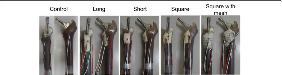
Initial stability of the Exeter stem (Exeter V40, 150 mm length, 44 mm offset, size no. 1, Stryker Orthopaedics, Mahwah, NJ) was investigated. A total of 25 composite Sawbones (medium left femur model 3403; Pacific Re-search Laboratories, Vashon, WA) were prepared. A standard femoral neck cut was made 1 cm proximal to the lesser trochanter. The bones were then broached using the appropriate Exeter broaching rasp. Following broaching, three kinds of proximal medial segmental
defects were made (Fig.1). In the Long group, the defect extended 5 cm distally from the resection level and had a width of 2 cm. This morphology was based on the method of Bolder et al. [3], and its length was equivalent to one-third of the stem length, creating a worst-case scenario in the distal direction. In the Short group, the defect morphology was defined as half of the length (2.5 cm) and the same width (2 cm) as the Long type defect. In the Square group, the defect morphology was a square with a side length of 3.2 cm, such that the area (theoretically 1024 mm2) was nearly equal to that of the Long type defect (theoretically 1000 mm2). This defect was created as a worst-case scenario in the anterior-pos-terior direction. Furthermore, the square type defect was reconstructed with a metal mesh (X-change Rim mesh-medium, Stryker Orthopaedics, Newbury, UK) as the Square with mesh group, which was fixed with 4 cerc-lage wires (diameter, 1.0 mm). After insertion of a ce-ment plug, retrograde insertion of cece-ment dough (CMW, DePuy Synthes, Warsaw, IN) was performed using a cement gun, followed by cement pressurization. The stem was then cemented, seated with the third marking at the level of the neck cut, using a stem cen-tralizer. In groups with defects, the defects were covered by hand with gauze during the insertion of cement dough and stems to keep containment of the proximal part of the femur. We also evaluated stability in a no-de-fect control model. We tested 5 specimens in each group.
Mechanical testing
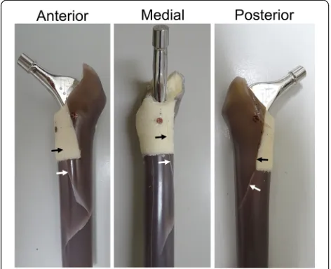
The load-displacement behavior of the stem and the prox-imal femur strain distribution were measured using an Instron ElectroPuls E10000 (Instron Systems, Norwood, MA, dynamic linear load capacity: ±10 kN, dynamic torque capacity: ±100 Nm) mechanical testing machine. For strain gauge measurements, three rosettes (KFG-1-120-D17-11L3M2S, Kyowa Electronic Instruments Co., Ltd., Tokyo, Japan, strain measurement range limit at room temperature: 5.0%) were externally bonded to anterior, pos-terior, and medial surfaces 17 mm distally from the prox-imal edge of the femoral neck cut [5]. The wires from the rosettes were connected to a data logger (UCAM-550A, Kyowa Electronic Instruments Co., Ltd., Tokyo, Japan).
to 3 kN, at which point the subsidence distance was measured. The femora were then tested in the rotational test while under compression. The compressive load of 3 kN was maintained, and the implant was internally ro-tated at a crosshead speed of 0.5°/sec up to 20° or the limit of complex failure. Complex failure was defined as fracture of the composite bone or fracture of the ce-ment. The torque and the strain distribution of the prox-imal femur were measured during internal rotation. Fracture torque was defined as the maximum measured torque with complex failure. Fracture energy was calcu-lated for the sample for which complex failure was ob-tained by numerically integrating the torque and angle measurements against time. Torsional stiffness was cal-culated from the torque-angle curve along the linear re-gion from 5 Nm to 65 Nm of torque.
Micro CT analysis
Following the experiment and after removing the stems, the proximal femur and cement complexes were evalu-ated with micro computed tomography (CT) (R mCT, Rigaku Corporation, Tokyo, Japan) at 20μm/pixel to confirm the failure mechanism. The imaging conditions of micro CT were 80 kV and 100μA.
Statistical analysis
The delta was calculated using data from our pilot study. Because the delta was calculated to be 1.8, a sample size of 5 femurs in each group was required to provide 80% power. The subsidence distance during the axial com-pression test up to 3 kN, the torsional stiffness with torque from 5 to 65 Nm, the rotation angle at torque of 70 Nm, and the maximum and minimum principal strain of the antero-cranial, postero-cranial, and medio-cranial regions of the femur at torque of 70 Nm were each sta-tistically compared. All statistical analyses were per-formed using JMP Pro 14 software (SAS Institute Japan, Tokyo, Japan). Pairwise comparisons among each of the 5 groups were performed using ANOVA with Tukey’s post hoc test. Data is presented as mean ± SD and the
corresponding 95% confidence intervals. A p value < 0.05 was considered significant.
Results
Axial compression test
In all groups, complex failure was not observed in any specimens. Mean subsidence distance under the axial compression test was not significantly different among the five groups (Table1).
Rotational test
Mean torsional stiffness with torque from 5 to 65 Nm in both the Long group (4.4 ± 0.3 [95% confidence interval {CI}, 4.0 to 4.7] Nm/deg.) and the Square group (4.3 ± 0.3 [95% CI, 4.0 to 4.7] Nm/deg.) was significantly smaller than in the Control group (4.8 ± 0.3 [95% CI, 4.5 to 5.2] Nm/deg.), although the stiffness did not differ among the Control group, the Short group (4.5 ± 0.2 [95% CI, 4.3 to 4.8] Nm/deg.) and the Square with mesh group (4.6 ± 0.2 [95% CI, 4.4 to 4.8] Nm/deg.) (Table 1, Fig. 3a). Mean internal rotation angles at torque of 70 Nm were not significantly different among the five groups (Table1, Fig.3a).
In the Square group, all specimens failed at the prox-imal part of the femur while rotation was increasing up to 20°. Mean fracture torque was 74.0 ± 3.7 (95% CI, 69.4 to 78.6) Nm and mean fracture energy was 13.6 ± 2.0 (95% CI, 11.1 to 16.0) J (Table 2). All fractures showed a spiral fracture pattern. In all Square group specimens, fracture lines were observed in the distal dir-ection continuing from the medio-cranial region of the bone cement into the composite bone (Fig. 4). In the other four groups, complex failure was not observed in any specimens.
[image:3.595.61.538.88.215.2]the rotational test also showed two or three steps that correspond to the steps on the rotational angle-torque curve (Fig.3b and c).
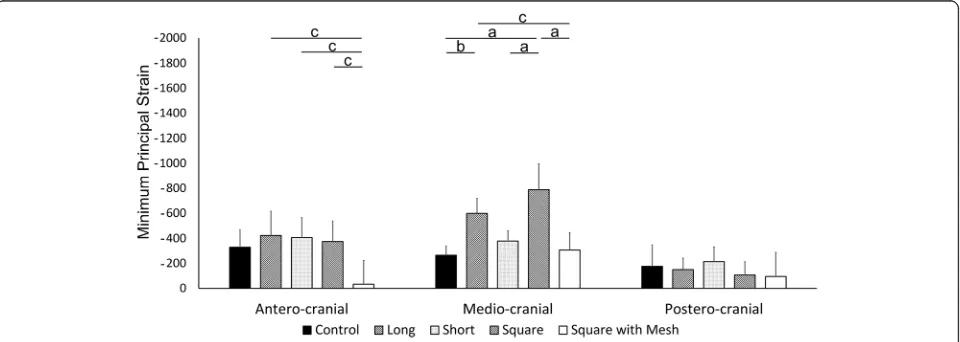
In the antero-cranial region, the magnitude of the maximum principal strain at torque of 70 Nm in the Long group (127.2 ± 133.0 [95% CI, −37.9 to 292.3]) was significantly the smallest among the five groups (Control group, 1229.6 ± 137.1 [95% CI, 1059.4 to 1399.8], p< 0.001; Short group, 950.6 ± 111.2 [95% CI, 812.4 to 1088.7], p < 0.001; Square group, 904.9 ± 244.8 [95% CI, 600.9 to 1208.9], p < 0.001; Square with mesh group, 955.1 ± 147.6 [95% CI, 771.9 to 1138.4], p < 0.001). This magnitude in the Square group was significantly smaller than the Control group (p= 0.034). However, there were no significant differences among the Short group, the Square group, and the Square with mesh group (Fig. 5). In the same region, the absolute value of the minimum principal strain in the Square with mesh group (−32.8 ± 190.3 [95% CI, −269.0 to 203.4]) was significantly smaller than in the Long group (−423.0 ± 192.0 [95% CI, − 661.3 to −184.6], p= 0.013), Short group (−406.9 ± 158.1 [95% CI, −603.2 to −210.6], p= 0.018), and Square group (−374.6 ± 165.5 [95% CI, −580.0 to −
169.1], p= 0.035). However, there were no significant differences among the Control (−328.3 ± 140.9 [95% CI, −503.2 to −153.4]), Long, Short, and Square groups (Fig. 6).
In the medio-cranial region, the magnitude of the maximum principal strain in the Square group (1176.4 ± 100.9 [95% CI, 1051.2 to 1301.7]) was significantly the largest among the five groups (Control group, 373.2 ± 129.5 [95% CI, 212.4 to 533.9], p< 0.001; Long group, 883.7 ± 153.3, [95% CI, 693.3 to 1074.1],p= 0.027; Short group, 434.5 ± 196.8, [95% CI, 190.2 to 678.9], p < 0.001; Square with mesh group, 256.9 ± 100.8, [95% CI, 131.8 to 382.0], p < 0.001) (Fig.5) and the absolute value of the minimum principal strain in the Square group (− 788.8 ± 208.1 [95% CI, −1047.2 to −530.3]) was signifi-cantly larger than in the Control group (−264.4 ± 75.0 [95% CI, −357.5 to −171.3], p< 0.001), Short group (− 379.2 ± 80.9, [95% CI,−479.6 to−278.7], p < 0.001), and Square with mesh group (−307.4 ± 139.0, [95% CI, − 480.0 to −134.8], p < 0.001) (Fig.6). In the same region, the magnitude of the maximum principal strain in the Long group was significantly larger than in the Control group (p < 0.001), the Short group (p= 0.001), and the Square with mesh group (p < 0.001) (Fig. 5). Addition-ally, the absolute value of the minimum principal strain in the Long group (−598.4 ± 118.7 [95% CI,−745.9 to− 451.0]) was significantly larger than in the Control group (p= 0.006) and the Square with mesh group (p= 0.019) (Fig.6).
In the postero-cranial region, the magnitude of the maximum principal strain in the Square group (555.7 ± 105.1 [95% CI, 425.2 to 686.1]) was significantly the smallest (Control group, 1440.3 ± 275.5 [95% CI, 1098.2 to 1782.3], p < 0.001; Long group, 1093.1 ± 347.0, [95% CI, 662.3 to 1524.0], p= 0.022; Short group, 1483.5 ± 288.4, [95% CI, 1125.4 to 1841.6], p < 0.001; Square with mesh group, 1288.7 ± 158.1, [95% CI, 1092.4 to 1485.0],
p= 0.002) (Fig. 5). In the same region, the absolute values of the minimum principal strains were not signifi-cantly different among the five groups (Control group,
−176.7 ± 171.5 [95% CI,−389.6 to 36.2]; Long group,− 150.2 ± 92.0 [95% CI, −264.4 to−36.0]; Short group,− 213.6 ± 115.8 [95% CI, −357.4 to−69.8]; Square group,
−106.5 ± 105.5 [95% CI, −237.6 to 24.5]; Square with mesh group, −122.2 ± 192.7 [95% CI, −361.5 to 117.1]) (Fig.6).
Micro CT evaluation
Micro CT showed some cement cracks different from the composite bone fracture lines in all Square group specimens (Fig. 7). In the other four groups, neither fracture lines nor cement cracks were observed in any specimens.
[image:4.595.57.291.83.387.2]Discussion
The purpose of this study was to evaluate the effects of a variety of segmental defects on load transmission to the proximal femur, in order to analyze initial polished tapered stem stability. Bolder et al. reported that the presence of a large medial segmental defect in the femur specifically reduces prosthetic stability, and that metal mesh was effective in creating a stable stem construction in a goat femur model [3]. However, the specific extent of a bone defect that can cause severe reduction of initial stem stability remains unclear. Therefore, it was un-known what type of defect requires reinforcement, such as metal meshes. In the present study, all specimens in the Square group failed while rotation was increasing up to 20°, and fracture lines were observed in the distal dir-ection continuing from the medio-cranial region of the bone cement into the composite bone. Additionally,
both the rotational angle-torque curve and the torque-strain curve showed two or three steps before complex failure, and the absolute values of both the maximum and minimum principal strains in the medio-cranial re-gion were significantly the largest in the Square group, except that the absolute value of the minimum principal strain showed no difference compared to the Long group. In addition, the mean torsional stiffness with torque from 5 to 65 Nm in the Square group was signifi-cantly smaller than in the Control group. Furthermore, micro CT showed some cracks in the cement mantle that were different from the composite bone fracture lines.
[image:5.595.56.541.110.199.2]The elastic modulus and ultimate strength of poly-methyl methacrylate are one-third to one-seventh that of cortical bone [9,10]. Considering this, it is reasonable to suppose that the cement cracks occurred during Table 1Subsidence distance during the axial compression test up to 3 kN, torsional stiffness with torque from 5 to 65 Nm under the rotational test, and internally rotated angle at torque of 70 Nm with 3 kN of compressive load
Defect morphology (length × width) (cm) (area) (mm2)
Control (none) (0)
Long (5 × 2) (1000)
Short (2.5 × 2) (500)
Square (3.2 × 3.2) (1024)
Square with Mesh (3.2 × 3.2) (1024)
Pvalue
Subsidence distance (mm) 2.6 ± 0.1 (2.4 to 2.8)
2.5 ± 0.1 (2.4 to 2.6)
2.5 ± 0.1 (2.4 to 2.7)
2.6 ± 0.3 (2.3 to 2.9)
2.5 ± 0.3 (2.2 to 2.9)
0.97
Torsional stiffness (Nm/deg.) 4.8 ± 0.3 (4.5 to 5.2)
4.4 ± 0.3* (4.0 to 4.7)
4.5 ± 0.2 (4.3 to 4.8)
4.3 ± 0.3* (4.0 to 4.7)
4.6 ± 0.2 (4.4 to 4.8)
0.03
Internally rotated angle (deg.) 15.0 ± 1.2 (13.5 to 16.4)
15.4 ± 0.6 (14.6 to 16.2)
15.7 ± 0.4 (15.2 to 16.2)
15.7 ± 0.5 (15.2 to 16.3)
15.1 ± 0.4 (14.7 to 15.6)
0.29
Values are expressed as mean ± SD with 95% confidence interval in parentheses; * significant compared with the Control group.
[image:5.595.57.543.448.683.2]rotation before breakage of the composite bone. Because the first step was observed at a mean torque of 37.6 ± 5.7 Nm, there is a possibility that cement cracking may occur even with weak torque in cases of wide defects such as the Square group defect, although mean fracture torque was 74.0 ± 3.7 Nm. Kaneuji et al. reported that the greatest compressive force at the bone-cement inter-face was observed at the proximal medial region in an in vitro polished tapered stem model [11, 12]. The present study suggests that, if revision THR is performed with-out reinforcement despite a morphologically wide bone defect, the mechanical stress is concentrated excessively in the bone cement, and this excessive mechanical stress may cause early periprosthetic fracturing, cement crack-ing, and/or lead to early mechanical loosening. These re-sults suggest that, even if a bone defect fits Paprosky classification type 2A [13, 14], when wide medial seg-mental defects more than 3 cm in width are present, reinforcement is necessary to prevent periprosthetic fracture or early mechanical loosening.
Although the areas of the defects of the Long group and the Square group were nearly equal, neither com-plex failure nor cement cracking was observed in the Long group. The present study showed that distally ex-tended bone defects categorized as Paprosky classifica-tion type 2C, such as the Long group defect, affect initial stem stability less than wider defects, such as the Square group defect.
In the medio-cranial region, the magnitude of the maximum principal strain in the Long group was signifi-cantly larger than in the Control group, the Short group, and the Square with mesh group. In the same region, the absolute value of the minimum principal strain in the Long group was also significantly larger than in the Control group, and the Square with mesh group. Mean-while, in the antero-cranial region, the magnitude of the maximum principal strain in the Long group was signifi-cantly the smallest among the five groups, although the absolute value of the minimum principal strain showed no difference except for the Square with mesh group.
It has been recognized that polished tapered stems slip in the cement and create considerable radial compres-sive loads at the bone-cement interface [11, 12, 15]. In the case of a distally extended defect, the tensile stress in the anterior region due to internal rotation may be absorbed at the bone defect region and not transmitted to the anterior part of the femur. Such imbalance of the stress distribution may cause stress shielding of the an-terior proximal bone, resulting in bone fragility [16,17]. The quality of periprosthetic bone determines the risk of periprosthetic fracture [7], and a high incidence of early periprosthetic fractures is associated with polished ta-pered stems [8,18–20]. Furthermore, our results showed that the mean torsional stiffness in the Long group was significantly smaller than in the Control group. If revision THR with defects that extend below the lower end of the lesser trochanter is performed without reinforcement, this bone defect may cause loosening or periprosthetic fracture postoperatively. The present re-sults suggest that it is preferable to perform IBG with circumferential metal mesh for defects with such morphology to obtain good long-term clinical results.
[image:6.595.55.292.122.190.2]The torsional stiffness in the Short group did not differ from that of the Control group. Additionally, in the medio-cranial region, the magnitude of the maximum principal strain in the Short group was significantly smaller than in the Long and the Square groups and the absolute value of the minimum principal strain was also significantly smaller than in the Square group. Further-more, the absolute values of both the maximum and minimum principal strains in the Short group showed no difference compared to the Control group in any re-gion. These results suggest that, if defect morphology is not wide and the distal end is at or above the lower end Table 2Number of steps observed in the rotation angle-torque
curve by complex failure, torque when the first step is observed, fracture torque, and fracture energy in Square group
Mean ± SD (95% CI)
Number of steps (specimens) 2 steps: 4, 3 steps: 1
Torque of the first step (Nm) 37.6 ± 5.7 (30.5 to 44.6)
Fracture torque (Nm) 74.0 ± 3.7 (69.4 to 78.6)
Fracture energy (J) 13.6 ± 2.0 (11.1 to 16.0)
Values are expressed as mean ± SD with 95% confidence interval in parentheses.
[image:6.595.56.292.481.673.2]of the lesser trochanter, attenuation of initial stem stabil-ity is minimal. This is consistent with reports that the extent of the bone defect affects the outcome of revision THR [2].
The procedure of femoral IBG is recognized as tech-nically demanding, and one of the reasons for this is the frequent occurrence of intraoperative periprosthetic femoral fractures [1, 21, 22]. Additionally, a higher inci-dence of postoperative femoral fractures, excessive fem-oral component subsidence, dislocation, and deep joint infections has been reported in femoral reconstruction with IBG compared to primary THR [21,23,24]. Hence, if bone defect morphology is limited, similar to the Short group defect, a surgical procedure to fill the bone defect region with bone cement without IBG can be consid-ered, especially for patients with high risk of surgical site infection, or patients who cannot tolerate large surgical invasion. Although evaluation of bone defects before surgery and preoperative planning are very important for revision THR [23], it is widely recognized that
preoperative evaluation of bone defects is difficult [25, 26], and that new bone defects may occur at the time of stem or bone cement removal [27, 28]. An IBG proced-ure is ideal in all cases of bone defect. However, circum-stances such as the absence of an experienced surgeon, or a lack of graft bone or materials required for IBG may occur. In such cases, it may be acceptable to fill the bone defect region with bone cement, if it is limited to a morphology comparable to our Short group defect. However, this procedure is not acceptable for cases with larger defects, as demonstrated by our Long and Square groups.
It is difficult to accurately reproduce actual clinical condi-tions with an experimental study. High torsional loads occur due to forces on the femoral head during stair climbing or rising from a chair, which can cause critical micromotion and may result in stem loosening [4,20,29]. Thus, it is im-portant to evaluate initial stem stability not only against axial load, but also against torsional load [30,31]. To reproduce actual clinical conditions as closely as possible, the axial load Fig. 5Magnitude of maximum principal strain at 70 Nm of torque with 3 kN of compressive load. a:p< 0.001, b:p< 0.01, c:p< 0.05
[image:7.595.61.539.87.259.2] [image:7.595.59.539.544.715.2]was applied and then maintained, and a rotational test with the femoral head center at the center of rotation was per-formed. This is a strength of the present study.
There were several limitations. First, a composite Saw-bone model was used in this study. Malkani et al. pointed out that torsional strength and stiffness were primarily dependent on variability in bone quality in in-dividual specimens [32]. Since inin-dividual differences among samples cannot be avoided in cadaveric bones and animal bones, a composite bone model was used to minimize intersample variability. Although this Sawbone is a composite bone for mechanical testing, it does not have cancellous bone, which may affect cementing and initial stability. Second, IBG was not performed in the Square with mesh group. Since the femoral IBG proced-ure is technically demanding, there is a risk of increasing intersample variability. Additionally, initial stem stability in IBG has already been sufficiently evaluated [3–5]. Since cortical bone has much stronger mechanical prop-erties than cancellous bone [33] or impacted bone [34, 35], proximal femoral cortical bone defects in particular affect initial stability. Although we recognized that tight packing of the bone graft may affect initial stability, the primary objective of this study was to clarify the effects of a variety of segmental defects on stem stability. Therefore, the focus was on evaluating the effect of seg-mental defect morphology. Third, mechanical stress dis-tribution on composite bone was not directly measured. The direct measurement of bone stress distribution, especially around cemented orthopedic implants, re-mains challenging. Kaneuji et al. reported a method for evaluating compressive force at the bone-cement inter-face, which has been recognized as excellent [11, 12].
However, this method requires complete fixation of the bone in the device, embedding the entire femur except for the proximal part. Hence, this set up could prevent femoral bone failure, which was the primary outcome variable for the present study. Additionally, drilling holes into the femur to insert a measuring rod would alter its structural integrity, thereby influencing the outcome var-iables of complex failure and bone strain. Considering that complex failure or internal rotation angle 20° were the endpoints of our rotation test, it is reasonable to as-sume that load transmission to the femur was evaluated by measuring the composite bone strain using a strain gauge.
[image:8.595.58.540.87.275.2]investigations under the same conditions are necessary to evaluate the effects of long type and short type defects on fracture torque and fracture energy.
Conclusions
The present study showed that the morphology of prox-imal femur bone defects greatly affected initial polished tapered stem stability and load transmission to the prox-imal femur. Even with proxprox-imal medial segmental bone defects of approximately the same size, wider defects in the anterior and posterior attenuate initial stability sig-nificantly more than vertically long defects. In contrast, if defect morphology is not wide and the distal end is at or above the lower end of the lesser trochanter, attenu-ation of load transmission and initial stem stability are minimal. In such cases, it may be acceptable to fill the bone defect region with bone cement. However, this pro-cedure is not acceptable for cases with bone defects ex-tending distally below the lower end of the lesser trochanter or wide defects with a width of 3 cm or more.
Abbreviations
CT:Computed tomography; IBG: Impaction bone grafting; THR: Total hip replacement
Acknowledgements
We would like to thank Allen Paul Heffel and Philip Malloy for English language editing.
Authors’contributions
TI conceived of the study, participated in its design, coordination and biomechanical testing, and helped to draft the manuscript. DT, AMT, and NI conceived of the study, and participated in its design and helped to draft the manuscript. TA, TS, and RA participated in study design and
biomechanical testing. YMI participated in the statistical analysis and helped to draft the manuscript. All of the abovementioned authors have read and approved the final version of the manuscript.
Funding
This work was partially supported by a research grant from Johnson & Johnson K.K. (Japan) to Hokkaido University. The funding sources had no role in: the design of the study; collection, analysis, or interpretation of the data; preparation of the manuscript; or its submission for publication.
Availability of data and materials
The datasets used in the present study are available from the corresponding author on reasonable request.
Ethics approval and consent to participate
Not applicable.
Consent for publication
Not applicable.
Competing interests
This work was partially supported by a research grant from Johnson & Johnson K.K. (Japan) to Hokkaido University.
Author details
1
Department of Orthopaedic Surgery, Faculty of Medicine and Graduate School of Medicine, Hokkaido University, Kita 15, Nishi 7, Kita-ku, Sapporo 060-8638, Japan.2Department of Biostatistics, Graduate School of Medicine,
Hokkaido University, Sapporo, Japan.
Received: 11 February 2019 Accepted: 11 July 2019
References
1. Iwase T, Otsuka H, Katayama N, Fujita H. Impaction bone grafting for femoral revision hip arthroplasty with Exeter universal stem in Japan. Arch Orthop Trauma Surg. 2012;132(10):1487–94.
2. Garcia-Cimbrelo E, Garcia-Rey E, Cruz-Pardos A. The extent of the bone defect affects the outcome of femoral reconstruction in revision surgery with impacted bone grafting: a five- to 17-year follow-up study. J Bone Joint Surg Br. 2011;93(11):1457–64.
3. Bolder SB, Schreurs BW, Verdonschot N, Ling RS, Slooff TJ. The initial stability of an Exeter femoral stem after impaction bone grafting in combination with segmental defect reconstruction. J Arthroplast. 2004;19(5):598–604. 4. Guala AJ, Buttaro M, Piccaluga F. Initial stability of circumferential meshes
with impacted bone allografts for massive femoral defects. Int Orthop. 2008; 32(5):605–9.
5. Takigami I, Otsuka H, Yamamoto K, Iwase T, Fujita H, Matsuda S, et al. Proximal femoral reconstruction with impaction bone grafting and circumferential metal mesh. J Orthop Sci. 2015;20(2):331–9.
6. Morita D, Iwase T, Ito T. Bone restoration with cemented Exeter universal stem - three-years longitudinal DEXA study in 165 hips for femur. J Orthop Sci. 2016;21(3):336–41.
7. Chandran P, Azzabi M, Andrews M, Bradley JG. Periprosthetic bone remodeling after 12 years differs in cemented and uncemented hip arthroplasties. Clin Orthop Relat Res. 2012;470(5):1431–5.
8. Morishima T, Ginsel BL, Choy GG, Wilson LJ, Whitehouse SL, Crawford RW. Periprosthetic fracture torque for short versus standard cemented hip stems: an experimental in vitro study. J Arthroplast. 2014;29(5):1067–71. 9. Kuhn KD. Bone cement: up-to-date comparison of physical and chemical
properties of commercial materials. Springer-Verlag, Berlin. 2000:60–6. 10. Aziz NA. Bone grafts and bone substitutes: Basic science and clinical
applications. Singapore: World Scientific; 2005. p. 31–7.
11. Kaneuji A, Yamada K, Hirosaki K, Takano M, Matsumoto T. Stem subsidence of polished and rough double-taper stems: in vitro mechanical effects on the cement-bone interface. Acta Orthop. 2009;80(3):270–6.
12. Numata Y, Kaneuji A, Kerboull L, Takahashi E, Ichiseki T, Fukui K, et al. Biomechanical behaviour of a French femoral component with thin cement mantle: The 'French paradox' may not be a paradox after all. Bone Joint Res. 2018;4;7(7):485–493.
13. Paprosky WG, Greidanus NV, Antoniou J. Minimum 10-year-results of extensively porous-coated stems in revision hip arthroplasty. Clin Orthop Relat Res. 1999;369:230–42.
14. Pak JH, Paprosky WG, Jablonsky WS, Lawrence JM. Femoral strut allografts in cementless revision total hip arthroplasty. Clin Orthop Relat Res. 1993;295:172–8. 15. Lee AJC, Parkins RD, Ling RSM. Time-dependent properties of
polymethylmethacrylate bone cement: the interaction of shape of femoral stems, surface finish and bone cement. Springer-Verlag, London. 1990:85–90. 16. Sumner DR. Long-term implant fixation and stress-shielding in total hip
replacement. J Biomech. 2015;18;48(5):797–800.
17. Sayyidmousavi A, Bougherara H. Investigation of stress shielding around the Stryker Omnifit and Exeter periprosthetic hip implants using an irreversible thermodynamic-based model. J Biomed Mater Res B Appl Biomater. 2012;100(5):1416–24. 18. Scott T, Salvatore A, Woo P, Lee YY, Salvati EA. Gonzalez Della Valle a.
polished, collarless, tapered, cemented stems for primary hip arthroplasty may exhibit high rate of Periprosthetic fracture at short-term follow-up. J Arthroplast. 2018;33(4):1120–5.
19. Brodén C, Mukka S, Muren O, Eisler T, Boden H, Stark A, et al. High risk of early periprosthetic fractures after primary hip arthroplasty in elderly patients using a cemented, tapered, polished stem. Acta Orthop. 2015;86(2):169–74.
20. Erhardt JB, Khoo PP, Stoffel KK, Yates PJ. Periprosthetic fractures around polished collarless cemented stems: the effect of stem design on fracture pattern. Hip Int. 2013;23(5):459–64.
21. Edwards SA, Pandit HG, Grover ML, Clarke HJ. Impaction bone grafting in revision hip surgery. J Arthroplast. 2003;18(7):852–9.
22. Farfalli GL, Buttaro MA, Piccaluga F. Femoral fractures in revision hip surgeries with impacted bone allograft. Clin Orthop Relat Res. 2007;462:130–6. 23. Leone WA Jr, Naughton M, Gratto-Cox G, Luland CM, Kilgore JE, Hill GE. The
patients with moderate to severe bone loss mean 4.7-year results. J Arthroplast. 2008;23(3):383–94.
24. Pekkarinen J, Alho A, Lepistö J, Ylikoski M, Ylinen P, Paavilainen T. Impaction bone grafting in revision hip surgery. A high incidence of complications. J Bone Joint Surg Br. 2000;82(1):103–7.
25. Parry MC, Whitehouse MR, Mehendale SA, Smith LK, Webb JC, Spencer RF, et al. A comparison of the validity and reliability of established bone stock loss classification systems and the proposal of a novel classification system. Hip Int. 2010;20(1):50–5.
26. Saleh KJ, Holtzman J, Gafni A, Saleh L, Davis A, Resig S, et al. Reliability and intraoperative validity of preoperative assessment of standardized plain radiographs in predicting bone loss at revision hip surgery. J Bone Joint Surg Am. 2001;83-A(7):1040–6.
27. Duymus TM, Solak Z, Ozturkmen Y, Azboy I, Mutlu S, Caniklioglu M. Mid-term results of previously cemented hip arthroplasties revised with uncemented modular femoral components: a retrospective study. J Orthop Surg Res. 2015;14;10:123.
28. Ornstein E, Atroshi I, Franzén H, Johnsson R, Sandquist P, Sundberg M. Early complications after one hundred and forty-four consecutive hip revisions with impacted morselized allograft bone and cement. J Bone Joint Surg Am. 2002;84-A(8):1323–8.
29. O'Connor DO, Burke DW, Jasty M, Sedlacek RC, Harris WH. In vitro measurement of strain in the bone cement surrounding the femoral component of total hip replacements during simulated gait and stair-climbing. J Orthop Res. 1996;14(5):769–77.
30. Crowninshield RD, Johnston RC, Andrews JG, Brand RA. A biomechanical investigation of the human hip. J Biomech. 1978;11(1–2):75–85. 31. Mjöberg B, Hansson LI, Selvik G. Instability, migration and laxity of total hip
prostheses. A roentgen stereophotogrammetric study. Acta Orthop Scand. 1984;55(2):141–5.
32. Malkani AL, Voor MJ, Fee KA, Bates CS. Femoral component revision using impacted morsellised cancellous graft. A biomechanical study of implant stability. J Bone Joint Surg Br. 1996;78(6):973–8.
33. Rho JY, Ashman RB, Turner CH. Young's modulus of trabecular and cortical bone material: ultrasonic and microtensile measurements. J Biomech. 1993; 26(2):111–9.
34. Cornu O, Schubert T, Libouton X. Particle size influence in an impaction bone grafting model. Comparison of fresh-frozen and freeze-dried allografts. J Biomech. 2009;16;42(14):2238–2242.
35. Dunlop DG, Brewster NT, Madabhushi SP, Usmani AS, Pankaj P, Howie CR. Techniques to improve the shear strength of impacted bone graft: the effect of particle size and washing of the graft. J Bone Joint Surg Am. 2003; 85-A(4):639–46.
36. Ginsel BL, Morishima T, Wilson LJ, Whitehouse SL, Crawford RW. Can larger-bodied cemented femoral components reduce periprosthetic fractures? A biomechanical study. Arch Orthop Trauma Surg. 2015;135(4):517–22.
Publisher’s Note