R E S E A R C H A R T I C L E
Open Access
Local biochemical and morphological differences
in human Achilles tendinopathy: a case control
study
Pingel J
1*, Fredberg U
2, Qvortrup K
3, Larsen JO
4, Schjerling P
1, Heinemeier K
1, Kjaer M
1and Langberg H
5Abstract
Background:The incidence of Achilles tendinopathy is high and underlying etiology as well as biochemical and morphological pathology associated with the disease is largely unknown. The aim of the present study was to describe biochemical and morphological differences in chronic Achilles tendinopathy. The expressions of growth factors, inflammatory mediators and tendon morphology were determined in both chronically diseased and healthy tendon parts.
Methods:Thirty Achilles tendinopathy patients were randomized to an expression-study (n= 16) or a structural-study (n= 14). Biopsies from two areas in the Achilles tendon were taken and structural parameters: fibril density, fibril size, volume fraction of cells and the nucleus/cytoplasm ratio of cells were determined. Further gene expressions of various genes were analyzed.
Results:Significantly smaller collagen fibrils and a higher volume fraction of cells were observed in the tendinopathic region of the tendon. Markers for collagen and its synthesis collagen 1, collagen 3, fibronectin, tenascin-c, transforming growth factor-bfibromodulin, and markers of collagen breakdown matrix metalloproteinase-2, matrix metalloproteinase-9 and metallopeptidase inhibitor-2 were significantly increased in the tendinopathic region. No altered expressions of markers for fibrillogenesis, inflammation or wound healing were observed. Conclusion:The present study indicates that an increased expression of factors stimulating the turnover of connective tissue is present in the diseased part of tendinopathic tendons, associated with an increased number of cells in the injured area as well as an increased number of smaller and thinner fibrils in the diseased tendon region. As no fibrillogenesis, inflammation or wound healing could be detected, the present data supports the notion that tendinopathy is an ongoing degenerative process.
Trial registration:Current Controlled Trials ISRCTN20896880
Keywords:Collagen, Gene expression, Patients, Growth factors, Tissue turnover
Background
Tendons connect muscle to bone and enable transmis-sion of forces from contracting muscle to bone, resulting in joint movement. They possess the ability to adapt to changes in loading [1] and studies have shown that col-lagen synthesis is increased as a result of both acute exer-cise [2,3] and prolonged physical training [4]. The
adaptation to loading can ultimately lead to increases in CSA and collagen content in chronically loaded tendons [5]. Despite this physiological ability to adapt, tendinopa-tic tendons represents a large and constantly growing clinical problem affecting both recreational and profes-sional athletes as well as people involved in repetitive labour [6,7]. Years of research have unfortunately not provided much insight into the pathogenesis of chronic tendinopathy [8]. Indeed, the etiology of tendinopathy has been related to repeated micro strain below the fail-ure threshold as an initiating stimulus for degenerative processes [9,10]. Other authors, however, have proposed
* Correspondence: [email protected]
1Institute of Sports Medicine, Department of Orthopedic Surgery M.
Bispebjerg Hospital and Center for Healthy Aging, Faculty of Health Sciences, University of Copenhagen, Copenhagen, Denmark
Full list of author information is available at the end of the article
that mechano-biological under-stimulation results in a degenerative cascade, through the production of a pat-tern of catabolic gene expression that leads ultimately to extracellular matrix degeneration [11]. Tendinopathy is characterized by activity-related pain, focal tendon ten-derness, and decreased local movement in the affected area [12,13]. The general opinion is that no inflammatory cells are present in the tendinopathic tissue [14] and that tendinopathy is the result of a degenerative process with collagen disorganization, collagen fibre separation, increased cellularity, neovascularization and focal necro-sis [15].
Previous studies have shown an altered concentration of certain matrix metalloproteinases MMPs, AdAMt’s and TIMP’s in normal and degenerate human Achilles tendon [16]. Additionally several cytokines [9,10] can be found in tendons and fibroblasts after cyclic mechanical stretching in healthy tendon tissue. However, the published data arises from the comparison of tendinopathic tissue with either control tissue from different anatomical tendons [17] or with tissues from identical anatomical tendons but from different subjects [18]. Since considerable micro-scopic structure differences have been demonstrated in anatomically different tendons [19], this limits the conclu-sions that may be drawn from these studies. Taking the aforementioned limitations into account, current data con-cerning local biochemical differences within tendinopathic tendons, seem to indicate that an altered expression of col-lagen [20], proteoglycans [21] and matrix metalloprotei-nases [16,22] exists in tendinopathic tendons. In addition the level of cytokines [23] VEGF and fibronectin [24] has been shown to be significantly different in the tendino-pathic area. However analyses of local biochemical differ-ences together with morphological differdiffer-ences are lacking.
The aim of the present study was to elucidate if any local structural differences are present in tendinopathic areas of human Achilles tendons compared to healthy areas in the same tendon. Furthermore, we wanted to investigate which proteoglycans, growth factors and cytokines that were involved in the local structural dif-ferences observed.
We hypothesize that several markers such as collagen 3 would be locally up regulated indicating formation of scar tissue with in the tendon [25] and higher concentrations of MMP-2 and MMP-9 indicating an enhanced degrada-tion of collagen structures in the tendinopathic area (t-area) when compared with the healthy area (h-(t-area) of the same tendon. Furthermore it is hypothesized that certain proteoglycans would have altered expression in the two tendon regions, e.g. an increased expression of decorin which might cause the collagen turnover to be increased also in chronic tendinopathic tendons. Additionally we hypothesize that growth factors like fibroblast growth
factor (bFGF) are decreased causing a reduced healing capacity in the injured area of the Achilles tendons.
Methods Design
Thirty patients with chronic Achilles tendon pain were included in this study approved by the local Ethical Com-mittee of the Capital Region Copenhagen (H-1-2009-114) and in compliance with the Helsinki Declaration. Addi-tionally the study was registered at Current Controlled Trials (ISRCTN20896880). Due to limitations in the amount of tissue gained from the tendon biopsies patients were randomly assigned to either a Structural study (n= 14) or a Biochemical study (n= 16) by the envelope method. All subjects were recreational athletes or workers with a long-term history of chronic Achilles tendon pain (> 1/2 year) (Table 4) and conventional con-servative treatments (eccentric rehabilitation, NSAIDs and corticosteroid injections) had been tried in all indivi-duals with no effect. Intake of NSAID or corticosteroid injection was not allowed 6 months prior to inclusion in the present study. All subjects were recruited from the Rheumatology Department, Silkeborg Hospital, Den-mark, and the biopsies from the Achilles tendons were taken as part of a standard procedure in order to examine for deposits of cholesterol, uric acid, and amyloid in the injured Achilles tendons.
Biopsy procedure
The subjects were locally anesthetized, in the peritendi-nous space from both the medial and lateral side of the tendon with injections of 2 × 10 ml 1% Lidocain, using ultrasound guidance. Biopsies were taken with a semi-automatic biopsy needle (14 GA, 9 cm; Angiotech) also using ultrasound (US) guidance. An initial tendon biopsy was taken in the maximally sick area evaluated using US (defined as the area with maximal increased tendon thick-ness, neovascularisation, hypoeccogenicity). This area was usually 3-5 cm above the attachment of the Achilles ten-don to the calcanaeus bone. A second biopsy was taken from the same tendon 4 cm proximal to the first biopsy in a region of the tendon tissue that was deemed normal using US.
Biopsy samples intended for analysis using Transmission Electron Microscopy were immersed in 2% glutaraldehyde in 0.05 M sodium buffer (pH 7.2), and the samples for gene expression were snap-frozen and stored at -80°C until analysis.
phosphate buffer (pH 7.2) the specimens were post-fixed in 1% OsO4 in 0.12 M sodium cacodylic buffer for 2 hours. The specimens were dehydrated in a graded series of ethanol (70%, 96% and 100%), transferred to propylene oxide and embedded in Epon (VWR Bie&Berntsen) in three steps according to standard procedures. For each biopsy one ultra thin section was cut approximately per-pendicular to the length axis of the tendon with a Reich-ert-Jung ultracut E microtome. The section was collected on a one-hole copper grid with a Formvar supporting membrane and stained with uranyl acetate and lead citrate. The sections were examined using a Phillips CM 100 transmission electron microscope operated at an accelerating voltage of 80 kV. Digital images were obtained with a MegaView II camera and an analysis software pack-age. From each ultra thin section the intercellular tissue was examined by taking a simple, random sample of ten digitized TEM images of the intercellular tissue. The cellu-lar component of the tendon was examined in eleven biopsy pairs by taking 6 times 6 images in three randomly positioned regions of the section. The 6 times 6 images were spliced into one image using multiple image align-ment (MIA) tools, so for each examined biopsy a total of three MIA images were obtained.
Stereology
The Stereological analyses of the images were carried out on a computer monitor onto which the digitized EM image was merged with a graphic representation of the stereological test systems for just 12 of the 14 biopsy pairs (2 biopsies was unfortunately not useable for stereology analyses) (C.A.S.T.-grid software, The Interna-tional Stereology Center at Olympus). The intercellular tissue was analyzed at a final magnification of 210.000 in the ten ordinary TEM images. The volume fraction (Vv) of collagen fibrils per intercellular tissue volume was esti-mated with the point counting technique as the number of points hitting collagen fibres divided by the number of points hitting the intercellular tissue (including collagen fibrils) using a point grid of 36 points. The number of collagen fibrils per cross sectional area of intercellular tis-sue (NA) was counted in 16 uniformly positioned, unbiased counting frames, each with an area of 0.0426 mm2(42.6μm2), and the individual diameters (d) of the sampled collagen fibrils were measured as the largest dia-meter perpendicular to the longest axis (i.e. the length of the minor axis of the ellipse) using the“measure-length” feature of the CAST-grid system. The unbiased counting frame ensures that all profiles, regardless of shape, size or orientation, have an equal probability of being sampled within area probe. The MIA images were analyzed at a final magnification of 115,000. The point counting tech-nique, using a point grid with approximately 1000 points, estimated the volume fractions of the cellular component
of the tendon tissue. The estimated parameters were: the volume fraction of cells within the tendon, the volume fraction of the nucleus within the cell, and the volume fraction of cytoplasm within the cell. A single experi-enced investigator performed all stereological analyses in a blinded fashion. The investigator was blinded for all subject characteristics, and whether the sample was obtained from the tendinopathic or the healthy region of the tendon.
RNA extraction and real time-PCR analysis
Total RNA isolation: Total RNA was extracted from fro-zen tendon samples from 16 subjects (sample weight: mean 23.2 ± 6.4 mg) by using 1 ml of TRI Reagent (Mole-cular Research Centre, Cincinnati, OH) 5 steel beads (2.3 mm) and 4 silica beads (1.0 mm Silicon Carbide Beads (454 grams) BioSpec Products Inc.). Glycogen was added (120μg per ml of TriReagent) to the tendon samples to improve RNA precipitation.
Extracted RNA was precipitated from the aqueous phase with isopropanol and was washed with ethanol [75%], dried and suspended in 10μl of nuclease-free water. The RNA concentration was determined using a RiboGreen RNA Quantitation kit 200-2000 Assays, Molecular Probes USA. RNA quality was determined on the basis of a RNA 6000 nano Chip assay kit, Agilent Technologies, Germany. The RNA samples were stored frozen at -20°C until subse-quent use in real-time RT-PCR procedures.
cDNA synthesis: 100 ng RNA was reverse transcribed for each tendon sample in a total volume of 20μl by using the QiagenOmniscript RT Kit at 37°C for 1 hour followed by 70°C for 15 minutes. The resulting cDNA was diluted twenty times in dilution buffer (10 mMTris EDTA buffer: Sigma Germany) + Salmon Testes DNA (1 ng/μl; Sigma Germany), and samples were stored at -20°C until used in the PCR reactions for specific mRNA analysis.
the melting temperature of DNA (95°C) for 10 minutes, followed by 50 cycles each of 15 seconds at 95°C, followed by the annealing step where optimal primer hybridization conditions were obtained by lowering the temperature to 58°C for 30 seconds, and the extension step, where the reaction mix was heated to 63°C for 90 seconds. Two housekeeping genes GAPDH and RPLP0 were used as reference genes. The RPLP0 gene had been chosen as an internal control, assuming RPLP0 to be constitutively expressed. To validate this assumption GAPDH mRNA was measured as another unrelated“constitutive”and nor-malized with RPLP0, showing no difference between the healthy and the tendinopathic region of the tendon (Figure 3).
Statistical analysis
The PCR data were log transformed and a Paired Students
t-test was performed to compare the results from the healthy area of the tendon with the tendinopathic area of the tendon, with exception of the results from IL-6, IL-1b, ki67 and HGF-1. These gene targets could not be detected in all samples. In these cases Chi2tests were performed. All PCR data are presented as the geo mean ± backtrans-formed SEM. The collagen fibril data were divided into area and diameter fractions, and a paired Studentst-test was performed to compare each fraction between the healthy and the tendinopathic area of the tendon. Like-wise, the volume fraction of cells and the volume fraction of the nucleus within the cell were compared using a Paired Studentt-test comparing the two areas of the ten-don. A P-value < 0.05 was considered to be significant and all data despite of the subject characteristics are shown as Mean ± SEM.
Results
Structural composition of the tendon
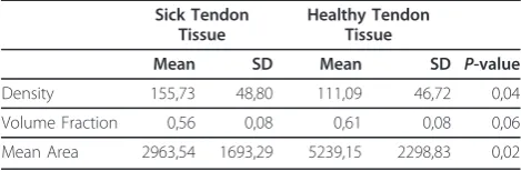
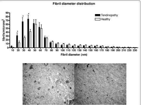
The density, volume fraction and mean area of the col-lagen fibrils were measured in biopsies from 14 of the ten-dinopathy patients (Table 1). The density of collagen fibrils was found to be significantly higher and the mean area of the collagen fibrils was significantly smaller in ten-dinopathic tendon region compared to that of healthy control region, additionally a trend towards significant dif-ference was found in volume fraction (Table 1). When analysing the individual bins in the diameter distribution
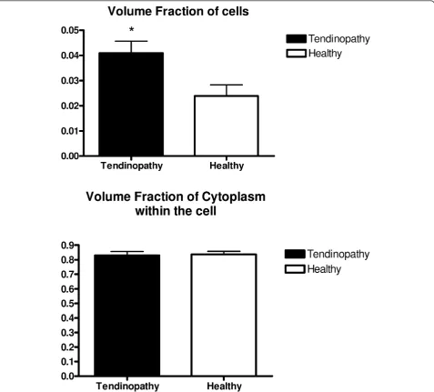
of the fibrils, a significantly higher number of fibrils with a diameter in the lower range (10-40 nm) was found in the tendinopathic area compared to the healthy area (diameter 10-20 nm: Tendinopathic area: 32 ± 7 fibrils/μm2; Healthy area 13 ± 4 fibrils/μm2; diameter: 20-30 nm: Tendino-pathic area: 68 ± 10 fibrils/μm2; Healthy area: 26 ± 4 fibrils/μm2; diameter 30-40 nm: Tendinopathic area: 74 ± 10 fibrils/μm2; Healthy area: 42 ± 5 fibrils/μm2; see Figure 1). In addition a significantly higher volume fraction of cells was observed in the tendinopathic area of the ten-don (Figure 2). The volume fraction of the cytoplasm within the cell was found to be identical in the two areas (Figure 2) implying an increased number of cells in the tendinopathic area.
Gene expression analysis
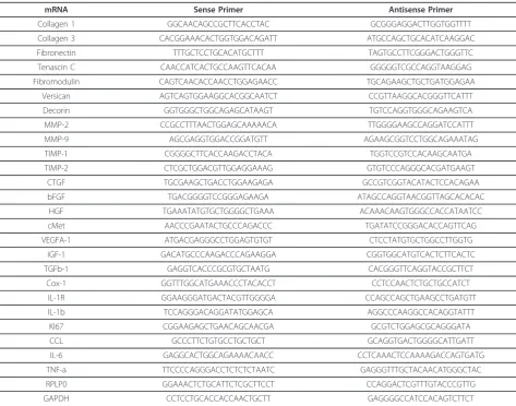
The gene expression of 24 genes was analyzed in the biopsies of 16 tendinopathy patients (Table 2). As men-tioned in the methods section, GAPDH mRNA served as a normalization factor for the genes of interest, and RPLP0 mRNA was used to validate the stability of the expression of GAPDH mRNA. Specific gene targets were selected covering the areas of: Extracellular matrix (ECM) components, degradation components, Growth factors, inflammatory markers and fibrillogenesis.
Extracellular matrix components: The expression of sev-eral structural components (collagen 1 and collagen 3) together with mRNA of fibronectin, tenascin-c and fibro-modulin was found to be significantly up regulated in the t-area (Figure 3). Versican was found to be unchanged and decorin tended to be decreased in the t-area compared to that of the h-area of the Achilles tendon (Figure 3).
Degradation factors: Expression of MMP-2, MMP-9 and TIMP-2 was significantly increased in the t-areas com-pared to that of the h-areas with no difference in expres-sion of TIMP-1 (Figure 3).
Growth factors: A significant up-regulation of TGF-b1 expression was observed in the t-area compared to that of the h-area, whereas bFGF and cmet expression in the same area was significantly decreased. No differences were observed in the expression of CTGF, VEGF-A1 and IGF-1 (Figure 4). The expression of HGF-1 could not be detected in all samples but no significant differences were found between the two regions of the tendon (Table 3).
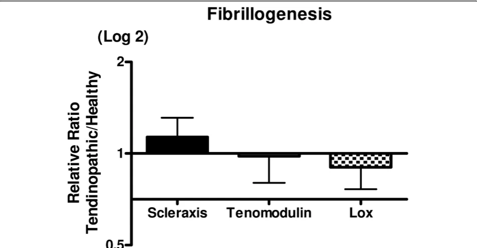
[image:4.595.56.291.654.731.2]Inflammatory markers: Data on IL-6, IL-1b and ki67 could not be detected in all samples and no differences were observed when the two regions were compared (chi squared test). Ki67 showed a significant decrease in expression in the tendinopathic areas. No expression could be detected of COX-2 and TNF-ain any of the samples (results not shown). COX-1, IL-1R and CCL2 was detected in all samples, but there was no significant differ-ence between the t-area and the h-area of the tendon (Fig-ure 4). Fibrillogenesis: There were no differences in
Table 1 Tendon fibril characteristics
Sick Tendon Tissue
Healthy Tendon Tissue
Mean SD Mean SD P-value
Density 155,73 48,80 111,09 46,72 0,04
Volume Fraction 0,56 0,08 0,61 0,08 0,06
expression of scleraxis, tenomodulin and Lysyloxidase (LOX) between the two areas (Figure 5)
Discussion
The major finding of the present study is that tendino-pathy causes focal biochemical and morphological differ-ences in the human Achilles tendon. The data obtained using TEM indicate that the structural composition of the t-area has a significantly increase in the number of smaller collagen fibrils compared to the h-area of the same tendon. This supports our hypothesis that loca-lized structural differences are present in tendinopathic Achilles tendons, potentially as a result of an increased turnover of the tissue in an attempt to heal potentially injured tissue. This fits with previous findings where a site-specific loss of larger collagen fibrils and an increase of fibrils with a small diameter was observed in Achilles tendons after tendon rupture [26]. However the present findings differ somewhat from the results observed in another study from our laboratory [27], in which a
[image:5.595.58.541.88.447.2]tendon, the limitation is that no control tissue is available from tendons that never had any symptoms. Changes that occur in the whole tendon can therefore be overlooked. A previous study has shown that histological changes in the tendon were not only present at the site of rupture but also in the macroscopical normal part of the tendon, indi-cating that local alterations of the tissue might not neces-sarily be local [29]. Whether tendinopathy shows the same pattern is unclear and needs further investigation. How-ever we believe that the present local differences between the t-area and the h-area are strong enough to justify the conclusions of the present study. To investigate if the increased number of small collagen fibrils could be due to genesis of collagen fibrils or degradation of previously much larger fibrils, the gene expression of scleraxis and
[image:6.595.60.538.89.520.2]structure, fibre arrangement, nuclear rounding and cellu-larity [15]. In the present study a significant increased volume fraction of cells was observed in the tendinopathic area of the tendon using TEM. This is in line with pre-vious animal studies of Soslowsky and colleagues [33-35], where rats ran with a velocity of 17 m/minute, 5 days/ week, 1 hour/day, either uphill or downhill for a period of between 2-16 weeks. In such experiments, a decreased col-lagen fibre organization and increased numbers of cell nuclei were observed [36,37]. The present TEM analysis did unfortunately not allow for distinguishing between the cell types that were counted, and thus it was not possible to exclude that other cell types than just fibroblasts might have migrated into the t-area of the tendon. The signifi-cantly higher mRNA expression of both collagen I and collagen III in the t-area shows a higher collagen synthesis of the tendon. At the same time indicates the higher expression of MMP-2 and MMP-9 in the t-area an increased collagen matrix degradation. Together these findings display a higher collagen turnover in the t-area of
[image:7.595.60.534.98.470.2]the tendon. It has previously been shown that normal ten-don tissue express matrix metalloproteinases and that a homeostatic turnover is necessary for tendon regeneration and maintenance [16]. A increased collagen turnover is usually associated with adaptation to exercise [2] or heal-ing of the tendon [38]. It is still puzzlheal-ing why the increased collagen turnover in the tendons of chronic patients like the present has not resulted in a decrease in symptoms or a healing of the tissue (symptoms range: 0.5-10 years Table 4). However these data are confirmed in other stu-dies also showing increased collagen turnover in tendino-pathic tendons [22,24] and in tendon ruptures [39]. Alterations in proteoglycans have previously been asso-ciated with tendon pathology [40,41]. Proteoglycans and glycoproteins are essential for the maintenance of homeos-tasis of the ECM of the tendon and achieve this by regulat-ing the collagen fibril assembly [42]. The upregulation of tenascin-C, and fibronectin is consistent with earlier find-ings [24,43]. The observed unchanged levels of versican contrasts earlier findings, where a significant down
Table 2 PCR primers
mRNA Sense Primer Antisense Primer
Collagen 1 GGCAACAGCCGCTTCACCTAC GCGGGAGGACTTGGTGGTTTT
Collagen 3 CACGGAAACACTGGTGGACAGATT ATGCCAGCTGCACATCAAGGAC
Fibronectin TTTGCTCCTGCACATGCTTT TAGTGCCTTCGGGACTGGGTTC
Tenascin C CAACCATCACTGCCAAGTTCACAA GGGGGTCGCCAGGTAAGGAG
Fibromodulin CAGTCAACACCAACCTGGAGAACC TGCAGAAGCTGCTGATGGAGAA
Versican AGTCAGTGGAAGGCACGGCAATCT CCGTTAAGGCACGGGTTCATTT
Decorin GGTGGGCTGGCAGAGCATAAGT TGTCCAGGTGGGCAGAAGTCA
MMP-2 CCGCCTTTAACTGGAGCAAAAACA TTGGGGAAGCCAGGATCCATTT
MMP-9 AGCGAGGTGGACCGGATGTT AGAAGCGGTCCTGGCAGAAATAG
TIMP-1 CGGGGCTTCACCAAGACCTACA TGGTCCGTCCACAAGCAATGA
TIMP-2 CTCGCTGGACGTTGGAGGAAAG GTGTCCCAGGGCACGATGAAGT
CTGF TGCGAAGCTGACCTGGAAGAGA GCCGTCGGTACATACTCCACAGAA
bFGF TGACGGGGTCCGGGAGAAGA ATAGCCAGGTAACGGTTAGCACACAC
HGF TGAAATATGTGCTGGGGCTGAAA ACAAACAAGTGGGCCACCATAATCC
cMet AACCCGAATACTGCCCAGACCC TGATATCCGGGACACCAGTTCAG
VEGFA-1 ATGACGAGGGCCTGGAGTGTGT CTCCTATGTGCTGGCCTTGGTG
IGF-1 GACATGCCCAAGACCCAGAAGGA CGGTGGCATGTCACTCTTCACTC
TGFb-1 GAGGTCACCCGCGTGCTAATG CACGGGTTCAGGTACCGCTTCT
Cox-1 GGTTTGGCATGAAACCCTACACCT CCTCCAACTCTGCTGCCATCT
IL-1R GGAAGGGATGACTACGTTGGGGA CCAGCCAGCTGAAGCCTGATGTT
IL-1b TCCAGGGACAGGATATGGAGCA AGGCCCAAGGCCACAGGTATTT
KI67 CGGAAGAGCTGAACAGCAACGA GCGTCTGGAGCGCAGGGATA
CCL GCCCTTCTGTGCCTGCTGCT GCAGGTGACTGGGGCATTGATT
IL-6 GAGGCACTGGCAGAAAACAACC CCTCAAACTCCAAAAGACCAGTGATG
TNF-a TTCCCCAGGGACCTCTCTCTAATC GAGGGTTTGCTACAACATGGGCTAC
RPLP0 GGAAACTCTGCATTCTCGCTTCCT CCAGGACTCGTTTGTACCCGTTG
regulation of versican was observed in both tendinopathic and ruptured tendon tissue [40]. This discrepancy might lie in the medical history of the patients. The present
[image:8.595.61.535.89.642.2]observed tendency to a decrease of decorin expression in the t-area of the tendon was contrary to our hypothesis. It has been previously shown that a down-regulation of dec-orin using anti-sense decdec-orin injections improved ligament healing in rabbits [44]. Whether the present finding of a
[image:9.595.57.539.86.632.2]matrix assembly leading to a delayed fibril formation and formation of thinner fibrils [45,46]. Treatment with injec-tions of growth factors for tendon injuries has received much attention in recent years. Growth factors are poly-peptide molecules that are decisive regulation of cell meta-bolism and cell proliferation and are associated with tendon healing [47-51]. Studies using local administration of bFGF [52,53], HGF [54,55] and IGF [56] have all shown beneficial effects in the healing process of tendon injuries, but not all injuries were tendinopathies though. Exogen-ous injections of bFGF in human patellar tendons have been shown to increase wound healing bothin vitroand
in vivoin patellar tendon models after surgery [52,53], and likewise, collagen type III and cell proliferation was increased after exogenous bFGF injections in patellar ten-dons after surgery in vivo [53]. Recently, our group showed that the cytokine IL-6 could act as a growth factor in tendon tissue [57]. Moreover, local injections of rhIL-6 have been shown to increase collagen synthesis in humans
[image:10.595.57.292.100.194.2]after one hour of exercise in the form of running, in healthy young men [57]. However, the issue as to whether local injections of IL-6 in tendinopathy patients may be beneficial to the healing process of a damaged tendon is still unknown. The pain that tendinopathy patients experi-ence has previously among other been related to Sub-stance-P, a neuro-peptide with various biological functions including pain transmission [58,59]. Since no difference in Substance P expression were observed between the two areas, the present data might indicate that other factors than substance P can be responsible for the pain in tendi-nopathic tendons. It is however also possible that the expression of Substance-P is increased in the whole ten-don and therefore overlooked due to the previously men-tioned limitations of the present design. Although the role of inflammation in tendinopathy is one that is often dis-cussed, it has long been known that tendinopathy is pri-marily a degenerative condition, in which inflammatory cells in or around the lesion are absent. All markers of inflammation that were measured showed no upregulated expression in the t-area of the tendon (Table 3). This underlines that long-term tendinopathy is not primarily an inflammatory process, but rather an ongoing degenerative process. Although inflammation is absent in tendinopathy at this late stage, it does not rule out that an inflammation insult was present at the initiation of the tendinopathic process [60,61]. In fact, various inflammatory mediators like TNF-alpha, IL-6, IL-15, IL-18 have been shown to play a role especially in wound healing after injury [62]
Table 3 Inflammatory markers
Healthy Tendinopathy Chi test
(detected/not detected) (detected/not detected)
IL-6 9/7 10/6 0.7
IL-1b 4/12 6/10 0.4
ki67 10/6 8/8 0.5
HGF-1 10/6 7/9 0.3
total 16 16
[image:10.595.60.536.453.700.2]and in early stages of tendinopathy [23,63]. However, since inflammation can not be detected in later stages of chronic tendinopathy, the present data may indicate that the use of anti-inflammatory treatments e.g. NSAIDs are not relevant for chronic tendinopathy patients.
Conclusion
The present study examined the differences in structural proteins, cellular volume densities and expression levels of various genes involved in regulation of matrix proteins in clinically and ultrasonographiclytendinopathic regions of the human Achilles tendon and healthy regions within the same Achilles tendons. The main findings were dif-ferences in the composition of collagen structures with the tendinopathic region containing significantly higher number of small size fibrils (diameter 10-40 nm) com-pared to the healthy region of the tendon. In addition, the tendinopathic region had a significantly higher volume fraction of cells, compatible with a greater num-ber of cells per unit volume. Furthermore, expression of several genes involved in both collagen synthesis and col-lagen degradation was significantly up-regulated, an observation that is consistent with an increased local turnover of collagen tissue in the affected tendinopathic area of the tendon. Gene expression was also influenced by the disease as several factors involved in wound heal-ing were expressed at a lower number in the tendino-pathic area. Lastly no sign of increased inflammation was found in the diseased region. Taken together these data indicate that local morphological and biochemical differ-ences are present within the tendon during Achilles ten-dinopathy. These findings may have implications in the choice of treatment for these patients.
Acknowledgements
We are grateful to Dr. A.P. Harrison, Faculty of LIFE Sciences, Copenhagen University, for reading and correcting this manuscript. This study was supported by grants from the Danish Rheumatism Association, The NovoNordic Foundation, the Danish Ministry of Culture Committee for Sports Research, the Danish Medical Research Counsel (22-04-0191) and the Nordea foundation (Healthy Aging grant).
Author details
1Institute of Sports Medicine, Department of Orthopedic Surgery M.
Bispebjerg Hospital and Center for Healthy Aging, Faculty of Health Sciences, University of Copenhagen, Copenhagen, Denmark.2Department of
Rheumatology, Silkeborg Hospital, Silkeborg, Denmark.3Department of Biomedical Sciences, University of Copenhagen, Copenhagen, Denmark.
4
Department of Neuroscience and Pharmacology, Faculty of Health Sciences,
University of Copenhagen, Copenhagen, Denmark.5Department of Public
Health, University of Copenhagen, Copenhagen, Denmark.
Authors’contributions
JP carried out the study, made the PCR analyses, made the statistics and drafted the manuscript. UF recruited all patients, took all biopsies. KQ made all the Transmission Electron Microscopy analyses, JOL made all stereology analyses and cell counts, PS and KH supervised the gene expression analyses and PS designed all PCR primers and supervised the statistical analyses. MK and HL participated in designing and coordinating the study and helped to draft the manuscript. All authors read and approved the final manuscript.
Competing interests
The authors declare that they have no competing interests.
Received: 5 December 2011 Accepted: 5 April 2012 Published: 5 April 2012
References
1. Couppe C, Kongsgaard M, Aagaard P, Hansen P, Bojsen-Moller J, Kjaer M, Magnusson SP:Habitual loading results in tendon hypertrophy and increased stiffness of the human patellar tendon.J ApplPhysiol2008,
105:805-810.
2. Langberg H, Skovgaard D, Petersen LJ, Bulow J, Kjaer M:Type I collagen synthesis and degradation in peritendinous tissue after exercise determined by microdialysis in humans.J Physiol1999,521(1):299-306. 3. Langberg H, Skovgaard D, Asp S, Kjaer M:Time pattern of
exercise-induced changes in type I collagen turnover after prolonged endurance exercise in humans.Calcif Tissue Int2000,67:41-44.
4. Langberg H, Ellingsgaard H, Madsen T, Jansson J, Magnusson SP, Aagaard P, Kjaer M:Eccentric rehabilitation exercise increases peritendinous type I collagen synthesis in humans with Achilles tendinosis.Scand J Med Sci Sports2007,17:61-66.
5. Kongsgaard M, Aagaard P, Kjaer M, Magnusson SP:Structural Achilles tendon properties in athletes subjected to different exercise modes and in Achilles tendon rupture patients.J ApplPhysiol2005,99:1965-1971. 6. Kannus P:Etiology and pathophysiology of chronic tendon disorders in
sports.Scand J Med Sci Sports1997,7:78-85.
7. Kvist M:Achilles tendon injuries in athletes.Sports Med1994,18:173-201. 8. Almekinders LC, Temple JD:Etiology, diagnosis, and treatment of tendonitis:
an analysis of the literature.Med Sci Sports Exerc1998,30:1183-1190. 9. Skutek M, Van GM, Zeichen J, Brauer N, Bosch U:Cyclic mechanical
stretching enhances secretion of Interleukin 6 in human tendon fibroblasts.Knee Surg Sports TraumatolArthrosc2001,9:322-326. 10. Skutek M, Van GM, Zeichen J, Brauer N, Bosch U:Cyclic mechanical
stretching modulates secretion pattern of growth factors in human tendon fibroblasts.Eur J ApplPhysiol2001,86:48-52.
11. Arnoczky SP, Lavagnino M, Egerbacher M:The
mechanobiologicalaetiopathogenesis of tendinopathy: is it the over-stimulation or the under-over-stimulation of tendon cells?Int J ExpPathol
2007,88:217-226, Volume 88, Issue 4, pages 217-226, August 2007. 12. Puddu G, Ippolito E, Postacchini F:A classification of Achilles tendon
disease.Am J Sports Med1976,4:145-150.
13. Riley G:Chronic tendon pathology: molecular basis and therapeutic implications.Expert Rev Mol Med2005,7:1-25.
14. Astrom M, Rausing A:Chronic Achilles tendinopathy. A survey of surgical and histopathologic findings.ClinOrthopRelat Res1995, 151-164. 15. Khan KM, Cook JL, Bonar F, Harcourt P, Astrom M:Histopathology of
[image:11.595.56.542.100.158.2]common tendinopathies.Update and implications for clinical management. Sports Med1999,27:393-408.
Table 4 Subjects characteristics
Age [yr] Gender M/F Weight [kg] Height [cm] BMI [kg/m2] History of symptoms [Y] (range)
Structural study (n= 14) 48 ± 12 11/3 86 ± 17 182 ± 8 26 ± 4 3 ± 2.5
(0.5-10 years)
Biochemical study (n= 16) 49 ± 10 10/6 85 ± 18 175 ± 10 28 ± 5 2 ± 1
(0.5-3 years)
16. Jones GC, Corps AN, Pennington CJ, Clark IM, Edwards DR, Bradley MM, Hazleman BL, Riley GP:Expression profiling of metalloproteinases and tissue inhibitors of metalloproteinases in normal and degenerate human achilles tendon.Arthritis Rheum2006,54:832-842.
17. Millar NL, Wei AQ, Molloy TJ, Bonar F, Murrell GA:Cytokines and apoptosis in supraspinatus tendinopathy.J Bone Joint Surg Br2009,91:417-424. 18. Jelinsky SA, Rodeo SA, Li J, Gulotta LV, Archambault JM, Seeherman HJ:
Regulation of gene expression in human tendinopathy.BMC MusculoskeletDisord2011,12:86.
19. Hadjicostas PT, Soucacos PN, Paessler HH, Koleganova N, Berger I:
Morphologic and histologic comparison between the patella and hamstring tendons grafts: a descriptive and anatomic study.Arthroscopy
2007,23:751-756.
20. Ireland D, Harrall R, Curry V, Holloway G, Hackney R, Hazleman B, Riley G:
Multiple changes in gene expression in chronic human Achilles tendinopathy.Matrix Biol2001,20:159-169.
21. Samiric T, Parkinson J, Ilic MZ, Cook J, Feller JA, Handley CJ:Changes in the composition of the extracellular matrix in patellar tendinopathy.Matrix Biol2009,28:230-236.
22. De MM, Van EB, DeGroot J, Jahr H, Van Schie HT, Van Arkel ER, Tol H, Heijboer R, Van Osch GJ, Verhaar JA:Achilles tendinosis: changes in biochemical composition and collagen turnover rate.Am J Sports Med
2007,35:1549-1556.
23. Millar NL, Hueber AJ, Reilly JH, Xu Y, Fazzi UG, Murrell GA, McInnes IB:
Inflammation is present in early human tendinopathy.Am J Sports Med
2010,38:2085-2091.
24. Alfredson H, Lorentzon M, Backman S, Backman A, Lerner UH:cDNA-arrays and real-time quantitative PCR techniques in the investigation of chronic Achilles tendinosis.J Orthop Res2003,21:970-975.
25. Williams IF, Heaton A, McCullagh KG:Cell morphology and collagen types in equine tendon scar.Res Vet Sci1980,28:302-310.
26. Magnusson SP, Qvortrup K, Larsen JO, Rosager S, Hanson P, Aagaard P, Krogsgaard M, Kjaer M:Collagen fibril size and crimp morphology in ruptured and intact Achilles tendons.Matrix Biol2002,21(4):369-377. 27. Kongsgaard M, Qvortrup K, Larsen J, Aagaard P, Doessing S, Hansen P, Kjaer M, Magnusson SP:Fibril Morphology and Tendon Mechanical Properties in Patellar Tendinopathy: Effects of Heavy Slow Resistance Training.Am J Sports MedApr2010,38(4):749-56.
28. Lichtwark GA, Bougoulias K, Wilson AM:Muscle fascicle and series elastic element length changes along the length of the human gastrocnemius during walking and running.J Biomech2007,40:157-164.
29. Maffulli N, Longo UG, Maffulli GD, Rabitti C, Khanna A, Denaro V:Marked pathological changes proximal and distal to the site of rupture in acute Achilles tendon ruptures.Knee Surg Sports TraumatolArthrosc2011,
19:680-687.
30. Eliasson P, Andersson T, Aspenberg P:Rat Achilles tendon healing: mechanical loading and gene expression.J ApplPhysiol2009,107:399-407. 31. Scott A, Sampaio A, Abraham T, Duronio C, Underhill TM:Scleraxis
expression is coordinately regulated in a murine model of patellar tendon injury.J Orthop Res2011,29:289-296.
32. Heinemeier KM, Olesen JL, Schjerling P, Haddad F, Langberg H, Baldwin KM, Kjaer M:Short-term strength training and the expression of myostatin and IGF-I isoforms in rat muscle and tendon: differential effects of specific contraction types.J ApplPhysiol2007,102:573-581. 33. Perry SM, McIlhenny SE, Hoffman MC, Soslowsky LJ:Inflammatory and
angiogenic mRNA levels are altered in a supraspinatus tendon overuse animal model.J Shoulder Elbow Surg2005,14:79S-83S.
34. Scott A, Cook JL, Hart DA, Walker DC, Duronio V, Khan KM:Tenocyte responses to mechanical loading in vivo: a role for local insulin-like growth factor 1 signaling in early tendinosis in rats.Arthritis Rheum2007,
56:871-881.
35. Soslowsky LJ, Thomopoulos S, Esmail A, Flanagan CL, Iannotti JP, Williamson JD III, Carpenter JE:Rotator cuff tendinosis in an animal model: role of extrinsic and overuse factors.Ann Biomed Eng2002,
30:1057-1063.
36. Glazebrook MA, Wright JR Jr, Langman M, Stanish WD, Lee JM:Histological analysis of achilles tendons in an overuse rat model.J Orthop Res2008,
26:840-2008.
37. Soslowsky LJ, Carpenter JE, DeBano CM, Banerji I, Moalli MR:Development and use of an animal model for investigations on rotator cuff disease.J Shoulder Elbow Surg1996,5:383-392.
38. Riley G:The pathogenesis of tendinopathy. A molecular perspective.
Rheumatology (Oxford)2004,43:131-142.
39. Karousou E, Ronga M, Vigetti D, Passi A, Maffulli N:Collagens, proteoglycans, MMP-2, MMP-9 and TIMPs in human achilles tendon rupture.ClinOrthopRelat Res2008,466:1577-1582.
40. Corps AN, Robinson AH, Movin T, Costa ML, Ireland DC, Hazleman BL, Riley GP:Versican splice variant messenger RNA expression in normal human Achilles tendon and tendinopathies.Rheumatology (Oxford)2004,
43:969-972.
41. Riley GP, Harrall RL, Constant CR, Chard MD, Cawston TE, Hazleman BL:
Glycosaminoglycans of human rotator cuff tendons: changes with age and in chronic rotator cuff tendinitis.Ann Rheum Dis1994,53:367-376. 42. Rees SG, Dent CM, Caterson B:Metabolism of proteoglycans in tendon.
Scand J Med Sci Sports2009,19:470-478.
43. Riley G:Tendinopathy-from basic science to treatment.Nat ClinPractRheumatol2008,4:82-89.
44. Nakamura N, Hart DA, Boorman RS, Kaneda Y, Shrive NG, Marchuk LL, Shino K, Ochi T, Frank CB:Decorin antisense gene therapy improves functional healing of early rabbit ligament scar with enhanced collagen fibrillogenesis in vivo.J Orthop Res2000,18:517-523.
45. Hedbom E, Heinegard D:Interaction of a 59-kDa connective tissue matrix protein with collagen I and collagen II.J BiolChem1989,264:6898-6905. 46. Vogel KG, Paulsson M, Heinegard D:Specific inhibition of type I and type
II collagen fibrillogenesis by the small proteoglycan of tendon.Biochem J
1984,223:587-597.
47. Fu SC, Wang W, Pau HM, Wong YP, Chan KM, Rolf CG:Increased expression of transforming growth factor-beta1 in patellar tendinosis.
ClinOrthopRelat Res2002,400:174-183.
48. Panossian V, Liu SH, Lane JM, Finerman GA:Fibroblast growth factor and epidermal growth factor receptors in ligament healing.ClinOrthopRelat Res1997,342:173-180.
49. Sahni A, Francis CW:Vascular endothelial growth factor binds to fibrinogen and fibrin and stimulates endothelial cell proliferation.Blood
2000,96:3772-3778.
50. Taipale J, Keski-Oja J:Growth factors in the extracellular matrix.FASEB J
1997,11:51-59.
51. Frazier K, Williams S, Kothapalli D, Klapper H, Grotendorst GR:Stimulation of fibroblast cell growth, matrix production, and granulation tissue formation by connective tissue growth factor.J Invest Dermatol1996,
107:404-411.
52. Chan BP, Chan KM, Maffulli N, Webb S, Lee KK:Effect of basic fibroblast growth factor. An in vitro study of tendon healing.ClinOrthopRelat Res
1997,342:239-247.
53. Chan BP, Fu S, Qin L, Lee K, Rolf CG, Chan K:Effects of basic fibroblast growth factor [bFGF] on early stages of tendon healing: a rat patellar tendon model.ActaOrthopScand2000,71:513-518.
54. Bennett NT, Schultz GS:Growth factors and wound healing: Part II Role in normal and chronic wound healing.Am J Surg1993,166:74-81. 55. Bennett NT, Schultz GS:Growth factors and wound healing: biochemical
properties of growth factors and their receptors.Am J Surg1993,
165:728-737.
56. Dahlgren LA, van der Meulen MC, Bertram JE, Starrak GS, Nixon AJ: Insulin-like growth factor-I improves cellular and molecular aspects of healing in a collagenase-induced model of flexor tendinitis.J Orthop Res2002,
20:910-919.
57. Andersen MB, Pingel J, Kjaer M, Langberg H:Interleukin-6: a growth factor stimulating collagen synthesis in human tendon.J ApplPhysiol2011,
110:1549-1554.
58. Brain SD:Sensory neuropeptides: their role in inflammation and wound healing.Immunopharmacology1997,37:133-152.
59. Lam FF, Yip AL:Unique gradual and sustained vasodilator response to substance P in the rabbit knee joint.Eur J Pharmacol2000,400:327-335. 60. Battery L, Maffulli N:Inflammation in overuse tendon injuries.Sports Med
Arthrosc2011,19:213-217.
61. Del BA, Battery L, Denaro V, Maccauro G, Maffulli N:Tendinopathy and inflammation: some truths.Int J ImmunopatholPharmacol2011,24:45-50. 62. Irie K, Uchiyama E, Iwaso H:Intraarticular inflammatory cytokines in acute
anterior cruciate ligament injured knee.Knee2003,10:93-96. 63. Hosaka Y, Kirisawa R, Yamamoto E, Ueda H, Iwai H, Takehana K:
Pre-publication history
The pre-publication history for this paper can be accessed here: http://www.biomedcentral.com/1471-2474/13/53/prepub
doi:10.1186/1471-2474-13-53
Cite this article as:Jet al.:Local biochemical and morphological differences in human Achilles tendinopathy: a case control study.BMC Musculoskeletal Disorders201213:53.
Submit your next manuscript to BioMed Central and take full advantage of:
• Convenient online submission
• Thorough peer review
• No space constraints or color figure charges
• Immediate publication on acceptance
• Inclusion in PubMed, CAS, Scopus and Google Scholar
• Research which is freely available for redistribution