Change of Distribution and Timing of Bite Force after
Botulinum Toxin Type A Injection Evaluated by a Computerized
Occlusion Analysis System
Ji Hee Song, Eunae S. Cho, Seong Taek Kim, and Hyung Joon Ahn
Department of Orofacial Pain and Oral Medicine, College of Dentistry, Yonsei University, Seoul, Korea.
Received: September 24, 2013 Revised: November 22, 2013 Accepted: November 22, 2013
Corresponding author: Dr. Hyung Joon Ahn, Department of Orofacial Pain and Oral Medicine, College of Dentistry, Yonsei University,
50-1 Yonsei-ro, Seodaemun-gu, Seoul 120-752, Korea.
Tel: 82-2-2228-3112, Fax: 82-2-393-5673 E-mail: [email protected]
∙ The authors have no financial conflicts of interest.
© Copyright:
Yonsei University College of Medicine 2014
This is an Open Access article distributed under the terms of the Creative Commons Attribution Non-Commercial License (http://creativecommons.org/ licenses/by-nc/3.0) which permits unrestricted non-commercial use, distribution, and reproduction in any medium, provided the original work is properly cited.
Purpose: The aim of this study was to determine the force distribution and pattern of mastication after injection of botulinum toxin type A (BTX-A) into both masse-ter muscles. The hypothesis to be tested was that the difference between right and left balance of occlusal force diminishes over time following BTX-A injection.
Materials and Methods: Fifteen patients were submitted to BTX-A injection ther-apy for subjective masseter hypertrophy. A total of 25 U of BTX-A (50 U in total) was injected into two points located 1 cm apart at the center of the lower one-third of both masseter muscles. All patients were examined using the T-Scan occlusion analysis system before and 4, 8, 12, and 24 weeks after BTX-A injection. Results:
A significant change in force balance was found between the right and left sides over time and the difference between the two sides decreased with the time post-injection, reaching a minimum at 12 weeks. Comparison of the force balance be-tween the anterior and posterior occlusions revealed no significant difference at any of the time points. The occlusion and disclusion times (right and left sides) did not differ significantly with time since BTX-A injection. Conclusion: A decline in the difference in the clenching force between the left and right sides was found with increasing time up to 12 weeks following BTX-A injection.
Key Words: Botulinum toxin type A, bite force, dental occlusion, masseter mus-cle, mastication, time factors
INTRODUCTION
The clinical use of botulinum toxin type A (BTX-A) has expanded into the field of dentistry over the past decade. It is used in the treatment of masticatory and facial muscle spasm, severe bruxism, facial tics, orofacial dyskinesias, dystonias, and id-iopathic hypertrophy of the masticatory muscles,1 as well as in the treatment of
temporomandibular disorders,2 myofascial pain syndrome,3 headaches such as
chronic migraine,4 recurrent dislocation of the temporomandibular joint (TMJ),
drooling, and Frey’s syndrome.5 Application of BTX-A to various orofacial
weak-Only subjects with normal occlusion conditions (class I) were included. Those with abnormal occlusion conditions that could affect the normal occlusion (e.g., missing teeth or severe tooth attrition) were excluded. Also those who had undergone dental treatment, temporomandibular disorder including TMJ osteoarthritis, and occlusal interference dur-ing eccentric movement were excluded because it could also disturb the normal occlusion. Additionally, those who were pregnant or had injection of BTX-A during the previ-ous 6 months were excluded because those condition could affect the results of the study.
BTX-A injection
The BTX-A used in this study was Botox (Allergan, Irvine, CA, USA); 100 U of Botox, obtained as a freeze-dried powder, was reconstituted to a concentration of 5 U/0.1 mL using 2 mL of 0.9% sterile, nonpreserved saline, and used immediately after reconstitution. A total of 25 units of BTX-A was injected into both masseter muscle bilaterally using a 1-mL syringe with a 29-gauge, 1/2-inch-long nee-dle. Areas of masseter prominence on clenching were marked, and injected at two points at the center of the lower one-third of the masseter muscle separated by 1 cm (Fig. 1). This site was chosen to avoid accidental toxin injection into the parotid gland, parotid duct, or facial artery.11 The
injection was conducted by a single person in order to re-duce error range.
T-Scan analysis
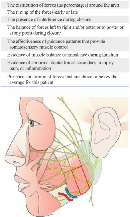
All patients were examined using the T-Scan occlusion analysis system (T-Scan III, Yours Dental, Seoul, Korea) before and 4, 8, 12, and 24 weeks after BTX-A injection. This T-Scan system comprises Microsoft-Windows-based software, the associated hardware, and patented paper-thin disposable sensors.10 The information provided on T-Scan
movies is listed in Table 1.
The sensor associated with the T-Scan system consists of two layers of Mylar (reinforced polyester film) in the form of a laminated pressure-sensitive ink grid. The film is cov-ered by a silver-thread grid, the intersection points of which are bathed in conductive ink. When a patient closes firmly on the sensor, the resultant reduction in electric resistance is converted to 8-bit digital values and translated into an im-age on the screen.12
The patient was seated in an upright position with be-cause the supine position can alter the contact position.13
The T-Scan III sensor and sensor support assembly were in-ness, and atrophy.
The control of mastication is dependent in large part upon sensory feedback, which involves epithelial mechano-receptors, periodontal, TMJ, and muscle afferents.6
Chang-es in the afferent input from a muscle caused by injection of BTX-A can modify the response of the cortex and the mo-tor neuron activity, and even initiate the activity of irrele-vant muscles. Such injections into masticatory muscles can subsequently influence mastication directly by inducing muscle weakness and atrophy, as well as indirectly by influ-encing the central pattern generator in the brainstem via modification of the sensory feedback from the masticatory muscle spindle.7
It has been often observed that BTX-A has analgesic ef-fects and reduces hyperactivity in the injected muscle.8
How-ever, in our clinic, some patients with pretreatment unilater-al chewing habits report an equunilater-alization of masticatory force after BTX-A injection. This effect of BTX-A has yet to be studied in detail.
The T-Scan occlusion analysis system is a dental tool that is used to analyze masticatory force. It was first devised in 1984 to measure occlusal forces and contact times as a prosthodontic adjunct in the treatment of occlusal problems and temporomandibular disorders. It has also been used as a measurement guide during prosthetic insertion and occlu-sal adjustment procedures. Data on occluocclu-sal forces and con-tact times are gathered by a recording sensor in the T-Scan system,9 and can subsequently be visualized in movie
for-mat, providing definitive diagnostic imaging of the force balance and function of the masticatory muscles.10
The aim of this study was to determine the force distribu-tion and pattern of masticadistribu-tion after injecdistribu-tion of BTX-A into the bilateral masseter muscles. The hypothesis to be tested was that the difference between right and left balance of occlusal force diminishes over time following BTX-A injection.
MATERIALS AND METHODS
Patient selection
dows statistics program (version 9.2, SAS Institute, Cary, NC, USA).
serted intraorally and positioned correctly. The T-Scan force-movie mode was then activated manually by pushing the button on the handle. The patients were instructed to bite in the habitual intercuspal position, and then to make excur-sive movements (not guided by the investigator). The right and left excursions were recorded separately. The patients were allowed a few minutes of rest between recordings. All of these procedures were repeated three times; the first two recordings were performed to familiarize the patient with the procedure, and the third record, made when the patient was completely familiar with the protocol, was used for the data analysis.
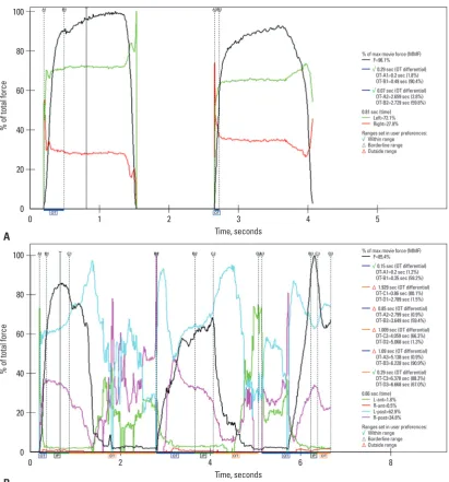
This study measured the distribution of forces between the left and right sides and between the anterior and posteri-or regions during clenching, and the change in occlusion and disclusion times after BTX-A injection (Figs. 2 and 3). The disclusion time, in seconds, is required to disclude the working and nonworking molar interferences and non-working premolar interferences from the habitual centric closure position to the completion of a mandibular excur-sion, and the occlusion time is the time in seconds, from the first contact of the teeth to the maximal intercuspation.14
Statistical analysis
Linear mixed modeling for longitudinal data was conduct-ed to analyze the change in the measurconduct-ed parameters over time. The interaction between times and groups was evalu-ated by compound symmetry covariance structure, banded Toeplitz, or autoregressive covariance structure. Bonferro-ni’s correction was used for post-hoc analysis. A p value of <0.01 was considered indicative of statistical significance.
All statistical analyses were performed using the SAS Win- Fig. 1. The red boxed area shows the botulinum toxin type A injection site. Table 1. The Information Provided on T-Scan Movies
The distribution of forces (as percentages) around the arch The timing of the forces-early or late
The presence of interference during closure
The balance of forces left to right and/or anterior to posterior at any point during closure
The effectiveness of guidance patterns that provide somatosensory muscle control
Evidence of muscle balance or imbalance during function Evidence of abnormal dental forces secondary to injury, pain, or inflammation
Presence and timing of forces that are above or below the average for this patient
Fig. 2. Balance of clenching force in a T-Scan movie (A: divided into two parts: left and right sides, B: divided into four parts labeled in clockwise direction: anterior left, anterior right, posterior right, posterior left).
[image:3.595.312.541.118.500.2] [image:3.595.100.510.529.710.2]Fig. 3. (A) Occlusion time. (B) Disclusion time. (A) A1 is the point the teeth start to occlude, B1 is the point the dentition is fully occluded, and the difference of A1 and B1 is occlusion time, marked as OT. (B) C1 is the point lateral movement starts, D1 is the point the dentition is fully separated and the difference is disclusion time, marked as DT. OT, occlusion time; DT, disclusion time.
Fig. 4. Force distribution on right and left sides before BTX-A injection (A) and 12 weeks thereafter (B). (A) In the left figure, before botuli-num toxin injection the distribution of the left and right force was uneven, 72.1% and 27.9%. 12 weeks post injection, (B) the right figure, the distribution evened as left 58.1% and right 27.9%. BTX-A, botulinum toxin type A.
Time, seconds
Time, seconds 0
0 20
20 40
40 60
60 80
80 100
100
%
o
f t
ot
al
fo
rc
e
%
o
f t
ot
al
fo
rc
e
0
0
1
2
2 3
4
4
6
5
8 A
B
% of max movie force (MMF) F=96.1% √ 0.29 sec (OT differential) OT-A1=0.2 sec (1.8%) OT-B1=0.49 sec (90.4%)
√ 0.07 sec (OT differential) OT-A2=2.659 sec (2.8%) OT-B2=2.729 sec (59.8%) 0.81 sec (time)
Left=72.1% Right=27.9% Ranges set in user preferences:
√ Within range
Δ Borderline range Δ Outside range
% of max movie force (MMF) F=85.4% √ 0.15 sec (OT differential) OT-A1=0.2 sec (1.2%) OT-B1=0.35 sec (59.2%) Δ 1.929 sec (DT differential) DT-C1=0.86 sec (80.1%) DT-D1=2.789 sec (1.5%)
Δ 0.85 sec (OT differential) OT-A2=2.799 sec (0.9%) OT-B2=3.649 sec (59.4%)
Δ 1.009 sec (DT differential) DT-C2=4.059 sec (66.3%) DT-D2=5.068 sec (1.3%)
Δ 1.09 sec (OT differential) OT-A3=5.138 sec (0.9%) OT-B3=6.228 sec (90.9%) √ 0.29 sec (DT differential) DT-C3=6.378 sec (88.3%) DT-D3=6.668 sec (67.0%) 0.66 sec (time)
L-ant=1.8% R-ant=0.5% L-post=62.9% R-post=34.8% Ranges set in user preferences:
√ Within range
Δ Borderline range Δ Outside range
[image:4.595.88.496.548.705.2]condition that can occur unilaterally or bilaterally. Unilater-al or bilaterUnilater-al hypertrophy of the masseter muscle is charac-terized by an increase in the volume of the muscle mass. This condition is benign, asymptomatic, and must be differ-entiated from parotid gland disease odontogenic problems, and rare neoplasms of muscular tissue. The reasons why patients request a medical consultation are predominantly related to aesthetics, especially if the hypertrophy is unilat-eral due to a noticeable asymmetry of the lower third of the face.16,17 This study was intended for patients who
com-plained only about masseter hypertrophy, and not other symptoms.
Botulinum toxin is a potent biological toxin produced by the Gram-positive bacterium Clostridium botulinum.18 It is
a presynaptic neurotoxin that causes dose-dependent weak-ness or paralysis in skeletal muscles by blocking the calci-um-mediated release of acetylcholine from motor nerve endings.19,20 BTX-A, one of seven subtypes of the toxin, is
a dichain protein consisting of a 50-kD light chain linked to a 100-kD heavy chain by a disulfide bond; it functionally denervates the affected portions of the muscle and it pri-marily affects α-motor neuron function, but it may also affect the γ motor neurons in the muscle spindles.21 Local paralysis
is reversed chiefly by neural sprouting, effectively reinnervat-ing the muscle.22 Long-term reductions in α, γ, and Ia
neuro-nal activity may have indirect effects on the central nervous system. This was demonstrated when Moreno-López, et al.23
who showed that single injections of BTX-A into the lateral rectus muscle of cats caused inhibition of abduction, altered electromyographic signals in the contralateral ocular mus-cles, and a disruption of the abducens motor neuron dis-charge patterns that lasted longer than 2 months.
BTX-A may also act directly or indirectly on nociceptors that affect transmission of sensory signals through A-δ and
RESULTS
Changes in the distribution of forces
A significant change in force balance was found between the right and left sides over time (i.e., preinjection and 4, 8, 12, and 24 weeks post-BTX-A injection) and at each mea-surement time point (p<0.0001). The difference between the two sides decreased with the time postinjection, reach-ing a minimum at 12 weeks (Fig. 4). These findreach-ings reflect the well-established time course of the actions of BTX-A.15
In other words, according to this result, the left and right clenching force becomes more balanced with the increasing effect of botulinum toxin injection.
Comparison of the force balance between the anterior and posterior occlusions revealed no significant difference at any of the time points (i.e., preinjection and 4, 8, 12, and 24 weeks postinjection; p>0.01 for all). Furthermore, com-parison of the preinjection force balance with those mea-sured at 4, 8, 12, and 24 weeks postinjection revealed no significant difference at any time point (p>0.05 for all) (Ta-ble 2). In other words, there is no correlation between the balance of anterior and posterior masticatory force and the increasing effect of botulinum toxin injection.
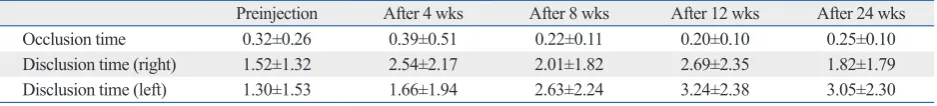
Changes in occlusion and disclusion times
The occlusion and disclusion times (right and left sides) did not differ significantly with time after BTX-A injection (p> 0.01) (Table 3).
DISCUSSION
[image:5.595.79.542.81.120.2]Benign masseter hypertrophy is a relatively uncommon
Table 2. Changes in the Force Distribution (%; Mean±SD) after Injection of BTX-A (n=15)
Preinjection After 4 wks After 8 wks After 12 wks After 24 wks Difference between left and right sides 25.5±10.1 15.7±10.3* 11.9±6.0* 7.1±5.6* 16.4±11.4* Difference between anterior and posterior 91.0±7.5 90.7±10.1 88.2±12.8 87.6±10.5 87.8±8.7
BTX-A, botulinum toxin type A. *p<0.01 (compared to preinjection).
Table 3. Change in Occlusion and Disclusion Times (in Seconds) with Time after BTX-A Injection (n=15)
Preinjection After 4 wks After 8 wks After 12 wks After 24 wks
Occlusion time 0.32±0.26 0.39±0.51 0.22±0.11 0.20±0.10 0.25±0.10
Disclusion time (right) 1.52±1.32 2.54±2.17 2.01±1.82 2.69±2.35 1.82±1.79 Disclusion time (left) 1.30±1.53 1.66±1.94 2.63±2.24 3.24±2.38 3.05±2.30
[image:5.595.71.540.166.219.2]values, were therefore analyzed since it was felt that the data obtained at the first and second recordings would be inaccurate. Second, since there was no control group, it was not possible to determine whether there was a placebo ef-fect. Future studies should address these limitations.
In conclusion, fifteen patients were examined using the T-Scan system before and after BTX-A injection into the masseter muscles. A decline in the difference in the clench-ing force between the left and right sides was found with increasing time (and hence increasing medicinal effect) up to 12 weeks following BTX-A injection.
REFERENCES
1. Clark GT. The management of oromandibular motor disorders and facial spasms with injections of botulinum toxin. Phys Med Rehabil Clin N Am 2003;14:727-48.
2. Freund B, Schwartz M, Symington JM. Botulinum toxin: new treatment for temporomandibular disorders. Br J Oral Maxillofac Surg 2000;38:466-71.
3. Lang AM. Botulinum toxin therapy for myofascial pain disorders. Curr Pain Headache Rep 2002;6:355-60.
4. Lovell BV, Marmura MJ. New therapeutic developments in chronic migraine. Curr Opin Neurol 2010;23:254-8.
5. Bhogal PS, Hutton A, Monaghan A. A review of the current uses of Botox for dentally-related procedures. Dent Update 2006;33: 165-8.
6. Soboįeva U, Lauriņa L, Slaidiņa A. The masticatory system--an overview. Stomatologija 2005;7:77-80.
7. Okeson JP. Management of temporomandibular disorders and oc-clusion. 6th ed. St. Louis, Missouri: Elsevier Mosby Publishers; 1993.
8. Fallah HM, Currimbhoy S. Use of botulinum toxin A for treat-ment of myofascial pain and dysfunction. J Oral Maxillofac Surg 2012;70:1243-5.
9. Kerstein RB, Lowe M, Harty M, Radke J. A force reproduction analysis of two recording sensors of a computerized occlusal anal-ysis system. Cranio 2006;24:15-24.
10. Montgomery MW, Shuman L, Morgan A. T-scan dental force analysis for routine dental examination. Dent Today 2011;30:112-4, 116.
11. Hu KS, Kim ST, Hur MS, Park JH, Song WC, Koh KS, et al. To-pography of the masseter muscle in relation to treatment with bot-ulinum toxin type A. Oral Surg Oral Med Oral Pathol Oral Radiol Endod 2010;110:167-71.
12. Kerstein RB. Combining technologies: a computerized occlusal analysis system synchronized with a computerized electromyog-raphy system. Cranio 2004;22:96-109.
13. Makofsky HW, Sexton TR, Diamond DZ, Sexton MT. The effect of head posture on muscle contact position using the T-Scan sys-tem of occlusal analysis. Cranio 1991;9:316-21.
14. Kerstein RB, Wright NR. Electromyographic and computer analy-ses of patients suffering from chronic myofascial pain-dysfunction syndrome: before and after treatment with immediate complete anterior guidance development. J Prosthet Dent 1991;66:677-86.
C fibers, and impact the detection of sensory signals through mechanoreceptors and chemo-nociceptors. These findings may also explain the changes in the distribution of clench-ing forces after BTX-A injection reported herein. It is possi-ble that the redistribution of clenching force after BTX-A injection is due to changes both in the chewing pattern at the central nervous system level and atrophy and weakness of the peripheral muscle region. However, more detailed re-search into the exact mechanism underlying the change in chewing pattern at the level of the central nervous system is needed to clarify this issue.
As mentioned above, as the medicinal effects of BTX-A increase, there is a concomitant decline in the difference in clenching force between the left and right sides post injec-tion. BTX-A injection shows maximum effect at 12 weeks postinjection, and after, that the effect decreases possibly creating an imbalance in force balance once again, which is suspected to be caused by the patients chewing habit.
Furthermore, there was no significant change in either oc-clusion or disoc-clusion time after BTX-A injection. The theory that decline in the difference in clenching force caused by the effect of botulinum toxin on sensory transmission by A-δ and C fibers, either directly or indirectly, is supported by the results of the present study. The durations of occlu-sion and discluocclu-sion times are determined by interference between the teeth. However, since botulinum toxin cannot influence that interference, it is perhaps not surprising that there was no significant change in either duration following BTX-A injection. Furthermore, the much lighter contact between the upper and lower anterior teeth than between the upper and lower posterior teeth in normal occlusion, and the absence of an effect of botulinum toxin on the con-tact between the anterior teeth explain why no significant difference was found between the anterior and posterior balance over time post-BTX-A injection. It may be that if all of the subjects had possessed a full-contact arch, a dif-ference may have been observed between the balance of anterior and posterior occlusions after BTX-A injection.
nature and preparation for clinical use. Eye (Lond) 1988;2(Pt 1): 16-23.
21. Filippi GM, Errico P, Santarelli R, Bagolini B, Manni E. Botuli-num A toxin effects on rat jaw muscle spindles. Acta Otolaryngol 1993;113:400-4.
22. Holds JB, Alderson K, Fogg SG, Anderson RL. Motor nerve sprouting in human orbicularis muscle after botulinum A injection. Invest Ophthalmol Vis Sci 1990;31:964-7.
23. Moreno-López B, de la Cruz RR, Pastor AM, Delgado-García JM. Botulinum neurotoxin alters the discharge characteristics of abducens motoneurons in the alert cat. J Neurophysiol 1994;72: 2041-4.
15. Aoki KR, Ranoux D, Wissel J. Using translational medicine to understand clinical differences between botulinum toxin formula-tions. Eur J Neurol 2006;13 Suppl 4:10-9.
16. Addante RR. Masseter muscle hypertrophy: report of case and lit-erature review. J Oral Maxillofac Surg 1994;52:1199-202. 17. Rispoli DZ, Camargo PM, Pires JL Jr, Fonseca VR, Mandelli KK,
Pereira MA. Benign masseter muscle hypertrophy. Braz J Otorhi-nolaryngol 2008;74:790-3.
18. Simpson LL. The origin, structure, and pharmacological activity of botulinum toxin. Pharmacol Rev 1981;33:155-88.
19. Drachman DB. Atrophy of skeletal muscle in chick embryos treat-ed with botulinum toxin. Science 1964;145:719-21.