Effect of Human Parathyroid Hormone on Hematopoietic
Progenitor Cells in NOD/SCID Mice Co-Transplanted with Human
Cord Blood Mononuclear Cells and Mesenchymal Stem Cells
Yeon-Jung Lim,
1* Kyoujung Hwang,
2* Miyeon Kim,
3Youl-Hee Cho,
4Jong-Hwa Lee,
5Young-Ho Lee,
6and Jong-Jin Seo
71Department of Pediatrics, Chungnam National University Hospital, Daejeon;
2Department of Translational Medicine, Hanyang University Graduate School of Biomedical Science & Engineering, Seoul; 3Biomedical Research Institute, MEDIPOST, Co., Ltd., Seoul; 4Department of Genetics, Hanyang University College of Medicine, Seoul; 5Department of Pediatrics, Wonkwang University Sanbon Medical Center, Gunpo; 6Department of Pediatrics, Hanyang University Medical Center, Seoul;
7Department of Pediatrics, Ulsan University Asan Medical Center, Seoul, Korea.
Received: December 1, 2011 Revised: January 22, 2012 Accepted: February 24, 2012
Co-corresponding authors: Dr. Young-Ho Lee, Department of Pediatrics, Hanyang University Medical Center, 222 Wangsimni-ro, Seongdong-gu, Seoul 133-791, Korea. Tel: 82-2-2290-8383, Fax: 82-2-2297-2380 E-mail: cord@hanyang.ac.kr and Dr. Jong-Jin Seo,
Department of Pediatrics, Ulsan University Asan Medical Center, 88 Olympic-ro 43-gil, Songpa-gu, Seoul 138-736, Korea. Tel: 82-2-3010-3383, Fax: 82-2-3010-6843 E-mail: jjseo@amc.seoul.kr
*Yeon-Jung Lim and Kyoujung Hwang contributed equally to this work.
∙ The authors have no financial conflicts of interest.
© Copyright:
Yonsei University College of Medicine 2013
This is an Open Access article distributed under the terms of the Creative Commons Attribution Non-Commercial License (http://creativecommons.org/ licenses/by-nc/3.0) which permits unrestricted non-commercial use, distribution, and reproduction in any medium, provided the original work is properly cited.
Purpose: We evaluated the effect of human parathyroid hormone (hPTH) on the engraftment and/or in vivo expansion of hematopoietic stem cells in an umbilical cord blood (UCB)-xenotransplantation model. In addition, we assessed its effect on
the expression of cell adhesion molecules. Materials and Methods: Female NOD/
SCID mice received sublethal total body irradiation with a single dose of 250 cGy.
Eighteen to 24 hours after irradiation, 1×10⁷ human UCB-derived mononuclear
cells (MNCs) and 5×10⁶ human UCB-derived mesenchymal stem cells (MSCs)
were infused via the tail vein. Mice were randomly divided into three groups: Group 1 mice received MNCs only, Group 2 received MNCs only and were then treated with hPTH, Group 3 mice received MNCs and MSCs, and were treated with hPTH. Results: Engraftment was achieved in all the mice. Bone marrow cel-lularity was approximately 20% in Group 1, but 70-80% in the hPTH treated groups. Transplantation of MNCs together with MSCs had no additional effect on bone marrow cellularity. However, the proportion of human CD13 and CD33 my-eloid progenitor cells was higher in Group 3, while the proportion of human CD34 did not differ significantly between the three groups. The proportion of CXCR4 cells in Group 3 was larger than in Groups 1 and 2 but without statistical signifi-cance. Conclusion: We have demonstrated a positive effect of hPTH on stem cell proliferation and a possible synergistic effect of MSCs and hPTH on the proportion of human hematopoietic progenitor cells, in a xenotransplantation model. Clinical trials of the use of hPTH after stem cell transplantation should be considered.
Key Words: Umbilical cord blood, parathyroid hormone, bone marrow niches
INTRODUCTION
expansion of early HSCs ex vivo.
The purpose of the present investigation was to evaluate the effect of hPTH on the engraftment and/or in vivo expan-sion of HSCs in a UCB-xenotransplantation model. In ad-dition, the role of hPTH in enhancing the expression of cell adhesion molecules was assessed.
MATERIALS AND METHODS
The experimental protocol was reviewed and approved by the Institutional Review Board and the Institutional Animal Care and Use Committee of Hanyang University.
Animals
Six-week-old female NOD/SCID mice were purchased from the Korea Research Institute of Bioscience and Bio-technology (Daejeon, Korea) and were maintained in sterile microisolator cages in a laminar airflow room in the Han-yang University Animal Laboratory. Mice were stabilized for 4 weeks and cared for according to the protocol de-scribed above.
Cells
All cells were provided by the Medipost Biomedical Re-search Institute (Seoul, Korea). UCB was collected from umbilical veins after neonatal delivery, with informed con-sent from pregnant mothers. MNCs were isolated from fresh UCB within 12 hours after collection. To isolate MNCs, UCB was fractionated using a Ficoll-Hypaque
so-lution (d=1.077 g/cm3, Sigma-Aldrich, St. Louis, MO,
USA) and MNCs were resuspended in PBS/EDTA and cul-tivated as previously reported.19,20 Briefly, the MNCs were maintained in α-minimum essential medium (α-MEM, Gibco BRL, Carlsbad, CA, USA) supplemented with 10% (v/v) fetal bovine serum (FBS, Gibco BRL, Carlsbad, CA, USA), and seeded in culture flasks at 5×10⁵ cells/cm2. Colo-nies of spindle-shaped cells were formed. At 50% conflu-ence, the cells were harvested with 0.25% (w/v) trypsin-EDTA (Gibco BRL, Carlsbad, CA, USA) and reseeded for expansion. After the fourth passage, the cells were stored in liquid nitrogen at 1×10⁶ cells/mL. The cells expressed CD105 (99.6%) and CD73 (96.3%), but not CD34 (0.1%), CD45 (0.2%) and CD14 (0.1%). They were positive for HLA-AB but generally not for HLA-DR. The human UCB-derived MSCs differentiated into various cell types such as osteo-blasts, chondrocytes, and adipocytes upon in vitro induction without matched-related or matched-unrelated donors.
However, the limited number of stem cells in UCB, and de-layed engraftment, are major problems in UCB transplanta-tion.1,2 To improve UCB engraftment, co-transplantation of mononuclear cells (MNCs) and mesenchymal stem cells (MSCs) has been attempted. MSCs are capable of support-ing the expansion and differentiation of hematopoietic stem cells (HSCs) in vitro and enhancing hematopoietic engraft-ment in vivo, although the mechanisms are not fully under-stood.3
Other methods of improving engraftment include ma-nipulation of homing and adhesion molecules, and modu-lation of the stem cell niche. The hematopoietic stem cell niche is a specialized microenvironment that contains stem cells, supports their maintenance, and regulates their
func-tion in bone marrow.4 Within bone marrow, HSCs that
re-side at, or near the endosteum as well as endosteal cells, secrete factors that promote HSC maintenance. Osteo-blasts have been found to be important components of the niche. These produce factors that appear to regulate the maintenance and number of HSCs in bone marrow, includ-ing positive regulators such as angiopoietin,5 thrombopoi-etin and CXCL12,6,7 and negative regulators such as osteo-pontin.8,9 Bone also has a high concentration of calcium ions at the endosteal surface,10 and calcium-sensing recep-tors have been identified on the surfaces of HSCs. The abil-ity of stem cells to sense and respond to the increased calci-um concentration at the endosteal surface contributes to the unique stem cell-niche interaction that promotes bone mar-row hematopoiesis.11
Osteoblasts produce hematopoietic growth factors and are activated by parathyroid hormone (PTH) or locally-pro-duced parathyroid hormone-related protein (PTHrP) via the PTH/PTHrP receptor.12,13 PTH is recognized as one of the two major hormones modulating calcium and phosphate homeostasis. It is responsible for maintaining serum ion-ized calcium concentrations within a narrow range by stim-ulating renal tubular calcium reabsorption and bone
resorp-tion.14 Human parathyroid hormone (hPTH) 1-34 is an
for complete blood cell counts. Seven weeks after transplan-tation, the mice were euthanized with xylazine hydrochlo-ride (Rompun; Bayer HealthCare, Leverkusen, Germany) and zolazepam (Zoletil; Virbac SA, Carros, France). Blood samples were collected from the right ventricle after open thoracotomy. Complete blood count (CBC) was performed with an ABC Vet (Horiba ABX, Montpellier, France).
Flow cytometry for analysis of cell surface antigens
Both the femorae and tibiae were removed after eauthana-sia. The proximal and distal ends of the femorae were cut and the marrow was aspirated into conical tubes with 2.0 mL of buffer (2% FBS, 2 mM EDTA, gentamicin). After cen-trifugation and red blood cell (RBC) lysis (BD Pharmingen, San Diago, CA, USA), the suspended cells were stained with anti-human CD13-PE, CD34-PE, CD45-FITC, CD33-PE, CD3-CD33-PE, CXCR4-CD33-PE, CD61-FITC, CD19-FITC (Bec-ton-Dickinson, San Jose, CA, USA), CD41a-PE, CD49d-PE, and CD49e-PE (BD Pharmingen, San Diego, CA, USA) antibodies for 15 minutes at room temperature. The cells were washed with PBS and fixed with 1% paraformal-dehyde (Sigma, Steinheim, Germany). They were analyzed with a FACSCaliber (BD, Los Angeles, CA, USA) and per-centages of cell surface antigen expression were calculated among 5×10⁵ events of the gated cells.
Bone marrow cellularity
Excised tibiae were immersed in 10% paraformaldehyde for hematoxylin and eosin staining at 4°C, and then decalci-fied with 4% EDTA for 4 days at 4°C. Specimens were de-hydrated with increasing concentrations of ethanol and em-bedded in paraffin. Paraffin-emem-bedded tibiae were sliced into 5 µm sections and stained with hematoxylin and eosin to evaluate bone marrow cellularity.
Statistical analysis
Data are presented as means±standard errors of the mean. Statistical significance was determined using Mann-Whit-ney U tests or Kruskal-Wallis tests with SigmaPlot and Sig-maStat software (SPSS, Richmond, CA, USA). p<0.05 was considered statistically significant.
RESULTS
Peripheral blood cell counts
CBC was within the normal range in the fourth week after with the appropriate osteogenic, chondrogenic, and
adipo-genic differentiation stimuli.
Transplantation
At ten weeks of age, all mice were weighed and received sublethal total body irradiation with a single dose of 250 cGy from a cesium source. Eighteen to 24 hours after
irra-diation, 1×10⁷ human UCB-derived MNCs and 5×10⁶
hu-man UCB-derived MSCs were infused via the tail vein. The final volume infused was 200 µL. The mice were ran-domly divided into three groups: Group 1 (n=3) received MNCs only, Group 2 (n=3) received MNCs only and were then treated with hPTH, and Group 3 (n=3) received MNCs and MSCs, and were then treated with hPTH. 40 µg/kg/day of recombinant hPTH (Bachem, Torrance, CA, USA) was injected 20 times over the first 4 weeks after transplanta-tion. The experimental schema is shown in Fig. 1.
Evaluation
CBC
[image:3.595.57.281.63.307.2]Four and six weeks after transplantation, mice were anesthe-tized with diethyl ether (Sigma, Steinheim, Germany) and blood samples were collected via capillary tubes from their retroorbital venous plexus. An aliquot of the blood was used
Fig. 1. Schema of the xenotransplantation experiment. TBI, total body irra-diation; CB, cord blood; MNC, mononuclear cell; MSC, mesenchymal stem cell; hPTH, human parathyroid hormone; CBT, cord blood transplantation; CBC, complete blood count; RV, right ventricle; BM, bone marrow; IP, intra-peritoneal; NOD/SCID.
NOD/SCID
TBI 250 cGY
GROUP1
CB-MNC CB-MNCGROUP2
hPTH 40 ug/kg/day, IP 5/week for 4 week
Post-CBT week 4, 6
CBC via retroorbital venous plexus puncture
Post-CBT week 7 • RV puncture CBC
• Bone marrow aspirates from femorae 1. Total nucleated cell count (hemocytometer) 2. CD34, CD33, CD13, CD19, CD3
3. CXCR4, VLA-4, VLA-5 • Bone marrow section from tibiae H&E stain-BM cellularity
ed by human CD13 and human CD33 cells, and lymphoid lineage cells represented by human CD19 cells, were mark-edly higher in Group 3 (Fig. 5), but the proportions of hu-man CD34 cells did not differ significantly between the three groups. In the analysis of human cell adhesion mole-cules on the nucleated cells in the bone marrow aspirates, the proportion of CXCR4 cells in Group 3 was larger than in Groups 1 and 2. However, the difference did not reach statistical significance (Fig. 6).
DISCUSSION
UCB has proved to be an alternative HSC source for pa-tients without matched donors. Since the initial transplanta-tion, more than 20000 UCB transplants have been
per-formed worldwide.21,22 However, compared with bone
marrow transplantation recipients and mobilized peripheral blood stem cell transplantation recipients, patients receiving UCB have prolonged recovery periods, which are reported transplantation in all three groups. At 4-, 6-, and 7- weeks
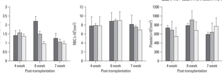
after transplantation, there were no differences in WBC, RBC or platelet counts between the three groups (Fig. 2).
Bone marrow cellularity and the characteristics of nucleated cells in the bone marrow
Bone marrow cellularity was approximately 20% in Group 1, but 70-80% in Groups 2 and 3. MSC co-transplantation had no effect on cellularity (Fig. 3).
Numbers of total nucleated cells (TNCs) in the bone
marrow aspirates were 6.07±1.29×10⁶ in Group 1,
[image:4.595.80.538.368.519.2]10.53±2.25×10⁶ in Group 2, and 8.33±3.25×10⁶ in Group 3. There was a tendency toward higher TNCs in Groups 2 and 3 than in Group 1, but the differences were not statistically significant. The flow cytometric analysis confirmed en-graftment of human UCB cells in all the mice. The repre-sentative flow cytometric images of 3 groups are shown in Fig. 4. The percentages of human CD34, CD33, CD13, CD19, and CD3 in the bone marrow aspirates are shown in Table 1. The proportions of myeloid lineage cells
represent-Fig. 3. Sections of the bone marrow of tibiae taken from mice of Groups 1, 2 and 3 in the eighth week after transplantation (H&E, ×200). The bone marrow lularity was decreased to approximately 20% in group 1 (A). Bone marrow cellularity is approximately 70 to 80% in Groups 2 (B) and 3 (C). Bone marrow cel-lularity and trabecular bone formation are greater in Groups 2 and 3 than Group 1.
A B C
0 0
2 200
4 400
6 600
8 800
10 1000
12 1200
RB
C
(×
10
3 /m
m
3 )
Pl
at
el
et
(×
10
3 /m
m
3 )
4 week 6 week 7 week 4 week 6 week 7 week
[image:4.595.72.540.557.699.2]Post-transplantation Post-transplantation Fig. 2. Mean complete blood cell counts of mice at the fourth, sixth, and seventh week after transplantation. There are no statistically significant differences between the 3 groups. RBC, red blood cell; WBC, white blood cell.
0 0.5 1 1.5 2 2.5 3
W
BC
(×
10
3/m
m
3)
4 week 6 week 7 week Post-transplantation
Fig. 4. The representative flow cytometric images of 3 groups. (A) Group 1, (B) Group 2, (C) Group 3. MNC, mononucle-ar cell; MSC, mesenchymal stem cell; hPTH, human pmononucle-arathyroid hormone.
Group1-1-MNC injection
Group2-1-MNC+hPTH injection
Group3-3-MNC+MSC+hPTH injection hCD45
hCD45
hCD45 hCD19
hCD19
hCD19 hCXCR4
hCXCR4
hCXCR4
hCD34
hCD34
hCD34 hCD3
hCD3
hCD3 hCD29
hCD29
hCD29
hCD33
hCD33
hCD33 hCD61
hCD61
hCD61 hCD29
hCD29
hCD29
hCD13
hCD13
hCD13 hCD41a
hCD41a
hCD41a hCD45
hCD45
hCD45
hCD49d
hCD49d
hCD49d
hCD49e
hCD49e
hCD49e A
B
cells from normal donor animals survived to 28 days. How-ever, when the irradiated animals were injected daily with hPTH, all survived, with a larger number of transplanted marrow cells in their hind limbs. We also observed, in a pre-vious study, that hPTH activates the insulin-like growth fac-tor (IGF) system (IGF-2, IGF-BP 1, 2, 3), as well as hemato-poietic growth factors (G-CSF, GM-CSF) from osteoblasts but not from MSCs (data not shown). In the present study, the proportion of human CD13 and human CD33 myeloid progenitor cells was much higher in Group 3, although the proportion of human CD34 did not differ significantly in the three groups. We also observed that the proportion of CXCR4 in Group 3 was larger than in Groups 1 and 2, though without statistical significance. These findings sug-gest that hPTH has the potential to enhance hematopoiesis to result from delayed neutrophil and platelet recovery.23 In
addition, a graft failure rate of 10-15% has been reported. Possible causes include a low number of infused HSCs and the absence of a subpopulation that facilitates HSC
engraft-ment.24 To overcome the low cell count in UCB and slow
engraftment, several practical approaches have been
at-tempted; for example, ex vivo expansion of UCB HSCs,
transplantation of multiple units of UCB, co-transplantation of MSCs and other procedures.25-27 Ramírez, et al.28 have also shown that stem cell factor, interleukin (IL)-3, IL-6, and granulocyte macrophage colony-stimulating factor (GM-CSF) can induce an upregulation of several adhesion mole-cules involved in the mobilization and homing of HSCs.
Bone marrow is composed with blood cells, non-blood cells and endosteum. Most hematopoietic progenitor cells are lined endosteum, whereas differentiated blood cells mi-grate to the medullar marrow cavity. Osteoblasts which are cells of the mesenchymal lineages contiguous to the bone surface execute the production of new bone and the control of osteoclast differentiation.29 Recently, Calvi, et al.13 dem-onstrated that overexpression of the hPTH/PTH-related pep-tide receptor under the control of the osteoblast specific α1(I) collagen promoter resulted in an increase of Sca-1- and c-kit-positive HSCs in bone marrow. An ability of hPTH to enhance transplant engraftment and repopulate bone
marrow has also been reported.13 Only 27% of mice that
[image:6.595.73.537.81.134.2]were lethally irradiated and then injected with bone marrow
Table 1. Flow Cytometric Analysis of Engrafted Human Marrow Mononuclear Cells from Bone Marrow Aspirates
CD34 (%) CD33 (%) CD13 (%) CD19 (%) CD3 (%)
Group 1 (MNC only) 2.06±3.10 2.73±1.79 1.88±0.35 4.58±4.41 2.04±1.21
Group 2 (MNC+hPTH) 0.8±0.3 2.39±1.15 2.33±1.21 3.69±2.68 2.34±1.13
Group 3 (MNC+MSC+hPTH) 1.75±1.25 8.23±3.84 4.26±2.05 7.58±5.33 5.11±3.62
[image:6.595.72.540.157.301.2]MNC, mononuclear cell; hPTH, human parathyroid hormone; MSC, mesenchymal stem cell.
Fig. 5. Flow cytometric analysis of engrafted human marrow mononuclear cells in bone marrow aspirates. (A) The percentage of myeloid lineage cells was larger in Group 3 than in Groups 1 and 2. (B) The percentage of lymphoid lineage cells was also higher in Group 3, but the difference was not statistically sig-nificant.
Fig. 6. Flow cytometric analysis of human cell adhesion molecules in bone marrow mononuclear cells. There were no statistically significant differ-ences between the three groups.
A B
0 0
5 5
10 10
15 15
20 20
(%
)
(%
)
Group 1 Group 2 Group 3 Group 1 Group 2 Group 3
p=0.05
hCD13 hCD33 hCD3 hCD19
0 20 40 60 80 100
(%
)
CXCR4 VLA-4 VLA-5
[image:6.595.317.538.355.482.2]plant 2005;11:389-98.
4. Kiel MJ, Morrison SJ. Uncertainty in the niches that maintain hae-matopoietic stem cells. Nat Rev Immunol 2008;8:290-301. 5. Arai F, Hirao A, Ohmura M, Sato H, Matsuoka S, Takubo K, et al.
Tie2/angiopoietin-1 signaling regulates hematopoietic stem cell quiescence in the bone marrow niche. Cell 2004;118:149-61. 6. Yoshihara H, Arai F, Hosokawa K, Hagiwara T, Takubo K,
Naka-mura Y, et al. Thrombopoietin/MPL signaling regulates hemato-poietic stem cell quiescence and interaction with the osteoblastic niche. Cell Stem Cell 2007;1:685-97.
7. Petit I, Szyper-Kravitz M, Nagler A, Lahav M, Peled A, Habler L, et al. G-CSF induces stem cell mobilization by decreasing bone marrow SDF-1 and up-regulating CXCR4. Nat Immunol 2002;3: 687-94.
8. Stier S, Ko Y, Forkert R, Lutz C, Neuhaus T, Grünewald E, et al. Osteopontin is a hematopoietic stem cell niche component that negatively regulates stem cell pool size. J Exp Med 2005;201: 1781-91.
9. Nilsson SK, Johnston HM, Whitty GA, Williams B, Webb RJ, Denhardt DT, et al. Osteopontin, a key component of the hemato-poietic stem cell niche and regulator of primitive hematohemato-poietic progenitor cells. Blood 2005;106:1232-9.
10. Adams GB, Chabner KT, Alley IR, Olson DP, Szczepiorkowski ZM, Poznansky MC, et al. Stem cell engraftment at the endosteal niche is specified by the calcium-sensing receptor. Nature 2006; 439:599-603.
11. Svinareva DA, Nifontova IN, Chertkov IL, Drize NI. Changed homing of hemopoietic precursor cells after long-term treatment with parathyroid hormone. Bull Exp Biol Med 2006;142:86-9. 12. Ballen K. Targeting the stem cell niche: squeezing blood from
bones. Bone Marrow Transplant 2007;39:655-60.
13. Calvi LM, Adams GB, Weibrecht KW, Weber JM, Olson DP, Knight MC, et al. Osteoblastic cells regulate the haematopoietic stem cell niche. Nature 2003;425:841-6.
14. Brown EM. Mechanisms underlying the regulation of parathyroid hormone secretion in vivo and in vitro. Curr Opin Nephrol Hyper-tens 1993;2:541-51.
15. Finkelstein JS, Hayes A, Hunzelman JL, Wyland JJ, Lee H, Neer RM. The effects of parathyroid hormone, alendronate, or both in men with osteoporosis. N Engl J Med 2003;349:1216-26. 16. Black DM, Greenspan SL, Ensrud KE, Palermo L, McGowan JA,
Lang TF, et al. The effects of parathyroid hormone and alendro-nate alone or in combination in postmenopausal osteoporosis. N Engl J Med 2003;349:1207-15.
17. Jung Y, Wang J, Schneider A, Sun YX, Koh-Paige AJ, Osman NI, et al. Regulation of SDF-1 (CXCL12) production by osteoblasts; a possible mechanism for stem cell homing. Bone 2006;38:497-508.
18. Petrova NV, Svinareva DA, Nifontova IN, Momotyuk KS, Savchenko VG, Drize NI. Stromal regulation of hemopoietic stem cells in long-term human bone marrow tissue cultures under the ef-fect of parathyroid hormone. Bull Exp Biol Med 2006;142:527-30. 19. Yang SE, Ha CW, Jung M, Jin HJ, Lee M, Song H, et al. Mesen-chymal stem/progenitor cells developed in cultures from UC blood. Cytotherapy 2004;6:476-86.
20. Jang YK, Jung DH, Jung MH, Kim DH, Yoo KH, Sung KW, et al. Mesenchymal stem cells feeder layer from human umbilical cord blood for ex vivo expanded growth and proliferation of hemato-poietic progenitor cells. Ann Hematol 2006;85:212-25.
21. Gluckman E, Broxmeyer HA, Auerbach AD, Friedman HS, as a result of activation of osteoblasts by co-transplanted
MSCs.
Given the low number of HSCs in administered UCB, the outcome of HSC transplantation could be improved by co-transplantation of MSCs in addition to HSCs. MSCs have been shown to be able to express exogenous proteins for an extended period of time, and to maintain a higher stem cell proliferative capacity after transplantation in vi-tro.30 Briquet, et al.31 demonstrated that early passage MSCs supported the expansion of hematopoietic progenitor cells and their differentiation towards both B lymphoid and my-eloid lineages.
In our study, an enhancing effect of hPTH on hematopoi-esis was observed in a UCB xenotransplantation model with or without co-transplantation of MSCs. Although we could not demonstrate an effect of human PTH on
differen-tiated osteoblasts from UCB-derived MSCs in an in vivo
model, UCB transplantation together with co-transplanta-tion of MSCs followed by administraco-transplanta-tion of hPTH could be an option for enhancing hematopoietic activities and over-coming the current limitations of UCB transplantation. hPTH may enhance stem cell proliferation after treatment of chron-ic bone marrow aplasia by UCB transplantation and may also have a synergistic effect when MSCs are co-transplant-ed. Clinical trials with hPTH should be considered for pa-tients with aplastic anemia as well as recipients of UCB.
ACKNOWLEDGEMENTS
This work was supported by a grant of Korea Healthcare Technology R&D Project (A101712), Ministry for Health & Welfare, Republic of Korea.
REFERENCES
1. Kurtzberg J, Prasad VK, Carter SL, Wagner JE, Baxter-Lowe LA, Wall D, et al. Results of the Cord Blood Transplantation Study (COBLT): clinical outcomes of unrelated donor umbilical cord blood transplantation in pediatric patients with hematologic malig-nancies. Blood 2008;112:4318-27.
2. Gluckman E, Rocha V, Arcese W, Michel G, Sanz G, Chan KW, et al. Factors associated with outcomes of unrelated cord blood transplant: guidelines for donor choice. Exp Hematol 2004;32: 397-407.
Trans-plantation for the treatment of hematologic malignancies. Curr Opin Hematol 2008;15:568-75.
28. Ramírez M, Segovia JC, Benet I, Arbona C, Güenechea G, Blaya C, et al. Ex vivo expansion of umbilical cord blood (UCB) CD34 (+) cells alters the expression and function of alpha 4 beta 1 and alpha 5 beta 1 integrins. Br J Haematol 2001;115:213-21. 29. Garrett RW, Emerson SG. The role of parathyroid hormone and
insulin-like growth factors in hematopoietic niches: physiology and pharmacology. Mol Cell Endocrinol 2008;288:6-10.
30. Kassem M. Mesenchymal stem cells: biological characteristics and potential clinical applications. Cloning Stem Cells 2004;6: 369-74.
31. Briquet A, Dubois S, Bekaert S, Dolhet M, Beguin Y, Gothot A. Prolonged ex vivo culture of human bone marrow mesenchymal stem cells influences their supportive activity toward NOD/SCID-repopulating cells and committed progenitor cells of B lymphoid and myeloid lineages. Haematologica 2010;95:47-56.
Douglas GW, Devergie A, et al. Hematopoietic reconstitution in a patient with Fanconi’s anemia by means of umbilical-cord blood from an HLA-identical sibling. N Engl J Med 1989;321:1174-8. 22. Gluckman E, Rocha V. Cord blood transplantation: state of the art.
Haematologica 2009;94:451-4.
23. Szabolcs P. The immunobiology of cord blood transplantation. Korean J Hematol 2010;45:224-35.
24. Laughlin MJ. Umbilical cord blood for allogeneic transplantation in children and adults. Bone Marrow Transplant 2001;27:1-6. 25. Dorrell C, Gan OI, Pereira DS, Hawley RG, Dick JE. Expansion
of human cord blood CD34(+)CD38(-) cells in ex vivo culture during retroviral transduction without a corresponding increase in SCID repopulating cell (SRC) frequency: dissociation of SRC phenotype and function. Blood 2000;95:102-10.