4155
Introduction
An important characteristic of stem cells is their ability to self-renew (Potten, 1992). Although many stem cells are believed to divide infinitely by self-renewal division, there is little evidence that demonstrates the infinite replicative potential of stem cell populations. Numerous studies have also shown that cells with a high replicative potential often exhibit many abnormalities when they are maintained in vitro. The serial propagation or aging of cells in culture not only induces chromosomal abnormalities, but also other degenerative cellular changes, including abnormal structures in the cytoplasm, changes in metabolism, replicative efficiency or growth rate that culminates in apoptosis or tumorigenic behavior (Rubin, 2002). Although stem cells are considered to have a special machinery to maintain their replicative potential without accumulating abnormalities (Cairns, 2002), stem cells derived from germline of embryos are sensitive to such stress, as embryonic stem (ES) or embryonic germ (EG) cells very often exhibit abnormalities in chromosome structure and genomic imprinting patterns after culture (Labosky et al., 1994;
Liu et al., 1997; Longo et al., 1997; Dean et al., 1998; Humpherys et al., 2001; Draper et al., 2004), which lead to loss of germline potential. However, owing to the lack of appropriate system, it remains unknown whether stem cells from postnatal tissues are similarly unstable in culture.
Spermatogonial stem cells are the founder cell population for spermatogenesis (Meistrich, 1993; de Rooij and Russell, 2002). Although spermatogonial stem cells are infrequent and divide slowly in vivo, the addition of GDNF to in vitro cultures of these cells induces self-renewing division of spermatogonial stem cells: the cells increase logarithmically in vitro without losing the capacity to produce spermatogenesis when transferred into the seminiferous tubules of infertile mouse testes (Kanatsu-Shinohara et al., 2003b; Ogawa et al., 2004; Kubota et al., 2004). Recipient mice that received grafts of transfected cultured spermatogonial stem cells sired transgenic offspring at a frequency approaching 50%, which is five- to 10-fold higher than the frequencies achieved using traditional methods based on oocytes or embryos (Kanatsu-Shinohara et al., 2005a). Owing to their unique characteristics, we Although stem cells are believed to divide infinitely by
self-renewal division, there is little evidence that demonstrates their infinite replicative potential. Spermatogonial stem cells are the founder cell population for spermatogenesis. Recently, in vitro culture of spermatogonial stem cells was described. Spermatogonial stem cells can be expanded in vitro in the presence of glial cell line-derived neurotrophic factor (GDNF), maintaining the capacity to produce spermatogenesis after transplantation into testis. Here, we examined the stability and proliferative capacity of spermatogonial stem cells using cultured cells. Spermatogonial stem cells were cultured over 2 years and achieved ~1085-fold expansion. Unlike other germline cells that often acquire genetic and epigenetic changes in vitro,
spermatogonial stem cells retained the euploid karyotype and androgenetic imprint during the 2-year experimental period, and produced normal spermatogenesis and fertile offspring. However, the telomeres in spermatogonial stem cells gradually shortened during culture, suggesting that they are not immortal. Nevertheless, the remarkable stability and proliferative potential of spermatogonial stem cells suggest that they have a unique machinery to prevent transmission of genetic and epigenetic damages to the offspring, and these characteristics make them an attractive target for germline modification.
Key words: Culture, Genomic imprinting, Karyotype, Spermatogenesis, Stem cell, Telomere
Summary
Genetic and epigenetic properties of mouse male germline stem
cells during long-term culture
Mito Kanatsu-Shinohara1,2, Narumi Ogonuki3, Tomohiko Iwano4, Jiyoung Lee2,5, Yasuhiro Kazuki6, Kimiko Inoue3, Hiromi Miki3, Masanori Takehashi2, Shinya Toyokuni7, Yoichi Shinkai4, Mitsuo Oshimura6, Fumitoshi Ishino5, Atsuo Ogura3and Takashi Shinohara2,*
1Horizontal Medical Research Organization, Graduate School of Medicine, Kyoto University, Kyoto 606-8501, Japan
2Department of Molecular Genetics Graduate School of Medicine, Kyoto University, Kyoto 606-8501, Japan
3RIKEN, Bioresource Center, Ibaraki 305-0074, Japan
4Experimental Research Center for Infectious Diseases, Institute for Virus Research, Kyoto University, Kyoto 606-8507, Japan
5Medical Research Institute, Tokyo Medical and Dental University, Tokyo 101-0062, Japan
6Department of Molecular and Cell Genetics, School of Life Sciences, Faculty of Medicine, Tottori University, Yonago,
Tottori 683-8503, Japan
7Department of Pathology and Biology of Diseases, Graduate School of Medicine, Kyoto University, Kyoto 606-8501, Japan
*Author for correspondence (e-mail: tshinoha@virus.kyoto-u.ac.jp)
Accepted 18 July 2005
Development 132, 4155-4163
Published by The Company of Biologists 2005 doi:10.1242/dev.02004
Research article
De
designated these cells ‘germline stem’ (GS) cells to distinguish them from ES cells or EG cells (Evans and Kaufman, 1981; Martin, 1981; Matsui et al., 1992; Resnick et al., 1992; Kanatsu-Shinohara et al., 2003b). Thus, GS cells created a new possibility to study spermatogonial stem cells.
In this study, we examined the replicative potential and stability of spermatogonial stem cells during long-term culture. Two independent GS cell cultures were maintained for 2 years, and the cultured cells were analyzed for their phenotypic and functional characteristics, including the germline potential.
Materials and methods
Cell culture
The GS cells were established from the testes of a newborn transgenic mouse from the C57BL6/Tg14(act-EGFP-OsbY01) line that was bred on the DBA/2 background (Kanatsu-Shinohara et al., 2003b), whose cells express enhanced green fluorescence protein (EGFP). A single-cell suspension was prepared using a two-step enzymatic digestion with collagenase and trypsin, and the cultures were initiated as previously described (Kanatsu-Shinohara et al., 2003b). The established GS cells were transferred onto a feeder cell layer of mitomycin C-treated mouse embryonic fibroblasts (MEFs). ES and multipotent germline stem (mGS) cells were cultured using standard ES cell culture conditions, as previously described (Kanatsu-Shinohara et al., 2004b). The cells were cryopreserved using Cellbanker (DIA-IATRON, Tokyo, Japan) (Kanatsu-Shinohara et al., 2003c).
Transplantation
The cultured cells were collected by trypsinization and were filtered through 30 m nylon mesh before transplantation. Approximately 3
l of the cell suspension were microinjected into the seminiferous tubules of the testes of 4- to 8-week-old WBB6F1-W/Wvrecipients (W mice; Japan SLC, Shizuoka, Japan) via the efferent duct (Ogawa et al., 1997). The injections filled 75 to 85% of the tubules in each recipient testis. The recipient W mice received anti-CD4 antibody injections to induce tolerance to the allogeneic germ cells (Kanatsu-Shinohara et al., 2003a). For the testing of tumor-forming potential, approximately 3⫻106 cells were injected into KSN nude mice (Japan SLC). The Institutional Animal Care and Use Committee of Kyoto University approved all of the animal experimentation protocols.
Analysis of testis
Donor cell colonization was analyzed by observation of fluorescence under UV light (Kanatsu-Shinohara et al., 2003a). This method allows the specific identification of transplanted cells, because the host testis does not fluoresce. The recipient testes were also fixed in 10% neutral buffered formalin, and processed for paraffin sectioning. All sections were stained with Hematoxylin and Eosin for histological analysis.
Antibodies and staining
The primary antibodies used were: rat anti-mouse EpCAM (G8.8), mouse anti-mouse SSEA-1 (MC-480; Developmental Studies Hybridoma Bank, University of Iowa), rat anti-human ␣6-integrin (CD49f) (GoH3), biotinylated hamster anti-rat 1-integrin (CD29) (Ha2/5), biotinylated rat anti-mouse CD9 (KMC8) and allophycocyanin (APC)-conjugated rat anti-mouse Kit (2B8; all from BD Biosciences, Franklin Lakes, NJ). APC-conjugated goat anti-rat-IgG (Cedarlane Laboratories, Ontario, Canada), APC-conjugated streptavidin (BD Biosciences) and Alexa Fluor 633-conjugated goat anti-mouse IgM (Molecular Probes, Eugene, OR) were used as secondary antibodies. The cell staining technique used for flow cytometry was as previously described (Shinohara et al., 1999). The stained cells were analyzed using a FACS-Calibur system (BD Biosciences), and only
EGFP-positive cells were gated for analysis. At least 10,000 events were acquired for each sample. Alkaline phosphatase staining was carried out using a Vector Blue substrate kit (Vector Laboratories, Burlingame, CA) according to manufacturer’s protocol.
Analysis of marker gene expression
Total RNA was isolated using Trizol reagent (Invitrogen, Carlsbad, CA). For reverse transcriptase-polymerase chain reaction (RT-PCR), first-strand cDNA was synthesized using Superscript™ II (RNase H– reverse transcriptase, Invitrogen). PCR was carried out using appropriate primer sets, as described previously (Kanatsu-Shinohara et al., 2004b; Kanatsu-Shinohara et al., 2005b; Falender et al., 2005).
Pulsed-field gel electrophoresis, terminal restriction fragments (TRF) analysis, and measurement of telomerase activity
The DNA was harvested from 2⫻106cells by salting out method, using a buffer containing 20 mM Tris-HCl pH 8.0, 10 mM EDTA, 400 mM NaCl, 0.5% SDS and 100 g/ml of proteinase K. The DNA was digested with HinfI overnight and separated by electrophoresis on a 1.1% agarose gel at 14°C, using a pulsed-field apparatus (BioRad, Hercules, CA). Pulsed-field electrophoresis was performed with 6 V/cm constant for 12 hours and a ramped pulse from 1 to 10 seconds. To examine the TRFs containing telomeric sequences, the gel was dried at 60°C for 3 hours, soaked in denaturing solution (1.5 M NaCl-0.5 M NaOH solution for 30 minutes, neutralized in 1.5 M NaCl, 0.5 M Tris-HCl (pH 8.0) buffer for 30 minutes, and probed with (C3TA2)4 telomeric DNA oligonucleotides at 37°C overnight.
Telomerase activity was measured using the TeloChaser detection kit (Toyobo, Osaka, Japan) as previously described (Tatematsu et al., 1996).
Microinsemination
Microinsemination was performed by intracytoplasmic injection (Kimura et al., 1995) of round spermatids from EGFP-positive spermatogenic colonies from donor testes into C57BL/6⫻DBA/2 F1 (BDF1) oocytes, collected from superovulated females. The embryos were transferred into the oviducts of pseudopregnant ICR females, 24 hours after in vitro culture. Live fetuses were retrieved on day 19.5 and were raised by lactating foster mothers.
Combined bisulfite restriction analysis (COBRA)
Genomic DNA was treated with sodium bisulfite, which deaminates unmethylated cytosines to uracils but does not affect 5-methylated cytosines. PCR amplification of differentially methylated regions (DMRs) from the bisulfite-treated genomic DNA was carried out using specific primers as previously described (Xiong and Laird, 1997; Kanatsu-Shinohara et al., 2004b). The amplified PCR products were digested with the indicated restriction enzymes, which have recognition sequences containing CpG in the original unconverted DNA. The intensity of the digested DNA bands was quantified using ImageGauge software (Fuji Photo Film, Tokyo, Japan).
Results
Long-term culture of GS cells
Neonatal testis cells from EGFP-transgenic mice were used to establish GS cells. After dissociation into single cell suspensions, testicular cells were cultured in the presence of a cytokine cocktail containing epidermal growth factor, basic fibroblast growth factor, leukemia inhibitory factor and GDNF. Under these conditions, the germ cells began to proliferate and form uniquely shaped colonies. After two to three passage in tissue culture, the germ cell colonies were transferred onto mitomycin C-treated MEFs.
De
To test the effect of long-term in vitro culture, we maintained two independent GS cells for 2 years (Fig. 1A), during which the cells continued logarithmic growth. The established GS cells were weakly positive for alkaline phosphatase (Fig. 1A), and they were regularly passaged every 5 to 6 days at a dilution of 1:3 to 1:6. Every 3 months during the 24-month experimental period, representative cell samples were cryopreserved. Flow cytometric analysis after 24 months in culture showed that the GS cells expressed the spermatogonial markers 1- and ␣6-integrins, CD9 and EpCAM (Shinohara et al., 1999; Kanatsu-Shinohara et al., 2004a; Ryu et al., 2004); the cells weakly expressed Kit, a marker of differentiated spermatogonia (Shinohara et al., 2000), and did not express SSEA-1, an ES cell marker (Solter and Knowles, 1978) (Fig. 1B). Similarly, RT-PCR analysis after 24 months of culture showed that, although the GS cells expressed the ES cell markers Oct4, Rex1 and ERas, they did not express other ES-specific markers, such as Nanog (Pesce and Schöler, 2001; Mitsui et al., 2003; Takahashi et al., 2003; Chambers et al., 2003). By contrast, many spermatogonia or germ cell markers, including neurogenin 3, Ret, Stra8 (spermatogonia markers) (Meng et al., 2000; Giuili et al., 2002; Yoshida et al., 2004), Mvh and Stella (germ cell markers) (Fujiwara et al., 1994; Saitou et al., 2002), were expressed in the GS cells. Both PLZF and TAF4b, transcription factors involved in spermatogonial stem cell renewal (Buaas et al., 2004, Costoya et al., 2004; Falender et al., 2005), were also expressed in the GS cells (Fig. 1C). Thus, the overall morphology and marker expression profiles of the GS cells, as examined by flow cytometry and RT-PCR, did not change after an approximately 1085-fold expansion by 139 passages over a 24-month period (Fig. 1E).
Although the GS cells continued to proliferate without noticeable changes in either of the two independent cultures over the
24-month experimental period, ES-like cells (mGS cells) developed in one case after freeze-thaw treatment (Kanatsu-Shinohara et al., 2004b) (Fig. 1A). In this case, the cultured cells were divided into two fractions at 51 days after the initiation of the culture. Some of the cells were maintained for the 2-year period without cryopreservation, whereas the remaining cells were cryopreserved. In one of the cultures derived from the frozen cell stocks, mGS cells appeared 46 days after thawing, a total of 91 days from the initiation of culture. However, we did not find ES-like cells in any other of the more than 30 freeze-thaw-treated GS cell cultures, suggesting that the freeze-thaw procedure per se does not always induce the production of mGS cells. Consistent with our previous study (Kanatsu-Shinohara et al., 2004b), the mGS
cells exhibited ES cell markers (Fig. 1C) and produced teratomas by subcutaneous injection into nude mice (data not shown).
Genetic and epigenetic stability of GS cells
[image:3.612.247.558.72.452.2]We next examined the stability of the karyotype and imprinting patterns of the GS cells. Hoechst 33258-stained GS cells from the two independent cultures were cytogenetically analyzed at 3, 12, 18 and 24 months after the initiation of cultures. More than 80% of the cells were euploid (38 paired autosomes and two sex chromosomes) after 24 months, and the proportion of euploid metaphase cells did not change significantly during long-term culture (Fig. 1D). We also examined whether the GS cells maintained normal telomere length, as continuous cell Fig. 1.Phenotypic analysis of GS cells. (A) Left, appearance of GS cells, 24 months after initiation of the cultures; middle, alkaline phosphatase activity in the GS cells; right, appearance of mGS cells that closely resemble ES cells. (B) Flow cytometric characterization of GS cells after 24 months in culture. Black line, control
immunoglobulin; red line, specific antibody. (C) RT-PCR analysis. Specific primers were used to amplify cDNA from GS and mGS cells. (D) Karyotype analysis of two separate cultures of GS cells. At least 50 cells were counted. (E) Cumulative growth curves for two separate cultures of GS cells. Cells were maintained for 2 years in which the total number of cells at each passage has been calculated. There is steady and consistent exponential increase in total cell number over time. Scale bar: 100 m in A.
De
division generally induces shortening of the telomeres of cultured cells, eventually leading to cell cycle arrest or apoptosis (Rubin, 2002). To monitor telomere shortening, we performed Southern blot analysis to examine the TRFs that contain the telomeric repeat sequences. The telomere length in the GS cells decreased progressively at a constant rate (~0.7 kb per month) during the experimental period and was reduced to ~40% of the original length after 24 months of continuous culture (Fig. 2A,B). This occurred despite evidence of telomerase activity in GS cells (Fig. 2C).
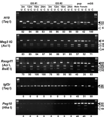
We also determined the genomic imprinting pattern in the GS cells, because the methylation patterns of germline cells tend to change during culture. The methylation patterns of the
DMRs of three paternally methylated regions [H19, Meg3 IG (Gtl2 – Mouse Genome Informatics) and Rasgrf1] and two maternally methylated regions (Igf2r and Peg10) were examined in GS cells harvested after 3, 12, 18 and 24 months of continuous culture (Fig. 3). Consistent with the findings of our previous study (Kanatsu-Shinohara et al., 2004b), COBRA analysis of the GS cells demonstrated androgenetic imprinting: methylation of the paternal DMRs and demethylation of the maternal DMRs. This androgenetic pattern was not altered in the two cultures at 24 months, indicating that GS cells are epigenetically stable. By contrast, the mGS cells that developed in a 3-month-old culture after freeze-thaw treatment had a different methylation pattern. Although paternally methylated regions in H19 and Rasgrf1 were highly methylated, DMRs in the Meg3 IG region were slightly undermethylated compared with those in the GS cells. Furthermore, while DMRs in Igf2rare unmethylated in the GS cells, they are hypermethylated in the mGS cells. These results support our previous observation that genomic imprinting is variable in mGS cells (Kanatsu-Shinohara et al., 2004).
Spermatogenesis and generation of offspring from GS cells after long-term culture
[image:4.612.43.270.231.741.2]To examine whether the cultured cells retained spermatogonial stem cell activity, we used a spermatogonial transplantation technique that allows competent spermatogonial stem cells to colonize the empty seminiferous tubules of infertile mouse testes (Brinster and Zimmermann, 1994). GS cells were recovered from the two separate cultures at different time points during the long-term culture period, and single cell suspensions of the cells were microinjected into the seminiferous tubules of immunosuppressed infertile W mice. W mice lack endogenous spermatogenesis, owing to mutations in the Kit receptor (Ohta et al., 2003); therefore, any spermatogenesis in the recipient testes must necessarily originate from the transplanted stem cells. Two months after transplantation, the recipient mice were sacrificed, and the testes were examined for the presence of EGFP-positive colonies under UV illumination. The transplanted GS cells colonized the seminiferous tubules of W recipient testes and produced EGFP-expressing spermatogenic colonies, regardless of the amount of time the cells had been cultured (Fig. 4A). Assuming 10% colonization efficiency (Nagano et al., 1999), the concentration of functional stem cells in the cultures ranged from 0.02% to 1.6% (Table 1). Overall, the number of stem cells expanded by approximately 1.8⫻1073-fold during 617 days in culture, during which time the total cell number increased 1.3⫻1073-fold. Given that the neonatal testis
Fig. 2.Analysis of telomere dynamics and telomerase activity. (A) Southern blot analysis of TRFs containing telomeric DNA sequences in five different ES (E14) cells and GS cells. Genomic DNAs from ES or GS cells were digested with HinfI, separated on a pulse-field gel and probed with the 5⬘-[32P](T
2AG3)3telomeric DNA
oligonucleotides. (B) Loss of telomeres in GS cells. Based on the median values of the TRF analysis shown in (A), shortening rates of the telomeric DNA sequences in GS cells were calculated for two separate cultures. (C) Telomerase activity in ES and GS cells. ES and GS cells were subjected to a standard telomeric repeat amplification assay. Extracts were pretreated with (H) or without heat inactivation (80°C, 10 minutes) prior to the telomerase assay. The total amount of cell lysates per assay is indicated.
De
normally contains approximately 34 stem cells per 105cells (Shinohara et al., 2001), this result also indicates that the stem cells expanded by ~1086-fold since the initiation of the cultures. Histological analysis confirmed the presence of normal-appearing spermatogenesis in the recipient testes (Fig. 4B).
Finally, to determine whether the germ cells were functionally normal, we attempted to produce offspring from long-term cultures of GS cells. We sacrificed W recipient mice that had received GS cells from 24-month-old cultures. The EGFP-expressing colonies were dissociated mechanically under UV light. Live spermatogenic cells were recovered by repeated pipetting of EGFP-positive seminiferous tubule fragments from three different recipient mice (Fig. 4C). Round spermatids were collected and microinjected into BDF1 oocytes. A total of 194 embryos were constructed, and 130 were transferred into the oviducts of pseudopregnant ICR females (Table 2). Of the 16 offspring born, 11 expressed the Egfp gene (Fig. 4C). The mean weight of the neonatal mice derived from GS cells was 1.7 g, and the mean placental weight was 0.15 g, both of which were within the normal ranges. COBRA analysis of tissue samples indicated that the offspring had normal imprinting patterns (Fig. 3). The offspring developed normally and became fertile as adults.
Discussion
Our study revealed the remarkable stability and proliferative capacity of spermatogonial stem cells during long-term culture. The genetic and epigenetic stability of spermatogonial stem cells is striking. Most mammalian somatic cells
have limited proliferative potential, and eventually undergo senescence after limited number of cell divisions (Rubin, 2002). Many cells that appear after degenerative phase are aneuploid, and it is from these that the permanent cells appear to evolve (Rubin, 2002). By contrast, the growth rate of the GS cells was constant during the 2 years in culture, and the euploid karyotype was retained for at least 139 passages. Our results also showed the functional stability of cultured spermatogonial stem cells; the production of fertile offspring from cells passaged continuously for 2 years demonstrates that the functional characteristics of spermatogonial stem cells are fully retained despite long-term in vitro expansion.
Our results contrast with previous reports that demonstrated instability of germline cells in vitro. Although ES cells have a spontaneous mutation frequency that is ~100-fold below that of somatic cells (Cervantes et al., 2002), culture of germline
[image:5.612.226.562.69.446.2]cells, such as preimplantation embryo, ES cells and EG cells, generally induces genetic and epigenetic changes. For example, previous studies have shown that ES cells progressively lose euploidy with increasing numbers of culture passages. More than 70% of ES cells became aneuploid by passages 25 (equivalent to more than 2 months of continuous culture), and these cells could no longer contribute to the germline by blastocyst injection (Longo et al., 1997). The aneuploid abnormalities induced by long-term culture appear to occur at specific chromosome loci: whereas mouse ES cells commonly develop trisomy eight after long-term culture (Liu et al., 1997), human ES cells have trisomy 17q and 12 (Draper et al., 2004). These karyotypic changes probably confer a growth advantage in vitro and hence represent a common cause of the loss of germline potential. In addition to their chromosomal instability, in vitro culture of preimplantation Fig. 3.COBRA of imprinted genes. PCR products of each DMR region were digested with restriction enzymes with a recognition sequence containing CpG in the original unconverted DNA. Where the enzyme sites are methylated, PCR products are fragmented; where PCR products are not fragmented, the enzyme sites are unmethylated. White arrowheads indicate the sizes of the unmethylated DNA fragments. Black arrowheads indicate the sizes of the methylated DNA fragments. The enzymes used to cleave each locus are indicated in parenthesis. BsiEI was used to analyze the Rasgrf1DMRs in the DNA from mouse pups and from mGS cells. Levels of percentage methylation, estimated by the intensity of each band, are indicated below the gels. U, uncleaved; C, cleaved.
De
embryo, ES cells and EG cells induces aberrant genomic imprinting, which can lead to morphological or functional abnormalities in embryo or offspring (Sasaki et al., 1995; Dean et al., 1998; Humpherys et al., 2001). Surprisingly,
composition of the medium can also influence the imprint pattern (Doherty et al., 2000; Khosla et al., 2002; Ecker et al., 2004). The instability of these embryonic germline cells probably reflects their embryonic origin, which is susceptible to subtle changes in the maternal environment. However, our results strongly suggest that spermatogonial stem cells have sophisticated repair mechanisms to prevent transmission of genetic or epigenetic damage to the progenitors, which may be similar to the those found in postnatal stem cells in other self-renewing tissues (Cairns, 2002; Potten et al., 2002; Gilbert, 2005).
[image:6.612.46.561.85.276.2]Despite their stability of spermatogonial stem cells, this study also demonstrated the shortening of telomeres during long-term culture, which suggests that these cells have limited proliferative potential. In most types of normal cells, the telomeric DNA erodes progressively with each round of cell division in the absence of telomerase activity (Blackburn, 2005). Shortened telomeres induce chromosome fusion and trigger a checkpoint that results in cell cycle arrest and apoptosis. By contrast, telomerase activity and maintenance of telomere length is associated with immortality in tumor and ES cells (Niida et al., 1998; Blackburn, 2005). As stem cells are considered to proliferate indefinitely by self-renewing division, the status of the telomeres in stem cells has been of great interest. Unlike most normal somatic cells, which do not have telomerase activity, low to moderate levels of telomerase expression have been described in adult stem cells from the skin, the gut and the hematopoietic system (Harle-Bachor et al., 1996; Kolquist et al., 1998; Allsopp et al., 2003). Telomerase plays an important role in maintaining the telomeres in male germ cells, as shown by the gradual loss of spermatogenic cells and eventual sterility in telomerase-deficient mice (Lee et al., 1998). However, our results show that telomere shortening occurs progressively during long-term culture of spermatogonial stem cells, despite the presence of telomerase activity. A similar phenomenon was also reported in hematopoietic stem cells, in which telomere shortening Table 1. Spermatogonial stem cell expansion during long-term culture
Days to GS cells Increase in Increase
transplant* injected/testis Colonies/ Stem cells/ GS cell number in stem cell
Culture (passage) (⫻104) Colonies/testis 105GS cells 105GS cells† (fold)‡ number (fold)‡,§
1 134 (24) 1.9 30.7±8.9 163.6±47.2 1635.7
216 (41) 7.5 1.5±0.6 2.00±0.8 20.0 1.7⫻108 2.1⫻106
301 (58) 5.0 32.0±11.3 64.0±22.7 640.0 1.4⫻1019 5.5⫻1018
366 (71) 2.7 17.7±7.5 65.4±27.9 654.4 4.7⫻1027 1.9⫻1027
461 (89) 3.0 19.7±0.3 65.6±1.1 655.7 6.0⫻1039 2.4⫻1039
550 (105) 1.5 9.7±1.2 64.5±8.0 644.7 5.7⫻1050 2.3⫻1050
638 (121) 1.5 1.6±0.9 10.7±6.2 106.7 8.6⫻1061 5.6⫻1060
730 (139) 1.5 5.5±2.3 36.7±15.0 366.7 9.1⫻1072 2.1⫻1072
2 113 (19) 3.0 8.7±2.1 28.9±7.1 289.0
216 (38) 3.75 4.7±1.0 12.5±2.6 124.5 2.1⫻108 9.0⫻107
301 (55) 5.0 9.8±5.7 19.6±11.5 196.0 2.5⫻1018 1.7⫻1018
366 (68) 2.7 22.0±3.5 81.5±13.0 814.8 8.2⫻1025 2.3⫻1026
461 (86) 3.0 24.8±3.4 82.5±11.2 825.0 5.1⫻1037 1.4⫻1038
550 (102) 1.5 9.8±1.7 65.3±11.1 653.3 1.8⫻1048 3.9⫻1048
638 (118) 1.5 1.3±0.8 8.3±5.0 83.3 6.3⫻1060 1.8⫻1060
730 (136) 1.5 5.8±1.5 38.3±9.9 383.3 1.3⫻1073 1.8⫻1073
Values are mean±s.e.m. Results from at least four recipient testes. *The number of days from initiation of culture to transplantation.
†It is assumed that 10% of transplanted stem cells can colonize the testis (Nagano et al., 1999). ‡The increase in GS or stem cell number from the first transplantation.
§(Increase in GS cell number at indicated time point)⫻(Stem cells/105GS cells at indicated time point)/(Stem cells/105GS cells at first transplantation).
Fig. 4. Spermatogenesis and the generation of offspring from GS cells. EGFP-expressing GS cells from 24-month-old cultures were transplanted into the seminiferous tubules of infertile W recipient mice. (A) A recipient testis showing the EGFP fluorescence in GS cell-derived colonies. Green tubules represent donor GS cell colonization. (B) Histological analysis (Hematoxylin and Eosin staining) of the recipient testis showing normal-appearing spermatogenesis. (C) Round spermatid released from a recipient testis, exhibiting EGFP fluorescence (arrow). (D) Offspring resulting from the microinjection of oocytes with round spermatids, exhibiting fluorescence. Scale bar: 1 mm.
De
[image:6.612.44.291.392.620.2]occurs after three to four rounds of serial transplantation, despite a high level of telomerase activity (Allsopp et al., 2003). These results indicate that postnatal stem cells might possess mechanisms for the regulation of telomere length that are different from those of ES cells.
Although our results show the remarkable stability of spermatogonial stem cells, our previous study has suggested that GS cells had multipotential characteristics (Kanatsu-Shinohara et al., 2004b). We reported that ES-like cells (mGS cells) appeared in the cultures during the initiation of GS cell cultures from neonatal testis cells. The origin of these mGS cells in neonatal testis cell culture is currently unknown (Kanatsu-Shinohara et al., 2004b); it is possible that they arise from a population of distinct undifferentiated pluripotent cells that persist in the testis from fetal stage. However, we previously demonstrated the direct conversion of GS cells into mGS cells in experiments using GS cells derived from p53 mutant mice that have a high frequency of teratoma (Lam and Nadeau, 2003; Kanatsu-Shinohara et al., 2004b). Based on this observation, we speculated that spermatogonial stem cells retain the ability to become multipotent cells during the early passages in vitro, an ability that may be lost during long-term cell culture (Kanatsu-Shinohara et al., 2004b). In this study, mGS cell colonies developed in 3-month-old cultures after a freeze-thaw treatment, but thereafter no other GS cells converted to mGS cells during the 2-year experimental period. This result confirms our previous observation that mGS cells appear in the early phase of neonatal testis cell culture, and also suggests that established GS cells are resistant to mGS cell conversion. Although current culture conditions support the germline potential in spermatogonial stem cells, they do not fully support the multilineage potential attributed to spermatogonial stem cells. Further studies will be necessary to determine whether mGS cells are derived from GS cells, and a special effort should be made to establish improved culture conditions that maintain the full developmental potential of spermatogonial stem cells.
Spermatogonial stem cells represent an ideal model in which to investigate the unique biology of stem cells. Spermatogonial stem cells can be transplanted between animals, genetically manipulated and cultivated under feeder-cell or serum-free conditions (Brinster and Zimmermann, 1994; Kubota et al., 2004; Kanatsu-Shinohara et al., 2005a, Kanatsu-Shinohara et al., 2005b). These advantages are not available for stem cells in other self-renewing systems. Using these techniques, spermatogonial stem cells can now be routinely expanded and subsequently examined for molecular and genetic characteristics. Future studies will determine the mechanism by which spermatogonial stem cells maintain the stable characteristics. In addition, the long-term stability of GS cells
will provide a strong advantage in practical application, and may complement ES cell technology. Although the shortening telomeres suggests that spermatogonial stem cells have limited proliferative potential, it would not be a serious concern in practical application of GS cells; assuming a constant rate of telomere loss, GS cells should continue to proliferate for 34 months and achieve ~10120-fold expansion before they undergo crisis. This should allow a sufficient time for in vitro genetic modification of GS cells for various purposes, including gene targeting. Thus, GS cells provide a new possibility in the study of stem cell biology and will be an attractive target of germline modification.
We thank Dr Y. Kaziro for encouragement and critical reading and Ms A. Wada for technical assistance. Financial support for this research was provided by the Ministry of Education, Culture, Sports, Science and Technology (MEXT) of Japan, and by grants from CREST and the Human Science Foundation (Japanese). This work was also supported in part by Special Coordination Funds for Promoting Science and Technology from MEXT.
References
Allsopp, R. C., Morin, G. B., Horner, J. W., DePinho, R., Harley, C. B. and Weissman, I. L.(2003). Effect of TERT over-expression on the long-term transplantation capacity of hematopoietic stem cells. Nat. Med. 9, 369-371.
Blackburn, E. H.(2005). Telomeres and telomerase: their mechanisms of action and the effects of altering their functions. FEBS Lett. 579, 859-862.
Brinster, R. L. and Zimmermann, J. W.(1994). Spermatogenesis following male germ-cell transplantation. Proc. Natl. Acad. Sci. USA 91, 11298-11302.
Buaas, F. W., Kirsch, A. L., Sharma, M., McLean, D. J., Morris, J. L., Griswold, M. D., de Rooij, D. G. and Braun, R. E.(2004). Plzf is required in adult male germ cells for stem cell self-renewal. Nat. Genet. 36, 647-652.
Cairns, J.(2002). Somatic stem cells and the kinetics of mutagenesis and carcinogenesis. Proc. Natl. Acad. Sci. USA99, 10567-10570.
Cervantes, R. B., Stringer, J. R., Shao, C., Tischfield, J. A. and Stambrook, P. J.(2002). Embryonic stem cells and somatic cells differ in mutation frequency and type. Proc. Natl. Acad. Sci. USA99, 3586-3590.
Chambers, I., Colby, D., Robertson, M., Nichols, J., Lee, S., Tweedie, S. and Smith, A. (2003). Functional expression cloning of Nanog, a pluripotency sustaining factor in embryonic stem cells. Cell113, 643-655.
Costoya, J. A., Hobbs, R. M., Barna, M., Cattoretti, G., Manova, K., Sukhwani, M., Orwig, K. E., Wolgemuth, D. J. and Pandolfi, P. P.(2004). Essential role of Plzf in maintenance of spermatogonial stem cells. Nat. Genet. 36, 653-659.
de Rooij, D. G. and Russell, L. D.(2000). All you wanted to know about spermatogonia but were afraid to ask. J. Androl. 21, 776-798.
Dean, W., Bowden, L., Aitchison, A., Klose, J., Moore, T., Menesses, J. J., Reik, W. and Feil, R.(1998). Altered imprinted gene methylation and expression in completely ES cell-derived mouse fetuses: association with aberrant phenotypes. Development125, 2273-2282.
Doherty, A. S., Mann, M. R. W., Tremblay, K. D., Bartolomei, M. S. and Schultz, R. M. (2000). Differential effects of culture on imprinted H19 expression in the preimplantation embryo. Biol. Reprod.62, 1526-1535.
[image:7.612.52.559.86.150.2]Draper, J. S., Smiths, K., Gokhale, P., Moore, H. D., Maltby, E., Johnson,
Table 2. Embryo development after microinsemination
Number of Number of Implantation Number of
Experiment reconstituted eggs Two cell (%) eggs transferred sites (%) offspring (%)
1 97 74 (76.3) 73 38 (52.1) 10 (13.7)
2 10 9 (90.0) 9 5 (55.6) 2 (22.2)
3 87 70 (80.5) 48 14 (29.2) 4 (8.3)
Total 194 153 (78.9) 130 57 (43.8) 16 (12.3)
Results from three separate experiments. Embryos were cultured for 24 hours and transferred into the pseudopregnant mother. Round spermatids were used for microinsemination.
De
J., Meisner, L., Zwaka, T. P., Thomson, J. A. and Andrews, P. W.(2004). Recurrent gain of chromosomes 17q and 12 in cultured human embryonic stem cells. Nat. Biotechnol. 22, 53-54.
Ecker, D. J., Stein, P., Xu, Z., Williams, C. J., Kopf, G. S., Bilker, W. B., Abel, T. and Schultz, R. M.(2004). Long-term effects of culture of preimplantation mouse embryos on behavior. Proc. Natl. Acad. Sci. USA
101, 1595-1600.
Evans, M. J. and Kaufman, M. H. (1981). Establishment in culture of pluripotential cells from mouse embryos. Nature292, 154-156.
Falender, A. E., Freiman, R. N., Geles, K. G., Lo, K. C., Hwang, K., Lamb, D. J., Morris, P. L., Tjian, R. and Richards, J. S.(2005). Maintenance of spermatogenesis requires TAF4b, a gonad-specific subunit of TFIID. Genes Dev. 19, 794-803.
Fujiwara, Y., Komiya, T., Kawabata, H., Sato, M., Fujimoto, H., Furusawa, M. and Noce, T.(1994). Isolation of a DEAD-family protein gene that encodes a murine homolog of Drosophila vasa and its specific expression in germ cell lineage. Proc. Natl. Acad. Sci. USA91, 12258-12262.
Gilbert, G. H.(2005). Label-retaining epithelial cells in mouse mammary gland divide asymmetrically and retain their template DNA strands. Development132, 681-687.
Giuili, G., Tomljenovic, A., Labrecque, N., Oulad-Abdelghani, M., Raaaoulzadegan, M. and Cuzin, F.(2002). Murine spermatogonial stem cells: targeted transgene expression and purification in an active state. EMBO Rep. 3, 753-759.
Harle-Bachor, C. and Boukamp, P. (1996). Telomerase activity in the regenerative basal layer of the epidermis in human skin and in immortal and carcinoma-derived skin keratinocytes. Proc. Natl. Acad. Sci. USA93, 6476-6481.
Humpherys, D., Eggan, K., Akutsu, H., Hochedlinger, K., Rideout III, W. M., Biniszkiewicz, D., Yanagimachi, R. and Jaenisch, R. (2001). Epigenetic instability in ES cells and cloned mice. Science 293, 95-97.
Kanatsu-Shinohara, M., Ogonuki, N., Inoue, K., Ogura, A., Toyokuni, S., Honjo, T. and Shinohara, T.(2003a). Allogeneic offspring produced by male germ line stem cell transplantation into infertile mouse testis. Biol. Reprod. 68, 167-173.
Kanatsu-Shinohara, M., Ogonuki, N., Inoue, K., Miki, H., Ogura, A., Toyokuni, S. and Shinohara, T.(2003b). Long-term proliferation in culture and germline transmission of mouse male germline stem cells. Biol. Reprod.
69, 612-616.
Kanatsu-Shinohara, M., Ogonuki, N., Inoue, K., Ogura, A., Toyokuni, S. and Shinohara, T.(2003c). Restoration of fertility in infertile mice by transplantation of cryopreserved male germline stem cells. Hum. Reprod.
18, 2660-2667.
Kanatsu-Shinohara, M., Toyokuni, S. and Shinohara, T.(2004a). CD9 is a surface marker on mouse and rat male germline stem cells. Biol. Reprod.
70, 70-75.
Kanatsu-Shinohara, M., Inoue, K., Lee, J., Yoshimoto, M., Ogonuki, N., Miki, H., Baba, S., Kato, T., Kazuki, Y., Toyokuni, S. et al.(2004b). Generation of pluripotent stem cells from neonatal mouse testis. Cell119, 1001-1012.
Kanatsu-Shinohara, M., Toyokuni, S. and Shinohara, T.(2005a). Genetic selection of mouse male germline stem cells in vitro: Offspring from single stem cells. Biol. Reprod.72, 236-240.
Kanatsu-Shinohara, M., Miki, H., Inoue, K., Ogonuki, N., Toyokuni, S., Ogura, A. and Shinohara, T.(2005b). Long-term culture of mouse male germline stem cells under serum- or feeder-free conditions. Biol. Reprod.
72, 985-991.
Khosla, S., Dean, W., Brown, D., Reik, W. and Feil. R.(2001). Culture of preimplantation mouse embryos affects fetal development and the expression of imprinted genes. Biol. Reprod. 64, 918-926.
Kimura, Y. and Yanagimachi, R. (1995). Mouse oocytes injected with testicular spermatozoa or round spermatids can develop into normal offspring. Development121, 2397-2405.
Kolquist, K. A., Ellisen, L. W., Counter, C. M., Meyerson, M., Tan, L. K., Weinberg, R. A., Harber, D. A. and Gerald, W. L.(1998). Expression of TERT in early premalignant lesions and a subset of cells in normal tissues. Nat. Genet. 19, 182-186.
Kubota, H., Avarbock, M. R. and Brinster, R. L.(2004). Growth factors essential for self-renewal and expansion of mouse spermatogonial stem cells. Proc. Natl. Acad. Sci. USA101, 16489-16494.
Labosky, P. A., Barlow, D. P. and Hogan, B. L. M.(1994). Mouse embryonic germ (EG) cell lines: transmission through the germline and differences in the methylation imprint of insulin-like growth factor 2 receptor (Igf2r) gene
compared with embryonic stem (ES) cell lines. Development120, 3197-3204.
Lam, M.-Y. J. and Nadeau, J. H.(2003). Genetic control of susceptibility to spontaneous testicular germ cell tumors in mice. Acta Pathol. Mic. Sc.111, 184-191.
Lee, H.-W., Blasco, M. A., Gottlieb, G. J., Horner, II, J. W., Greider, C. W. and DePinho, R. A.(1998). Essential role of mouse telomerase in highly proliferative organs. Nature392, 569-574.
Liu, X., Wu, H., Loring, J., Hormuzdi, S., Disteche, C. M., Bornstein, P. and Jaenisch, R.(1997). Trisomy eight in ES cells is a common potential problem in gene targeting and interferes with germ line transmission. Dev. Dyn. 209, 85-91.
Longo, L., Bygrave, A., Grosveld, F. G. and Pandolfi, P. P.(1997). The chromosome make-up of mouse embryonic stem cells is predictive of somatic and germ cell chimaerism. Transgenic Res. 6, 321-328.
Martin, G. R.(1981). Isolation of a pluripotent cell line from early mouse embryos cultured in medium conditioned by teratocarcinoma stem cells. Proc. Natl. Acad. Sci. USA78, 7634-7638.
Matsui, Y., Zsebo, K. and Hogan, B. L. M. (1992). Derivation of pluripotential embryonic stem cells from murine primordial germ cells in culture. Cell70, 841-847.
Meistrich, M. L. and van Beek, M. E. A. B.(1993). Spermatogonial stem cells. In Cell and Molecular Biology of the Testis(ed. C. Desjardins and L. L. Ewing), pp. 266-295. New York: Oxford University Press.
Meng, X., Lindahl, M., Hyvönen, M. E., Parvinen, M., de Rooij, D. G., Hess, M. W., Raatikainen-Ahokas, A., Sainio, K., Rauvala, H., Lakso, M. et al. (2000). Regulation of cell fate decision of undifferentiated spermatogonia by GDNF. Science287, 1489-1493.
Mitsui, K., Tokuzawa, Y., Itoh, H., Segawa, K., Murakami, M., Takahashi, K., Maruyama, M., Maeda, M. and Yamanaka, S. (2003). The homeoprotein Nanog is required for maintenance of pluripotency in mouse epiblast and ES cells. Cell113, 631-642.
Nagano, M., Avarbock, M. R. and Brinster, R. L. (1999). Pattern and kinetics of mouse donor spermatogonial stem cell colonization in recipient testes. Biol. Reprod. 60, 1429-1436.
Niida, H., Matsumoto, T., Satoh, H., Shiwa, M., Tokutake, Y., Furuichi, Y. and Shinkai, Y.(1998). Severe growth defect in mouse cells lacking the telomerase RNA component. Nat. Genet. 19, 203-206.
Ogawa, T., Aréchaga, J. M., Avarbock, M. R. and Brinster, R. L.(1997). Transplantation of testis germinal cells into mouse seminiferous tubules. Int. J. Dev. Biol.41, 111-122.
Ogawa, T., Ohmura, M., Tamura, Y., Kita, K., Ohbo, K., Suda, T. and Kubota, Y.(2004). Derivation and morphological characterization of mouse spermatogonial stem cell lines. Arch. Histol. Cytol. 67, 297-306.
Ohta, H., Tohda, A. and Nishimune, Y. (2003). Proliferation and differentiation of spermatogonial stem cells in the W/Wv mutant mouse
testis. Biol. Reprod. 69, 1815-1821.
Pesce, M. and Schöler, H. R.(2001). Oct-4: gatekeeper in the beginning of mammalian development. Stem Cells19, 271-278.
Potten, C. S.(1992). Cell lineages. In Oxford Textbook of Pathology(ed. J. O. McGee, P. G. Isaacson and N. A. Wright), pp. 43-52. Oxford: Oxford University Press.
Potten, C. S., Owen, G. and Booth, D.(2002). Intestinal stem cells protect their genome by selective segregation of template DNA strands. J. Cell. Sci.
115, 2381-2388.
Resnick, J. L., Bixler, L. S., Cheng, L. and Donovan, P. J.(1992). Long-term proliferation of mouse primordial germ cells in culture. Nature359, 550-551.
Rubin, H.(2002). The disparity between human cell senescence in vitro and lifelong replication in vivo. Nat. Biotechnol. 20, 675-681.
Ryu, B.-Y., Orwig, K. E., Kubota, H., Avarbock, M. R. and Brinster, R. L.(2004). Phenotypic and functional characteristics of male germline stem cells in rats. Dev. Biol.274, 158-170.
Saitou, M., Barton, S. C. and Surani, M. A.(2002). A molecular program for the specification of germ cell fate in mice. Nature418, 293-300.
Sasaki, H., Ferguson-Smith, A. C., Shum, A. S. W., Barton, S. C. and Surani, M. A.(1995). Temporal and spatial regulation of H19imprinting in normal and uniparental mouse embryos. Development121, 4195-4202.
Shinohara, T., Avarbock, M. R. and Brinster, R. L.(1999). 1- and ␣ 6-integrin are surface markers on mouse spermatogonial stem cells. Proc. Natl. Acad. Sci. USA96, 5504-5509.
Shinohara, T., Orwig, K. E., Avarbock, M. R. and Brinster, R. L.(2000). Spermatogonial stem cell enrichment by multiparameter selection of mouse testis cells. Proc. Natl. Acad. Sci. USA97, 8346-8351.
De
Shinohara, T., Orwig, K. E., Avarbock, M. R. and Brinster, R. L.(2001). Remodeling of the postnatal mouse testis is accompanied by dramatic changes in stem cell number and niche accessibility. Proc. Natl. Acad. Sci. USA98, 6186-6191.
Solter, D. and Knowles, B. B.(1978). Monoclonal antibody defining a stage-specific mouse embryonic antigen (SSEA-1). Proc. Natl. Acad. Sci. USA75, 5565-5569.
Takahashi, K., Mitsui, K. and Yamanaka, S. (2003). Role of Eras in promoting tumor-like properties in mouse embryonic stem cells. Nature414, 122-128.
Tatematsu, T., Nakayama, J., Danbara, M., Shionoya, S., Sato, H., Omine, M. and Ishikawa, F.(1996). A novel quantitative ‘stretch PCR assay’, that detects a dramatic increase in telomerase activity during the progression of myeloid leukemias. Oncogene13, 2265-2274.
Xiong, Z. and Laird, P. W.(1997). COBRA: a sensitive and quantitative DNA methylation assay. Nucleic Acids Res. 25, 2532-2534.
Yoshida, S., Takakura, A., Ohbo, K., Abe, K., Wakabayashi, J., Yamamoto, M., Suda, T. and Nabeshima, Y. (2004). Neurogenenin3 delineates the earliest stages of spermatogenesis in the mouse testis. Dev. Biol. 269, 447-458.