R E S E A R C H A R T I C L E
Open Access
Structural brain changes in first episode
Schizophrenia compared with Fronto-Temporal
Lobar Degeneration: a meta-analysis
Bayanne Olabi
1*, Ian Ellison-Wright
2, Ed Bullmore
3,4and Stephen M Lawrie
1Abstract
Background:The authors sought to compare gray matter changes in First Episode Schizophrenia (FES) compared with Fronto-Temporal Lobar Degeneration (FTLD) using meta-analytic methods applied to neuro-imaging studies.
Methods:A systematic search was conducted for published, structural voxel-based morphometric MRI studies in patients with FES or FTLD. Data were combined using anatomical likelihood estimation (ALE) to determine the extent of gray matter decreases and analysed to ascertain the degree of overlap in the spatial distribution of brain changes in both diseases.
Results:Data were extracted from 18 FES studies (including a total of 555 patients and 621 comparison subjects) and 20 studies of FTLD or related disorders (including a total of 311 patients and 431 comparison subjects). The similarity in spatial overlap of brain changes in the two disorders was significant (p = 0.001). Gray matter deficits common to both disorders included bilateral caudate, left insula and bilateral uncus regions.
Conclusions:There is a significant overlap in the distribution of structural brain changes in First Episode Schizophrenia and Fronto-Temporal Lobar Degeneration. This may reflect overlapping aetiologies, or a common vulnerability of these regions to the distinct aetio-pathological processes in the two disorders.
Keywords:Schizophrenia/pathology, FTLD/pathology, Meta-analysis, Magnetic resonance imaging/methods, Humans, Brain/pathology, Brain mapping, Imaging processing, Computer-Assisted/methods
Background
Schizophrenia is a disorder characterised by positive symptoms (hallucinations and delusions), thought dis-order and negative symptoms (such as apathy). Brain imaging studies have identified structural changes both early in the presentation of the illness and more exten-sive changes later in the course of the illness [1]. The distribution of changes has been replicated between studies [2], and may be considered a ‘structural signa-ture’of schizophrenia within the brain. However, an ad-equate explanation for this spatial distribution remains elusive. Models have been proposed involving genetic factors coding neuro-protein variants resulting in abnor-mal development of limbic and
frontal-temporal-subcortical networks [3]. However, it remains uncertain which neuro-chemical pathways are responsible and how they cause the spatial changes.
FTLD is characterised by declines in social function, interpersonal conduct, emotional blunting, speech and language abnormalities and loss of insight [4]. It is asso-ciated with degeneration of the prefrontal and anterior temporal cortices [5]. However, the topographical distri-bution of structural brain changes is heterogeneous amongst different patient groups, reflecting behavioral and pathological variants. Although the most common histological feature is tauopathy [6], over half of all patients, including those with a family history of the condition, have no abnormality in the tau gene or pro-tein [7], consistent with pathological and aetiological heterogeneity.
In this study, we investigate whether there is overlap in the distribution of brain changes in First Episode * Correspondence:[email protected]
1
Division of Psychiatry, School of Molecular and Clinical Medicine, University of Edinburgh, Royal Edinburgh Hospital, Edinburgh EH10 5HF, UK Full list of author information is available at the end of the article
Schizophrenia (FES) and Fronto-Temporal Lobar Degen-eration (FTLD). There is evidence for similarities in clin-ical, neuropsychological and neuroimaging findings in patients with FTLD and schizophrenia [8]. In some cases, patients with FTLD have been diagnosed with a schizophrenia-like psychotic illness years before the de-mentia diagnosis is made [9]. Echopraxia, echolalia, aprosody of speech, utilisation behavior,‘negative’ symp-toms, self-neglect, and bizarre, compulsive, and stereo-typed behaviors are well recognised in both disorders. Executive dysfunction with relative preservation of visual perception and spatial skills [7,10] and deficits in social cognition, theory of mind, empathy and affect recogni-tion have been identified in both disorders [11].
Frontal, temporal and hippocampal atrophy [12,13] and regionally specific reductions in the anterior corpus callosum [14,15] and anterior hippocampus [16,17] have been described in MRI studies of both FTLD and schizo-phrenia. Frontal hypoperfusion on single photon emis-sion tomography or positron emisemis-sion tomography constitutes one of the imaging criteria for the diagnosis of frontotemporal dementia [18], and is also one of the most robust functional imaging findings in the schizo-phrenia literature in patients with chronic and first-episode illness [19]. There is also recent evidence that schizophrenia and FTLD co-occur in some families, sug-gesting the possibility of a common vulnerability to these disorders [20]. While the pathology of schizophre-nia remains uncertain, there have been considerable advances in elucidating the complex and heterogeneous pathology of FTLD [21,22]. We have chosen to examine FES rather than chronic schizophrenia because the structural changes in FES may more accurately reflect the pathological changes of the disorder, and may minimize the confounding effects of long-term medica-tion and other aspects of chronic illness. Different anti-psychotics have individual volumetric effects on brain structure [23,24] and therefore, patients with FES were used in order to reduce heterogeneity.
The aim of this review is to determine the distribution of brain changes in FTLD and FES, by employing an established meta-analytic technique (anatomical likeli-hood estimation, ALE) [25] that is widely used for coordinate-based meta-analyses of neuroimaging data by converting the co-ordinates of peak gray matter changes from multiple published studies into spatial probability maps. However, the accuracy and extent of these maps is dependent on the total number of peak co-ordinates available from published studies. Therefore, this study employs a new statistical approach to investigate the de-gree of spatial correspondence between the two disor-ders, taking into account the greater availability of data co-ordinates for FTLD than FES. The comparison of brain changes between an individual MRI scan and maps
for different disorders may become increasingly import-ant for early diagnosis, as currently, diagnoses of psychi-atric disorders are made on the basis of clinical manifestations and associated psychosocial disturbances. There are current initiatives to encourage the classifica-tion of mental disorders for research purposes, such as the RDoC (Research Domain Criteria) approach [26]. Several MRI-based studies have attempted to distinguish between patients and healthy subjects with high accur-acy (ranging from 75 to 92%) [27]. Therefore, the statis-tical technique described in this paper for assessing spatial overlap may have wider clinical utility in the future.
Methods
The PRISMA (preferred reporting items for systematic reviews and meta-analyses) guidelines were followed to conduct this review [28].
Literature search
A comprehensive keyword search of EMBASE (from 1980), PsycINFO (from 1801) and Medline (from 1950), was conducted using the following search strategy. The following Boolean phrase was used: {([Magnetic Reson-ance Imaging] OR [MRI]) AND ([Schizophrenia] OR [schizo*] OR [FTLD] OR [Fronto-Temporal Lobar De-generation) AND ([Voxel] OR [VBM])}. Both free-text and expanded medical subject headings were be used. The search strategy was supplemented using a cited reference search, and by inspecting the reference lists of included articles. The search was conducted in January 2011. No time span was specified for date of publication.
Criteria for inclusion/exclusion
Studies were considered for the review using the follow-ing inclusion criteria: 1) they were published in English as a peer-reviewed article (rather than a letter, abstract, or case report); 2) they compared a sample of formally diagnosed subjects with a group of unrelated healthy control subjects; 3) they utilized voxel-based analysis of gray matter in structural MRI scans to investigate differ-ences in whole-brain; and 4) they reported the three-dimensional co-ordinates of changes in stereotactic space.
diagnosis without administration of prior antipsychotic medication in order to be included in our meta-analysis.
Studies were excluded if: 1) there were insufficient data to extract the number of subjects in each group; 2) there were fewer than five subjects in either the patient group or the comparison group; 3) the comparison groups consisted of patients with minor non-psychiatric illnesses; 4) the patient group in the schizophrenia stud-ies consisted of subjects with child-onset schizophrenia or chronic schizophrenia beyond the stage of first-episode psychosis; 5) the studies used region-of-interest volumetric analyses, or the deformation- or tensor-based volumetric methods for measuring regional brain volumes; and 6) the data contributed to another publica-tion, in which case the publication with the largest group size under study was selected.
Data abstraction
Data were extracted from two independent investigators (BO and IEW) and were double-checked. Information gathered from the studies included the authors, year of publication, demographic variables (number of subjects, age at baseline and gender), illness variables (diagnosis and duration of illness), and the reported stereotactic coordinates related to the comparisons between struc-tural MRI scans of patients and controls from every selected study. Co-ordinates that were reported in the stereotactic space of the Montreal Neurological Institute (MNI) were converted to Talairach coordinates using the Lancaster transform (icbm2tal) in the GingerALE 2.0 program [29]. Talairach co-ordinates that had been gen-erated by the Brett transform applied to statistical para-metric mapping MNI co-ordinates were transformed back to MNI space in GingerALE and then to Talairach space using the Lancaster transform.
Statistical analysis
Meta-analyses were performed using the Talairach stereotactic coordinates derived from the included stud-ies. Meta-analyses were carried out using GingerALE 2.0 [29]. This uses the technique of Activation Likelihood Estimation (ALE) [25] and permits weighting of studies in the meta-analysis, e.g. based on sample size. This modification of ALE treats the spatial conjunction of co-ordinates from separate studies as more significant than conjunction of co-ordinates from a single study. The probability values can then be interpreted on an image-wide basis after correction for multiple testing using the False Discovery Rate, a method which controls the pro-portion of type 1 errors (false positives) among signifi-cant results [30].
Descriptive meta-analyses were then performed to identify the distribution of brain changes in FES and
FTLD, when compared to control subjects. The overlap of these two distributions was measured.
Spatial overlap testing
We tested the overlap of the FTLD co-ordinates with the FES spatial map generated by ALE using a random-isation method based on our previous work hybridising ALE spatial techniques with GSMA (Genome Scan Meta-Analysis) statistical methods [31].
For each FTLD study, the reported loci of maximal anatomical difference were modeled as the peaks of three-dimensional Gaussian probability density functions with full-width half-maximum of 7 mm, within a brain mask of size N of linear dimension 2 mm. The voxels in this probability image were then ranked from N (highest probability) to 1 (lowest probability), giving voxels of equal probability a mean rank. This created a rank image for each study which was smoothed with a 7 mm Gauss-ian filter. This image was masked with the First Episode Schizophrenia spatial map generated by ALE and the total value of ranks within the mask was calculated. The total for all studies of ranks within the mask was calcu-lated (‘FES Mask Total’).
A null distribution for the FES Mask Total result was derived by 1000 permutations of the same process, but using an equal number of co-ordinates for each study derived from a random uniform distribution of coordi-nates within the brain mask. The probability of a FES Mask Total under the null hypothesis was calculated as the proportion of permutations giving a value equal or greater than the actual value.
The data set being tested was included in the ranking of all known outcomes. The significance threshold was set at p < 0.05.
Results
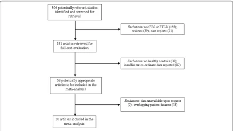
The electronic literature search of the three databases yielded 396 articles, of which 181 were retrieved in full-text format. 56 studies were identified as being poten-tially appropriate to be included in the meta-analysis, and the inclusion and exclusion criteria was used throughout the selection process to yield 38 articles ap-propriate for use in the meta-analysis. Figure 1 displays a flow diagram that shows the reasons for exclusion at each stage of the selection process.
largest size sample was 68 schizophrenia patients [40]. Among the FTLD patient group, the smallest size sam-ple was 6 patients [50,62], and the largest size samsam-ple was 51 patients [60]. All included studies were published from 2000 to 2010.
ALE meta-analyses
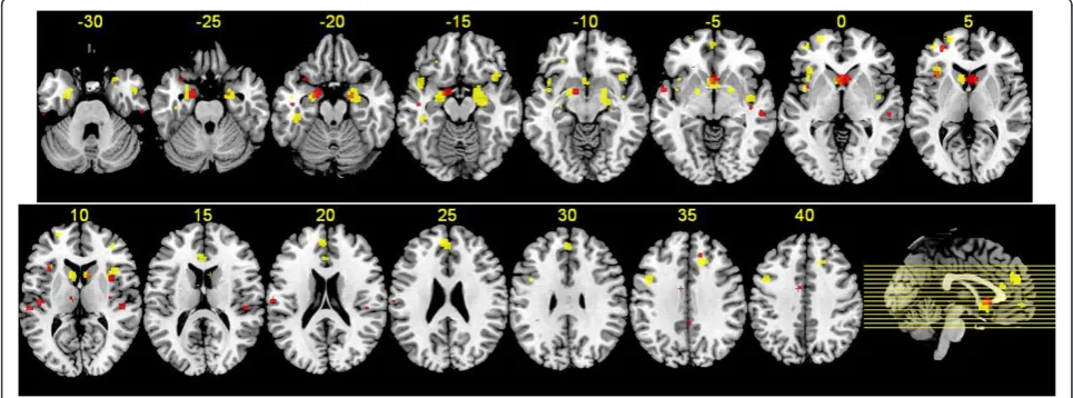
Eighteen FES studies included 185 co-ordinates of gray matter decreases. Meta-analysis of this data identified gray matter decreases in regions including bilateral head of caudate nucleus, bilateral insula, bilateral amygdala/ uncus region and bilateral superior temporal region (Figure 2).
Twenty FTLD studies provided 260 co-ordinates of gray matter decreases. Meta-analysis of this data identi-fied gray matter decreases in regions including bilateral head of caudate nucleus, bilateral insula, bilateral amyg-dala/uncus region and bilateral superior temporal region (Table 2, Figure 2).
Common changes in FES and FTLD
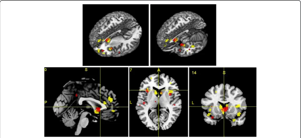
Gray matter decreases were present in both disorders in regions including bilateral caudate head, left insula and bilateral uncus region (Figure 3).
The spatial congruence between the disorders was estimated by percentage overlap. The FTLD map of gray matter decreases comprised 2450 voxels in a brain mask of 201069 voxels. The FES map of gray matter decreases comprised 888 voxels in a brain mask of 201069 voxels.
The overlap between the FTLD and FES maps was 124 voxels. Therefore the overlap of FTLD within FES was 14% (compared to an expected overlap of 1.2% if the FTLD map was randomly distributed in the brain mask). The overlap of FES within FTLD was 5% (compared to an expected overlap of 0.4% if the FES mask was ran-domly distributed in the brain mask). Applying the spatial overlap testing method, the overlap of the FTLD co-ordinates with the FES spatial map generated by ALE was significant (p = 0.001).
Discussion
In this study, we identified overlap in the distribution of brain changes in First Episode Schizophrenia (FES) and Fronto-Temporal Lobar Degeneration (FTLD). We found that the two disorders involved gray matter defi-cits in common regions (p = 0.001). These included the basal ganglia (bilateral caudate head), paralimbic (left in-sula) and limbic (bilateral uncus) regions, as shown in Figure 4. To our knowledge, this is the first meta-analysis that compares neuro-structural profiles between FES and FTLD. Our study presents a novel method using ALE analyses to derive a statistical test for the chance overlap of the spatial distribution of the two disorders.
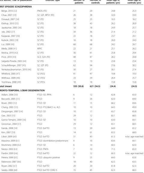
[image:4.595.57.541.88.358.2]Table 1 Studies included in the Meta-analyses
First author, Year of publication Disorder type Number of patients
Number of controls
Mean age of patients (yrs)
Mean age of controls (yrs)
FIRST EPISODE SCHIZOPHRENIA
Berge, 2010 [32] PNOS (FE) 21 20 24.8 25.3
Chua, 2007 [33] SZ, SZF, BPSY (FE) 26 38 32.0 33.0
Douaud, 2007 [34] SZ (FE) 25 25 16.5 16.2
Ebdrup, 2010 [35] SZ (FE) 38 43 26.2 26.9
Jayakumar, 2005 [36] SZ (FE) 18 18 24.9 25.7
Job, 2002 [37] SZ (FE) 34 36 21.4 21.2
Kasparak, 2007 [38] SZ (FE) 22 18 23.7 24.1
Kubicki, 2002 [39] SZ (FE) 16 18 26.0 24.0
Lui, 2009 [40] SZ (FE) 68 68 24.2 24.7
Meda, 2008 [41] WPIC 22 21 25.1 26.2
Molina, 2010 [42] SZ (FE) 30 41 25.8 29.4
Price, 2010 [43] SCZ, SZA (FE) 48 47 26.2 24.8
Salgado-Pineda, 2003 [44] SZ (FE) 13 13 23.8 23.4
Schaufelberger, 2007 [45] SZ, SZF (FE) 62 94 27.6 30.2
Venkatasubramanian, 2010 [46] SZ (FE) 30 27 30.1 27.4
Whitford, 2005 [47] SZ (FES) 41 47 19.8 19.3
Witthaus, 2009 [48] SZ (FES) 23 29 26.4 25.7
Yoshihara, 2008 [49] SZ (FES) 18 18 15.8 15.8
Total (mean) 555 (30.8) 621 (34.5) (24.4) (24.5)
FRONTO-TEMPORAL LOBAR DEGENERATION
Adlam, 2006 [50] FTLD: SD, fPPA 6 12 62.8 65.0
Boccardi, 2005 [51] FTLD 9 26 62.0 69.0
Boxer, 2003 [52] FTLD: SD 11 15 66.2 69.6
Chang, 2005 [53] FTLD: FTLD&ALS vs. ALS 10 10 64.5 49.9
Desgranges, 2007 [54] FTLD: SD 10 17 65.7 65.8
Gee, 2003 [55] FTLD 29 12 65.1 68.5
Gorno-Tempini, 2004 [56] FTLD: SD 10 10 63.0 69.1
Grossman, 2004 [57] FTLD 29 12 65.1 68.5
Kanda, 2008 [58] FTLD: bvFTD 13 20 64.9 65.2
Kim, 2007 [59] FTLD 14 61 63.3 68.0
Libon 2009 [60] FTLD: bvFTD 51 43 61.3 n/av: age-matched
Massimo 2009 [61] FTLD: disinhibition-predominant 4 10 63.6 64.1
Mummery 2000 [62] FTLD: SD 6 14 60.5 62.0
Nestor 2003 [63] FTLD: PNFA 7 10 71.5 65.9
Pardini 2009 [64] FTLD: bvFTD 22 12 60.3 n/av: age-matched
Pereira 2009 [65] FTLD: ubiquitin positive 9 25 64.0 63.8
Rabinovici 2007 [66] FTLD 18 40 62.5 63.5
Rosen 2002 [67] FTLD: bvFTD 20 20 61.8 62.3
neuro-psychiatric disorders with genetic or syndromal links to schizophrenia. For example, there is evidence that bipolar disorder and schizophrenia share suscepti-bility genes, and gray matter deficits of bipolar disorder overlap with those of schizophrenia but are more lim-ited to paralimbic regions involved in emotion regula-tion [70-72]. Temporal lobe epilepsy is also associated with psychotic episodes with similarities to schizophre-nia and is associated with gray matter deficits in limbic regions, overlapping with those of schizophrenia [73-75]. Autistic spectrum disorders and schizophrenia share gray matter deficits in the limbo-striato-thalamic circuitry [76], possibly reflecting shared genetic [77,78] and environmental [79-82] risk factors.
Previous results of meta-analyses and structural neuro-imaging studies of FES and FTLD
These results confirm those of previous meta-analyses of the two disorders, using smaller samples of primary studies. Several previous meta-analyses using‘ region-of-interest’ analyses have examined gray matter deficits in schizophrenia [83,84], with consistent findings of medial temporal lobe atrophy [85-88]. Patients with FES also have smaller whole-brain volume, with greater lateral ventricular volume [89]. Frontal [90,91] and temporal
[37] volumes have been reported to be smaller at first episode, and basal ganglia are also affected early in the illness [33,92]. The ALE method has also been used to investigate brain structural abnormalities in schizophre-nia derived from studies using voxel-based morpometry analyses [1,93]. In a recent meta-analysis by Chanet al., patients with FES had lower gray matter volumes in frontal, temporal, striatal, and cerebellar regions com-pared with both control subjects and people who are at genetically high-risk of developing schizophrenia [94]. They mapped progressive changes from the high-risk stage to the first-episode stage, clarifying potential mar-kers for disease risk (anterior cingulate and right insula volume reduction) and for disease onset (caudate vol-ume reduction) [94].
[image:6.595.56.539.506.685.2]In FTLD, a meta-analysis of voxel-based morphometry studies found predominant frontal and temporal lobe in-volvement, with specific patterns of atrophy in the three clinical subtypes of FTLD, namely, FTD (frontotemporal dementia), SD (semantic dementia) and PNFA (progres-sive non-fluent aphasia) [95]. Correspondingly, diagnos-tic criteria for FTLD create a‘triple dissociation’of these subtypes with a high diagnostic accuracy clinically [96]. The clinical characteristics of each subtype of FTLD cor-respond well with the neuroanatomical deficits found.
Figure 2Regions of gray matter change in FES and FTLD subjects.Regions of gray matter decreases in FES subjects compared with controls (red), regions of gray matter decreases in FTLD subjects compared with controls (yellow), and overlapping regions of gray matter decreases controls (orange), displayed on a brain template. The left side of the image represents the left side of the brain. The Talairach level (z co-ordinate) is given above each horizontal slice.
Table 1 Studies included in the Meta-analyses(Continued)
Williams 2005 [69] FTLD 18 17 60.8 61.7
Total (mean) 311 (15.6) 431 (21.6) (63.6) (65.0)
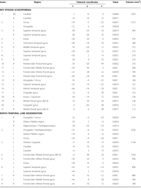
Table 2 Regions of gray matter decreases in FES and FTLD subjects compared with controls
Cluster Region Talairach coordinates Value Volume (mm3)
x y z
FIRST EPISODE SCHIZOPHRENIA
1 R/L Caudate 2 14 0 0.0046 1624
R Caudate 10 10 12 0.0017
2 L Uncus −18 −2 −22 0.0031 1272
L Amygdala −12 0 −14 0.0028
3 R Superior temporal gyrus 48 −24 10 0.0019 400
R Superior temporal gyrus 48 −26 16 0.0018
4 L Insula −36 20 6 0.0025 376
5 L Transverse temporal gyrus −46 −18 10 0.0021 288
6 R Middle temporal gyrus 54 −26 −2 0.0022 272
7 L Superior temporal gyrus −58 −26 12 0.0021 272
8 L Superior temporal gyrus −56 2 −4 0.0021 216
9 R Insula 36 8 10 0.0021 216
10 R Parietal lobe: Postcentral gyrus 54 −20 44 0.0022 216
11 L Frontal lobe: Middle frontal gyrus −30 50 4 0.002 200
12 L Frontal lobe: Inferior frontal gyrus −22 28 −6 0.0018 184
13 L Parietal lobe: Postcentral gyrus −60 −18 20 0.002 184
14 R Amygdala / Uncus 20 −4 −22 0.002 168
15 L Superior temporal gyrus −32 14 −22 0.0019 152
16 L Inferior temporal gyrus −46 −14 −18 0.002 152
17 L Cingulate gyrus −8 −4 38 0.002 152
18 R Insula / Claustrum 34 −14 12 0.0018 136
19 R Medial frontal gyrus (BA 8) 14 34 36 0.0017 136
20 L Cingulate gyrus 0 −42 38 0.0016 112
21 R Medial frontal gyrus (BA 6) 6 6 48 0.0017 112
FRONTO-TEMPORAL LOBE DEGENERATION
1 R Amygdala / Uncus 22 −4 −20 0.0029 3744
R Globus Pallidus region 20 2 −4 0.0024
R Hippocampus / Parahippocampus 28 −20 −10 0.0015
2 L Amygdala / Parahippocampus −22 −6 −16 0.0027 2336
L Globus Pallidus region −18 0 −6 0.0025
L Uncus −26 −4 −28 0.002
3 L Anterior cingulate −2 10 −4 0.0024 2144
L Caudate −8 10 10 0.0022
L Caudate −6 2 14 0.0015
4 L Frontal lobe: Medial frontal gyrus (BA 9) −4 48 24 0.003 1256
5 L Frontal lobe: Inferior frontal gyrus −36 24 4 0.0022 936
L Insula −38 14 0 0.0016
6 L Superior temporal gyrus −42 12 −14 0.0021 896
L Superior temporal gyrus −44 2 −12 0.0018
7 R Frontal lobe: Inferior frontal gyrus 42 16 −12 0.003 880
8 L Frontal lobe: Middle frontal gyrus −44 8 38 0.0032 784
Functional significance of implicated regions and symptoms of FES and FTLD
The involvement of the basal ganglia (bilateral gray matter decreases in the caudate head in both disor-ders) agrees with Middleton and Strick’s prediction that abnormalities within cortico-striatal circuits may
[image:8.595.68.538.97.367.2]underlie neuropsychiatric symptoms [97,98]. Anatom-ical studies have revealed discrete connections between the basal ganglia and the cerebral cortex, reciprocally interconnecting a large and diverse set of cortical areas [99]. Dysregulation and abnormal dopaminergic trans-mission in these loops may contribute to hyperkinetic
[image:8.595.57.539.474.696.2]Figure 3Gray matter deficits in FES subjects (red) and FTLD subjects (yellow). Congruent changes in both disorders are shown in orange.Brain changes are shown in three-dimensional views with brain sections removed (A, B), a sagittal plane (C), an axial plane (D) and a coronal plane (E). The Talairach co-ordinate for each plane is given above each image (C, D, E) (P = posterior, A = anterior, S = superior, L = left).
Table 2 Regions of gray matter decreases in FES and FTLD subjects compared with controls(Continued)
10 L Frontal lobe: Superior frontal gyrus −22 60 2 0.0023 704
L Frontal lobe: Superior frontal gyrus −24 56 12 0.0017
11 R Cingulate gyrus 16 26 36 0.0025 576
12 R Insula 42 −6 −4 0.0021 568
13 L Temporal lobe: fusiform gyrus −42 −30 −18 0.0027 520
14 L Frontal lobe: Middle frontal gyrus −22 18 50 0.0027 496
15 L Anterior cingulate −2 32 16 0.002 400
16 R Caudate 8 12 12 0.0024 376
17 R Middle temporal gyrus 48 2 −28 0.0024 312
18 L Frontal lobe: Inferior frontal gyrus −40 52 2 0.0024 232
19 L Frontal lobe: Middle frontal gyrus −28 34 −14 0.002 224
20 L Insula −42 2 0 0.0017 216
21 L Parietal lobe (BA 7) −20 −46 58 0.0023 216
22 L Frontal lobe: Medial frontal gyrus (BA 10) 0 54 −4 0.0018 192
23 R Superior temporal gyrus 28 14 −30 0.0016 176
24 L Uncus −34 −16 −34 0.0017 136
25 R Cerebellum: Anterior lobe 48 −36 −28 0.0023 136
26 R Frontal lobe: Middle frontal gyrus 36 44 8 0.0017 120
movements as well as cognitive impairments [100-102]. Robbins [103] proposed that the heterogeneous range of core symptoms associated with psychosis, appearing to be associated with a range of structural and func-tional abnormalities, might be explained by a frontal-striatal hypothesis, where an altered balance in the flow of information between through the basal ganglia could explain the seemingly disparate symptoms and char-acteristics of schizophrenia and psychotic episodes in FTLD. Our results indicate that the basal ganglia loop in particular incorporating the head of the caudate nu-cleus, are affected by FES and FTLD, which may at least partly explain their shared symptoms.
Our findings reveal that both disorders are charac-terised by reduced volumes of various paralimbic and limbic structures. Neuroanatomists and cytoarchitectolo-gists have grouped the regions shown to be affected in FES and FTLD, namely the superior temporal gyrus (temporal pole), rostral and caudal anterior cingulate, posterior cingulate, orbital frontal cortex, insula, and parahippocampal regions, into the paralimbic cortex [104,105]. There is documented evidence to support paralimbic dysfunction in various psychiatric disorders, namely psychopathy [106], bipolar disorder [72], psych-otic symptoms in depressive disorder [107] and attention deficit hyperactivity disorder [108]. Our results also indi-cate that both disorders are characterised by reduced caudate size, lesions of which cause impairments in learning, memory [109] and behaviour through the se-lection of appropriate sub-goals based on an evaluation of action-outcomes [110]. This may reflect the common symptomatology between the two disorders.
Explanation for congruence
Various explanations need to be considered for the over-lap in gray matter deficits between the two disorders. Firstly, the result could be due to coincidence. However, we have applied a statistical test to the spatial congru-ence which suggests that this is highly unlikely (the p value for the null hypothesis of a random distribution of FTLD co-ordinates within the FES spatial map was p = 0.001). Secondly, the symptoms of neuropsychiatric disorders are related to the anatomy of the brain path-ology. Therefore, the selection of schizophrenia and FTLD, which share certain symptoms (as described above) may constrain the anatomical findings towards certain brain regions, in the absence of any other more meaningful connection between the neuropathology of the two disorders. The evidence that schizophrenia and FTLD co-occur in some families [6] suggests the possi-bility of a more fundamental connection between the two disorders. Thirdly, the neuropathology of the two disorders may be (at the neurochemical pathway level) distinct but the common network identified in this study may be ‘a final common pathway’ in the pathological process of both disorders, or a common reaction to such processes.
Limitations
[image:9.595.56.540.89.312.2]extensive as there is improved power to detect changes. Thirdly, there are alternative approaches for investigat-ing spatial overlap of two disorders. For example, Yu et al., used a post-hoc meta-analytic estimation of the extent to which gray matter compares with controls in bipolar disorder and schizophrenia by using a modifica-tion of the ALE method [71]. Lastly, the mean age of patients with FES was 24.4 years, whereas in FTLD, the mean age was 63.6 years (Table 1). There are no vali-dated methods to account for these differences in an ALE meta-analysis, and it is possible that some of the results may have been affected by “normal” structural brain aging processes [111]. Anatomical likelihood esti-mation analyses are a relatively novel and changing technique, and as time progresses, standardisation of meta-analysis techniques will help researchers evaluate results from different studies more objectively.
Future directions
In the future, localized gray matter deficits detected via the above analyses may be combined with those identi-fied in activation studies of cognitive deficits in schizo-phrenia and FTLD, in order to understand the correlation between functional and structural connectiv-ity in both disorders. For example, the structural MRI gray matter deficits could be used as nodes for a net-work analysis [112], as can now be done on individual scans [113], which may be utilized to investigate and compare functional connectivity changes in FES and FTLD.
Conclusions
In summary, we reviewed data from 18 FES studies and 20 studies of FTLD that used voxel-based morphometry to identify common structural brain abnormalities. The brain regions found to be significantly affected included gray matter deficits in the bilateral caudate, left insula and bilateral uncus regions. The overlap in distribution of the disorders does not necessarily indicate a funda-mental sharing of neurochemical pathology between FES and FTLD. However, we propose that the emerging gen-etic, pathological and clinical typology of FTLD may provide a model for the deconstruction of subtypes of schizophrenia.
Competing interests
BO, IEW and SML disclose that they have no competing interests. EB is employed half-time by GlaxoSmithKline (GSK) and half-time by the University of Cambridge and is a stockholder in GSK.
Authors’contributions
BO and IEW devised the study and wrote the first draft of the manuscript. BO ascertained the studies and extracted the data. IEW checked and analysed the data. EB and SML participated in the design and coordination and helped to draft the manuscript. All authors read and approved the final manuscript.
Acknowledgements
The listed authors were solely involved the in designing the study, data acquisition, analysis and interpretation, as well as drafting and revising the manuscript, with no external contributions. SML has received financial support from Pfizer (formerly Wyeth) in relation to imaging studies of people with schizophrenia, but this played no role in influencing this study.
Author details
1Division of Psychiatry, School of Molecular and Clinical Medicine, University of Edinburgh, Royal Edinburgh Hospital, Edinburgh EH10 5HF, UK.2Avon and Wiltshire Mental Health Partnership NHS Trust, Salisbury, UK.3Department of Psychiatry, Behavioral & Clinical Neuroscience Institute, University of Cambridge, Cambridge, UK.4Cambridgeshire & Peterborough NHS Foundation Trust, Cambridge, UK.
Received: 29 December 2011 Accepted: 31 July 2012 Published: 7 August 2012
References
1. Ellison-Wright I, Glahn DC, Laird AR, Thelen SM, Bullmore E:The anatomy of first-episode and chronic schizophrenia: an anatomical likelihood estimation meta-analysis.Am J Psychiatry2008,165(8):1015–23.
2. Keshavan MS, Tandon R, Boutros NN, Nasrallah HA:Schizophrenia, "just the facts": what we know in 2008 Part 3: neurobiology.Schizophr Res2008, 106(2–3):89–107.
3. Goghari VM, Sponheim SR, MacDonald AW 3rd:The functional neuroanatomy of symptom dimensions in schizophrenia: a qualitative and quantitative review of a persistent question.Neurosci Biobehav Rev 2010,34(3):468–86.
4. Neary D, Snowden JS, Gustafson L, Passant U, Stuss D, Black S, Freedman M, Kertesz A, Robert PH, Albert M, Boone K, Miller BL, Cummings J, Benson DF: Frontotemporal lobar degeneration: a consensus on clinical diagnostic criteria.Neurology1998,51:1546–54.
5. Mohandas E, Rajmohan V:Frontotemporal dementia: An updated overview.Indian J Psychiatry2009,51(Suppl 1):S65–9.
6. Taniguchi S, McDonagh AM, Pickering-Brown SM, Umeda Y, Iwatsubo T, Hasegawa M, Mann DM:The neuropathology of frontotemporal lobar degeneration with respect to the cytological and biochemical characteristics of tau protein.Neuropath Appl Neurobiol2004,30:1–18. 7. Neary D, Snowden J, Mann D:Frontotemporal dementia. Lancet Neurol2005,
4:771–80.
8. Momeni P, Wickremaratchi MM, Bell J, Arnold R, Beer R, Hardy J, Revesz T, Neal JW, Morris HR:Familial early onset frontotemporal dementia caused by a novel S356T MAPT mutation, initially diagnosed as schizophrenia. Clin Neurol Neurosurg2010,112(10):917–20.
9. Velakoulis D, Walterfang M, Mocellin R, Pantelis C, McLean C:
Frontotemporal dementia presenting as schizophrenia-like psychosis in young people: clinicopathological series and review of cases.Br J Psychiatry2009,194(4):298–305.
10. Pantelis C, Barnes TR, Nelson HE, Tanner S, Weatherley L, Owen AM, Robbins TW:Frontal-striatal cognitive deficits in patients with chronic
schizophrenia.Brain1997,120(Pt 10):1823–43.
11. Shamay-Tsoory SG, Aharon-Peretz J, Levkovitz Y:The neuroanatomical basis of affective mentalizing in schizophrenia: comparison of patients with schizophrenia and patients with localized prefrontal lesions. Schizophr Res2007,90(1–3):274–83.
12. Lawrie SM, Abukmeil SS:Brain abnormality in schizophrenia. A systematic and quantitative review of volumetric magnetic resonance imaging studies.Br J Psychiatry1998,172:110–20.
13. Velakoulis D, Wood SJ, Wong MT, McGorry PD, Yung A, Phillips L, Smith D, Brewer W, Proffitt T, Desmond P, Pantelis C:Hippocampal and amygdala volumes according to psychosis stage and diagnosis: a magnetic resonance imaging study of chronic schizophrenia, first-episode psychosis, and ultra-high-risk individuals.Arch Gen Psychiatry2006, 63(2):139–49.
14. Kaufer DI, Miller BL, Itti L, Fairbanks LA, Li J, Fishman J, Kushi J, Cummings JL:Midline cerebral morphometry distinguishes frontotemporal dementia and Alzheimer's disease.Neurology1997,48(4):978–85. 15. Walterfang M, Wood AG, Reutens DC, Wood SJ, Chen J, Velakoulis D,
stages of schizophrenia: cross-sectional study in first episode and chronic illness.Br J Psychiatry2008,192(6):429–34.
16. Laakso MP, Frisoni GB, Kononen M, Mikkonen M, Beltramello A, Geroldi C, Bianchetti A, Trabucchi M, Soininen H, Aronen HJ:Hippocampus and entorhinal cortex in frontotemporal dementia and Alzheimer’s disease: a morphometric MRI study.Biol Psychiatry2000,47:1056–63.
17. Velakoulis D, Stuart GW, Wood SJ, Smith DJ, Brewer WJ, Desmond P, Singh B, Copolov D, Pantelis C:Selective bilateral hippocampal volume loss in chronic schizophrenia.Biol Psychiatry2001,50:531–9.
18. McKhann GM, Albert MS, Grossman M, Miller B, Dickson D, Trojanowski JQ: Clinical and pathological diagnosis of frontotemporal dementia: report of the Work Group on Frontotemporal Dementia and Pick’s Disease.Arch Neurol2001,58:1803–9.
19. Velakoulis D, Pantelis C:What have we learned from functional imaging studies in schizophrenia? The role of frontal, striatal and temporal areas. Aust N Z J Psychiatry1996,30:195–209.
20. Schoder D, Hannequin D, Martinaud O, Opolczynski G, Guyant-Maréchal L, Le Ber I, Campion D:Morbid risk for schizophrenia in first-degree relatives of people with frontotemporal dementia.Br J Psychiatry2010, 197:28–35.
21. Pickering-Brown SM:The complex aetiology of frontotemporal lobar degeneration.Exp Neurol2007,206(1):1–10.
22. Aswathy PM, Jairani PS, Mathuranath PS:Genetics of frontotemporal lobar degeneration.Ann Indian Acad Neurol2010,13(Suppl 2):S55–62. 23. Goghari VM, Lang DJ, Khorram B, Götz J, Vandorpe RA, Smith GN, Kopala
LC, Barr AM, Honer WG:Anterior internal capsule volumes increase in patients with schizophrenia switched from typicalantipsychotics to olanzapine.J Psychopharmacol2011,25(5):621–9.
24. Molina V, Reig S, Sanz J, Palomo T, Benito C, Sánchez J, Pascau J, Desco M: Changes in cortical volume with olanzapine in chronic schizophrenia. Pharmacopsychiatry2007,40(4):135–9.
25. Eickhoff SB, Laird AR, Grefkes C, Wang LE, Zilles K, Fox PT:Coordinate-based activation likelihood estimation meta-analysis of neuroimaging data: A random-effects approach based on empirical estimates of spatial uncertainty.Hum Brain Mapp2009,30:2907–2926.
26. Morris SE, Cuthbert BN:Research Domain Criteria: cognitive systems, neural circuits and dimensions of behaviour.Dialogues Clin Neurosci2012, 14(1):29–37.
27. Takayanagi Y, Takahashi T, Orikabe L, Mozue Y, Kawasaki Y, Nakamura K, Sato Y, Itokawa M, Yamasue H, Kasai K, Kurachi M, Okazaki Y, Suzuki M: Classification of first-episode schizophrenia patients and healthy subjects by automated MRI measures of regional brain volume and cortical thickness.PLoS One2011,6(6):e21047.
28. Liberati A, Altman DG, Tetzlaff J, Mulrow C, Gøtzsche PC, Ioannidis JP, Clarke M, Devereaux PJ, Kleijnen J, Moher D:The PRISMA statement for reporting systematic reviews and meta-analyses of studies that evaluate healthcare interventions: explanation and elaboration.BMJ2009,339: b2700.
29. Laird AR, Fox PM, Price CJ, Glahn DC, Uecker AM, Lancaster JL, Turkeltaub PE, Kochunov P, Fox PT:ALE meta-analysis: controlling the false discovery rate and performing statistical contrasts.Hum Brain Mapp2005, 25:155–164.
30. Verhoeven KJF, Simonsen KL, McIntyre LM:Implementing false discovery rate control: increasing your power.Oikos2005,108:643–647. 31. Ellison-Wright I, Ellison-Wright Z, Bullmore E:Structural brain change in
Attention Deficit Hyperactivity Disorder identified by meta-analysis.BMC Psychiatry2008,8:51.
32. Bergé D, Carmona S, Rovira M, Bulbena A, Salgado P, Vilarroya O:Gray matter volume deficits and correlation with insight and negative symptoms in first-psychotic-episode subjects.Acta Psychiatr Scand2011, 123(6):431–9.
33. Chua SE, Cheung C, Cheung V, Tsang JT, Chen EY, Wong JC, Cheung JP, Yip L, Tai KS, Suckling J, McAlonan GM:Cerebral grey, white matter and CSF in never-medicated, first-episode schizophrenia.Schizophr Res2007, 89:12–21.
34. Douaud G, Smith S, Jenkinson M, Behrens T, Johansen-Berg H, Vickers J, James S, Voets N, Watkins K, Matthews PM, James A:Anatomically related grey and white matter abnormalities in adolescent-onset schizophrenia. Brain2007,130:2375–86.
35. Ebdrup BH, Glenthøj B, Rasmussen H, Aggernaes B, Langkilde AR, Paulson OB, Lublin H, Skimminge A, Baaré W:Hippocampal and caudate volume
reductions in antipsychotic-naive first-episode schizophrenia.J Psychiatry Neurosci2010,35(2):95–104.
36. Jayakumar PN, Venkatasubramanian G, Gangadhar BN, Janakiramaiah N, Keshavan MS:Optimized voxel-based morphometry of gray matter volume in first-episode, antipsychotic-naive schizophrenia.Prog Neuropsychopharmacol Biol Psychiatry2005,29:587–91.
37. Job DE, Whalley HC, McConnell S, Glabus M, Johnstone EC, Lawrie SM: Structural gray matter differences between first-episode schizophrenics and normal controls using voxel-based morphometry.Neuroimage2002, 17:880–9.
38. Kasparek T, Prikryl R, Mikl M, Schwarz D, Ceskova E, Krupa P:Prefrontal but not temporal grey matter changes in males with first-episode schizophrenia.Prog Neuropsychopharmacol Biol Psychiatry2007,31:151–7. 39. Kubicki M, Shenton ME, Salisbury DF, Hirayasu Y, Kasai K, Kikinis R, Jolesz FA,
McCarley RW:Voxel-based morphometric analysis of gray matter in first episode schizophrenia.Neuroimage2002,17(4):1711–9.
40. Lui S, Deng W, Huang X, Jiang L, Ma X, Chen H, Zhang T, Li X, Li D, Zou L, Tang H, Zhou XJ, Mechelli A, Collier DA, Sweeney JA, Li T, Gong Q: Association of cerebral deficits with clinical symptoms in antipsychotic-naive first-episode schizophrenia: an optimized voxel-based
morphometry and resting state functional connectivity study.Am J Psychiatry2009,166(2):196–205.
41. Meda SA, Giuliani NR, Calhoun VD, Jagannathan K, Schretlen DJ, Pulver A, Cascella N, Keshavan M, Kates W, Buchanan R, Sharma T, Pearlson GD:A large scale (N = 400) investigation of gray matter differences in schizophrenia using optimized voxel-based morphometry.Schizophr Res 2008,101(1–3):95–105.
42. Molina V, Sanz J, Villa R, Pérez J, González D, Sarramea F, Ballesteros A, Galindo G, Hernández JA:Voxel-based morphometry comparison between first episodes of psychosis with and without evolution to schizophrenia.Psychiatry Res2010,181(3):204–10.
43. Price G, Cercignani M, Chu EM, Barnes TR, Barker GJ, Joyce EM, Ron MA: Brain pathology in first-episode psychosis: magnetization transfer imaging provides additional information to MRI measurements of volume loss.Neuroimage2010,49(1):185–92.
44. Salgado-Pineda P, Baeza I, Perez-Gomez M, Vendrell P, Junque C, Bargallo N, Bernardo M:Sustained attention impairment correlates to gray matter decreases in first episode neuroleptic-naive schizophrenic patients. Neuroimage2003,19:365–75.
45. Schaufelberger MS, Duran FL, Lappin JM, Scazufca M, Amaro E Jr, Leite CC, De Castro CC, Murray RM, McGuire PK, Menezes PR, Busatto GF:Grey matter abnormalities in Brazilians with first-episode psychosis.Br J Psychiatry Suppl2007,51:s117–22.
46. Venkatasubramanian G:Neuroanatomical correlates of psychopathology in antipsychotic-naïve schizophrenia.Indian J Psychiatry2010, 52(1):28–36.
47. Whitford TJ, Farrow TF, Gomes L, Brennan J, Harris AW, Williams LM:Grey matter deficits and symptom profile in first episode schizophrenia. Psychiatry Res2005,139:229–38.
48. Witthaus H, Kaufmann C, Bohner G, Ozgürdal S, Gudlowski Y, Gallinat J, Ruhrmann S, Brüne M, Heinz A, Klingebiel R, Juckel G:Gray matter abnormalities in subjects at ultra-high risk for schizophrenia and first-episode schizophrenic patients compared to healthy controls.Psychiatry Res2009,173(3):163–9.
49. Yoshihara Y, Sugihara G, Matsumoto H, Suckling J, Nishimura K, Toyoda T, Isoda H, Tsuchiya KJ, Takebayashi K, Suzuki K, Sakahara H, Nakamura K, Mori N, Takei N:Voxel-based structural magnetic resonance imaging (MRI) study of patients with early onset schizophrenia.Ann Gen Psychiatry2008, 7:25.
50. Adlam AL, Patterson K, Rogers TT, Nestor PJ, Salmond CH, Acosta-Cabronero J, Hodges JR:Semantic dementia and fluent primary progressive aphasia: two sides of the same coin?Brain2006,129(Pt 11):3066–80.
51. Boccardi M, Sabattoli F, Laakso MP, Testa C, Rossi R, Beltramello A, Soininen H, Frisoni GB:Frontotemporal dementia as a neural system disease. Neurobiol Aging2005,26(1):37–44.
52. Boxer AL, Rankin KP, Miller BL, Schuff N, Weiner M, Gorno-Tempini ML, Rosen HJ:Cinguloparietal atrophy distinguishes Alzheimer disease from semantic dementia.Arch Neurol2003,60(7):949–56.
54. Desgranges B, Matuszewski V, Piolino P, Chételat G, Mézenge F, Landeau B, de la Sayette V, Belliard S, Eustache F:Anatomical and functional alterations in semantic dementia: a voxel-based MRI and PET study. Neurobiol Aging2007,28(12):1904–13.
55. Gee J, Ding L, Xie Z, Lin M, DeVita C, Grossman M:Alzheimer's disease and frontotemporal dementia exhibit distinct atrophy-behavior correlates: a computer-assisted imaging study.Acad Radiol2003,10(12):1392–401. 56. Gorno-Tempini ML, Dronkers NF, Rankin KP, Ogar JM, Phengrasamy L, Rosen
HJ, Johnson JK, Weiner MW, Miller BL:Cognition and anatomy in three variants of primary progressive aphasia.Ann Neurol2004,55(3):335–46. 57. Grossman M, McMillan C, Moore P, Ding L, Glosser G, Work M, Gee J:What's
in a name: voxel-based morphometric analyses of MRI and naming difficulty in Alzheimer's disease, frontotemporal dementia and corticobasal degeneration.Brain2004,127(Pt 3):628–49.
58. Kanda T, Ishii K, Uemura T, Miyamoto N, Yoshikawa T, Kono AK, Mori E: Comparison of grey matter and metabolic reductions in frontotemporal dementia using FDG-PET and voxel-based morphometric MR studies.Eur J Nucl Med Mol Imaging2008,35(12):2227–34.
59. Kim EJ, Rabinovici GD, Seeley WW, Halabi C, Shu H, Weiner MW, DeArmond SJ, Trojanowski JQ, Gorno-Tempini ML, Miller BL, Rosen HJ:Patterns of MRI atrophy in tau positive and ubiquitin positive frontotemporal lobar degeneration.J Neurol Neurosurg Psychiatry2007,78(12):1375–8. 60. Libon DJ, McMillan C, Gunawardena D, Powers C, Massimo L, Khan A,
Morgan B, Farag C, Richmond L, Weinstein J, Moore P, Coslett HB, Chatterjee A, Aguirre G, Grossman M:Neurocognitive contributions to verbal fluency deficits in frontotemporal lobar degeneration.Neurology 2009,73(7):535–42.
61. Massimo L, Powers C, Moore P, Vesely L, Avants B, Gee J, Libon DJ, Grossman M:Neuroanatomy of apathy and disinhibition in frontotemporal lobar degeneration.Dement Geriatr Cogn Disord2009, 27(1):96–104.
62. Mummery CJ, Patterson K, Price CJ, Ashburner J, Frackowiak RS, Hodges JR: A voxel-based morphometry study of semantic dementia: relationship between temporal lobe atrophy and semantic memory.Ann Neurol2000, 47(1):36–45.
63. Nestor PJ, Graham NL, Fryer TD, Williams GB, Patterson K, Hodges JR: Progressive non-fluent aphasia is associated with hypometabolism centred on the left anterior insula.Brain2003,126(Pt 11):2406–18. 64. Pardini M, Huey ED, Cavanagh AL, Grafman J:Olfactory function in
corticobasal syndrome and frontotemporal dementia.Arch Neurol2009, 66(1):92–6.
65. Pereira JM, Williams GB, Acosta-Cabronero J, Pengas G, Spillantini MG, Xuereb JH, Hodges JR, Nestor PJ:Atrophy patterns in histologic vs clinical groupings of frontotemporal lobar degeneration.Neurology2009, 72(19):1653–60.
66. Rabinovici GD, Seeley WW, Kim EJ, Gorno-Tempini ML, Rascovsky K, Pagliaro TA, Allison SC, Halabi C, Kramer JH, Johnson JK, Weiner MW, Forman MS, Trojanowski JQ, Dearmond SJ, Miller BL, Rosen HJ:Distinct MRI atrophy patterns in autopsy-proven Alzheimer's disease and frontotemporal lobar degeneration.Am J Alzheimers Dis Other Demen2007, 22(6):474–88.
67. Rosen HJ, Gorno-Tempini ML, Goldman WP, Perry RJ, Schuff N, Weiner M, Feiwell R, Kramer JH, Miller BL:Patterns of brain atrophy in
frontotemporal dementia and semantic dementia.Neurology2002, 58(2):198–208.
68. Seeley WW, Crawford R, Rascovsky K, Kramer JH, Weiner M, Miller BL, Gorno-Tempini ML:Frontal paralimbic network atrophy in very mild behavioral variant frontotemporal dementia.Arch Neurol2008,65(2):249–55. 69. Williams GB, Nestor PJ, Hodges JR:Neural correlates of semantic and
behavioural deficits in frontotemporal dementia.Neuroimage2005, 24(4):1042–51.
70. Bora E, Fornito A, Yücel M, Pantelis C:The effects of gender on grey matter abnormalities in major psychoses: a comparative voxelwise meta-analysis of schizophreniaand bipolar disorder.Psychol Med2011,11:1–13. 71. Yu K, Cheung C, Leung M, Li Q, Chua S, McAlonan G:Are Bipolar Disorder and Schizophrenia Neuroanatomically Distinct?An Anatomical Likelihood Meta-analysis. Front Hum Neurosci2010,4:189.
72. Ellison-Wright I, Bullmore E:Anatomy of bipolar disorder and schizophrenia: a meta-analysis.Schizophr Res2010,117(1):1–12. 73. Sundram F, Cannon M, Doherty CP, Barker GJ, Fitzsimons M, Delanty N,
Cotter D:Neuroanatomical correlates of psychosis in temporal lobe
epilepsy: voxel-based morphometry study.Br J Psychiatry2010, 197(6):482–92.
74. Cascella NG, Schretlen DJ, Sawa A:Schizophrenia and epilepsy: is there a shared susceptibility?Neurosci Res2009,63(4):227–35.
75. Labate A, Cerasa A, Gambardella A, Aguglia U, Quattrone A:Hippocampal and thalamic atrophy in mild temporal lobe epilepsy: a VBM study. Neurology2008,71(14):1094–101.
76. Cheung C, Yu K, Fung G, Leung M, Wong C, Li Q, Sham P, Chua S, McAlonan G:Autistic disorders and schizophrenia: related or remote? An anatomical likelihood estimation.PLoS One2010,5(8):e12233.
77. Burbach JP, van der Zwaag B:Contact in the genetics of autism and schizophrenia.Trends Neurosci2009,32:69–72.
78. Goodman AB:A family history study of schizophrenia spectrum disorders suggests new candidate genes in schizophrenia and autism.Psychiatr Q 1994,65:287–297.
79. McGrath J, Eyles D, Mowry B, Yolken R, Buka S:Low maternal vitamin D as a risk factor for schizophrenia: a pilot study using banked sera.Schizophr Res2003,63:73–78.
80. Cannell JJ:Autism and vitamin D.Med Hypotheses2008,70:750–759. 81. Brown AS:Prenatal infection as a risk factor for schizophrenia.Schizophr
Bull2006,32:200–202.
82. Chess S:Autism in children with congenital rubella.J Autism Child Schizophr1971,1:33–47.
83. Sommer I, Ramsey N, Kahn R, Aleman A, Bouma A:Handedness, language lateralisation and anatomical asymmetry in schizophrenia: meta-analysis. Br J Psychiatry2001,178:344–351.
84. Konick LC, Friedman L:Meta-analysis of thalamic size in schizophrenia. Biol Psychiatry2001,49:28–38.
85. Wright IC, Rabe-Hesketh S, Woodruff PWR, David AS, Murray RM, Bullmore ET:Meta-analysis of regional brain volumes in schizophrenia.Am J Psychiatry2000,157:16–25.
86. Zakzanis KK, Poulin P, Hansen KT, Jolic D:Searching the schizophrenic brain for temporal lobe deficits: a systematic review and meta-analysis. Psychol Med2000,30:491–504.
87. Nelson MD, Saykin AJ, Flashman LA, Riordan HJ:Hippocampal volume reduction in schizophrenia as assessed by magnetic resonance imaging: a meta-analytic study.Arch Gen Psychiatry1998,55:433–440.
88. Chance SA, Esiri MM, Crow TJ:Amygdala volume in schizophrenia: post-mortem study and review of magnetic resonance imaging findings.Br J Psychiatry2002,180:331–338.
89. Vita A, De Peri L:Hippocampal and amygdala volume reductions in first-episode schizophrenia.Br J Psychiatry2007,190:271.
90. Shenton ME, Dickey CC, Frumin M, McCarley RW:A review of MRI findings in schizophrenia.Schizophr Res2001,49:1–52.
91. Ananth H, Popescu I, Critchley HD, Good CD, Frackowiak RSJ, Dolan RJ: Cortical and subcortical gray matter abnormalities in schizophrenia determined through structural magnetic resonance imaging with optimized volumetric voxel-based morphometry.Am J Psychiatry2002, 159:1497–1505.
92. Salgado-Pineda P, Baeza I, Pérez-Gómez M, Vendrell P, Junqué C, Bargalló N, Bernardo M:Sustained attention impairment correlates to gray matter decreases in first episode neuroleptic-naive schizophrenic patients. Neuroimage2003,19:365–375.
93. Leung M, Cheung C, Yu K, Yip B, Sham P, Li Q, Chua S, McAlonan G:Gray matter in first-episode schizophrenia before and after antipsychotic drug treatment.Anatomical likelihood estimation meta-analyses with sample size weighting. Schizophr Bull2011,37(1):199–211.
94. Chan RC, Di X, McAlonan GM, Gong QY:Brain anatomical abnormalities in high-risk individuals, first-episode, and chronic schizophrenia: an activation likelihood estimation meta-analysis of illness progression. Schizophr Bull2011,37(1):177–88.
95. Schroeter ML, Raczka K, Neumann J, YvesVon Cramon D:Towards a nosology for frontotemporal lobar degenerations - a meta-analysis involving 267 subjects.Neuroimage2007,36(3):497–510.
96. Lund T, Groups M:Clinical and neuropathological criteria for
frontotemporal dementia.J Neurol Neurosurg Psychiatry1994,57:416–418. 97. Middleton FA, Strick PL:Basal ganglia output and cognition: evidence
from anatomical, behavioral, and clinical studies.Brain Cogn2000, 42:183–200.
99. Delong MR, Wichman T:Circuits and circuit disorders of the basal ganglia.Arch Neurol2007,64(1):20–24.
100. Gao WJ, Wang Y, Goldman-Rakic PS:Dopamine modulation of perisomatic and peridendritic inhibition in prefrontal cortex.J Neurosci2003, 23:1622–1630.
101. Cassady SL, Adami H, Moran M, Kunkel R, Thaker GK:Spontaneous dyskinesia in subjects with schizophrenia spectrum personality.Am J Psychiatry1998,155(1):70–5.
102. Fish B, Marcus J, Hans SL, Auerbach JG, Purdue S:Infants at risk for schizophrenia: Sequelae of a genetic neurointegrative defect.Arch Gen Psychiatry1992,49:221–235.
103. Robbins TW:The case of frontostriatal dysfunction in schizophrenia. Schizophr Bull1990,16(3):391–402.
104. Garey LJ:Brodmann's Localisation in the Cerebral Cortex. London: Smith-Gordon; 1994.
105. Mesulam MM:Principles of Behavioural and Cognitive Neurology. New York: Oxford University Press; 2000.
106. Kiehl KA:A cognitive neuroscience perspective on psychopathy: evidence or paralimbic system dysfunction.Psychiatry Res2006, 142(2–3):107–28.
107. Skaf CR, Yamada A, Garrido GE, Buchpiguel CA, Akamine S, Castro CC, Busatto GF:Psychotic symptoms in major depressive disorder are associated with reduced regional cerebral blood flow in the subgenual anterior cingulate cortex: a voxel-based single photon emission computed tomography (SPECT) study.J Affect Disord2002, 68(2–3):295–305.
108. Rubia K:"Cool" inferior frontostriatal dysfunction in attention-deficit/ hyperactivity disorder versus "hot" ventromedial orbitofrontal-limbic dysfunction in conduct disorder: a review.Biol Psychiatry2011, 69(12):e69–87.
109. Grahn JA, Parkinson JA, Owen AM:The role of the basal ganglia in learning and memory: neuropsychological studies.Behav Brain Res2009, 199(1):53–60.
110. Grahn JA, Parkinson JA, Owen AM:The cognitive functions of the caudate nucleus.Prog Neurobiol2008,86(3):141–55.
111. Vemuri P, Wiste HJ, Weigand SD, Knopman DS, Trojanowski JQ, Shaw LM, Bernstein MA, Aisen PS, Weiner M, Petersen RC, Jack CR Jr:Alzheimer's disease neuroimaging initiative: Serial MRI and CSF biomarkers in normal aging, MCI, and AD.Neurology2010,75(2):143–51.
112. Zalesky A, Fornito A, Bullmore ET:Network-based statistic: identifying differences in brain networks.Neuroimage2010,53(4):1197–207. 113. Tijms BM, Seriès P, Willshaw DJ, Lawrie SM:Similarity-based extraction of
individual networks from gray matter MRI scans.Cereb Cortex2012, 22(7):1530–41.
doi:10.1186/1471-244X-12-104
Cite this article as:Olabiet al.:Structural brain changes in first episode Schizophrenia compared with Fronto-Temporal Lobar Degeneration: a meta-analysis.BMC Psychiatry201212:104.
Submit your next manuscript to BioMed Central and take full advantage of:
• Convenient online submission
• Thorough peer review
• No space constraints or color figure charges
• Immediate publication on acceptance
• Inclusion in PubMed, CAS, Scopus and Google Scholar
• Research which is freely available for redistribution