http://dx.doi.org/10.4236/scd.2014.44012
How to cite this paper: Verma, V., Mehta, A., Pal, S., Kumar, M., Singh, B., Kumar, A. and Gautam, S. (2014) In Pursuit of Porcine Pluripotent Stem Cells for Autologous Cell Therapy. Stem Cell Discovery, 4, 107-124.
http://dx.doi.org/10.4236/scd.2014.44012
In Pursuit of Porcine Pluripotent Stem Cells
for Autologous Cell Therapy
Vinod Verma
1*, Ashish Mehta
2, Sanjay Pal
1, Manoj Kumar
3, Birbal Singh
4, Ashok Kumar
5,
Sanjeev Gautam
61Centre of Biotechnology, Nehru Science Complex, University of Allahabad, Allahabad, India 2National Heart Research Institute Singapore, National Heart Centre, Singapore
3Department of Microbiology & Immunology, National Institute of Nutrition, Hyderabad, India 4Indian Veterinary Research Institute, Regional Station, Palampur, India
5Department of Zoology, MLK Post Graduate College, Balrampur, India 6Department of Biotechnology, Kurukshetra University, Kurukshetra, India
Email:∗vinodverma29@rediffmail.com, ∗vverma@allduniv.ac.in
Received 16 July 2014; revised 10 August 2014; accepted 7 September 2014
Copyright © 2014 by authors and Scientific Research Publishing Inc.
This work is licensed under the Creative Commons Attribution International License (CC BY). http://creativecommons.org/licenses/by/4.0/
Abstract
Treatments to repair the human heart following regenerative diseases remain a challenge for medical science. Unlike lower vertebrate species the human heart lacks a regenerative pathway meaning that research has to be focused on cell transplantation. Porcines (Sus scrofa) are excel-lent models for cardiovascular disease and pluripotent stem cells (PSCs) generated from porcines will provide important clinical insights for cardiac cell therapy. This could open a new avenue of research into degenerative conditions as porcine is a more effective human proxy to work with. However, bona fide PSCs are currently available only in rodents (mouse, rat) and primates (mon-key, human). Attempts to derive pluripotent stem cells (PSCs) from porcine have been going on for more than two decades with slow progress. Despite the fact that the porcine stem cells are under increasing glare of publicity due to milestone achievements in this area of research. Advances in stem cell technology, especially the genetic engineering, innovative cell culturing and induced pluripotency to generate stem cells has dramatically revolutionized the basic and applied investi-gations and applications of porcine stem cells. This review attempts to summarize the major sig-naling pathways involved in maintenance of pluripotency and the state of the art conceptual and technical progress for generating bona fide porcine PSCs.
Keywords
Porcine Model, Signaling Pathways, Stem Cells, Induced Pluripotent Stem Cells, Pluripotency, Regenerative Medicine, Cell Therapy, Epigenetics
*
1. Introduction
Stem cells have huge potential for alleviating suffering for many diseases of humans for which effective therapy is currently not available. The PSCs are the unicellular equivalent to a whole animal derived from either pre-(em- bryonic stem cells or ESCs) or post-(epiblast stem cells or EpiSCs) implantation embryos [1]. Under precise culture conditions, the PSCs are theoretically capable of differentiating into all cell types of an adult animal.
In the late 1960s, Gordon’s pioneering work on nuclear transfer using amphibian cells demonstrated that dif-ferentiation was a reversible process [2]. Decades later in 2006, Yamanaka demonstrated that ectopic expression of transcriptional factors (Oct3/4, Sox2, Klf4 and c-Myc) could induce pluripotency in terminally differentiated somatic cells, first in mouse [3], then in humans [3]. Rigorous testing using molecular and functional studies have clearly demonstrated that iPSC are similar to ESCs including germ line competence [4].
While, mouse and human embryonic stem cells have been in the limelight for the last two decades, limited efforts have been on porcine embryonic stem cells despite reports of first isolation in early 1990s [5] and sub- sequent numerous attempts to develop porcine ESCs (pESCs). This is mainly because the pig is anatomically and physiologically more similar to humans than the rodents or other model animals. Although the information is available on porcine ESCs (pESCs) after pioneering work on porcine and murine ESCs (mESCs), the stem cell lines developed were poorly defined [6]. In addition, the stem cells reported were found to lose pluripotency over a relatively shorter number of passages [7]. It was suggested that slow epiblast development with prolonged pre-implantation stages in comparison to murine or human, might hinder the growth and pluripotency of porcine stem cells [7].
Recent advances in with reprogramming technology have provided ability to generate porcine iPSCs. Putative porcine iPS cells fulfilling several criteria of true pluripotency were reported after reprogramming fibroblasts with viral vectors [8]-[10]. Notably, in one study, chimeric offspring were also reported. It has been demonstrated that human factors could be used in porcine mesenchymal stem cells (pMSCs) to generate iPSCs capable of generating chimeras with germline transmission. After breeding of chimeric porcine, 2 out of 43 F-1 offspring carried some of the reprogramming factors in their genome, suggesting germ line transmission [11]. However, both piglets with germline transmitted reprogramming factors died around birth. Moreover, lack of true porcine ES cell lines pre-vents the unequivocal identification of stem cells after reprogramming towards iPS cells in this species. Reports are scarce on bona fide piPSC, and their maintenance by endogenous factors with silenced exogenous repro-gramming factors [12].
The paucity of information related to morphology, pluripotency markers, and differentiation capabilities ham- pers a thorough evaluation of the validity of putative stem cell lines. Data from the literature suggests that similar regulatory pathways are likely to exist among different species. Coupling of these pathways with their distinct expression patterns, the relative concentrations of pluripotency-related molecules, and timing of embryo devel-opment, along with supportive micro-environmental conditions, would appear to vary in a species-specific manner. It is envisaged that the understanding of signaling pathways and other subtle, but consequential diversities may endow with valuable information for isolating the genuine porcine PSCs.
2. Signaling Pathways Involved in Maintenance of Pluripotency and Self-Renewal
in Murine, Humans and Porcines
The two most important and distinguishing traits that categorize PSCs, are pluripotency and self-renewal that allow the stem cells to divide continually in an undifferentiated manner. Both the traits orchestrate by a complex cascade of signaling pathways. However, literature indicates the involvement of some common signaling path-ways among mice, primates and porcine stem cells, though the growth factors equilibrium may differ among different species [13]. Understanding of various regulatory pathways involved in pluripotency of porcine PS cells is still not clear. Till date bon fide porcine pluripotent stem cells have not been established. Therefore, the exposition and understanding of the mechanism underlying pluripotency of human and mice PS cells would be the stepping stone towards the successful establishment of the culture conditions for the bona fide PS cells.
2.1. Murine and Human
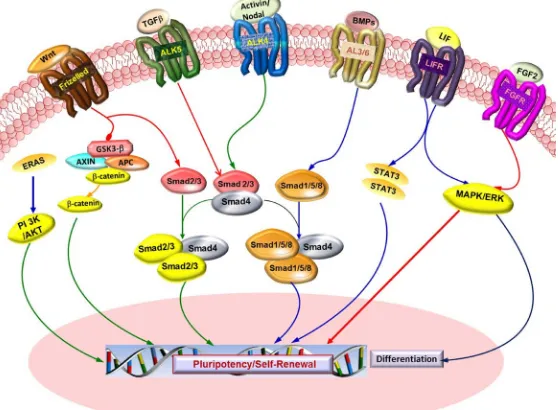
to specific LIF receptor (LIFR-α) at cellular surface, which forms a heterodimer with GP130 signal transducing unit (Figure 1). This leads to the activation of the JAK/STAT (Janus kinase/signal transducer and activator of transcription) and mitogen-activated protein kinase (MAPK) cascade. Once activated, the STAT3 is phosphory-lated and subjected to dimerization, before being translocated to the nucleus where it acts as a transcription fac-tor [15]. Withdrawal of LIF in culture medium pushes stem cells en route for differentiation, but they somehow maintain pluripotency. Hence, it is necessary to supplement the culture media with LIF to maintain murine stem cells in undifferentiated state. Genetically manipulated stem cells may remain undifferentiated in absence of LIF. For mouse ES cell pluripotency and self-renewal, STAT3 is indispensable [16] [17], and the downstream target of STAT3 is c-Myc [18]. LIF can sustain self-renewal only in the presence of serum (cheap proxy for BMP4). It means that key factor controlling this process is BMP (Bone Morphogenic Protein, a polypeptide related to the TGF-β superfamily of proteins). BMP4 binds to two distinct types of serine-threonine kinase receptors known as BMPR1 and BMPR2. Signal transduction via these receptors takes place via Smad and MAP kinase pathways to upshot transcription of its target genes. The downstream effectors of BMP4 are well-characterized SMAD1, 5 and 8 proteins. The role of BMP4 has been usually expressed more as controlling cell differentiation rather than promoting self-renewal [19] [20].
Modified form of murine stem cell culture conditions were effectively used to derive first monkey [21] and then human ESCs [22]. But so far, only a limited success has been achieved in other mammalian species. To understand the molecular mechanisms that contribute to pluripotency in vitro, mouse and primate PSCs are used as the main reference.
The LIF is not required for culturing and maintaining human ESCs [23]. However, LIF phosphorylates STAT3 in primate ES cells as in mouse ESCs [24]. In human PSCs, the TGF-β signaling pathway plays a key role in maintaining pluripotency and self-renewal (Figure 1). They signal via SMAD1/5 and activin/nodal branch which signal through SMAD 2/3/4. SMAD 1/5 branch transduces via type I receptors [Anaplastic lymphoma kinase (ALK) also known as ALK tyrosine kinase receptor], ALK1, ALK2, ALK3 and ALK6 (ACVERL1, ACVER1, BMPRIA and BMPR1B). TGF-β/activin/nodal involves activation of SMAD2/3 via ALK4, ALK5 and ALK7 (TGFBR1 and ACVR1C). On activation, via phosphorylation and alliance with common SMAD4, the receptor-activated SMADs enters into the nucleus where they regulate transcription of downstream genes [25].
[image:3.595.174.453.456.661.2]In disparity to TGF-Beta/Activin/Nodal signaling, BMP signaling is incapable to sustain self-renewal and is linked with differentiation. In human PSCs, the BMP2 promotes extra-embryonic endoderm differentiation,
whereas the BMP4 induces differentiation into mesoderm and ectoderm. FGF2 (fibroblast growth factor-2) is proficient in maintaining human PSCs without serum and feeder cells via repression of BMP. FGF2 promotes self-renewal of Human PSCs by activating the PI3K pathway [26] [27].
Wnts (Wingless-Type MMTV Integration Site Family Members) proteins are also supposed to play signifi-cant role in maintaining pluripotency. In Canonical Wnt signaling, Wnt ligands bind to Frizzled receptor, which in turn activates Dsh (Dishevelled) that leads to inactivation of GSK-3β (glycogen synthase kinase-3β). GSK-3β negatively regulates the degradation of β-catenin which accumulates in cell nucleus and forms a transcriptional-ly active complex with T-cell specific factors (TCF) that activates target genes [28].
However, human PSCs cannot be maintained during long-term culture by activation of the Wnt pathway solely, and requires other extrinsic signals [29].
Apart from that, Nanog (NK2-family homeobox transcription factor) is reported to be vital for self-renewal in human PSCs. In comparison to Oct3/4 and Sox2, Nanog expression is elevated in human stem cells and is down regulated as cells undergo differentiation. Oct3/4 and Sox2 bind to the Nanog promoter to regulate Nanog tran-scription. Human PSCs display various signaling pathways implicated in self-renewal and pluripotency that are mutually dependent and exhibit array of cross-talk mechanisms [30].
Stem cells pluripotency and self-renewal capacity in both mouse and human are regulated by conserved tran-scription factors. However, downstream regulators are apparently not well conserved as compared to conven-tional pluripotency factors Oct4/Sox2 in both mouse and humans [31]. Substantial dissimilarities occur in the transcriptional networks and signaling pathways that regulates mouse and human PSC self-renewal and lineage development.
2.2. Porcine
Particular cell signaling pathways have been revealed to direct pluripotency in mouse and primates. The existing observations in porcine induced pluripotency and maintenance in murine and human stem cells advocate that, similar regulatory pathways might be conserved among them. However, species-related differences in the me-chanisms controlling pluripotency are apparent. The cells isolated from primate pre-implantation of blastocyst, presumably originating from naive epiblast, as it occurs in mouse, spontaneously progress to the primed epiblast
in vitro before giving rise to stable cell lines that despite the fact that have been named ESCs, are actually EpiSCs. EpiSC are different form typical ES cells in various aspects as listed in Table 1.
Pig epiblast has been shown to be dependent on activin/nodal signaling for self-renewal, as previously shown for human ESCs, indicating that maintenance of pluripotency by this signaling mechanism is conserved in mammals [32]. These authors demonstrated that pig EpiSCs express core pluripotency markers and maintain pluripotency via activin/nodal signaling pathway. Moreover, these cells could be induced to differentiate toward trophectoderm and to germ cell precursors in response to BMP4 [32]. Blomberg et al., 2008, demonstrated consistent expression of LIF receptors in porcine epiblast cells cultured for 24 hr after separating them from in-ner cell mass (ICM) of the embryo [33]. Hall et al., 2009, confirmed that LIFR and bFGF are not expressed in epiblast, but within the trophectoderm [34]. These findings revealed that cell signaling associated with main-taining pluripotency in human ESCs indicates the dispensability of LIF in supporting pluripotency in porcine species. However, Brevini et al., have shown that LIF is a key factor and it supports both attachment and self- renewal of stem cells [35]. They further hypothesized that LIF is unlikely to act through the GP130/LIFR/ STAT3 signaling pathway, but rather via an alternative cascade involving phosphoinositide-3 kinase (PI 3K), serine/threonine protein kinase (AKT) (a key effector in the PI3K pathway) and phosphatase and tensin homolog deleted on chromosome 10 (PTEN) (a negative regulator of the same pathway), known to be responsive to LIF and has been previously shown to trigger the expression of NANOG and to facilitate efficient proliferation and survival of murine ESCs [36]. bFGF has been reported to be indispensable for proliferation, self-renewal and pluripotency of pig epiblast cells [37].
Table 1. Comparative behaviour of naive and primed pluripotent state [1].
Culture cell Naive Primed
Embryonic tissue of origin Mouse pre-implantation ICM Mouse post implantation epiblast, primate pre-implantation ICM
Cell line Rodent ESC hESC, rodent EpiSC
Pluripotency in vitro Yes Yes
Teratomas Yes Yes
Colony morphology Domed Flattened
Rapid self-renewal Yes No
Slow self-renewal No Yes
Response to LIF/BMP4 Self-renewal None
Response to FGF2/ERK Differentiation None
Chimera formation Yes No
Pluripotency markers Oct4, Nanog, Sox2, Klf2, Klf4 Oct4, Sox2, Nanog
Single cell dissociation Yes No
Clonigenecity High Low
X-inactivation No Yes
Response to 2i Self-renewal Cell death
potent cells. Altogether these revelations suggested that Nanog might be able to maintain pig pluripotent cells in an undifferentiated state, and may thus serve a vital molecule for self-renewal in porcine [32].
Evaluation of crucial signaling pathways like WNT, NOTCH, TGFB1, VEGF and JAK-STAT in porcine, murine and human PSCs have indicated that the principal transcriptional network to uphold pluripotency and self-renewal in porcine had noteworthy resemblances to human but were diverse from that in murine. More- over, porcine PSCs were found to be positive for prime state markers of Otx2andFabp7, which is the characte- ristic feature of human ESC and murine EpiSCs. However, porcine PSCs were lacking expression of specific naïve state markers like KLF2/4/5 [39].These finding indicates that porcine PS cells are more like prime state cells.
Apart from that the bunch of imprinted genes were muzzled in porcine PSCs as earlier witnessed in murine PSCs that have inadequate potential to contribute to chimaeras [39]. These significant dissimilarities in imprint-ing and naïve state gene expression advocates that so far recognized poricne PS cell lines could be more ana-logous to primed state cells. Hence, the porcine PS cells seem to have the less potential to give rise to chimeras and cloned offspring’s [39].
3. Generation of Pluripotent Stem Cells (PSCs) from Porcine
Isolation of putative PSCs have been demonstrated from porcine blastocyst [33]. However, there is accord that these cell lines do not complete the needs of standard ESCs [40]. Alongside porcine ES cells, depiction of flat-tened cuboidal shape epithelial-like cells were also found [41]. The first few endeavors to establish primary cul-tures of epiblastic cells from 7 to 11 days post-conception blastocyst were carried out [42]. The lack of vimentin expression [42], high nuclear to cytoplasmic ratio and tendency to form clumps [5] [43] were used as the first gauge to assess presumptive ESCs phenotype. Alkaline phosphatase (AP), an established and reliable marker is used for the characterization of undifferentiated ESCs in various livestock species [44]-[46]. However, Wheeler
et al., 1994 demonstrated the most stringent criterion of confirming pluripotency in the presumptive PSCs was their ability to produce chimeras [47]. This criteria to evaluate contribution of putative stem cell lines in chimera formation was not full proof, as they used coat color contribution and average daily gain in grams [47]. Interes-tingly, porcine ES cells isolated from day 24 - 25 embryos when cultured on inactivated MEFs (Murine em-bryonic fibroblasts) showed morphological resemblance to murine ESCs and differentiated into a wide range of cell types in vitro. In suspension culture, porcine ES cells formed embryoid bodies (EBs). The ES cells were able to differentiate and contribute to tissues of a chimeric piglet [48].
hatched blastocysts and as well as isolated ICM (inner cell mass) of intermediate and late-hatched blastocysts. Furthermore, in vivo pluripotency of these cells was verified by birth of a chimeric piglet, as well as by pigmen-tation and DNA markers, and the ability to direct the development of nuclear-transfer embryos to the blastocyst stage. However, only one live piglet was born from 131 embryos transferred, which was chimeric in different tissues assessed by microsatellite markers. Unfortunately germ line contribution was not observed [49]. Primary explants were isolated from in vitro produced (IVF) blastocysts, and the cells were injected into the blastocysts. The injected cells could contribute to embryos at the blastocyst stage [50].
Furthermore, pESCs have also been generated from epiblast of elongating embryos [32] as well as from ICM of early blastocysts using a cocktail of growth factors [51].However, the status of these cells (naive or primed) and their germline transmission has not been demonstrated. ICM cells derived porcine ESC lines have been estab-lished using inhibitors of glycogen synthase kinase 3β and MAPK1 [52]. These lines were derived in standard hESC culture medium but later transferred to ESC medium supplemented with glycogen synthase kinase 3β and MAPK1. Morphology of these colonies was reported to be analogous to mouse ESCs.
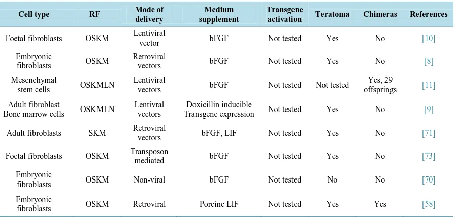
While till date there is no established bona fide pESC reported, multiple reports have shown derivation of piPSCs from a variety of somatic tissues as listed in Table 2. Interestingly, porcine induced pluripotent stem cells (piPSC) have been generated using mouse [8] or human [9]-[11] transcription factors as mentioned in Table 2.
The piPSC generated from different tissue by using different approaches (Figure 2) behave differently de-pending on the culture conditions. In mice and rats, naive stem cells can be generated with LIF and 2 signal transduction inhibitors-2i (MAPK/extracellular signal regulated kinase inhibitor, PD0325901 and glycogen Synthase kinase 3β inhibitor, CHIR99021) [53] [54].
[image:6.595.92.540.494.708.2]It has been shown that human PSCs in primed state can be transformed to the naive like state by using molecules like 2i, LIF and Forskolin [55] [56] or continuous or sustained expression of the exogenous OSKM and nanog genes [57]. Similarly, Fujishiro et al., and his colleagues demonstrated that generation of putative naive porcine iPSCs with similar characteristics to mouse naive PSC, had the potential to form chimeras [58]. However, it is interesting to note that comparing the expression pattern of piPSCs in different reports suggest consistent ex- pression of Sox2, Oct4 and Nanog [9]-[11] but the expression of other pluripotency markers is debatable. Such observations may be highly valuable in understanding the differences between intra-species iPSC variations. For instance, Oct4 is regarded as the gate keeper of cellular pluripotency and majority of mammalian species express Oct4 and associated transcriptional factors. In humans and mouse Oct4 is restricted to ICM cells of developing embryos [59] [60] and ESCs [49]. However, in farm animal like cattle, this gene is detected both in ICM and trophoblast cells, suggesting that its expression is not limited to pluripotent cells of the early embryo [61]. Simi-
Table 2. Summary of porcine induced pluripotent stem cells (iPSCs) generated from various somatic cell. The abbreviations used have already been explained in the text.
Cell type RF Mode of
delivery
Medium supplement
Transgene
activation Teratoma Chimeras References
Foetal fibroblasts OSKM Lentiviral
vector bFGF Not tested Yes No [10]
Embryonic
fibroblasts OSKM
Retroviral
vectors bFGF Not tested Yes No [8]
Mesenchymal
stem cells OSKMLN
Lentiviral
vectors bFGF Not tested Not tested
Yes, 29
offsprings [11]
Adult fibroblast
Bone marrow cells OSKMLN
Lentivral vectors
Doxicillin inducible
Transgene expression Not tested Yes No [9]
Adult fibroblasts SKM Retroviral
vectors bFGF, LIF Not tested Yes No [71]
Foetal fibroblasts OSKM Transposon
mediated bFGF Not tested Yes No [73]
Embryonic
fibroblasts OSKM Non-viral bFGF Not tested No No [70]
Embryonic
fibroblasts OSKM Retroviral Porcine LIF Not tested Yes Yes [58]
Figure 2. Numerous methods of generating induced pluripotent stem cells. Only few representative methods are shown here.
larly, in pig the Oct4 is present in the ICM, trophectoderm, and during hatching it is expressed in the epiblast component of the embryonic disc [59] [62]. Apart from this there is a gradual down-regulation of Oct4 expres-sion noted in porcine ESCs [63].
Like Oct-4, Nanog too shows marked differences in porcine species as compared to human or rodent systems. In porcine, Nanog expression is not only limited to the ICM and epiblast [64] but its expression could be de-tected in day E7.5 to day E10.5 embryos, which is five days later blastocyst formation [34]. Unlike the mouse, expression of Nanog is restricted to only a few cells of the ICM around implantation [65]. These observations clearly suggest that different species have different timing of Nanog expression. Nevertheless, chimeric animals can be generated by injecting porcine ICM clumps into blastocyst which suggests that few cells in porcine ICM are pluripotent or attain pluripotency [66]. Somehow, putative porcine pluripotent stem cells lose their pluripo-tency over a relatively short number of passages as compared to their mouse or human counterparts. These ob-servations clearly suggest that other factors may be responsible for the maintenance of self-renewal of these cells in an undifferentiated status.
Porcine iPS cells derived from embryonic fibroblasts in serum containing medium morphologically resemble human ES cells with flatten, compact colonies that stain positively for AP, SSEA-4, Nanog and Rex1 [8]. Por-cine induced pluripotent stem cells (piPSCs) derived from primary ear fibroblasts or bone marrow cells and ESC-specific culture medium without bFGF, also express human ES cell surface markers [9]. However, iPS cells generated form porcine fetal fibroblasts cultured in hESC-specific medium supplemented with bFGF stain positive for AP and SSEA-1, and negative for SSEA-3, SSEA-4, TRA1-60 and TRA1-81 [10], which is very unusual compared to other stem cells cultured in hESC medium. The patterns of methylation in iPSCs generated also resemble the patterns observed in ESCs, especially for genes related to pluripotency, such as Oct4. Never-theless, all these iPSCs differentiate into cells representing the three germ layers via EB and teratoma formation. The use of episomal factors in combination with 2i has also proven successful for piPSCs generation [67]. The cells display similarities to their LIF-dependent ESCs counterparts [68]. In spite of that, the cell lines showed either vector integration or persistence of episomal plasmids even after several passages [67].
The most challenging criterion of pluripotency was accomplished by West, and his colleagues, where 30 of 36 pigs produced were chimeric, displaying varying levels of chimerism. In this study, the forced expression of transcription factors enabled the derivation and establishment of iPSCs in pig that have displayed pluripotency [11] [69]. LIF-dependent naive pluripotent cells were generated from cultured porcine ICM, using combinations of DOX-driven Oct4, Klf4 human transgenes and stringent culture conditions but there was no evidence of germ-line transmission.
Studies have demonstrated that piPSCs generated from fibroblast using OSKM, displayed mES like mor-phology when they were maintained in LIF/STAT3-dependent manner and they exhibited potential of generating chimeric and reconstructed NT (Nuclear Transfer) embryos [72]. However, in this study, the transgenes were clearly down regulated, but were not silent completely, and it was in agreement with previous studies [70] [71].
Kues et al., displayed the utility of porcine Oct4-EGFP cells and transposon mediated reprogramming for the derivation of porcine iPS cells. However, this study also indicated the limitation of culture conditions and cul-ture media to support pluripotency [73]. Petkov et al.,demonstrated that the outcome of porcine somatic cell re-programming experiments depends on the selection of expression vector promoter because it controls the ex-pression levels of the reprogramming transcription factors. In this study EF1a and CAG promoters were found to be more efficient than doxycycline-induced tet operator (TetO) promoters [74].
These finding suggest that the endogenous pluripotent genes and their networks could not sustain pluripotency of putative porcine PSCs. Thus, establishment and optimization of culture conditions specific for porcine plu- ripotent stem cells is prerequisite for any further advancement in porcine stem cell research.
3.1. Culture Conditions
It is evident from earlier reports that culture conditions required to sustain pPSCs have not been well established. The stumbling block faced by laboratories worldwide has been that putative PSCs lose their pluripotency over a relatively short number of passages. Bona fide Porcine PSCs in which pluripotency maintained by the endogen-ous pluripotency factors have not yet been reported [12]. In other words, the incessant expression of the ex-ogenous transgenic transcription factors is still obligatory to sustain the pluripotent phenotype of porcine iPS- like cells [9]-[11].
Over the past few decades, all endeavour’s to generate livestock PSCs have failed. Researchers have used mouse or human protocols as reference for culturing of ungulates PS cells, which include feeders, serum and supplementation with LIF [45] [75] [76] or FGF2 [75], epidermal growth factor and stem cell factor (SCF) [76].
Usually for the pPSC culturing, standard human stem cell medium [69] or mouse and human standard cultur-ing medium are mixed in 1:1 ratio [8] [77] because of the paucity of the information related to presence of re-ceptors on pPSCs [35] demonstrated that porcine pluripotent cell lines do not express LIF rere-ceptors [13]. How-ever, they do express FGFR-2 [13]. Apart from that their finding also suggests that LIF is not essential for the maintenance of pluripotency, but its presence inhibits the differentiation process. They also indicated that LIF do not act through LIF receptor signaling pathway.
Researchers also tried to culture ICM and epiblast in feeder layer and serum-free culture conditions. A study was based on modified TeSR medium and matrigel. Almost half of the primary cultures got differentiated within 3 - 4 passages. They used mechanical passaging for sub-culturing [64]. However, mTeSR1 based on Ludwig et al., is most widely used feeder and serum free culture medium for hESCs and iPSCs [78].
Vassiliev et al., cultured ICM cells in MEM medium supplemented with EGF (epidermal growth factor) and activin [51]. They were able to generate chimeras by using putative porcine ES cells. But it was not vivid from their study whether they were able to keep the putative pESCs in undifferentiated self-renewal state beyond 14 passages [51]. Various type of culture conditions were used to culture ICM/epiblast primary out growths [26]. But, none of the culture conditions supported to keep the putative porcine ES cells in undifferentiated state after certain passages.
A very different culture regime i.e., 2i/LIF medium led to the first isolation and establishment of bona fide ESCs from rat embryos [53] [54], and noncompliant mouse strains [79]. Human embryo-derived stem cells cannot be maintained in 2i/LIF alone [80]. However, it facilitated to establish mouse ESC-like cells in presence of forskolin [55]. Consequently, human cells with an epigenetic and transcriptional profile analogous to naïve ESC lines could be established.
Haraguchi et al., and colleagues were able to generate presumptive porcine ES cell lines from the ICM of porcine embryos by using inhibitors of glycogen synthase kinase 3β and MAPK1. They were able to maintain the cell lines over 100 passages only [52].
β-catenin and c-Myc. CHIR99021, a GSK3β inhibitor, effects Wnt signaling activation [81] [82]. These findings were in conjugation with several previous reports, where they have shown that CHIR99021 augments the repro-gramming efficiency [82].
Rudriguez et al., demonstrated that blocking MEK signaling increases the proportion of Nanog expressing cells in the ICM of porcine embryos [83]. From their findings, it is apparent that FGF signaling participates in the formation of the founders of the ICM. PDO325901 (MEK pathway inhibitor), CHIR (GSK3β inhibitior) and LIF supplementation were used to modulate pluripotency in piPSCs cells to acquire features of naive/ground pluripotency. It was demonstrated that small molecule inhibitors can be used to increase the homogeneity of iPSC cultures [83]. Oxygen tension is also an imperative trait for PSC’s maintenance and differentiation. The iPSCs induction executed in hypoxic conditions (5% O2) demonstrated augmentation of the reprogramming
ef-ficiency [84].
3.2. Epigenetic Modifiers
Apart from mRNA and episomal vector approach to safely reprogram somatic cells, another approach is usage of concoction of small molecules that are allied with epigenetic modifiers and key signaling pathways listed in Table 3. Small molecules that alter chromatin and gene expression enhance the reprogramming efficiency in iPSC generation. Inhibitors of histone deacetylases (HDACs), histone demethylases (HDMs), and histone me-thyltransferases (HMTs), which regulate chromatin remodeling, have been recognized as small molecules for terminally differentiated cells reprogramming. For instance, Huangfu et al., used chromatin modification inhibi-tors, the 5’-azacytidine (5-aza) (DNA methyltransferase inhibitor), dexamethazone (synthetic glucocorticoid) and valproic acid (VPA, a histone deacetylase inhibitor) to investigate their effects on reprogramming efficiency [85]. It was inferred that 5-aza, dexamethazone and VPA enhanced the efficiency of cellular reprogramming by 10-fold, 2.6-fold and >100-fold respectively. The effect of VPA was found to be much stronger than that of 5’-azacytidine and other HDAC inhibitors tested [85]. Other noticeable epigenetic modifiers which are used during the reprogramming are, parnate, a histone demethylase inhibitor [86], 5-aza and RG108, a DNA methyl-transferase inhibitors [87]. Butyrate (a HDAC inhibitor) also improves iPSC generation by augumenting acety-lation of histone H3 and demethyacety-lation of genes promoter, related to of pluripotency [88] [89].
Lee et al., demonstrated that Sirtuin 1 (SIRT1) (a member of the sirtuin family of NAD+-dependent protein deacetylases) a class III HDAC assists in generation of iPSCs from MEFs via miR-34a and p53 Pathways [90]. It was hypothesized that SIRT1 works as a positive epigenetic regulator in reprogramming somatic cells.
Korean scientists discovered a new molecular compound, RSC133 (inhibits histone deacetylase and DNA methyltransferase) that could enhance the reprogramming efficiency of human adult cell to iPSCs. It was in-ferred that compound derivative acts as the “booster of pluripotency”, and it potently improves the reprogram-ming of human somatic cells into a pluripotent state and aids the growth and maintenance of human pluripotent stem cells [91].
Even in livestock, there are several reports that suggest that the reprogramming process is mired by faulty ep-igenetic modifications during the reprogramming process, resulting into low survival rates among clones [92] and inadequacy of iPS reprogramming. Histone deacetylation is considered to be an unwanted event in nuclear reprogramming assays. Trichostatin A (TSA) has been used to improve cloning efficiency by as much as 75% in various species [92].
Kim et al., have shown that the usage of decitabine (5-aza-2’-deoxycytydine) in ESC culture improves the number of colonies obtained [93]. Treatment of early pre-implantation murine and bovine embryos with 5-AzaC exhibited a simple and efficient method for isolation of putative bovine PS cells [94]. However, there are still safety concerns regarding the usage of epigenetic modifiers or cocktail of small molecules as they could activate cell growth pathway or inhibit tumor suppressor pathways.
3.3. Post-Translational Modulation of Pluripotency Genes
Table 3. List of small molecules and epigenetic modifiers used for somatic cell reprogramming.
Names Mode of action References
Forskolin Stimulates adenylate cyclase activity and increases cAMP [58]
Valproic acid Histone deacetylase inhibitor [85]
BIO
(6-Bromoindirubin-3’-oxime) Inhibitor for GSK-3 a/β in the Wnt signaling pathway. [94]
Lithium GSK3 Inhibition (Wnt+) [26]
CHIR99021 GSK3 inhibitor [46]
Trichostatin A histone deacetylase inhibitor [85]
Sodium Butyrate Sodium butyrate is a known inhibitor of histone deacetylases and inhibits both the
mRNA and protein content of cyclin D1 [85]
Parnate Histone demethylase inhibitor [86]
5-azacytidine DNA methyltransferase inhibitor [85]
SC1 (Pluripotin) Can act as a LIF replacement molecule for mouse embryonic (mES) cell
self-renewal [113]
RG108 DNA methyltransferase inhibitor [87]
PD0325901 Selectively binds and inhibits MEK [46]
SIRT1 A class III HDAC assists in Generation of iPSCs [90]
RSC133 Inhibits histone deacetylase and DNA methyltransferase [91]
RepSox Tgfbr1 kinase inhibitor [114]
Etoposide Notch1 upregulation [109]
Cytochalasin D Wnt5a upregulation [109]
Vitamin C Nutrient vital that lower reactiveoxygen species [110]
Chaetocin Histone Methyltransferase Inhibition [109]
Pifithrin-α Inhibitor of p53-dependent apoptosis. Moreover, reduced p53 activity augments
reprogramming efficiency of human and mouse somatic cells [111]
Pifithrin-μ
Inhibitor of p53 mitochondrial pathway by reducing its affinity for antiapoptotic proteins Bcl-2 and Bcl-XL but does not affect any additional transcriptional
functions of p53
[112]
DNMT1 Inhibition promote fully reprogramming [79]
HDACs Inhibition increase the efficiency of reprogramming [85]
G9a Increase the efficiency of reprogramming [53]
modifiers, the miRNAs play a significant part in the reprogramming of terminally differentiated cells.
Several miRNAs have been shown to enhance iPSC reprogramming when expressed along with combinations of the OSKM factors. Introduction of miRNAs specific to ESCs improve the generation of iPSCs in mouse. The miRNAs miR-291-3p, miR-294 and miR-295 augment the reprogramming efficiency [98]. The miRNAs did not improve reprogramming efficiency in the presence of c-Myc transgene; it suggests that they could be down-stream target of c-Myc. These miRNA seems to have the ability to control cell cycle, to effect the augmentation of iPSC reprogramming [98].
reprogram-ming and stimulates expression of Oct4 gene, and that suppression/degradation of HDAC2 by using Valproic acid is also vital [100]. As far as usage of miRNA for reprogramming is concerned, there is no authentic report available.
3.4. Differentiation Potential of Presumptive Porcine PS Cells
Despite non-stabilized defined state of pluripotency of pPSCs [74], in vitro differentiation of presumptive porcine iPSC into rod photoreceptors and their integration into the retinas of recipient pigs has been reported [101]. The impending therapeutic benefits of stem cell-based treatment to recover the cardiac function have been customary in small animal models of myocardial infarction (MI) [102]-[106]. Human PSCs transplanted to treat immune- deficient porcine or murine models of acute myocardial infarction (AMI) [107], but intra-myocardial injection of livestock stem cells, particularly porcine PS cells in a pig model of AMI has not yet been gauged. Animal models that imitate human cardiac disease, such as MI, AMI and ischemia-reperfusion (IR) that induces heart failure are useful models to study cardiovascular disease (CVD). Recently, a group of researchers examined the possibility of using porcine PS cells to prevent LV dysfunction and post in infarction left ventricular remodelling in hearts suffering from AMI. They demonstrated that porcine PS cell could be an effective treatment for myocardial in-farction (MI). In this study, engrafted porcine PS cells showed the differentiation potential by differentiating into vessel cells, which further augmented the formation of new vessels in infarcted heart [108]. However, no observe myocyte generation was observed. piPSCs may have the prospective to be used in ischemic cardiac diseases. This study had some serious limitations and further research is required to translate cell therapy form bench to bed side. However, a porcine acute myocardial infarction (AMI) model can fill the space between treatment for small animals and treatment for humans.
4. Future Directions/Opportunities and Challenges
It is clear that pig and humans share far more anatomical, histological, biochemical and physiological properties than do the mice and humans. As a result, the pig has emerged as an excellent model for cardiovascular research and many other basic and applied biomedical applications.
The supply of human organs and tissues will be scarce to satisfy burgeoning demands of organ transplantation, making xenotransplantation a viable alternative. In this concern, pig is a promising effective translational model, and represents a candidate species for studying CVD, besides several other applications in regenerative medicine. Bona fide stem cells generated from porcine could be used in multiple applications such as creating models for human genetic diseases, engineering organs for transplantation therapies.
Understanding and unravelling the basic cellular and molecular mechanisms that regulate cell-mediated tissue regeneration is the key to harness this potential. Future research in the field could open novel prospects of re-search into degenerative conditions as pig is a more effective human proxy to work with. The rere-search on por-cine stem cells will lay a foundation for future studies of stem cell transplantation and regenerative medipor-cine. In vitro culturing of porcine stem cell-derived cardiomyocytes could be transplanted into porcine MI model, which would give the researchers and clinicians valuable insights for cell therapy. There is an urgent need to develop validated reagents and cell culture methods to enhance use of porcine stem cells in regenerative medicine. To bestow important clinical insights for cell therapy or transplantation, necessary investigation is required in large animals.
The general ethical and logistical issues impede the use of humans or human-derived tissues for research and discovery pertaining to biomedical applications, which highlight the need for research model that closely mimic human anatomy, physiology, disease and injury processes. In view of significant differences between rodents and humans, the murine (rat and mice) confound or prohibit their use in translational studies.
The pig will continue to be ideal species to serve this purpose. The pigs conserve immunological and physio- logical attributes of human hearts apart from resemblance with the human heart. Hence, they can provide a po-tent research tool for pre-clinical study and particularly for transplantation medicine.
Table 4. Synopsis of the characterization of porcine iPSCs (induced pluripotent stem cells) from various laboratories.
Mode of delivery and
characterization of porcine iPSCs [8] [10] [9] [11] [70] [68] [71] [74]
* [74]#
Delivery method Viral Viral Viral Viral Plasmid Viral Viral Viral Viral
Pluripotency markers
Oct4 ND + + + + + + + +
Sox2 + + + + + + + + +
Nanog + + + + + + + + +
Rex1 + + + ND + ND ND ND +
CDH1 ND + + ND ND ND ND ND +
Surface markers
SSEA1 ND + − + − + ND − −
SSEA3 ND − + + − − ND ND ND
SSEA4 + − + − + + + + −
Tra-1-60 ND ± + + + ND + ND ND
Tra-1-81 ND − + − − ND + ND ND
Functional assays
Teratoma + + + + ND + + − −
EBs ND + + + + + + + +
Chimera ND ND ND + ND ND ND ND ND
*Tet O gp; #Ef1a and CAG gp; ND: Not Determined.
the globe. So far, generation of bona fide porcine PSCs has proven elusive, despite overwhelming knowledge and availability of numerous types of epigenetic modifiers, small molecules, specialized reagents and culture details from mouse and human stem cells work. Presumably, this information cannot directly extrapolate to oth-er species as such. Building on the available knowledge, we can innovate on techniques and ideas voth-ery specific to porcine species.
5. Conclusion
The establishment of methods for generating porcine stem cells for clinical applications is still a work in pro- gress. Porcine pre and peri-implantation process as well as mechanism behind different cell fate specification is considerably dissimilar from mouse and human ones, that’s why presumptive porcine PS cells behave different-ly from existing human and mouse stem cell lines. Consequentdifferent-ly, a comprehensive understanding of species specific developmental processes, signaling pathways and other related transcription factors would tender a roadmap for the generation of bona fide PSCs and their in vitro differentiation into cardiomyocytes. Until now none of the research group has documented successful differentiation of pPSCs into cardiomyocytes. To recapi-tulate, porcine PSCs will fill the vacuum between mouse and human pluripotent stem cells. However, notewor-thy technical obstacles for the generation of bona fide porcine PS cells still persists that will only prevail over via ages of exhaustive research.
Disclosure of Potential Conflicts of Interest
The authors indicate no potential conflicts of interest.Acknowledgements
This work is supported by a grant from DST (SERB/LS-310/2013).
References
http://dx.doi.org/10.1016/j.stem.2009.05.015
[2] Gurdon, J.B., Elsdale, T.R. and Fischberg, M. (1958) Sexually Mature Individuals of Xenopus laevis from the Trans-plantation of Single Somatic Nuclei. Nature, 182, 64-65. http://dx.doi.org/10.1038/182064a0
[3] Takahashi, K. and Yamanaka, S. (2006) Induction of Pluripotent Stem Cells from Mouse Embryonic and Adult Fi-broblast Cultures by Defined Factors. Cell, 126, 663-676. http://dx.doi.org/10.1016/j.cell.2006.07.024
[4] Bilic, J. and Belmonte, J.C.I. (2012) Concise Review: Induced Pluripotent Stem Cells Versus Embryonic Stem Cells: Close Enough or Yet Too Far Apart? Stem Cells, 30, 33-41. http://dx.doi.org/10.1002/stem.700
[5] Strojek, R.M., Reed, M.A., Hoover, J.L. and Wagner, T.E. (1990) A Method for Cultivating Morphologically Undiffe-rentiated Embryonic Stem Cells from Porcine Blastocysts. Theriogenology, 33, 901-913.
http://dx.doi.org/10.1016/0093-691X(90)90825-E
[6] Notarianni, E., Laurie, S., Moor, R.M. and Evans, M.J. (1990) Maintenance and Differentiation in Culture of Pluripo-tential Embryonic Cell Lines from Pig Blastocysts. Journal of Reproduction Fertility-Supplement, 41, 51-56.
[7] Hall, V. (2008) Porcine Embryonic Stem Cells: A Possible Source for Cell Replacement Therapy. Stem Cell Reviews, 4, 275-282. http://dx.doi.org/10.1007/s12015-008-9040-2
[8] Esteban, M.A., Xu, J., Yang, J., Peng, M., Qin, D., Li, D., Jiang, Z., Chen, J., Deng, K., Zhong, M., Cai, J., Lai, L. and Pei, J. (2009) Generation of Induced Pluripotent Stem Cell Lines from Tibetan Miniature pig. The Journal of Biologi-cal Chemistry, 284, 17634-17640. http://dx.doi.org/10.1074/jbc.M109.008938
[9] Wu, Z., Chen, J., Ren, J., Bao, L., Liao, J., Cui, C., Rao, L., Li, H., Gu, Y., Dai, H., Zhu, H., Teng, X., Cheng, L. and Xiao, L. (2009) Generation of Pig Induced Pluripotent Stem Cells with a Drug-Inducible System. Journal of Molecular Cell Biology, 1, 46-54. http://dx.doi.org/10.1093/jmcb/mjp003
[10] Ezashi, T., Telugu, V.P.V.L., Alexenko, A.P., Sachdev, S., Sinha, S. and Roberts, R.M. (2009) Derivation of Induced Pluripotent Stem Cells from Pig Somatic Cells. Proceedings of the National Academy of Sciences of USA, 106, 10993- 10998. http://dx.doi.org/10.1073/pnas.0905284106
[11] West, F.D., Uhl, E.W., Liu, Y., Stowe, H., Lu, Y., Yu, P., Gallegos-Cardenas, A., Pratt, S.L. and Stice, S.L. (2011) Brief Report: Chimeric Pigs Produced from Induced Pluripotent Stem Cells Demonstrate Germline Transmission and No Evidence of Tumor Formation in Young Pigs. Stem Cells, 29, 1640-1643. http://dx.doi.org/10.1002/stem.713
[12] Nowak-Imialek, M., Kues, W., Carnwath, J.W. and Niemann, H. (2011)Pluripotent Stem Cells and Reprogrammed Cells in Farm Animals. Microscopy and Microanalysis, 17, 474-497. http://dx.doi.org/10.1017/S1431927611000080
[13] Brevini, T.A., Pennarossa, G., Attanasio, L., Vanelli, A., Gasparrini, B. and Gandolfi, F. (2010) Culture Conditions and Signalling Networks Promoting the Establishment of Cell Lines from Parthenogenetic and Biparental Pig Embryos.
Stem Cell Reviews and Reports, 6, 484-495. http://dx.doi.org/10.1007/s12015-010-9153-2
[14] Smith, A.G., Nichols, J., Robertson, M. and Rathjen, P.D. (1992) Differentiation Inhibiting Activity (DIA/LIF) and Mouse Development. Developmental Biology, 151, 339-351. http://dx.doi.org/10.1016/0012-1606(92)90174-F
[15] Niwa, H., Burdon, T., Chambers, I. and Smith, A. (1998) Self-Renewal of Pluripotent Embryonic Stem Cells Is Me-diated via Activation of STAT3. Genes Development, 12, 2048-2060. http://dx.doi.org/10.1101/gad.12.13.2048
[16] Matsuda, T., Nakamura, T., Nakao, K., Arai, T., Katsuki, M., Heike, T. and Yokota, T. (1999)STAT3 Activation Is Sufficient to Maintain an Undifferentiated State of Mouse Embryonic Stem Cells. The EMBO Journal, 18, 4261-4269.
http://dx.doi.org/10.1093/emboj/18.15.4261
[17] Raz, R., Lee, C.K., Cannizzaro, L.A., d’Eustachio, P. and Levy, D.E. (1999) Essential Role of STAT3 for Embryonic Stem Cell Pluripotency. Proceedings of the National Academy of Sciences of the United States of America, 96, 2846- 2851. http://dx.doi.org/10.1073/pnas.96.6.2846
[18] Cartwright, P., McLean, C., Sheppard, A., Rivett, D., Jones, K. and Dalton, S. (2005) LIF/STAT3 Controls ES Cell Self-Renewal and Pluripotency by a Myc-Dependent Mechanism. Development, 132, 885-896.
http://dx.doi.org/10.1242/dev.01670
[19] Ying, Q.L., Stavridis, M., Griffiths, D., Li, M. and Smith, A. (2003) Conversion of Embryonic Stem Cells into Neu-roectodermal Precursors in Adherent Monoculture. Nature Biotechnology, 21, 183-186.
http://dx.doi.org/10.1038/nbt780
[20] Winnier, G., Blessing, M., Labosky, P.A. and Hogan, B.L. (1995) Bone Morphogenetic Protein-4 Is Required for Me-soderm Formation and Patterning in the Mouse. Genes Development, 9, 2105-2116.
http://dx.doi.org/10.1101/gad.9.17.2105
[21] Thomson, J.A., Kalishman, J., Golos, T.G., Durning, M., Harris, C.P., Becker, R.A. and Hearn, J.P. (1995) Isolation of a Primate Embryonic Stem Cell Line. Proceedings of the National Academy of Sciences of the United States of Ameri-ca, 92, 7844-7848. http://dx.doi.org/10.1073/pnas.92.17.7844
Embryonic Stem Cell Lines Derived from Human Blastocysts. Science, 282, 1145-1147.
http://dx.doi.org/10.1126/science.282.5391.1145
[23] Kawahara, Y., Manabe, T., Matsumoto, M., Kajiume, T. and Yuge, L. (2009) LIF-Free Embryonic Stem Cell Culture in Simulated Microgravity. PLoS ONE, 4, e6343. http://dx.doi.org/10.1371/journal.pone.0006343
[24] Humphrey, R.K., Beattie, G.M., Lopez, A.D., Bucay, N., King, C.C., Firpo, M.T., Rose-John, S. and Hayek, A. (2004) Maintenance of Pluripotency in Human Embryonic Stem Cells Is STAT3 Independent. Stem Cells, 22, 522-530.
http://dx.doi.org/10.1634/stemcells.22-4-522
[25] James, D., Levine, A.J., Besser, D. and Hemmati-Brivanlou, A. (2005) TGFbeta/Activin/Nodal Signaling Is Necessary for the Maintenance of Pluripotency in Human Embryonic Stem Cells. Development, 132, 1273-1282.
http://dx.doi.org/10.1242/dev.01706
[26] Wang, G., Zhang, H., Zhao, Y., Li, J., Cai, J., Wang, P., Meng, S., Feng, J., Miao, C., Ding, M., Li, D. and Deng, H. (2005) Noggin and bFGF Cooperate to Maintain the Pluripotency of Human Embryonic Stem Cells in the Absence of Feeder Layers. Biochemical and Biophysical Research Communications, 330, 934-942.
http://dx.doi.org/10.1016/j.bbrc.2005.03.058
[27] Xu, C., Rosler, E., Jiang, J., Lebkowski, J.S., Gold, J.D., O’Sullivan, C., Delavan-Boorsma, K., Mok, M., Bronstein, A. and Carpenter, M.K. (2005) Basic Fibroblast Growth Factor Supports Undifferentiated Human Embryonic Stem Cell Growth without Conditioned Medium. Stem Cells, 23, 315-323. http://dx.doi.org/10.1634/stemcells.2004-0211
[28] Davidson, K.C., Jamshidi, P., Daly, R., Hearn, M.T., Pera, M.F. and Dottori, M. (2007) Wnt3a Regulates Survival, Expansion, and Maintenance of Neural Progenitors Derived from Human Embryonic Stem Cells. Molecular and Cel-lular Neuroscience, 36, 408-415. http://dx.doi.org/10.1016/j.mcn.2007.07.013
[29] Dravid, G., Ye, Z., Hammond, H., Chen, G., Pyle, A., Donovan, P., Yu, X. and Cheng, L. (2005) Defining the Role of Wnt/β-Catenin Signaling in the Survival, Proliferation and Self-Renewal of Human Embryonic Stem Cells. Stem Cells,
23, 1489-1501. http://dx.doi.org/10.1634/stemcells.2005-0034
[30] Yasuda, S.Y., Tsuneyoshi, N., Sumi, T., Hasegawa, K., Tada, T., Nakatsuji, N. and Suemori, H. (2006) NANOG Main- tains Self-Renewal of Primate ES Cells in the Absence of a Feeder Layer. Genes to Cells, 11, 1115-1123.
http://dx.doi.org/10.1111/j.1365-2443.2006.01000.x
[31] Ying, Q.L., Wray, J., Nichols, J., Batlle, M.L., Doble, B., Woodgett, J., Cohen, P. and Smith, A. (2008) The Ground State of Embryonic Stem Cell Self-Renewal. Nature, 453, 519-523. http://dx.doi.org/10.1038/nature06968
[32] Alberio, R., Croxall, N. and Allegrucci, A. (2010) Pig Epiblast Stem Cells Depend on Activin/Nodal Signaling for Plu-ripotency and Self-Renewal. Stem Cells and Development, 19, 1627-1636. http://dx.doi.org/10.1089/scd.2010.0012
[33] Blomberg, L.A., Schreier, L.L. and Talbot, N.C. (2008)Expression Analysis of Pluripotency Factors in the Undifferen-tiated Porcine Inner Cell Mass and Epiblast During in Vitro Culture. Molecular Reproduction and Development, 75, 450-463. http://dx.doi.org/10.1002/mrd.20780
[34] Hall, V.J., Christensen, J., Gao, Y., Schmidt, M.H. and Hyttel, P. (2009) Porcine Pluripotency Cell Signaling Develops from the Inner Cell Mass to the Epiblast during Early Development. Developmental Dynamics, 238, 2014-2024.
http://dx.doi.org/10.1002/dvdy.22027
[35] Brevini, T.A., Antonini, S., Pennarossa, G., Maffei, S. and Gandolfi, F. (2012) Pluripotency Network in Porcine Em-bryos and Derived Cell Lines. Reproduction in Domestic Animals, 47, 86-91.
http://dx.doi.org/10.1111/j.1439-0531.2012.02060.x
[36] Welham, M.J., Storm, M.P., Kingham, E. and Bone, H.K. (2007) Phosphoinositide 3-Kinases and Regulation of Em-bryonic Stem Cell Fate. Biochemical Society Transactions, 35, 225-228.
[37] Mummery, C.L., van Rooyen, M., Bracke, M., van den Eijnden-van Raaij, J., van Zoelen, E.J. and Alitalo, K. (1993) Fibroblast Growth Factor-Mediated Growth Regulation and Receptor Expression in Embryonal Carcinoma and Em-bryonic Stem Cells and Human Germ Cell Tumours. Biochemical and Biophysical Research Communications, 191, 188-195. http://dx.doi.org/10.1006/bbrc.1993.1201
[38] Jirmanova, L., Afanassieff, M., Gobert-Gosse, S., Markossian, S. and Savatier, P. (2002) Differential Contributions of ERK and PI3-Kinase to the Regulation of Cyclin D1 Expression and to the Control of the G1/S Transition in Mouse Embryonic Stem Cells. Oncogene, 21, 5515-5528. http://dx.doi.org/10.1038/sj.onc.1205728
[39] Ma, Y., Yang, J.Y., Cheng, D., Liu, X., Ma, X., West, F.D. and Wang, H. (2014) Comparative Gene Expression Sig-nature of Pig, Human and Mouse Induced Pluripotent Stem Cell Lines Reveals Insight into Pig Pluripotency Gene Networks. Stem Cell Reviews and Reports, 10, 162-176. http://dx.doi.org/10.1007/s12015-013-9485-9
[40] Brevini, T.A., Pennarossa, G. and Gandolfi, F. (2010) No Shortcuts to Pig Embryonic Stem Cells. Theriogenology, 74, 544-550. http://dx.doi.org/10.1016/j.theriogenology.2010.04.020
http://dx.doi.org/10.1016/0093-691X(90)90558-B
[42] Evans, M.J., Notarianni, E., Laurie, S. and Moor, R.M. (1990)Derivation and Preliminary Characterization of Pluripo-tent Cell Lines from Porcine and Bovine Blastocysts. Theriogenology, 33, 125-128.
http://dx.doi.org/10.1016/0093-691X(90)90603-Q
[43] Piedrahita, J.A., Anderson, G.B. and Bondurant, R.H. (1990) On the Isolation of Embryonic Stem Cells: Comparative Behavior of Murine, Porcine and Ovine Embryos. Theriogenology, 34, 879-901.
http://dx.doi.org/10.1016/0093-691X(90)90559-C
[44] Talbot, N.C., Rexroad Jr., C.E., Pursel, V.G. and Powell, A.M. (1993) Alkaline Phosphatase Staining of Pig and Sheep Epiblast Cells in Culture. Molecular Reproduction and Development, 36, 139-147.
http://dx.doi.org/10.1002/mrd.1080360204
[45] Verma, V., Gautam, S.K., Singh, B., Manik, R.S., Palta, P., Singla, S.K., Goswami, S.L. and Chauhan, M.S. (2007) Isolation and Characterization of Embryonic Stem Cell-Like Cells from in Vitro-Produced Buffalo (Bubalus bubalis) Embryos. Molecular Reproduction and Development, 74, 520-529. http://dx.doi.org/10.1002/mrd.20645
[46] Verma, V., Huang, B., Kallingappa, P.K. and Oback, B. (2013)Dual Kinase Inhibition Promotes Pluripotency in Finite Bovine Embryonic Cell Lines. Stem Cells and Development, 22, 1728-1742. http://dx.doi.org/10.1089/scd.2012.0481
[47] Wheeler, M.B. (1994) Development and Validation of Swine Embryonic Stem Cells: A Review. Reproduction, Fertil-ity and Development, 6, 563-568. http://dx.doi.org/10.1071/RD9940563
[48] Shim, H., Gutierrez-Adan, A., Chen, L.R., BonDurant, R.H., Behboodi, E. and Anderson, G.B. (1997) Isolation of Pluripotent Stem Cells from Cultured Porcine Primordial Germ Cells. Biology of Reproduction, 57, 1089-1095.
http://dx.doi.org/10.1095/biolreprod57.5.1089
[49] Chen, L.R., Shiue, Y.L., Bertolini, L., Medrano, J.F., BonDurant, R.H. and Anderson, G.B. (1999) Establishment of Pluripotent Cell Lines from Porcine Preimplantation Embryos. Theriogenology, 52, 195-212.
http://dx.doi.org/10.1016/S0093-691X(99)00122-3
[50] Miyoshi, K., Taguchi, Y., Sendai, Y., Hoshi, H. and Sato, E. (2000) Establishment of a Porcine Cell Line from in Vi-tro-Produced Blastocysts and Transfer of the Cells into Enucleated Oocytes. Biology of Reproduction, 62, 1640-1646.
http://dx.doi.org/10.1095/biolreprod62.6.1640
[51] Vassiliev, I., Vassilieva, S., Beebe, L.F., Harrison, S.J., McIlfatrick, S.M. and Nottle, M. B. (2010) In Vitro and in Vivo
Characterization of Putative Porcine Embryonic Stem Cells. Cellular Reprogramming, 12, 223-230.
http://dx.doi.org/10.1089/cell.2009.0053
[52] Haraguchi, S., Kikuchi, K., Nakai, M. and Tokunaga, T. (2012) Establishment of Self-Renewing Porcine Embryonic Stem Cell-Like Cells by Signal Inhibition. Journal of Reproduction and Development, 58, 707-716.
http://dx.doi.org/10.1262/jrd.2012-008
[53] Buehr, M., Meek, S., Blair, K., Yang, J., Ure, J., Silva, J., McLay, R., Hall, J., Ying, Q.L. and Smith, A. (2008) Cap-ture of Authentic Embryonic Stem Cells from Rat Blastocysts. Cell, 135, 1287-1298.
http://dx.doi.org/10.1016/j.cell.2008.12.007
[54] Li, P., Tong, C., Mehrian-Shai, R., Jia, L., Wu, N., Yan, Y., Maxson, R.E., Schulze, E.N., Song, H., Hsieh, C.L., Pera, M.F. and Ying, Q.L. (2008) Germline Competent Embryonic Stem Cells Derived from Rat Blastocysts. Cell, 135, 1299-1310. http://dx.doi.org/10.1016/j.cell.2008.12.006
[55] Hanna, J., Cheng, A.W., Saha, K., Kim, J., Lengner, C.J., Soldner, F., Cassady, J.P., Muffat, J., Carey, B.W. and Jae-nisch, R. (2010) Human Embryonic Stem Cells with Biological and Epigenetic Characteristics Similar to Those of Mouse ESCs. Proceedings of the National Academy of Sciences of the United States of America, 107, 9222-9227.
http://dx.doi.org/10.1073/pnas.1004584107
[56] Pomp, O., Dreesen, O., Leong, D.F., Meller-Pomp, O., Tan, F., Zhou, T.F. and Colman, A. (2011) Unexpected X Chromosome Skewing during Culture and Reprogramming of Human Somatic Cells Can Be Alleviated by Exogenous Telomerase. Cell Stem Cell, 9, 156-165. http://dx.doi.org/10.1016/j.stem.2011.06.004
[57] Buecker, C., Chen, H.H., Polo, J.M., Daheron, L., Bu, L., Barakat, T.S., Okwieka, P., Porter, A., Gribnau, J., Hoched-linger, K. and Geijsen, N. (2010) A Murine ESC-Like State Facilitates Transgenesis and Homologous Recombination in Human Pluripotent Stem Cells. Cell Stem Cell, 6, 535-546. http://dx.doi.org/10.1016/j.stem.2010.05.003
[58] Fujishiro, S.H., Nakano, K., Mizukami, Y., Azami, T., Arai, Y., Matsunari, H., Ishino, R., Nishimura, T., Watanabe, M., Abe, T., Furukawa, Y., Umeyama, K., Yamanaka, S., Ema, M., Nagashima, H. and Hanazono, Y. (2013) Genera-tion of Naive-Like Porcine-Induced Pluripotent Stem Cells Capable of Contributing to Embryonic and Fetal Develop-ment. Stem Cells and Development, 22, 473-482. http://dx.doi.org/10.1089/scd.2012.0173
[59] Kirchhof, N., Carnwath, J.W., Lemme, E., Anastassiadis, K., Scholer, H. and Niemann, H. (2000) Expression Pattern of Oct-4 in Preimplantation Embryos of Different Species. Biology of Reproduction, 63, 1698-1705.
[60] Mitalipov, S.M., Kuo, H.-C., Hennebold, J.D. and Wolf, D.P. (2003) Oct-4 Expression in Pluripotent Cells of the Rhesus Monkey. Biology of Reproduction, 69, 1785-1792. http://dx.doi.org/10.1095/biolreprod.103.019455
[61] van Eijk, M.J., van Rooijen, M.A., Modina, S., Scesi, L., Folkers, G., van Tol, H.T., Bevers, M.M., Fisher, S.R., Lewin, H.A., Rakacolli, D., Galli, C., de Vaureix, C., Trounson, A.O., Mummery, C.L. and Gandolfi, F. (1999) Molecular Clon- ing, Genetic Mapping, and Developmental Expression of Bovine POU5F1. Biology of Reproduction, 60, 1093-1103.
http://dx.doi.org/10.1095/biolreprod60.5.1093
[62] Vejlsted, M., Du, Y., Vajta, G. and Maddox-Hyttel, P. (2006) Post-Hatching Development of the Porcine and Bovine Embryo—Defining Criteria for Expected Development in Vivo and in Vitro. Theriogenology, 65, 153-165.
http://dx.doi.org/10.1016/j.theriogenology.2005.09.021
[63] Wolf, X.A., Rasmussen, M.A., Schauser, K., Jensen, A.T., Schmidt, M. and Hyttel, P. (2011) OCT4 Expression in Outgrowth Colonies Derived from Porcine Inner Cell Masses and Epiblasts. Reproduction in Domestic Animals, 46, 385-392. http://dx.doi.org/10.1111/j.1439-0531.2010.01675.x
[64] du Puy, L., Lopes, S.M., Haagsman, H.P. and Roelen, B.A. (2011) Analysis of Co-Expression of OCT4, NANOG and SOX2 in Pluripotent Cells of the Porcine Embryo, in Vivo and in Vitro. Theriogenology, 75, 513-526.
http://dx.doi.org/10.1016/j.theriogenology.2010.09.019
[65] Silva, J., Nichols, J., Theunissen, T.W., Guo, G., van Oosten, A.L., Barrandon, O., Wray, J., Yamanaka, S., Chambers, I. and Smith, A. (2009) Nanog Is the Gateway to the Pluripotent Ground State. Cell, 138, 722-737.
http://dx.doi.org/10.1016/j.cell.2009.07.039
[66] Nagashima, H., Giannakis, C., Ashman, R.J. and Nottle, M.B. (2004) Sex Differentiation and Germ Cell Production in Chimeric Pigs Produced by Inner Cell Mass Injection into Blastocysts. Biology of Reproduction, 70, 702-707.
http://dx.doi.org/10.1095/biolreprod.103.022681
[67] Telugu, B.P.V.L., Ezashi, T. and Roberts, R.M. (2010) Porcine Induced Pluripotent Stem Cells Analogous to Naive and Primed Embryonic Stem Cells of the Mouse. The International Journal of Developmental Biology, 54, 1703-1711.
http://dx.doi.org/10.1387/ijdb.103200bt
[68] Telugu, B.P.V.L., Ezashi, T., Sinha, S., Alexenko, A.P., Spate, L., Prather, R.S. and Roberts, R.M. (2011) Leukemia Inhibitory Factor (LIF)-Dependent, Pluripotent Stem Cells Established from Inner Cell Mass of Porcine Embryos. The Journal of Biological Chemistry, 286, 28948-28953. http://dx.doi.org/10.1074/jbc.M111.229468
[69] West, F.D., Terlouw, S.L., Kwon, D.J., Mumaw, J.L., Dhara, S.K., Hasneen, K., Dobrinsky, J.R. and Stice, S.L. (2010) Porcine Induced Pluripotent Stem Cells Produce Chimeric Offspring. Stem Cells and Development, 19, 1211-1220.
http://dx.doi.org/10.1089/scd.2009.0458
[70] Park, J.K., Kim, H.S., Uh, K.J., Choi, K.H., Kim, H.M., Lee, T., Yang, B.C., Kim, H.J., Ka, H.H., Kim, H. and Lee, C.K. (2013) Primed Pluripotent Cell Lines Derived from Various Embryonic Origins and Somatic Cells in Pig. PLoS ONE, 8, e52481. http://dx.doi.org/10.1371/journal.pone.0052481
[71] Montserrat, N., de Onate, L., Garreta, E., Gonzalez, F., Adamo, A., Eguizabal, C., Hafner., S, Vassena, R. and Izpisua Belmonte, J.C. (2012) Generation of Feeder-Free Pig Induced Pluripotent Stem Cells without Pou5f1. Cell Transplan-tation, 21, 815-825.
[72] Cheng, D., Guo, Y., Li, Z., Liu, Y., Gao, X., Gao, Y., Cheng, X., Hu, J. and Wang, W. (2012) Porcine Induced Pluri-potent Stem Cells Require LIF and Maintain Their Developmental Potential in Early Stage of Embryos. PLoS ONE, 7, e51778. http://dx.doi.org/10.1371/journal.pone.0051778
[73] Kues Wilfried, A., Herrmann, D., Barg-Kues, B., Haridoss, S., Nowak-Imialek, M., Buchholz, T., Streeck, M., Grebe, A., Grabundzija, I., Merkert, S., Martin, U., Hall, V.J., Rasmussen, M.A., Ivics, Z., Hyttel, P. and Niemann, H. (2013) Derivation and Characterization of Sleeping Beauty Transposon-Mediated Porcine Induced Pluripotent Stem Cells.
Stem Cells and Development, 22, 124-135. http://dx.doi.org/10.1089/scd.2012.0382
[74] Petkov, S., Hyttel, P. and Niemann, H. (2013) The Choice of Expression Vector Promoter Is an Important Factor in the Reprogramming of Porcine Fibroblasts into Induced Pluripotent Cells. Cellular Reprogramming, 15, 1-8.
[75] Strelchenko, N. (1996) Bovine Pluripotent Stem Cells. Theriogenology, 45, 131-140.
http://dx.doi.org/10.1016/0093-691X(95)00362-C
[76] Saito, S., Sawai, K., Ugai, H., Moriyasu, S., Minamihashi, A., Yamamoto, Y., Hirayama, H., Kageyama, S., Pan, J., Murata, T., Kobayashi, Y., Obata, Y. and Yokoyama, K.K. (2003) Generation of Cloned Calves and Transgenic Chi-meric Embryos from Bovine Embryonic Stem-Like Cells. Biochemical and Biophysical Research Communications,
309, 104-113. http://dx.doi.org/10.1016/S0006-291X(03)01536-5
[77] Montserrat, N., Bahima, E.G., Batlle, L., Häfner, S., Rodrigues, A.M., González, F. and Izpisúa Belmonte, J.C. (2013) Generation of Pig iPS Cells: A Model for Cell Therapy. Journal of Cardiovascular Translational Research, 6, 295-297.
http://dx.doi.org/10.1007/s12265-012-9403-6
ent Culture of Human Embryonic Stem Cells. Nature Methods, 3, 637-646. http://dx.doi.org/10.1038/nmeth902
[79] Nichols, J., Jones, K., Phillips, J.M., Newland, S.A., Roode, M., Mansfield, W., Smith, A. and Cooke, A. (2009) Vali-dated Germline-Competent Embryonic Stem Cell Lines from Nonobese Diabetic Mice. Nature Methods, 15, 814-818.
http://dx.doi.org/10.1038/nm.1996
[80] Li, J., Wang, G., Wang, C., Zhao, Y., Zhang, H., Tan, Z., Song, Z., Ding, M. and Deng, H. (2007)MEK/ERK Signal-ing Contributes to the Maintenance of Human Embryonic Stem Cell Self-Renewal. Differentiation, 75, 299-307.
http://dx.doi.org/10.1111/j.1432-0436.2006.00143.x
[81] Li, W., Wei, W., Zhu, S., Zhu, J., Shi, Y., Lin, T., Hao, E., Hayek, A., Deng, H. and Ding, S. (2009) Generation of Rat and Human Induced Pluripotent Stem Cells by Combining Genetic Reprogramming and Chemical Inhibitors. Cell Stem Cell, 4, 16-19. http://dx.doi.org/10.1016/j.stem.2008.11.014
[82] Marson, A., Foreman, R., Chevalier, B., Bilodeau, S., Kahn, M., Young, R.A. and Jaenisch, R. (2008) Wnt Signaling Promotes Reprogramming of Somatic Cells to Pluripotency. Cell Stem Cell, 3, 132-135.
http://dx.doi.org/10.1016/j.stem.2008.06.019
[83] Rodriguez, A., Allegrucci, C. and Alberio, R. (2012) Modulation of Pluripotency in the Porcine Embryo and iPS Cells.
PLoS ONE, 7, e49079. http://dx.doi.org/10.1371/journal.pone.0049079
[84] Yoshida, Y., Takahashi, K., Okita, K., Ichisaka, T. and Yamanaka, S. (2009)Hypoxia Enhances the Generation of In-duced Pluripotent Stem Cells. Cell Stem Cell, 5, 237-241. http://dx.doi.org/10.1016/j.stem.2009.08.001
[85] Huangfu, D., Maehr, R., Guo, W., Eijkelenboom, A., Snitow, M., Chen, A.E. and Melton, D.A. (2008) Induction of Pluripotent Stem Cells by Defined Factors Is Greatly Improved by Small-Molecule Compounds. Nature Biotechnology,
26, 795-797. http://dx.doi.org/10.1038/nbt1418
[86] Li, W., Zhou, H., Abujarour, R., Zhu, S., Joo Young, J., Lin, T., Hao, E., Scholer, H.R., Hayek, A. and Ding, S. (2009) Generation of Human-Induced Pluripotent Stem Cells in the Absence of Exogenous Sox2. Stem Cells, 27, 2992-3000. [87] Mikkelsen, T.S., Hanna, J., Zhang, X., Ku, M., Wernig, M., Schorderet, P., Bernstein, B.E., Jaenisch, R., Lander, E.S.
and Meissner, A. (2008) Dissecting Direct Reprogramming through Integrative Genomic Analysis. Nature, 454, 49-55.
http://dx.doi.org/10.1038/nature07056
[88] Mali, P., Chou, B.K., Yen, J., Ye, Z., Zou, J., Dowey, S., Brodsky, R.A., Ohm, J.E., Yu, W., Baylin, S.B., Yusa, K., Bradley, A., Meyers, D.J., Mukherjee, C., Cole, P.A. and Cheng, L. (2010) Butyrate Greatly Enhances Derivation of Human Induced Pluripotent Stem Cells by Promoting Epigenetic Remodeling and the Expression of Pluripotency-As- sociated Genes. Stem Cells, 28, 713-720. http://dx.doi.org/10.1002/stem.402
[89] Liang, G., Taranova, O., Xia, K. and Zhang, Y. (2010) Butyrate Promotes Induced Pluripotent Stem Cell Generation.
The Journal of Biological Chemistry, 285, 25516-25521. http://dx.doi.org/10.1074/jbc.M110.142059
[90] Lee, Y.L., Peng, Q., Fong, S.W., Chen, A.C., Lee, K.F., Ng, E.H., Nagy, A. and Yeung, W.S. (2012) Sirtuin 1 Facili-tates Generation of Induced Pluripotent Stem Cells from Mouse Embryonic Fibroblasts through the miR-34a and p53 Pathways. PLoS ONE, 7, e45633. http://dx.doi.org/10.1371/journal.pone.0045633
[91] Lee, J., Xia, Y., Son, M.Y., Jin, G., Seol, B., Kim, M.J., Son, M.J., Do, M., Lee, M., Kim, D., Lee, K. and Cho, Y.S. (2012) A Novel Small Molecule Facilitates the Reprogramming of Human Somatic Cells into a Pluripotent State and Supports the Maintenance of an Undifferentiated State of Human Pluripotent Stem Cells. Angewandte Chemie Interna-tional Edition, 51, 12509-12513. http://dx.doi.org/10.1002/anie.201206691
[92] Zhao, J., Hao, Y., Ross, J.W., Spate, L.D., Walters, E.M., Samuel, M.S., Rieke, A., Murphy, C.N. and Prather, R.S. (2010) Histone Deacetylase Inhibitors Improve in Vitro and in Vivo Developmental Competence of Somatic Cell Nuc-lear Transfer Porcine Embryos. Cellular Reprogramming, 12, 75-83. http://dx.doi.org/10.1089/cell.2009.0038
[93] Kim, C., Amano, T., Park, J., Carter, M.G., Tian, X. and Yang, X. (2009) Improvement of Embryonic Stem Cell Line Derivation Efficiency with Novel Medium, Glucose Concentration, and Epigenetic Modifications. Cloning and Stem Cells, 11, 89-100. http://dx.doi.org/10.1089/clo.2008.0053
[94] Lim, M.L., Vassiliev, I., Richings, N.M., Firsova, A.B., Zhang, C. and Verma, P.J. (2011) A novel, Efficient Method to Derive Bovine and Mouse Embryonic Stem Cells with in Vivo Differentiation Potential by Treatment with 5-Azacyti- dine. Theriogenology, 76, 133-142. http://dx.doi.org/10.1016/j.theriogenology.2011.01.027
[95] Lee, R.C., Feinbaum, R.L. and Ambros, V. (1993) The C. elegans Heterochronic Gene lin-4 Encodes Small RNAs with Antisense Complementarity to lin-14. Cell, 75, 843-854. http://dx.doi.org/10.1016/0092-8674(93)90529-Y
[96] Wang, Y., Medvid, R., Melton, C., Jaenisch, R. and Blelloch, R. (2007) DGCR8 Is Essential for microRNA Biogenesis and Silencing of Embryonic Stem Cell Self-Renewal. Nature Genetics, 39, 380-385. http://dx.doi.org/10.1038/ng1969
[97] Kanellopoulou, C., Muljo, S.A., Kung, A.L., Ganesan, S., Drapkin, R., Jenuwein, T., Livingston, D.M. and Rajewsky, K. (2005) Dicer-Deficient Mouse Embryonic Stem Cells Are Defective in Differentiation and Centromeric Silencing.
Genes Development, 19, 489-501. http://dx.doi.org/10.1101/gad.1248505
![Table 1. Comparative behaviour of naive and primed pluripotent state [1]](https://thumb-us.123doks.com/thumbv2/123dok_us/8111465.791131/5.595.89.524.100.329/table-comparative-behaviour-naive-primed-pluripotent-state.webp)