Hyperexpression of mitogen-activated protein
kinase in human breast cancer.
V S Sivaraman, … , G J Nuovo, C C Malbon
J Clin Invest.
1997;
99(7)
:1478-1483.
https://doi.org/10.1172/JCI119309
.
Mitogen-activated protein (MAP) kinases act as transducers of extracellular signaling via
tyrosine kinase-growth factor receptors and G-protein-linked receptors to elements
regulating transcription. The activity, abundance, and localization of MAP kinase was
investigated in normal and malignant neoplasia of the breast. In carcinoma of the breast,
MAP kinase was heavily phosphorylated on tyrosyl residues and its activity elevated
5-10-fold over benign conditions, such as fibroadenoma and fibrocystic disease. By in situ
reverse transcription-polymerase chain reaction, hyperexpression of MAP kinase mRNA
can be localized to malignant, epithelial cells. Metastatic cells within involved lymph nodes
of patients with breast cancer also display hyperexpression of MAP kinase. In spite of
persistent activation via phosphorylation, MAP kinase expression is upregulated 5-20-fold
and this hyperexpression may be a critical element to initiation as well as the metastatic
potential of various forms of human breast cancer.
Research Article
Find the latest version:
J. Clin. Invest.
© The American Society for Clinical Investigation, Inc. 0021-9738/97/04/1478/06 $2.00
Volume 99, Number 7, April 1997, 1478–1483
Rapid Publication
Hyperexpression of Mitogen-activated Protein Kinase in Human Breast Cancer
Vimala S. Sivaraman,* Hsien-yu Wang,‡ Gerard J. Nuovo,§ and Craig C. Malboni
*Department of Surgery, ‡Department of Physiology and Biophysics, §Department of Pathology, and iDepartment of Molecular
Pharmacology, University Medical Center, SUNY/Stony Brook, Stony Brook, New York 11794
Abstract
Mitogen-activated protein (MAP) kinases act as transducers
of extracellular signaling via tyrosine kinase–growth factor
receptors and G-protein–linked receptors to elements
regu-lating transcription. The activity, abundance, and
localiza-tion of MAP kinase was investigated in normal and
malig-nant neoplasia of the breast. In carcinoma of the breast,
MAP kinase was heavily phosphorylated on tyrosyl residues
and its activity elevated 5–10-fold over benign conditions,
such as fibroadenoma and fibrocystic disease. By in situ
re-verse transcription-polymerase chain reaction,
hyperexpres-sion of MAP kinase mRNA can be localized to malignant,
epithelial cells. Metastatic cells within involved lymph nodes
of patients with breast cancer also display hyperexpression
of MAP kinase. In spite of persistent activation via
phos-phorylation, MAP kinase expression is upregulated 5–20-fold
and this hyperexpression may be a critical element to
initia-tion as well as the metastatic potential of various forms of
human breast cancer. (
J. Clin. Invest.
1997. 99:1478–1483.)
Key words: MAP kinase
•breast
•carcinoma
•in situ
poly-merase chain reaction
•hyperexpression
Introduction
Breast cancer is one of the most common malignancies affect-ing women (1). The etiology and pathogenesis of breast carci-noma remain unclear, but several observations suggest a prominent role for the mitogen-activated protein (MAP)1
ki-nase regulatory network (2). Regulation of MAP kiki-nase is rel-egated to a cascade of protein kinases, culminating in dual-specificity kinases that phosphorylate MAP kinase on threonyl and tyrosyl residues (3, 4). Upstream regulators of MAP ki-nase, such as the small molecular weight G-protein oncogene product ras (5) and Raf-1 (6), as well as protein kinase C (7)
have been associated with breast cancer. To what extent MAP kinase represents a common point of activation by agents pro-moting breast cell proliferation or whether MAP kinase is it-self a critical element in the etiology or pathogenesis of breast carcinoma was investigated directly in human tissues obtained from patients undergoing surgery for both benign and malig-nant conditions of the breast.
Methods
Assurances. The protocol and patient consent forms for these studies were reviewed and approved by the Institutional Review Board (Committee on Research Involving Human Subjects) of the State University of New York at Stony Brook.
Tissue preparation. Tissue was excised, sectioned, frozen, stored temporarily at 2808C, and placed in liquid nitrogen. Frozen samples were placed in liquid nitrogen while mechanically pulverized. The re-sultant powder was reconstituted into a lysis buffer [70 mM b -glyc-erophosphate (pH 7.2), 0.1 mM sodium vanadate, 2 mM MgCl2, 1 mM
EGTA, 1 mM dithiothreitol, 0.5% (vol/vol) Triton X-100, 0.2 mM phenylmethylsulfonyl fluoride, 5 mg/ml Leupeptin, and 2 mg/ml apro-tinin] and the MAP kinase activity was measured using EGF receptor peptide as the substrate (8). MAP kinase activity of human breast tis-sue stored, maintained, and assayed under these conditions was sta-ble for at least 2 mo.
MAP kinase assay. MAP kinase activity was measured using EGF receptor peptide as the substrate, as described (8).
Immunoblotting and immunohistochemistry. Tissue was excised, sectioned, frozen, and prepared as described above. Samples were processed further in one of two manners. The samples (50 mg protein/ lane) to be used in direct immunoblotting were subjected to SDS-PAGE on 10% acrylamide separating gels. The samples to be sub-jected to immunoprecipitation followed by immunoblotting were im-munoprecipitated from whole cell extracts (0.6 mg protein) with a murine monoclonal antibody to MAP kinase (Zymed Laboratories, Inc., South San Francisco, CA) and the immunoprecipitate was sub-jected to SDS-PAGE on 10% acrylamide separating gels. The re-solved proteins from direct SDS-PAGE (see Figs. 2 and 7) or from the immunoprecipitation (see Fig. 6) were transferred to nitrocellu-lose blots and stained either with an antibody specific for human MAP kinase or with an antiphosphotyrosine antibody (Transduction Laboratories, Lexington, KY), made visible by alkaline phosphatase– conjugated second antibody staining of the immune complexes (9). Briefly, the blots were prepared, stained with primary antibodies, washed, stained with the second antibody, and then incubated at 228C with substrate solution [5.0 ml of 50 mM glycine (pH 9.6), 0.17 mg/ml
p-nitro blue tetrazolium chloride, 7 mM MgCl2, and 0.08 mg/ml
5-bromo-4-chloro-3-indoyl phosphate] until bands were visible (usually 30 s). The reaction was terminated by washing free the substrate solu-tion and rinsing with distilled water. Immunostaining of the dually phosphorylated “active” form of MAP kinase was performed with rabbit polyclonal antibodies (Promega, Madison, WI). The mobility of MAP kinase was established by protein markers. For immunohis-tochemical analysis of MAP kinase, a mouse monoclonal antibody raised against the human MAP kinase was used. Immune complexes Address correspondence to Craig C. Malbon, Department of
Molec-ular Pharmacology, Diabetes and Metabolic Diseases Research Pro-gram, University Medical Center, SUNY/Stony Brook, Stony Brook, NY 11794-8651. Phone: 516-444-7873; FAX: 516-444-7696; E-mail: craig@pharm.som.sunysb.edu
Received for publication 8 August 1996 and accepted in revised form 17 January 1997.
in immunohistochemical staining were made visible by use of a sec-ond, biotinylated antibody, followed by alkaline phosphatase–conju-gated Streptavidin and a fast red substrate (10).
In situ polymerase chain reaction. Thin (4-mm) sections of paraf-fin blocks of primary breast carcinoma tissue were subjected to in situ reverse transcription-polymerase chain reaction (RT-PCR) (11, 12). Tissue was stained with eosin and hematoxylin. Negative controls used PCR primers for an unrelated hepatitis C viral RNA (sense ori-entation, TCCGCGGCCGCACTCCACCATGAATCACTCCCC; anti-sense orientation, AGTCTTGCGGCCGCAGCGCCAAATC) after DNAse digestion. Positive controls used PCR primers for MAP ki-nase (sense orientation, GCAGGTGTTCGACGTGGG; antisense orientation, GTGCAGAACGTTAGCTGAAT) and genomic DNA in the absence of pretreatment with DNAse. For analysis of MAP ki-nase mRNA in situ, samples were treated with DNAse, subjected to reverse transcription, and then PCR. Digoxigenin (in the form of dUTP) was used as the reporter molecule for PCR and an antidigoxi-genin antibody coupled to alkaline phosphatase was used with a chro-mogen to make the PCR products visible (blue staining).
Results and Discussion
MAP kinase activity was assayed in 37 breast tissue samples from the following patients: five with normal breast tissue, one
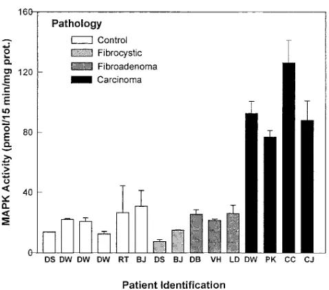
with gynecomastia, four with benign fibroadenoma, five with fibrocystic disease, one with fibrocystic disease and fibroade-noma, two with chronic inflammatory disease, eleven subse-quently identified to have primary breast carcinoma, and one patient with carcinosarcoma. The activity of MAP kinase, as-sayed in extracts using the EGF receptor peptide as substrate, was markedly elevated in all 11 patients with breast cancer. Three are shown in Fig. 1. Four samples of malignant and nor-mal tissue from one patient (DW) with cancer of the breast were analyzed. The pathology identified carcinoma in one sample from DW, the one displaying elevated MAP kinase ac-tivity. The three samples from DW that were negative for ma-lignancy displayed four- to fivefold lower activity. MAP kinase activity (pmol/min/mg protein) was 1.4060.19 (mean6SEM, n5 6) for tissue from the control study group as compared with 6.3960.71 (P# 0.05 for the difference) for the tissue from patients with primary breast carcinoma.
Since enhanced activity may reflect activation of MAP nase via upstream signaling elements, the amount of MAP ki-nase was determined in breast tissue extracts from patients with fibrocystic disease, benign fibroadenoma, and carcinoma of the breast. Equal amounts of cellular protein were subjected to SDS-PAGE, immunoblotting, and staining with an antibody to human MAP kinase (Fig. 2). Immunostaining of the blots displays marked hyperexpression of MAP kinase in breast cancer samples specifically. Tissue samples of benign fibroade-noma and fibrocystic disease displayed little if any staining of MAP kinase under these same conditions.
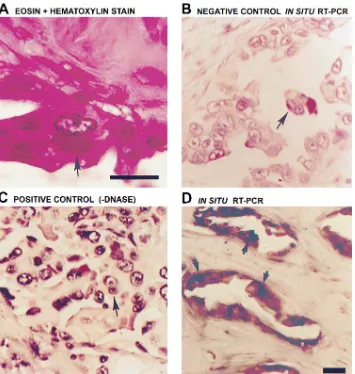
[image:3.612.58.297.55.266.2]Breast tissue is heterogeneous with respect to cell type and therefore it was critical to identify the cell type(s) responsible for the elevated expression of MAP kinase. In view of the lim-ited amounts of tissue available and the need for high sensitiv-ity, RT-PCR was adapted in situ for the studies (Fig. 3). Tissue sections from the primary breast carcinomas analyzed for MAP kinase activity and expression were stained with hema-toxylin and eosin (Fig. 3 A); representative cancer cells are highlighted by arrows. Analysis using PCR primers for an un-related hepatitis C RNA that would not be present in breast tissue provides a negative control (Fig. 3 B). In the absence of the treatment with DNAse, amplifying genomic DNA with the primers for MAP kinase demonstrates nuclear staining and provides a positive control (Fig. 3 C). Digestion with DNAse followed by in situ RT-PCR provided the first evidence (blue staining) for high levels of MAP kinase mRNA in the cyto-plasm of cancerous epithelial cells (arrows), but not in sur-rounding stromal and adipose cells (Fig. 3, D and F). In addi-tion, samples from the same patient, one normal specimen (Fig. 3 E), the other harboring malignancy (Fig. 3 F), were an-alyzed by in situ RT-PCR using the primers for MAP kinase. Intense staining indicates expression of MAP kinase mRNA in
Figure 1. MAP kinase activity is increased markedly in extracts of breast tissue from patients with carcinoma as compared with normal, benign fibroadenoma, and fibrocystic disease. 30 tissue samples were analyzed, each in triplicate, each on more than one occasion. Repre-sentative results are displayed for each of the four groups defined by pathological analysis of the tissue.
[image:3.612.56.555.630.719.2]the cytoplasm of malignant cells. For benign fibroadenoma-tous tissue, only a weak signal was observed on occasion in the epithelial cells (not shown).
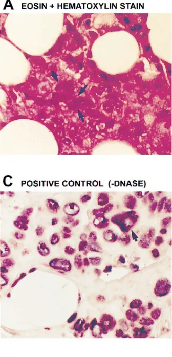
Regional lymph node metastasis of breast carcinoma is a frequent event. Examination of tissue from lymph nodes pro-vided an opportunity to determine whether hyperexpression of MAP kinase persists in the metastatic cells of involved lymph nodes (Fig. 4). Hematoxylin and eosin staining of tissue sections obtained from lymph node metastases of primary breast carcinoma reveals cancer cells (Fig. 4 A, arrow). Per-forming RT-PCR with primers for hepatitis C RNA after prior digestion of the samples with DNAse provided a negative
[image:4.612.57.225.63.412.2]con-trol for in situ analysis, displaying no signal (Fig. 4 B). In the absence of DNAse treatment, amplification of genomic DNA with PCR primers for MAP kinase displayed intense nuclear staining (blue) of all cells, providing the positive control (Fig. 4 C). When in situ RT-PCR was performed using primers for MAP kinase after DNAse digestion, intense staining was ob-served in the metastatic cancer cells, but not in the surround-ing stromal cells within the lymph node (Fig. 4 D). Although not quantitative, results from the in situ RT-PCR showed prominent signal from the cancerous epithelial cells and virtu-ally no signal from surrounding stromal cells, reflecting hyper-expression of MAP kinase mRNA in the cancer cells.
Tissues from primary breast cancer and lymph node me-tastases were subjected to immunohistochemical analysis. An-tibodies specific for MAP kinase were used to stain the tissue and a fast red substrate for the secondary, alkaline phos-phatase–conjugated antibody made visible MAP kinase in pri-mary and metastatic breast cancer (Fig. 5, A and B,
respec-tively). Intense red staining was observed in the cytoplasm of cancerous epithelial cells at both the primary and metastatic sites. By criteria of enzyme activity, immunoblotting, in situ PCR, and immunohistochemical localization, MAP kinase clearly is overexpressed in the epithelial cells of primary breast cancer as well as at a distant metastatic site. Thus,
[image:5.612.61.416.58.432.2]overexpres-Figure 4. MAP kinase mRNA is highly expressed in lymph node me-tastasis of primary breast carcinoma: analysis by in situ RT-PCR. Thin (4-mm) sections of paraffin blocks of lymph node metastasis of primary breast carcinoma tissue were sub-jected to in situ RT-PCR. Tissue was stained with eosin and hematoxylin. Histopathology reveals metastatic cancer cells, as indicated with an ar-row (A). A negative control using PCR primers for an unrelated hepati-tis C viral RNA after DNAse diges-tion (B), a positive control using PCR primers for MAP kinase and genomic DNA without prior DNAse treatment (C), and the test using PCR primers for MAP kinase after treatment with DNAse, followed by RT-PCR (D). Digoxigenin (in the form of dUTP) was used as the re-porter molecule for PCR and an anti-digoxigenin antibody coupled to al-kaline phosphatase used with a chromogen to make the PCR prod-ucts visible (blue staining). In all pan-els, arrows identify representative cancer cells. Bar, 5.0 mm.
[image:5.612.58.429.551.741.2]sion of MAP kinase occurs in human breast cancer cells, at pri-mary and metastatic sites, i.e., an involved lymph node.
Immunoblotting of MAP kinase reveals overexpression in tissue samples of patients with primary breast cancer (Fig. 6 A). Both the 42,000-Mr and 44,000-Mr forms of MAP kinase
were detected in the samples, although in many cases the two
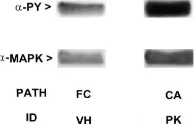
[image:6.612.317.509.60.182.2]forms are not readily apparent, perhaps owing to comigration after differential phosphorylation (Fig. 2). In many instances, for samples from patients with fibrocystic disease as well as from normal patients, MAP kinase expression was not readily detected (Fig. 2), whereas in samples from other patients (Fig. 6 A) MAP kinase was detected. The samples from primary breast cancer, when subjected to immunoprecipitation and then to immunoblotting with an antibody to phosphotyrosine, display increased phosphotyrosine staining (Fig. 6 B). Staining of immunoblots with antibodies specific for the dually phos-phorylated, “active” form of MAP kinase also reveal increased staining in those samples from primary breast cancer patients compared with normal controls and patients with fibrocystic disease (Fig. 6 C). Further immunoblotting analysis of samples from patient VH with fibrocystic disease as compared with that of patient PK with primary breast cancer confirms that not only is the amount of MAP kinase increased, but also its phos-phorylation state is increased in primary breast cancer (Fig. 7). MAP kinase plays a pivotal role in cell proliferation and its activity is regulated by diverse extracellular signals and by products of several protooncogenes(3–6). Reversible protein phosphorylation is the established mechanism of regulation of MAP kinase (3), an activity controlled by a family of dual specificity protein kinases and a complex upstream cascade from tyrosine kinase–linked and G-protein–linked receptors (4). Attenuation in response to chronic stimulation, a para-digm in cell signaling(13), would be expected to dampen MAP kinase activation and expression. Activation of MAP kinase is intimately associated with cell proliferation (14) and constitu-tively active MAP kinase induces oncogenicity when MAP ki-nase kiki-nase is expressed in fibroblasts (15). The first in vivo analysis of MAP kinase activity in human breast cancer, re-ported herein, reveals sharply elevated activities of this key protein kinase. The activation of MAP kinase observed in pri-mary breast carcinoma cannot be ascribed solely to phosphor-ylation of the protein. Immunoblotting, in fact, revealed a marked increase in the amount of MAP kinase in primary breast cancer when compared with normal tissue as well as to tissue from patients suffering from nonmalignant diseases of the breast. Analysis by in situ RT-PCR confirms these data
Figure 6. MAP kinase is overexpressed in primary breast cancer. Samples were prepared as described in the legend to Fig. 2. Samples were immunoprecipitated (IP, B) and/or directly subjected to SDS-PAGE (50 mg/lane) and the resolved proteins were transferred elec-trophoretically to nitrocellulose blots for immunoblot staining (IB, A
and C). For A, immunoblots (IB) were stained for MAP kinase pro-tein with a murine monoclonal antibody against MAP kinase. For B, samples were immunoprecipitated with a rabbit polyclonal anti– MAP kinase antisera (IP) and the immunoprecipitates were sub-jected to SDS-PAGE and immunostaining with an antiphosphoty-rosine antibody (IB). For C, immunoblots (IB) were stained with a rabbit polyclonal antibody specific for the dually phosphorylated (serine and tyrosine), active form of MAP kinase.
[image:6.612.54.299.64.503.2]and clearly establishes the malignant epithelial cells as overex-pressing MAP kinase mRNA. In addition, expression of MAP kinase in the metastatic cells within the involved lymph node was markedly elevated. Hyperexpression of MAP kinase may be a critical event in the initiation/progression of human breast carcinoma and derivative metastases.
Acknowledgments
We acknowledge the invaluable assistance of Dr. Alan Heimann in tissue procurement and of Dr. Roger Grimson for statistical analysis of the data.
This work was supported in part by a grant from the American Cancer Society (to C.C. Malbon), by an award (to V.S. Sivaraman) from the William and Florence Catacosinos Cancer Fund, and by in-stitutional support from the Department of Surgery, University Med-ical Center-SUNY/Stony Brook, the Diabetes and Metabolic Disease Research Program at Stony Brook, and the Center for Advanced Technology (Biotechnology) of the State of New York. This work could not have been accomplished without the generous support from the Department of Pathology and the University Hospital-SUNY/Stony Brook.
References
1. Wingo, P.A., T. Tong, and S. Bolden. 1995. Cancer statistics 1995. CA Cancer J. Clin. 45:8–30.
2. Dickson, R.B., D.S. Salomon, and M.E. Lippman. 1992. Tyrosine kinase-receptor protooncogene interactions in breast cancer. Cancer Treat. Res. 61:
249–273.
3. Blenis, J. 1994. Signal transduction via MAP kinase: proceed at your own RSK. Proc. Natl. Acad. Sci. USA. 90:5889–5892.
4. Cobb, M.H., and E.J. Goldsmith. 1995. How MAP kinases are regulated. J. Biol. Chem. 270:14843–14846.
5. Janes, P.W., R.J. Daly, A. Defazio, and R.J. Sutherland. 1994. Activation of the Ras pathway in breast cancer cells. Oncogene. 9:3601–3608.
6. Callans, L.S., H. Naama, M. Khandelwal, R. Plotkin, and L. Jardin. 1995. Raf-1 expression in human breast cancer cells. Ann. Surg. Oncol. 2:38–42.
7. Arteaga, C.L., M.D. Johnson, G. Todderud, R.J. Coffey, G. Carpenter, and D.L. Page. 1991. Elevated content of tyrosine kinase substrate phospholi-pase C-gamma in human breast carcinomas. Proc. Natl. Acad. Sci. USA. 88: 10435–10439.
8. Gupta, S.K., C. Gallego, G.L. Johnson, and L.E. Heasley. 1992. MAP ki-nase is constitutively active in gip2 and src transformed rat 1a cells. J. Biol. Chem. 267:7987–7990.
9. Moxham, C.P., S.T. George, M.P. Graziano, H.J. Brandwein, and C.C. Malbon. 1986. Mammalian beta1- and beta2-adrenergic receptors, immunologi-cal and structural comparisons. J. Biol. Chem. 261:14562–14570.
10. Nuovo, G.J., A. Forde, P. MacConnell, and R. Farhenwald. 1993. In situ detection of PCR-amplified HIV-1 nucleic acids and tumor necrosis factor cDNA in cervical tissues. Am. J. Pathol. 143:40–48.
11. Nuovo, G.J., P.B. MacConnell, A. Simsir, F. Valea, and D.L. French. 1995. Correlation of the in situ detection of polymerase chain reaction-ampli-fied metalloproteinase complementary DNAs and their inhibitors with progno-sis in cervical carcinoma. Cancer Res. 55:267–275.
12. Nuovo, G.J. 1994. In Situ Hybridization. Protocols and Applications. 2nd Ed. Raven Press, New York.
13. Hadcock, J.H., and C.C. Malbon. 1993. Agonist regulation of gene ex-pression of adrenergic receptors and G-proteins. J. Neurochem. 60:1–9.
14. Brunet, A., J.M. Brondello, G. L’Allemain, P. Lenormand, F. McKen-zie, G. Pages, and J. Pouyssegur. 1995. MAP kinase module: role in the control of cell proliferation. Comptes Rendus Sci. Soc. Biol. 189:43–57.