Programmed cell death in amyotrophic lateral
sclerosis
Christelle Guégan, Serge Przedborski
J Clin Invest.
2003;
111(2)
:153-161.
https://doi.org/10.1172/JCI17610
.
Amyotrophic lateral sclerosis (ALS) is a relentless fatal paralytic disorder confined to the
voluntary motor system (1). Its prevalence is about three to five in 100,000 individuals,
making it the most frequent paralytic disease in adults. Although ALS can strike anyone at
any age, generally the onset of the disease is in the fourth or fifth decade of life. Common
clinical features of ALS include muscle weakness, fasciculations, brisk (or depressed)
reflexes, and extensor plantar responses. Even though motor deficit usually predominates in
the limbs, bulbar enervation can also be severely affected, leading to atrophy of the tongue,
dysphagia, and dysarthria. Other cranial nerves (e.g., oculomotor nerves) are usually
spared. The progressive decline of muscular function results in paralysis, speech and
swallowing disabilities, emotional disturbance, and, ultimately, respiratory failure causing
death among the vast majority of ALS patients within 2–5 years after the onset of the
disease. Pathologically, ALS is characterized by a loss of upper motor neurons in the
cerebral cortex and of the lower motor neurons in the spinal cord. Often, there is also a
profound degeneration of the corticospinal tracts, which is most evident at the level of the
spinal cord. The few remaining motor neurons are generally atrophic, and many
demonstrate abnormal accumulation of neurofilament, in both their cell bodies and axons.
To date, only a […]
Perspective
Find the latest version:
Amyotrophic lateral sclerosis (ALS) is a relentless fatal paralytic disorder confined to the voluntary motor system (1). Its prevalence is about three to five in 100,000 individuals, making it the most frequent par-alytic disease in adults. Although ALS can strike any-one at any age, generally the onset of the disease is in the fourth or fifth decade of life. Common clinical features of ALS include muscle weakness, fascicula-tions, brisk (or depressed) reflexes, and extensor plan-tar responses. Even though motor deficit usually pre-dominates in the limbs, bulbar enervation can also be severely affected, leading to atrophy of the tongue, dysphagia, and dysarthria. Other cranial nerves (e.g., oculomotor nerves) are usually spared. The progres-sive decline of muscular function results in paralysis, speech and swallowing disabilities, emotional distur-bance, and, ultimately, respiratory failure causing death among the vast majority of ALS patients with-in 2–5 years after the onset of the disease. Pathologi-cally, ALS is characterized by a loss of upper motor neurons in the cerebral cortex and of the lower motor neurons in the spinal cord. Often, there is also a pro-found degeneration of the corticospinal tracts, which is most evident at the level of the spinal cord. The few remaining motor neurons are generally atrophic, and many demonstrate abnormal accumulation of neuro-filament, in both their cell bodies and axons. To date, only a few approved treatments (e.g., mechanical ven-tilation and riluzole) prolong survival in ALS patients to some extent. However, the development of more effective neuroprotective therapies remains impeded by our limited knowledge of the actual mechanisms by which neurons die in ALS, and of how the disease progresses and propagates.
ALS, like other common neurodegenerative disorders, is sporadic in the vast majority of patients, and familial in only a few (1). The clinical and pathological expres-sions of ALS are almost indistinguishable between the familial and sporadic forms, although often in the for-mer the age at onset is younger, the course of the disease more rapid, and the survival after diagnosis shorter (1). The cause of sporadic ALS remains unknown, while that of at least some familial forms has been identified (see below). Although the identified gene defects responsible for ALS account for a minute fraction of cases, most experts believe that unraveling the molecu-lar basis by which those mutant gene products cause neurodegeneration may shed light on the etiopatho-genesis of the common sporadic form of ALS.
Genetic forms of ALS
Although familial ALS is often referred to as a single entity, genetic evidence actually reveals at least four dif-ferent types that have been assigned to distinct loci of the human genome (2). This review will focus on a form that is responsible for the disease in approximately 20% of all familial cases and that is linked to mutations in the gene for the cytosolic free radical–scavenging enzyme superoxide dismutase-1 (SOD1) (3, 4). To date, approximately 100 different point mutations in SOD1 throughout the entire gene have been identified in ALS families, and all but one is dominant. Many of these mutations lead to the substitution of an amino acid within regions of the enzyme with very distinct struc-tural and functional roles. It is thus fascinating to note that so many discrete SOD1 alterations share a similar clinical phenotype, even though the disease duration and, to a lesser extent, age at onset vary among patients with different SOD1 mutations (5). Also astonishing is the fact that SOD1 mutations, which are present at birth and, by virtue of SOD1’s ubiquitous expression, in all tissues, produce a rapidly progressive adult-onset degenerative condition in which motor neurons are almost exclusively affected.
Most of these mutations have apparently reduced enzymatic activity (3, 6), a finding that has prompted investigators to test whether a loss of SOD1 activity can kill neurons. It was unequivocally shown that reducing SOD1 activity to about 50% using antisense
Programmed cell death in amyotrophic lateral sclerosis
Christelle Guégan
1,2and Serge Przedborski
1,3,41Department of Neurology, Columbia University, New York, New York, USA
2Institut National de la Santé et de la Recherche Médicale, Unit 421, Institut Mondor de Médecine Moléculaire, Créteil, France 3Department of Pathology, and
4Center of Neurobiology and Behavior, Columbia University, New York, New York, USA
J. Clin. Invest.111:153–161 (2003). doi:10.1172/JCI200317610.
PERSPECTIVE
Neurodegeneration | Serge Przedborski, Series Editor
Address correspondence to:Serge Przedborski, BB-307, Columbia University, 650 West 168th Street, New York, New York 10032, USA. Phone: (212) 305-1540; Fax: (212) 305-5450; E-mail: SP30@columbia.edu.
Conflict of interest:The authors have declared that no conflict of interest exists.
oligonucleotides kills pheochromocytoma-12 (PC-12) cells and motor neurons in spinal cord organotypic cultures (7, 8). However, mutant mice deficient in SOD1 do not develop any motor neuron disease (9), and the transgenic expression of different SOD1 mutants in both mice (10–12) and rats (13) causes an ALS-like syndrome in these animals, whether SOD1 free radical–scavenging catalytic activity is increased, normal, or almost absent (10–14). These observations provide compelling evidence that the cytotoxicity of mutant SOD1 is mediated not by a loss-of-function but rather by a gain-of-function effect (15).
Transgenic mutant SOD1 mouse model of ALS
As indicated above, the transgenic expression of differ-ent SOD1 mutants in both mice (10–12) and rats (13) produces a paralytic syndrome in these animals that replicates the clinical and pathological hallmarks of ALS. The age at onset of symptoms and the lifespan of these transgenic rodents vary among the different lines, depending on the mutation expressed and its level of expression, but when they become symptomatic they invariably show motor abnormalities that progress with the same pattern (11, 16).
The first motor abnormality, at least in mice, is the development of a fine tremor in at least one limb when the animal is held in the air by the tail (16). Thereafter, weakness and atrophy of proximal muscles, predomi-nantly in the hind limbs, develop progressively. At the end stage, transgenic mutant SOD1 mice are severely paralyzed and can no longer feed or drink on their own (16). Neither their nontransgenic littermates nor age-matched transgenic mice expressing wild-type SOD1 enzyme develop any of these motor abnormalities.
The first neuropathological changes seen in trans-genic mutant SOD1 mice are perikarya, axonal and dendritic vacuoles in motor neurons with little involve-ment in the surrounding neuropil, and undetectable neuronal loss or gliosis (11, 17). In the transgenic mutant SOD1 mice that express a glycine-to-alanine substitution at position 93 (G93A) (10), these changes are observed in 4- to 6-week-old asymptomatic animals. By the time the first symptom, fine limb tremor, arises (about 90 days), vacuolization is prominent, and some neuronal loss, especially of large motor neurons (>25
µm), is observed in the spinal cord. At the end stage, dramatic paralysis (about 140 days), there is still some degree of vacuolization, but the prominent features are the dramatic loss of motor neurons (∼50%), an abun-dance of dystrophic neurites, a marked gliosis (18), some globular Lewy body–like intracellular inclusions, and a dearth of motor neurons filled with phosphory-lated neurofilaments (10, 16, 17, 19). Despite the close similarities between the phenotype of transgenic mutant SOD1 rodents and ALS, this experimental model departs from the human disease in a few impor-tant ways. First, vacuolar degeneration has not been a well-recognized component of motor neuron patholo-gy in ALS. Second, neurofilamentous accumulation in cell bodies and proximal axons is infrequent in the lines of transgenic animals that express mutant SOD1, while
it is conspicuous in ALS. Third, none of the transgenic lines show degeneration in the rodent equivalent of the human corticospinal tract. Notably, these transgenic animals replicate in rodents the effect of mutant SOD1, but how relevant this is to the sporadic form of ALS — which is not linked to SOD1 mutations — is unknown. Despite these imperfections and limitations, transgenic mutant SOD1 rodents unquestionably rep-resent an excellent experimental model of ALS, one which has already generated valuable insights into the pathogenesis of ALS and opened new therapeutic avenues for this dreadful disease.
Hypothesis for mutant SOD1 cytotoxicity
Despite the explosion of ALS research engendered by the discovery of the SOD1 mutations, the actual nature of the gained function by which mutant SOD1 kills motor neurons in ALS remains elusive. Multiple mech-anisms have been implicated in the demise of motor neurons in ALS (20), but only a few may be directly rel-evant to the form linked to mutant SOD1. For instance, the known free radical–scavenging function of SOD1 led researchers to believe that mutant SOD1–induced neurodegeneration was due to an oxidative stress. This idea was, at least initially, received with enthusiasm due to the fact that a variety of mark-ers of oxidative damage are indeed increased in ALS spinal cords (20). Currently, it is thought that, if SOD1 mutants were to generate oxidative stress, they could do so by two distinct and not mutually exclusive mech-anisms. In the first mechanism, the point mutations would relax SOD1 conformation, hence allowing abnormal kinds or amounts of substrates to reach and react with the transitional metal — copper — contained in the catalytic site of the enzyme. Among the aberrant substrates to be proposed are peroxynitrite (21) and hydrogen peroxide (22), both of which can directly or indirectly mediate serious tissue damage. In the second mechanism, it is speculated that SOD1 mutations are associated with a labile binding of zinc to the protein (23), and that, by having lost zinc, mutant SOD1, in the presence of nitric oxide, will catalyze the production of peroxynitrite (24), which can inflict serious oxidative damage to virtually all cellular elements.
Alternatively, mutant SOD1 cytotoxicity may result from the propensity of this mutant protein to form intracellular proteinaceous aggregates (25), which are a prominent pathological feature of several of the trans-genic lines (12, 13, 19), and of various cultured cell types expressing mutant SOD1, including motor neurons (26). As in other neurodegenerative disorders with intra-cellular inclusions, whether or not these proteinaceous aggregates are actually noxious remains uncertain. Nev-ertheless, it may be speculated that their presence in the cytosol of motor neurons may be deleterious, by, for example, impairing the microtubule-dependent axonal transport of vital nutriments, or by perturbing the nor-mal turnover of intracellular proteins (27).
SOD1 cDNA were dying by apoptosis (28), a form of programmed cell death (PCD). Similar observations were subsequently made in transfected PC-12 cells (29) and in primary neurons grown from transgenic mice expressing mutant SOD1 (30). Collectively, these in vitro data have led many investigators to consider that mutant SOD1 may kill motor neurons by activating PCD, a term that we here use in the sense of cell death mediated by specific signaling pathways. The possible implication of PCD in ALS has been rather appealing to the field of motor neuron diseases ever since the neuronal apoptosis inhibitory protein (NAIP) was identified as a candidate gene for an inherited ALS-related disorder, spinal muscular atrophy (31). The remainder of this review will focus on PCD in ALS. Because most of the published mechanistic investiga-tions of that topic have been performed in transgenic mutant SOD1 mice, this appraisal will emphasize this mouse model of ALS, but human data will be cited whenever possible to support the relevance of the ani-mal findings to the human condition.
Morphology of dying motor neurons
In light of the presumed proapoptotic properties of mutant SOD1 observed in vitro, it may be wondered whether, in transgenic mutant SOD1 mice, dying spinal cord motor neurons would also exhibit features of apoptosis, whose morphological hallmarks include cytoplasmic and nuclear condensation, compaction of nuclear chromatin into sharply circumscribed masses along the inside of the nuclear membrane, and struc-tural preservation of organelles (at least until the cell is broken into membrane-bound fragments called apoptotic bodies that are phagocytized). This ques-tion has been examined in several careful morpholog-ical studies performed in transgenic mutant SOD1 mice (17, 32–34). In these animals, most of the sick
neurons are atrophic, and their cytoplasm is occupied with vacuoles corresponding to dilated rough ER, Golgi apparatus, and mitochondria (17). From our own ultrastructural studies in these mice (S. Przed-borski, unpublished observations), we can add that many sick neurons have diffusely condensed cyto-plasm and nuclei and irregular shapes. Although the actual type of this cell death remains to be deter-mined, these dying neurons exhibit a rather non-apoptotic morphology with some features reminis-cent of autophagic or cytoplasmic neuronal death (35). Yet, in our experience, definitely apoptotic cells are seen but are rare in the spinal cord of affected transgenic mutant SOD1 mice (Figure 1a). For instance, in end-stage transgenic SOD1G93A mice,
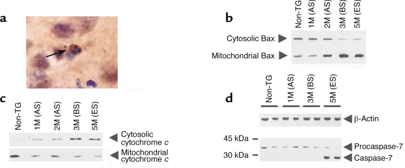
[image:4.576.96.505.489.659.2]which have lost about 50% of their anterior-horn motor neurons, it can be estimated that about two apoptotic cells will be seen per 40-µm-thick section of the lumbar spinal cord. We have also observed that the vast majority of these apoptotic cells no longer exhib-it definexhib-ite morphological characteristics or express phenotypic markers that allow their identification as neurons or glia. However, some (less than 15%) of the spinal cord apoptotic cells are still immunoreactive for specific proteins such as neurofilament or glial fibril-lary acid protein (33), suggesting that both neuronal and glial cells are dying by apoptosis in the mutant SOD1 model (34). In our opinion, the paucity of apoptotic dying motor neurons in this mouse model of ALS reflects the difficulty in detecting these cells by morphological means due to the presumed low daily rate of motor neuron loss (16) and the notoriously rapid disappearance of apoptotic cells. Forms of PCD with morphological features distinct from apoptosis also exist (35–37), making it difficult to exclude the possibility that a nonapoptotic form of PCD underlies mutant SOD1–related cellular degeneration.
Figure 1
Illustrations of PCD alterations in spinal cord of transgenic mutant SOD1 mice. (a) Photomicrograph of definitely apoptotic cells found in the anterior horn of an end-stage transgenic SOD1G93Amouse. The arrow shows several typical round chromatin clumps. (b) Western blot
In human ALS cases, using morphological criteria including size, shape, and aggregates of Nissl substance, Martin (38) has arranged residual spinal cord motor neurons in ALS postmortem samples in three categories that he believes reflect different stages of degeneration. In the chromatolysis stage, motor neurons still resem-ble their normal counterparts except that the cell body appears swollen and round, the Nissl substance dis-persed, and the nucleus eccentrically placed. Some chro-matolytic neurons have prominent cytoplasmic hyaline body inclusions. In the attritional stage, the cytoplasm and the nucleus appear homogenous and condensed, and the cell body appears shrunken and with hazy mul-tipolar shape. In the so-called apoptotic stage, the affect-ed motor neuron is approximately one-fifth of its nor-mal diameter, the cytoplasm and nucleus are extremely condensed, and the cell body adopts a fusiform or round shape devoid of any process. Notably, in none of the three stages do residual motor neurons show appre-ciable cytoplasmic vacuoles or nuclear condensation
accompanied by round chromatin clumps. Taken together, these findings suggest that, while degenerating neurons in both human ALS and its experimental models do exhib-it some features reminiscent of apoptosis, the vast majority of dying cells cannot con-fidently be labeled as typical apoptotic.
Expression of apoptotic markers
[image:5.576.65.330.61.257.2]Besides exhibiting singular morphologi-cal features, apoptotic cells may also show a variety of cellular alterations. The detec-tion of internucleosomal DNA cleavage by either gel electrophoresis or in situ methods has emerged as a popular means of supporting the occurrence of apopto-sis in all sorts of pathological situations, including ALS. However, like many of these apoptotic markers, DNA fragmen-tation detected by in situ methods (e.g., terminal deoxynucleotidyl transferase-mediated nick end labeling) is now well recognized as also occurring in nonapop-totic cell death, including necrosis (35). So the value of DNA cleavage evidenced by in situ techniques as a specific marker of apoptosis may be limited. In addition to this caveat, the search for DNA frag-mentation in ALS postmortem samples has generated conflicting results. In one autopsy study, DNA fragmentation was detected by an in situ method in spinal cord motor neurons in ALS but not in control specimens (39). In two other sim-ilar studies, DNA fragmentation was detected not only in the motor cortex and spinal cord of ALS specimens, but also, though to a lesser degree, in control spec-imens (40, 41). In a subsequent study, internucleosomal DNA fragmentation was detected in affected (e.g., motor cor-tex and spinal cord) but not in spared brain regions (e.g., somatosensory cortex) of ALS cases (38), and, in diseased motor neurons, only at the somatodendrit-ic attrition and apoptotsomatodendrit-ic stages and not at the chro-matolytic stage (38). The author of that study has also documented DNA fragmentation in anterior-horn gray matter of the spinal cord and motor cortex of ALS cases by gel electrophoresis (38), a technique not frequently used in the nervous system to identi-fy apoptosis since, in many neurological situations, it is difficult to obtain samples with a sufficiently high proportion of dying cells. In contrast to all these positive findings, other groups, using similar tech-niques and tissue samples, have failed to provide any evidence of internucleosomal cleavage of DNA in postmortem tissue from human ALS cases or from animal models of the disease (32, 41, 42). Although the actual reason for these divergent results is unclear, they cast doubt on the reliability and even the specificity of such findings.
Figure 2
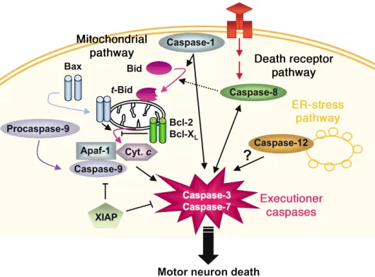
Molecular pathways of PCD. To date, at least three different PCD molecular pathways have been recognized: the mitochondrial pathway (also called intrinsic), the death receptor pathway (also called extrinsic), and the ER pathway. In the mitochondrial PCD pathway, translocation of the proapoptotic protein Bax and the BH3-domain-only protein Bid from the cytosol to the mitochondria promotes cell death by induc-ing the release of cytochrome c(Cyt. c) from the mitochondria to the cytosol; although both full-length and truncated Bid (t-Bid) translocate to the mitochondria, t-Bid is the most biologically active form. Once in the cytosol, cytochrome cactivates caspase-9 in the presence of Apaf-1, which, in turn, activates downstream executioner cas-pases. This pathway can be inhibited by the antiapoptotic protein Bcl-2 and by the protein caspase inhibitor X chromosome–linked inhibitor of apoptosis (XIAP). In the death receptor pathway, caspase-8 is activated by death receptors (members of the TNFR family) in the plasma membrane via the intermediary of adapter proteins. Death receptors include Fas (CD95) and the low-affinity neurotrophin receptor (p75NTR).
Two other apoptotic markers, the LeYantigen (43) and
fractin (44), were also studied in ALS, and here the pic-ture seems less ambiguous. Neither marker was detect-ed in spinal cords of controls, but were highly expressdetect-ed in spinal cords of, respectively, ALS cases (39) and trans-genic SOD1G93Amice (34). Likewise, the levels of the
apoptosis-related protein prostate apoptosis response-4 (45) were increased in spinal cord samples from both ALS patients and transgenic mutant SOD1 mice com-pared with their respective controls (46). Together with the morphological data summarized above, these find-ings support the view that apoptosis occurs in ALS. What all of these studies fail to do, however, is to provide definite mechanistic insights into the significance of these alterations in the pathogenesis of ALS.
Activation of apoptotic molecular pathways
Given the ambiguous results of the morphological studies, it appears that a more convincing approach to evaluating the role of apoptosis in ALS may be to deter-mine whether the neurodegenerative process in trans-genic mutant SOD1 mice, irrespective of the morphol-ogy of the dying cells, involves known molecular mediators of PCD, and whether targeting such key fac-tors can affect the course of the disease.
PCD is a multistep machinery (Figure 2) that involves a complex interaction between survival pathways, acti-vated by trophic factors, and death pathways, actiacti-vated by various stresses. So far, the two pathways that have been most implicated in neuronal survival are the PI3K pathway, which activates Akt (also known as protein kinase B) to suppress the activation of proapoptotic proteins, and the extracellular signal–regulated kinase/MAPK (ERK/MAPK) pathway, which activates antiapoptotic proteins (47).
The best-known PCD-mediating pathways are those involved in the activation of caspase-3. The caspases are a family of cysteine-aspartate proteases (Figure 2; see below for details), many of which are involved in PCD either at the level of upstream signaling (notably cas-pase-8 and caspase-9) or more downstream at the effec-tor level (notably caspase-3). Caspase-8 and caspase-9 both cleave procaspase-3 to activate it. Caspase-9 is acti-vated by a signal derived from mitochondria under the control of the Bcl-2 family of proteins (Figure 2; see below for details). Caspase-8 is activated by death recep-tors (members of the TNF receptor family) in the plas-ma membrane via the intermediary of adapter proteins (48). Death receptors include the low-affinity neu-rotrophin receptor (p75NTR) and Fas (CD95); the latter
seems to participate in the death of embryonic motor neurons in primary cultures (49), but whether Fas con-tributes to the death of mature motor neurons and to the neurodegenerative process in transgenic mice expressing mutant SOD1 remains to be demonstrated. Other key molecules in PCD signaling include ceramide, MAPKs (JNK and p38), and the transcription factors activator protein-1 and NF-κB (47, 48).
In light of the presumed proapoptotic properties of mutant SOD1 (28), it is tempting to suggest that the mutant protein may be a death-signaling molecule in
itself, either directly, by setting in motion the PCD cas-cade, or indirectly, by interacting with a variety of intra-cellular targets such as trophic factors, Bcl-2 family members, or even mitochondria. Mitochondria are a particularly appealing target, because they not only contain mutant SOD1 (50) but are structurally and functionally altered in transgenic mutant SOD1 mice (51, 52), and because they play a pivotal role in PCD (53). Also relevant to the issue of death and survival sig-nals in the mutant SOD1 model are the Western blot and immunohistochemical demonstrations of the weakening surviving signal mediated by PI3K/Akt in spinal cords of transgenic mutant SOD1 mice even before overt neuropathological features arise (54). Once the mutant SOD1–mediated neurodegenerative process has been initiated, several secondary alterations develop in spinal cords of transgenic mutant SOD1 mice, including microglial cell activation (18) and T cell infiltration (55), both of which may release a plethora of cytokines and other pro-PCD mediators. Accord-ingly, while the nature of the initial death signal in transgenic mutant SOD1 mice remains elusive, in a more advanced stage of the disease the increased expression of several extracellular inflammation-relat-ed factors such as IL-1β, IL-6, and TNF-α(56) may amplify the death signals that are already reaching motor neurons in this mouse model of ALS, by activat-ing death receptors such as Fas (49). IL-1βcontent is also elevated in human ALS spinal cords (57).
The role of the Bcl-2 family in motor-neuronal cell death in ALS
The Bcl-2 family, implicated in the regulation of PCD (Figure 2), is composed of both cell-death suppressors such as Bcl-2 and Bcl-XLand promoters such as Bax,
Bad, Bak, and Bcl-xS (58). Many of these molecules are present and active within the nervous system and appear to be potent modulators of neuronal death. In human ALS cases and affected transgenic SOD1G93A
mice, Bcl-2 mRNA content appears significantly decreased and Bax mRNA content significantly increased in the lumbar cord compared with those of controls (59, 60). This is consistent with the finding in both human ALS cases and symptomatic transgenic SOD1G93Amice that the spinal cord expressions of the
antiapoptotic proteins Bcl-2 and Bcl-XL are either
unchanged (40, 61) or decreased (38, 60), whereas that of the proapoptotic Bax and Bad proteins is increased (38, 40, 60). Different SOD1 mutations do not cause exactly the same neuropathology. It is important to note that a very similar pattern of changes of selected pro– and anti–cell death Bcl-2 family members was found in spinal cords of affected transgenic SOD1G86R
mice compared with their wild-type counterparts (62). None of these alterations, however, is seen in young asymptomatic transgenic SOD1G93A mice; but they
expressed mainly in its active homodimeric confor-mation (38, 60). As illustrated in Figure 1b, it is markedly relocated from the cytosol to the mitochon-dria (38, 63); this relocation is, in many cellular set-tings, a prerequisite to the recruitment of the mito-chondria-dependent apoptosis pathway. So it seems that, in ALS, during the neurodegenerative process, the fine-tuned balance between cell-death antagonist and agonist of the Bcl-2 family is upset in favor of pro–cell death forces. In support of this view is the finding that overexpression of Bcl-2, presumably by buffering some of the pro–cell death drive (60), miti-gates neurodegeneration and prolongs survival in transgenic SOD1G93Amice (64); a similar beneficial
effect of Bcl-2 was reported in mutant SOD1–trans-fected PC-12 cells (29).
Other meaningful Bcl-2 family members that appear to be in play in ALS include Bid and Harakiri, two potent pro-PCD peptides, which can participate in the cell death process, either directly or indirectly, by potentiat-ing the effect of Bax. Bid appears to be highly expressed in the spinal cord of transgenic SOD1G93Amice and is
cleaved into its most active form during the progression of the disease (65). Harakiri’s expression has been detect-ed in motor neurons of ALS, but not of control, spinal cord specimens, specifically in spared neurons, of which some exhibited an abnormal morphology reminiscent of that labeled by Troost et al. (61) as apoptotic (66).
The quest to elucidate how Bcl-2 family members are deregulated in ALS is fascinating. As in other patho-logical situations, it is unlikely that mutant SOD1 directly produces the observed changes in Bax. It is more likely that mutant SOD1 ignites intracellular sig-naling pathways, which, in turn, cause Bax upregula-tion and translocaupregula-tion. This scenario would be consis-tent with what we currently know about the regulation of Bax and how Bax is usually brought into action in PCD. The tumor suppressor protein p53 counts among the rare molecules known to regulate Bax expression (67). In normal situations, p53 basal levels in the cell are very low, but upon activation, as seen in pathological situations, there is a rapid rise in p53 mRNA and protein levels, as well as posttranslational modifications that stabilize the protein (68). Activation of the p53 pathway in ALS is evidenced by the demon-stration that p53 is increased in the nuclear fraction of affected brain regions in ALS patients (69), as is p53 immunostaining in neuron nuclei of transgenic SOD1G86Rmice (62). Despite the compelling evidence
that p53 is activated in ALS, two independent studies have failed to provide any supportive data for an actu-al role for this transcriptionactu-al factor in mutant SOD1–mediated neurodegeneration (70, 71).
Caspases in the ALS neurodegenerative process
Caspases are members of a distinct family of cysteine proteases that share the ability to cleave their substrates after specific aspartic acid residues and that are present in cells as inactive zymogens, called procaspases. So far, 14 different mammalian caspases have been identified that differ in primary sequence and substrate specificity.
An instrumental role for caspases in ALS neurodegen-eration is supported by the demonstration that the irreversible broad-caspase inhibitor benzyloxycarbonyl-Val-Ala-Asp(O-methyl)-fluoromethylketone attenuates mutant SOD1–mediated cell death in transfected PC-12 cells (29) and in transgenic SOD1G93Amice (57).
All of the identified caspases are grouped based on their function. One group includes caspases -1, -4, -5, -11, -12, and -14, which are now believed to play a pri-mary role in cytokine maturation. Among these, in ALS, the lion’s share of attention until now has been given to caspase-1, the key enzyme responsible for the activation of IL-1. Procaspase-1 is highly expressed in spinal cord motor neurons, and its activation in the spinal cord of transgenic mutant SOD1 mice coincides with the development of the glial response and with the very beginning of the loss of motor neurons (33, 34, 57, 63, 72). Despite caspase-1’s likely indirect role in PCD, chronic inhibition of caspase-1 by a dominant negative mutant of the enzyme has been proven effective in pro-longing the life of transgenic SOD1G93Amice (73). So
far, the status of the other members of the caspase-1 subfamily in ALS is unknown. Some preliminary inves-tigations show that caspase-12, which is known to be activated following ER stress (74), is expressed in motor neurons of nontransgenic mice, and even more so in those of symptomatic transgenic SOD1G93Amice (C.
Guégan et al., unpublished observations). In sympto-matic transgenic SOD1G93Amice, most of the motor
neurons immunopositive for caspase-12 appear con-densed, shrunken, and vacuolized. Although more work on caspase-12 remains to be done in this model of ALS, our preliminary data argue that sick cells are the site of an ER stress whose occurrence could well contribute to the overall cascade of deleterious events that ultimately underlies the demise of spinal cord motor neurons in the mutant SOD1 model.
By contrast, caspases -2, -3, -6, -7, -8, -9, and -10 have been implicated in apoptosis per se, although their roles can be further divided into “initiator” and “effector.”
Initiator caspases include procaspases -2, -8, -9, and -10, all of which have long prodomains and protein-protein interaction motifs, such as the death-effector domain and the caspase-activation and -recruitment domain, that contribute to the transduction of various signals into proteolytic activity. Procaspase-8 is acti-vated after ligation of certain cell surface receptors, such as the TNF receptors. Interestingly, while signifi-cant glial response and production of IL-1βoccur early in transgenic mutant SOD1 mice (see above), activa-tion of procaspase-8, like inducactiva-tion of TNF-α(56), is only detected in spinal cords near the end stage (65). This suggests that, in this ALS model, the TNF/cas-pase-8 machinery may be a late contributor to the degenerative process. Caspase-2 is another initiator of PCD whose activation occurs in the spinal cord of affected transgenic mutant SOD1 mice (S. Vukosavic et al., unpublished observations). Yet ablation of cas-pase-2 in transgenic SOD1G93Amice has been reported
ALS, it is dispensable. A third caspase initiator is cas-pase-9, whose role is pivotal in the so-called mitochon-dria-dependent PCD pathway (53). Here, after a death stimulus, released mitochondrial cytochrome c inter-acts in the cytosol with apoptotic protease-activating factor-1 (Apaf-1) in the presence of dATP, which stim-ulates the processing of procaspase-9 into its active form, which in turn can activate the downstream exe-cutioner caspases (see below). Evidence of prominent recruitment of this mitochondrial pathway has been documented in spinal cord specimens of both ALS patients and transgenic SOD1G93Amice (63). In that
study, it is shown that, while cytochrome cis confined to the mitochondria in cells in the control samples, it is diffusely distributed in the cytosol in several of the spared cells, especially neurons, in the pathological samples. It is also demonstrated, at least in transgenic mutant SOD1G93A mice, that the mitochondrial
cytochrome ctranslocation to the cytosol occurs at the same time as the cytosolic Bax translocation to the mitochondria and activation of procaspase-9, and before activation of downstream caspase executioners such as procaspase-3 and procaspase-7 (Figure 1, b–d). Because caspase-9 is thought to be so critical in many cell-death settings, it is very likely that the observed translocation of cytochrome cand activation of pro-caspase-9 in ALS represent significant pathological events. Consistent with this view is the finding that pre-vention of mitochondrial cytochrome crelease length-ens the lifespan of transgenic SOD1G93Amice (76).
Effector caspases include procaspases -3, -6, and -7, all of which have short prodomains and lack intrinsic enzymatic activity. However, upon their cleavage, which is triggered by, for example, initiator caspases, effector caspases acquire the capacity to cleave a large number of intracellular substrates, which probably results in the eventual death of the cell. Consistent with this scenario, it has been reported that key effec-tor caspases such as caspase-3 and caspase-7 (see Fig-ure 1d) are indeed activated in spinal cords of trans-genic mutant SOD1 mice in a time-dependent manner that parallels the time course of the neurodegenerative process (33, 34); activation of procaspase-3 has also been observed in spinal cord samples from ALS patients (38). Yet current data on the sequence of events in the PCD cascade indicate that, once effector caspases have been activated, the cell death process, at least in certain pathological settings, has reached a point of no return. This would suggest that, in these specific conditions, the death commitment point is situated upstream of these caspases, and, consequent-ly, interventions aimed at inhibiting these downstream caspases may fail to provide any real neuroprotective benefit (77). Whether this applies to the demise of motor neurons in ALS remains to be determined.
Conclusion
In this review we have described evidence that numer-ous key molecular components of PCD are recruited in ALS. We have also shown that, while precious data on PCD in ALS have been obtained thanks to the
study of postmortem human samples, information regarding the temporal relationships of these changes and their significance in the pathological cascade emanates essentially from the use of transgenic mutant SOD1 mouse models. In light of the above-described PCD-related changes, it would appear that this active form of cell death is not the sole patholog-ical mediator of cell demise in ALS but rather one key component within a coalition of deleterious factors ultimately responsible for the degenerative process. As discussed above, however, the actual relationships between mutant SOD1 and the various other pre-sumed culprits represented by protein aggregates, oxi-dant production, and PCD activation are still unknown, and a better understanding of the patho-genic cascade in ALS will require their elucidation.
In our opinion, one of the most important take-home messages from the body of work summarized above is that an apoptotic morphology should not be used as the sole criterion of whether molecular path-ways of PCD have been recruited. Indeed, we can not stress enough that the PCD molecular pathways may be activated in a neurodegenerative process such as that seen in ALS, even when the prevalent morphology of the dying cells is nonapoptotic. Relatedly, caspase-9 is instrumental in paraptosis (37), a specific morpholog-ical form of nonapoptotic cell death.
Apart from the question of whether the morphology of dying neurons in ALS is apoptotic, but still relevant to our discussion, is the contrast between the paucity of morphologically identified dying cells and the rather robust spinal cord molecular PCD alterations. How can this striking discrepancy be reconciled? First, it is possi-ble that the morphological expression of PCD is much more ephemeral than its molecular translation. There-fore, since in ALS the degenerative process is asynchro-nous, small lasting differences in the expression of these markers may have significant impact on the total num-ber of cells that exhibit a given marker at a given time point. Second, it is also possible that, since apoptotic morphological features are confined to the cell body while PCD molecular alterations may be found not only in cell but also in cell processes, axons, and nerve termi-nals. Thus, the detection of PCD morphology may be much more challenging than the detection of PCD molecular events. Third, the molecular tools used in all of the cited studies see not only the rare cells that are truly dying but also the numerous sick cells that may or may not ultimately die and that thus may or may not show the typical apoptotic morphology.
Whether PCD is also activated in neuron and glial cells in the forms of ALS that are not linked to mutant SOD1 is unknown at this point.
Clearly, the overall mechanism of neurodegeneration in ALS is still incompletely known. Nevertheless, the available evidence indicates that PCD is in play in ALS and thus warrants further investigation of the role of the PCD cascade in ALS pathogenesis and treatment. The most effective therapeutic strategies tested so far in transgenic mutant SOD1 mice target very distinct molecular pathways. We can therefore imagine that, ultimately, the best therapy for ALS will come from a combination of several interventions and not from a single treatment. In keeping with this view, unraveling the sequence of key PCD factors recruited during ALS neurodegeneration should enable us to identify the most significant molecules to be targeted by this ther-apeutic cocktail to produce optimal neuroprotection.
Acknowledgments
The authors wish to thank Robert E. Burke and Miquel Vila for their insightful comments on the manuscript, and Pat White and Brian Jones for their help in its preparation. The authors also acknowledge the support of National Institute of Neurological Disorders and Stroke grants R29 NS37345, RO1 NS38586, NS42269, and P50 NS38370, US Department of Defense grant DAMD 17-99-1-9471, the Lowenstein Foundation, the Lillian Goldman Charitable Trust, the Parkinson’s Dis-ease Foundation, the Muscular Dystrophy Association, the ALS Association, and Project ALS.
1. Rowland, L.P. 1995. Hereditary and acquired motor neuron diseases. In
Merritt’s textbook of neurology. L.P. Rowland, editor. Williams & Wilkins. Philadelphia, Pennsylvania, USA. 742–749.
2. Brown, R.H., Jr. 1995. Amyotrophic lateral sclerosis: recent insights from genetics and transgenic mice. Cell.80:687–692.
3. Deng, H.-X., et al. 1993. Amyotrophic lateral sclerosis and structural defects in Cu,Zn superoxide dismutase. Science.261:1047–1051. 4. Rosen, D.R., et al. 1993. Mutations in Cu/Zn superoxide dismutase gene
are associated with familial amyotrophic lateral sclerosis. Nature. 362:59–62.
5. Cudkowicz, M.E., et al. 1997. Epidemiology of mutations in superoxide dismutase in amyotrophic lateral sclerosis. Ann. Neurol.41:210–221. 6. Przedborski, S., et al. 1996. Blood superoxide dismutase, catalase and
glutathione peroxidase activities in familial and sporadic amyotrophic lateral sclerosis. Neurodegeneration.5:57–64.
7. Troy, C.M., and Shelanski, M.L. 1994. Down-regulation of copper/zinc superoxide dismutase causes apoptotic death in PC12 neuronal cells.
Proc. Natl. Acad. Sci. USA.91:6384–6387.
8. Rothstein, J.D., Bristol, L.A., Hosler, B., Brown, R.H., Jr., and Kuncl, R.W. 1994. Chronic inhibition of superoxide dismutase produces apoptotic death of spinal neurons. Proc. Natl. Acad. Sci. USA.91:4155–4159. 9. Reaume, A.G., et al. 1996. Motor neurons in Cu/Zn superoxide
dismu-tase-deficient mice develop normally but exhibit enhanced cell death after axonal injury. Nat. Genet.13:43–47.
10. Gurney, M.E., et al. 1994. Motor neuron degeneration in mice that express a human Cu, Zn superoxide dismutase mutation. Science.264:1772–1775. 11. Wong, P.C., et al. 1995. An adverse property of a familial ALS-linked SOD1 mutation causes motor neuron disease characterized by vacuolar degeneration of mitochondria. Neuron.14:1105–1116.
12. Bruijn, L.I., et al. 1997. ALS-linked SOD1 mutant G85R mediated dam-age to astrocytes and promotes rapidly progressive disease with SOD1-containing inclusions. Neuron.18:327–338.
13. Nagai, M., et al. 2001. Rats expressing human cytosolic copper-zinc superoxide dismutase transgenes with amyotrophic lateral sclerosis: associated mutations develop motor neuron disease. J. Neurosci. 21:9246–9254.
14. Subramaniam, J.R., et al. 2002. Mutant SOD1 causes motor neuron dis-ease independent of copper chaperone-mediated copper loading. Nat. Neurosci.5:301–307.
15. Brown, R.H., Jr. 1995. Superoxide dismutase in familial amyotrophic lat-eral sclerosis: models for gain of function. Curr. Opin. Neurobiol.5:841–846. 16. Chiu, A.Y., et al. 1995. Age-dependent penetrance of disease in a trans-genic mouse model of familial amyotrophic lateral sclerosis. Mol. Cell. Neurosci.6:349–362.
17. Dal Canto, M.C., and Gurney, M.E. 1995. Neuropathological changes in two lines of mice carrying a transgene for mutant human Cu,Zn SOD, and in mice overexpressing wild type human SOD: a model of familial amyotrophic lateral sclerosis (FALS). Brain Res.676:25–40.
18. Almer, G., Vukosavic, S., Romero, N., and Przedborski, S. 1999. Inducible nitric oxide synthase upregulation in a transgenic mouse model of famil-ial amyotrophic lateral sclerosis. J. Neurochem.72:2415–2425. 19. Tu, P.H., et al. 1996. Transgenic mice carrying a human mutant
super-oxide dismutase transgene develop neuronal cytoskeletal pathology resembling human amyotrophic lateral sclerosis lesions. Proc. Natl. Acad. Sci. USA.93:3155–3160.
20. Cleveland, D.W., and Rothstein, J.D. 2001. From Charcot to Lou Gehrig: deciphering selective motor neuron death in ALS. Nat. Rev. Neurosci. 2:806–819.
21. Beckman, J.S., Carson, M., Smith, C.D., and Koppenol, W.H. 1993. ALS, SOD and peroxynitrite. Nature.364:584.
22. Wiedau-Pazos, M., et al. 1996. Altered reactivity of superoxide dismutase in familial amyotrophic lateral sclerosis. Science.271:515–518. 23. Crow, J.P., Sampson, J.B., Zhuang, Y.X., Thompson, J.A., and Beckman,
J.S. 1997. Decreased zinc affinity of amyotrophic lateral sclerosis-asso-ciated superoxide dismutase mutants leads to enhanced catalysis of tyro-sine nitration by peroxynitrite. J. Neurochem.69:1936–1944.
24. Estevez, A.G., et al. 1999. Induction of nitric oxide-dependent apoptosis in motor neurons by zinc-deficient superoxide dismutase. Science. 286:2498–2500.
25. Bruijn, L.I., et al. 1998. Aggregation and motor neuron toxicity of an ALS-linked SOD1 mutant independent from wild-type SOD1. Science. 281:1851–1854.
26. Durham, H.D., Roy, J., Dong, L., and Figlewicz, D.A. 1997. Aggregation of mutant Cu/Zn superoxide dismutase proteins in a culture model of ALS. J. Neuropathol. Exp. Neurol.56:523–530.
27. Johnson, W.G. 2000. Late-onset neurodegenerative diseases: the role of protein insolubility. J. Anat.196:609–616.
28. Rabizadeh, S., et al. 1995. Mutations associated with amyotrophic later-al sclerosis convert superoxide dismutase from an antiapoptotic gene to a proapoptotic gene: studies in yeast and neural cells. Proc. Natl. Acad. Sci. USA.92:3024–3028.
29. Ghadge, G.D., et al. 1997. Mutant superoxide dismutase-1-linked famil-ial amyotrophic lateral sclerosis: molecular mechanisms of neuronal death and protection. J. Neurosci.17:8756–8766.
30. Mena, M.A., et al. 1997. Effects of wild-type and mutated copper/zinc superoxide dismutase on neuronal survival and L-DOPA-induced toxi-city in postnatal midbrain culture. J. Neurochem.69:21–33.
31. Roy, N., et al. 1995. The gene for neuronal apoptosis inhibitory protein is partially deleted in individuals with spinal muscular atrophy. Cell. 80:167–178.
32. Migheli, A., et al. 1999. Lack of apoptosis in mice with ALS. Nat. Med. 5:966–967.
33. Pasinelli, P., Houseweart, M.K., Brown, R.H., Jr., and Cleveland, D.W. 2000. Caspase-1 and -3 are sequentially activated in motor neuron death in Cu,Zn superoxide dismutase-mediated familial amyotrophic lateral sclerosis. Proc. Natl. Acad. Sci. USA.97:13901–13906.
34. Vukosavic, S., et al. 2000. Delaying caspase activation by Bcl-2: a clue to disease retardation in a transgenic mouse model of amyotrophic lateral sclerosis. J. Neurosci.20:9119–9125.
35. Clarke, P.G.H. 1999. Apoptosis versus necrosis. In Cell death and diseases of the nervous system. V.E. Koliatsos and R.R. Ratan, editors. Humana Press. Totowa, New Jersey, USA. 3–28.
36. Yaginuma, H., et al. 1996. A novel type of programmed neuronal death in the cervical spinal cord of the chick embryo. J. Neurosci.16:3685–3703. 37. Sperandio, S., de Belle, I., and Bredesen, D.E. 2000. An alternative, non-apoptotic form of programmed cell death. Proc. Natl. Acad. Sci. USA. 97:14376–14381.
38. Martin, L.J. 1999. Neuronal death in amyotrophic lateral sclerosis is apoptosis: possible contribution of a programmed cell death mecha-nism. J. Neuropathol. Exp. Neurol.58:459–471.
39. Yoshiyama, Y., Yamada, T., Asanuma, K., and Asahi, T. 1994. Apoptosis related antigen, Le(Y) and nick-end labeling are positive in spinal motor neurons in amyotrophic lateral sclerosis. Acta Neuropathol. (Berl.) 88:207–211.
40. Ekegren, T., Grundstrom, E., Lindholm, D., and Aquilonius, S.M. 1999. Upregulation of Bax protein and increased DNA degradation in ALS spinal cord motor neurons. Acta Neurol. Scand.100:317–321. 41. Migheli, A., Cavalla, P., Marino, S., and Schiffer, D. 1994. A study of
42. He, B.P., and Strong, M.J. 2000. Motor neuronal death in sporadic amy-otrophic lateral sclerosis (ALS) is not apoptotic. A comparative study of ALS and chronic aluminium chloride neurotoxicity in New Zealand white rabbits. Neuropathol. Appl. Neurobiol.26:150–160.
43. Hiraishi, K., Suzuki, K., Hakomori, S., and Adachi, M. 1993. Le(y) anti-gen expression is correlated with apoptosis (programmed cell death).
Glycobiology.3:381–390.
44. Suurmeijer, A.J., van der Wijk, J., van Veldhuisen, D.J., Yang, F., and Cole, G.M. 1999. Fractin immunostaining for the detection of apoptotic cells and apoptotic bodies in formalin-fixed and paraffin-embedded tissue.
Lab. Invest.79:619–620.
45. Rangnekar, V.M. 1998. Apoptosis mediated by a novel leucine zipper protein Par-4. Apoptosis.3:61–66.
46. Pedersen, W.A., Luo, H., Kruman, I., Kasarskis, E., and Mattson, M.P. 2000. The prostate apoptosis response-4 protein participates in motor neuron degeneration in amyotrophic lateral sclerosis. FASEB J. 14:913–924.
47. Harper, S.J., and LoGrasso, P. 2001. Signaling for survival and death in neurones. The role of stress-activated kinases, JNK and p38. Cell. Signal. 13:299–310.
48. Gupta, S. 2001. Molecular steps of death receptor and mitochondrial pathways of apoptosis. Life Sci.69:2957–2964.
49. Raoul, C., et al. 2002. Motoneuron death triggered by a specific pathway downstream of Fas. Potentiation by ALS-Linked SOD1 mutations.
Neuron.35:1067–1083.
50. Higgins, C.M., Jung, C., Ding, H., and Xu, Z. 2002. Mutant Cu, Zn super-oxide dismutase that causes motoneuron degeneration is present in mitochondria in the CNS. J. Neurosci.22:RC215.
51. Kong, J.M., and Xu, Z.S. 1998. Massive mitochondrial degeneration in motor neurons triggers the onset of amyotrophic lateral sclerosis in mice expressing a mutant SOD1. J. Neurosci.18:3241–3250.
52. Browne, S.E., et al. 1998. Metabolic dysfunction in familial, but not spo-radic, amyotrophic lateral sclerosis. J. Neurochem.71:281–287. 53. Kroemer, G., and Reed, J.C. 2000. Mitochondrial control of cell death.
Nat. Med.6:513–519.
54. Warita, H., et al. 2001. Early decrease of survival signal-related proteins in spinal motor neurons of presymptomatic transgenic mice with a mutant SOD1 gene. Apoptosis.6:345–352.
55. Alexianu, M.E., Kozovska, M., and Appel, S.H. 2001. Immune reactivity in a mouse model of familial ALS correlates with disease progression.
Neurology.57:1282–1289.
56. Nguyen, M.D., Julien, J.P., and Rivest, S. 2001. Induction of proinflam-matory molecules in mice with amyotrophic lateral sclerosis: no require-ment for proapoptotic interleukin-1beta in neurodegeneration. Ann. Neurol.50:630–639.
57. Li, M., et al. 2000. Functional role of caspase-1 and caspase-3 in an ALS transgenic mouse model. Science.288:335–339.
58. Chao, D.T., and Korsmeyer, S.J. 1998. BCL-2 family: regulators of cell death. Annu. Rev. Immunol.16:395–419.
59. Mu, X., He, J., Anderson, D.W., Trojanowski, J.Q., and Springer, J.E. 1996.
Altered expression of bcl-2 and bax mRNA in amyotrophic lateral scle-rosis spinal cord motor neurons. Ann. Neurol.40:379–386.
60. Vukosavic, S., Dubois-Dauphin, M., Romero, N., and Przedborski, S. 1999. Bax and Bcl-2 interaction in a transgenic mouse model of familial amyotrophic lateral sclerosis. J. Neurochem.73:2460–2468.
61. Troost, D., Aten, J., Morsink, F., and De Jong, J.M.B.V. 1995. Apoptosis in amyotrophic lateral sclerosis is not restricted to motor neurons. Bcl-2 expression is increased in unaffected post-central gyrus. Neuropathol. Appl. Neurobiol.21:498–504.
62. Gonzalez de Aguilar, J.L., et al. 2000. Alteration of the Bcl-x/Bax ratio in a transgenic mouse model of amyotrophic lateral sclerosis: evidence for the implication of the p53 signaling pathway. Neurobiol. Dis.7:406–415. 63. Guégan, C., Vila, M., Rosoklija, G., Hays, A.P., and Przedborski, S. 2001. Recruitment of the mitochondrial-dependent apoptotic pathway in amyotrophic lateral sclerosis. J. Neurosci.21:6569–6576.
64. Kostic, V., Jackson-Lewis, V., De Bilbao, F., Dubois-Dauphin, M., and Przedborski, S. 1997. Bcl-2: prolonging life in a transgenic mouse model of familial amyotrophic lateral sclerosis. Science.277:559–562. 65. Guégan, C., et al. 2002. Instrumental activation of Bid by caspase-1 in a
transgenic mouse model of ALS. Mol. Cell. Neurosci.20:553–562. 66. Shinoe, T., et al. 2001. Upregulation of the pro-apoptotic BH3-only
pep-tide harakiri in spinal neurons of amyotrophic lateral sclerosis patients.
Neurosci. Lett.313:153–157.
67. Miyashita, T., et al. 1994. Tumor suppressor p53 is a regulator of bcl-2 and bax gene expression in vitro and in vivo. Oncogene.9:1799–1805. 68. Appella, E., and Anderson, C.W. 2001. Post-translational modifications
and activation of p53 by genotoxic stresses. Eur. J. Biochem. 268:2764–2772.
69. Martin, L.J. 2000. p53 is abnormally elevated and active in the CNS of patients with amyotrophic lateral sclerosis. Neurobiol. Dis.7:613–622. 70. Prudlo, J., et al. 2000. Motor neuron cell death in a mouse model of FALS is not mediated by the p53 cell survival regulator. Brain Res. 879:183–187.
71. Kuntz, C., Kinoshita, Y., Beal, M.F., Donehower, L.A., and Morrison, R.S. 2000. Absence of p53: no effect in a transgenic mouse model of familial amyotrophic lateral sclerosis. Exp. Neurol.165:184–190.
72. Pasinelli, P., Borchelt, D.R., Houseweart, M.K., Cleveland, D.W., and Brown, R.H.J. 1998. Caspase-1 is activated in neural cells and tissue with amyotrophic lateral sclerosis-associated mutations in copper-zinc super-oxide dismutase. Proc. Natl. Acad. Sci. USA.95:15763–15768.
73. Friedlander, R.M., Brown, R.H., Gagliardini, V., Wang, J., and Yuan, J. 1997. Inhibition of ICE slows ALS in mice. Nature.388:31.
74. Nakagawa, T., et al. 2000. Caspase-12 mediates endoplasmic-reticulum-specific apoptosis and cytotoxicity by amyloid-beta. Nature.403:98–103. 75. Bergeron, L., et al. 1998. Defects in regulation of apoptosis in
caspase-2-deficient mice. Genes Dev.12:1304–1314.
76. Zhu, S., et al. 2002. Minocycline inhibits cytochrome c release and delays progression of amyotrophic lateral sclerosis in mice. Nature.417:74–78. 77. Zheng, T.S., et al. 2000. Deficiency in caspase-9 or caspase-3 induces