Alan R. Brash
J Clin Invest.
2001;
107(11)
:1339-1345.
https://doi.org/10.1172/JCI13210
.
Arachidonic acid is a slippery molecule that owes its mobility to its four cis double bonds.
These are the source of its flexibility, keeping the pure fatty acid liquid, even at subzero
temperatures, and helping to give mammalian cell membranes their correct fluidity at
physiological temperatures. The double bonds are also the key to the propensity of
arachidonic acid to react with molecular oxygen. This can happen nonenzymatically,
contributing to oxidative stress, or through the actions of three types of oxygenase:
cyclooxygenase (COX), lipoxygenase (LOX), and cytochrome P450. While the products of
these enzymes and of the nonenzymatic transformations have well-substantiated
bioactivities, unchanged arachidonic acid itself has biological activity. This will be a main
focus this review. Before dealing with bioactivity, I will consider the solubility properties of
the molecule, which are crucial to understanding the availability within the cell of
endogenous and exogenous arachidonic acid. I then discuss two controversial issues,
arachidonic acid transport into cells and the accessibility of added arachidonic acid to
endogenous cellular compartments, and finally turn to selected biological actions of this
lipid. The enzymes of arachidonic acid release have been well covered in specialized
reviews and are introduced here only in passing. Physical properties and their relevance to
the distribution of arachidonic acid The sodium salt of arachidonic acid is a soap, the same
[…]
Perspective
Find the latest version:
Arachidonic acid is a slippery molecule that owes its mobility to its four cis double bonds. These are the source of its flexibility, keeping the pure fatty acid liq-uid, even at subzero temperatures, and helping to give mammalian cell membranes their correct fluidity at physiological temperatures. The double bonds are also the key to the propensity of arachidonic acid to react with molecular oxygen. This can happen nonenzymati-cally, contributing to oxidative stress, or through the actions of three types of oxygenase: cyclooxygenase (COX), lipoxygenase (LOX), and cytochrome P450. While the products of these enzymes and of the nonen-zymatic transformations have well-substantiated bioac-tivities, unchanged arachidonic acid itself has biologi-cal activity. This will be a main focus this review. Before dealing with bioactivity, I will consider the solubility properties of the molecule, which are crucial to under-standing the availability within the cell of endogenous and exogenous arachidonic acid. I then discuss two con-troversial issues, arachidonic acid transport into cells and the accessibility of added arachidonic acid to endogenous cellular compartments, and finally turn to selected biological actions of this lipid. The enzymes of arachidonic acid release have been well covered in spe-cialized reviews and are introduced here only in passing.
Physical properties and their relevance to the distribution of arachidonic acid
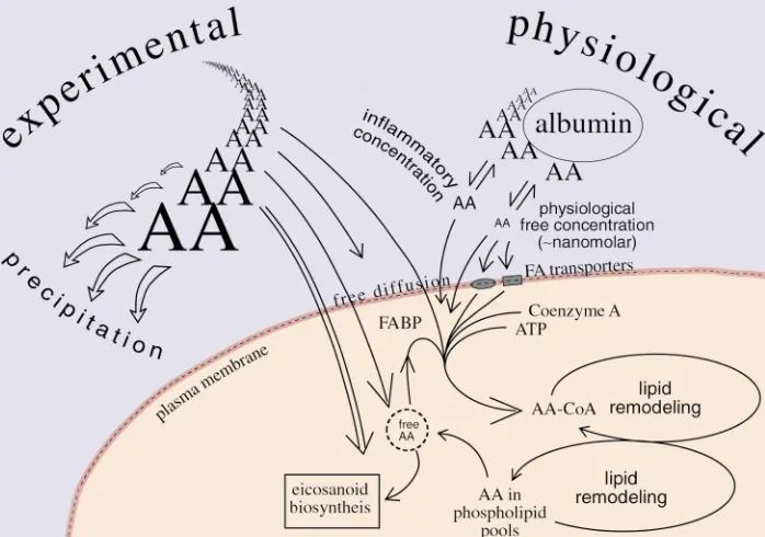
The sodium salt of arachidonic acid is a soap, the same as might be prepared for any other long-chain fatty acid. It can be dissolved fairly readily in aqueous solu-tion. This is in complete contrast to arachidonic free acid, which is an insoluble oil. Interconversion of the salt (ionic) and nonionic forms of arachidonic acid occurs in the range of normal physiological pH. The high pKa of arachidonic acid is crucial, as it sets the solubility properties and the possible distribution of the unesterified fatty acid in cells. It is also central to many of the topics of this review, as most studies of enzymatic transformations and biological activities of arachidonic acid are dependent on addition of exoge-nous arachidonic acid to cells and tissues.
What is the solubility of this added arachidonic acid, and where will it distribute? Cells have hydrophobic (membrane and protein) sites and aqueous/polar sites, so “choices” are available for the different ionic forms of the fatty acid. Like other fatty acids (and certain other membrane components, including acylated
pro-teins and phospholipids), arachidonic acid is amphi-pathic, and its hydrophobic tail can remain in a lipid bilayer while its polar carboxyl group (charged or uncharged) can emerge into the aqueous environment outside the membrane.
Surprisingly, the physical chemistry of solutions of polyunsaturated fatty acids such as linoleic and arachi-donic acids is not completely defined. If arachidonate sodium salt is dissolved in a weakly alkaline solution and then titrated with HCl, the clear solution starts to become cloudy. The observed pKa in the titration, the point of 50% ionization, is noted around pH 8 (as reported for linoleic acid, ref. 1). At this stage, a 1 mil-limolar (0.3 mg/ml) solution of arachidonate Na salt would be almost opaque, as half the molecules are con-verted to the insoluble free acid. Strangely, when the same experiment is carried out with more dilute solu-tions of polyunsaturated fatty acid, the apparent pKa falls towards pH 7 (2). This trend in decreasing pKa implies that at a constant pH (e.g., pH 7.4) the lower the concentration of the fatty acid, the better its solubility. The explanation for this change in apparent pKa may relate to a tendency for the long carbon chains of dif-ferent molecules to bunch together in an aqueous sys-tem, an effect that would be less prevalent at dilute concentrations. This may change the accessibility or reactivity of the carboxyl group to acid and alkali. These properties have practical significance in that they determine the aqueous solubility of arachidonic acid in the concentrations ranges likely to be used in biologi-cal experiments. The ionic environment also influences solubility: for example, the calcium salts of long-chain fatty acids are water insoluble, as clearly evidenced by the appearance of a scum when hard-water (containing CaCO3) is mixed with soap. Similarly, solutions of
arachidonic acid salts will tend to precipitate in the presence of millimolar solutions of calcium ions.
Protein binding can increase the overall concentration of arachidonic acid that can be present in an aqueous environment by effectively decreasing the concentration of free molecules in solution. Albumin, in particular, binds specifically to fatty acids (3). Because of its high concentration in human plasma (35 mg/ml, 0.6 mM), this protein greatly reduces the effective concentration of free fatty acid molecules and permits millimolar con-centrations to be stabilized in an aqueous environment. Similarly, the extracellular fluid has an albumin con-centration of 0.1 mM, and physiologically, this is also
Arachidonic acid as a bioactive molecule
Alan R. Brash
critical to the presentation of low concentrations of the free fatty acid (unbound) to cells. In the presence of albumin, the concentrations of free fatty acids are like-ly kept well below 0.1 µM (3, 4) (Figure 1).
Mechanisms of cellular uptake of exogenous arachidonic acid: the transfer issue
The need for protein-mediated transport of long-chain fatty acids into the cell is hotly debated, and the reviews make interesting reading (e.g., refs. 4–9). One line of argument is that the rates of flip-flop of free fatty acids across lipid bilayers are sufficiently fast that protein transfer mechanisms are not required to explain phys-iological uptake (7, 8). On the other hand, there is evi-dence that several proteins facilitate long-chain fatty acid uptake (e.g., ref. 6). There is also evidence of sat-urability of fatty acid uptake and of competitive inhi-bition, which would appear to suggest that specific fatty acid receptors or transporters are involved. The counter argument is that these findings may be explained by physical limits to the uptake process relat-ed to the partitioning of fatty acids between albumin and the cell membranes (8). Proponents of the fatty acid transport systems concede that simple diffusion may account for a portion of the observed rates of fatty acid uptake (4, 5). It is also agreed that this may become the major mechanism if cells are suddenly flooded with high micromolar concentrations of fatty acids, as could occur under experimental conditions. The proponents of passive diffusion seem prepared to give less ground at this stage, noting that passive transfer is fast enough
to account for observed rates of fatty acid uptake, and going so far as to challenge that “these observations ... make it difficult to imagine why nature would use resources for a purpose not needed” (8).
Transport of long-chain fatty acid definitely does occur in Escherichia coli, where it is tightly linked to CoA-esterification via the acyl CoA synthetase (10). In mam-malian and yeast cells, it appears that the acyl CoA syn-thetase merely enhances uptake indirectly. Formation of the polar CoA ester effectively traps the fatty acid in the cell and thus functions as part of a facilitated dif-fusion process. One of the CoA synthetases (ligases) is specific for arachidonic acid and other C20 fatty acids, although there is no evidence of its specific involve-ment in uptake (11). The membrane proteins implicat-ed in fatty acid uptake in mammalian cells include FAT/CD36 (fatty acid translocase), FATP (fatty acid transport protein) and FABPpm (fatty acid binding protein-plasma membrane) (6). Two of these proteins (FAT and FABPpm) have separate and well-character-ized functions, and FATP has homology to long-chain fatty acid CoA synthetase. FAT is homologous to CD36, a lipoprotein receptor and mediator of platelet aggregation and adhesion (12). FABPpm is a 40 kDa protein with ubiquitous expression that is identical in sequence to mitochondrial aspartate aminotransferase. There is clear evidence, nonetheless, that FABPpm is localized to the plasma membrane and increases fatty acid uptake when expressed in 3T3 fibroblasts (13).
It is relevant to note that the vast literature on the binding and uptake of long-chain fatty acids is
dom-Figure 1
[image:3.576.189.538.116.361.2]inated by an interest in fatty acids as fuel. Hence the best-studied substrates are the saturated fatty acids along with oleate. The research is also directed main-ly at tissues such as heart, liver, and muscle, rather than the epithelial or endothelial cell, fibroblast, leukocyte, and platelet that are the focus of most studies on eicosanoids.
Competition of exogenous arachidonic acid with endogenous stores
The extensive evidence that exogenously added radio-labeled arachidonic acid can be converted to radiola-beled products by COX, P450, and LOX oxygenases still leaves open the question of how effectively the exoge-nous substrate competes with endogeexoge-nous arachido-nate for access to these enzymes, many of which are located on the endoplasmic reticulum or nuclear mem-brane. Measurements of specific activity are required to address this question. Fortunately, mass spectrometry — using deuterated or 14C-labeled arachidonic acid to
introduce a difference in mass between the exogenous and the endogenous substrates — allows for highly pre-cise measurement of specific activity. This approach has been used to follow the fate of arachidonic acid in leukocytes and platelets (14, 15).
In an analysis of 5-LOX transformations of arachi-donic acid in washed suspensions of ionophore-stim-ulated leukocytes (with no extracellular albumin to modify unbound substrate concentration), we found that arachidonic acid at 5 µM was used by 5-LOX about equally with endogenously released substrate (14). At 50 µM exogenous substrate, the natural arachidonic acid release appeared to be suppressed, and nearly all the 5-LOX metabolism used the added substrate (14). Based on our current knowledge of leukotriene biosynthesis, this 5-LOX metabolism occurs on the nuclear membrane (16).
In platelets, the story also has a few interesting twists. Sautebin et al. (15) added varying concentrations of [14C]arachidonic acid to washed platelets (again with no
albumin) and showed that its highest percent utiliza-tion compared with endogenously released arachidon-ic acid corresponded to the lowest concentrations added. This shows that low levels of exogenous arachi-donic acid (1–5 µM) can penetrate into the platelet to the site of COX metabolism and have productive access to the enzyme. At all higher concentrations of added arachidonic acid (10–100 µM), the exogenous substrate accounted for about one-third of the thromboxane syn-thesis. Presumably, the release of arachidonic acid was not stimulated at the lowest levels of added substrate (hence no competition), while the higher levels generat-ed enough thromboxane for a paracrine/autocrine acti-vation of endogenous substrate release.
In complete contrast, in the same experiments these authors showed that the 12-LOX of platelet cytosol used almost exclusively the exogenous substrate over
the whole range of added arachidonic acid concentra-tions (15). This indicates that the 12-LOX did not have access to the arachidonic acid released from internal platelet membranes. These results are supported and extended by studies on substrate utilization by platelets in a more physiological setting. In plasma, collagen-stimulated platelets do not utilize arachidonate from plasma lipids for thromboxane synthesis, although they do use it for 12–hydroxyeicosatetraenoic acid (12-HETE) synthesis by the 12-LOX in the cytosol (17).
In principle, exogenous arachidonic acid might be metabolized by cellular oxygenases either in its original form or following esterification and release. To address the question of whether the added substrate is oxidized directly or after cycling through the membrane lipids, we have again used mass spectrometry (14). The method takes advantage of the fact that when cells are incubat-ed in water labelincubat-ed with the heavy isotope of oxygen, H218O, any fatty acid released from esterified stores will
pick up an 18O label in its carboxyl group:
The 18O label is completely stable under physiological
conditions and under routine conditions of lipid extrac-tion and analysis, including mass spectrometric analy-sis (18). Therefore, by measuring the molecular weight of the free fatty acid (or its prostaglandin or leukotriene products), it is readily apparent whether the product was formed from an ester or from a free fatty acid that had never been esterified. We employed this method to examine the fate of exogenous arachidonic acid during leukotriene biosynthesis in leukocytes (14). We found, as expected, that in the absence of added arachidonic acid the 5-HETE product of the leukocyte 5-LOX con-tained an 18O label. This confirms that stored
arachi-donate ester was hydrolyzed to form the substrate for the LOX. By contrast, when 5 µM or 50 µM deuterated arachidonic acid was added along with the ionophore, the deuterated 5-HETE formed from the exogenous substrate contained only a small percentage of 18O label,
indicating that mainly the added arachidonic acid was used directly, without any prior esterification (14).
This mass spectrometric methodology is technically challenging, but it offers unique insights into the mobilization of fatty acids in cells. It was employed recently to measure the cycling of fatty acids through esterification and release in plant lipid turnover (19).
mass assay. Using a mass per volume calculation, the concentration of esterifiedarachidonate in resting or unactivated human platelets (∼20 µl volume and ∼30 µg arachidonate/109 platelets) corresponds to
approxi-mately 5 mM. This calculated concentration represents an average for whole cells; the actual value in particular subcellular compartments, such as the plasma mem-brane, will no doubt be substantially higher.The release of 1% of this reserve of esterified arachidonate could give up to 50 µM local concentrations prior to its release from the cell. In fact, the percent of platelet arachido-nate stores released on activation is around 10% (20). In the inflamed skin tissue of psoriasis, free arachi-donic acid is abundant, at approximately 30 µg/g (100
µM); in the uninvolved skin it is still the most abun-dant eicosanoid at approximately 4 µg/g (13 µM) (21). Even resting leukocytes contain around 3 pmol/106
cells, or 0.5–1 µM on a per volume basis (22). In isolat-ed islets of Langerhans, by comparison, (taking the vol-ume of the 2,000 cells of an islet as 2 nanoliters), the resting level of arachidonic acid is measured by mass spectrometric assay at about 15 µM (23). The level dou-bles when the cells are stimulated with glucose and increases tenfold with carbacol, each associated with a physiological response postulated to be mediated by the released arachidonic acid (23). More analyses of this type are needed to help substantiate the implication that arachidonic acid is a bioactive molecule. We need more numbers for individual cell types.
The issue of what is a relevant biological concentra-tion of arachidonic acid hits full force upon the assess-ment of bioactivity. Leaving aside the ester and amide derivatives that act on cannabinoid receptors and the multiple activities that depend on transformation by oxygenase enzymes (COX, LOX, and P450), it usually takes at least a 1–10 µM concentration of arachidonic acid to elicit effects on cells. Is 10 µM of added arachi-donic acid too high a concentration? Some activities require 100–300 µM, but at these concentrations,
arachidonic acid may not even be in solution; solubility will be influenced by the pH, the ionic environment, the cell concentration (since high cell numbers equate with more available hydrophobic membranes), and the pres-ence of albumin or other fatty acid binding proteins.
Bioactivities of arachidonic acid
Most of the effects of arachidonic acid are attributable to its conversion by oxygenases (COX, LOX, and P450) to prostaglandins, leukotrienes, and other bioactive prod-ucts. The extent to which this accounts for the essential nature of the essential fatty acids has been recently reviewed (24). In his review, Spector notes that the need for arachidonic acid products is the main reason that the omega-6 fatty acids are essential, while the corresponding need for the omega-3 acids (see Serhan and Oliw, this Per-spective series, ref. 25) is yet to be clarified but may involve an irreplaceable structural role in selected tissues (24).
A limitation in some investigations of arachidonic acid bioactivity is an undue reliance on oxygenase inhibitors in invoking a biological role for the sup-posed “products” of oxygenase metabolism. In fact, it is not uncommon to find cell lines that do not metab-olize added arachidonic acid and that are essentially lacking in the ability to generate oxygenase metabo-lites. In particular, studies that rely on the nonselective antioxidant class of LOX inhibitors can be misleading, suggesting an involvement of LOX enzyme activity when the fatty acids themselves may mediate the bio-logical activity in question.
There are specific G-protein–coupled receptors for lipids such as lysophosphatidic acid and platelet-acti-vating factor, and the bioactions of phorbol ester are said to mimic the role of natural diglycerides. Similar-ly, there are specific receptors for the esters and amides of arachidonate, namely the cannabinoid receptors for 2-arachidonylglycerol and arachidonylethanolamide (anandamide). The biochemistry and pharmacology of these derivatives have been well reviewed, and here I will concentrate on free arachidonic acid.
Like other fatty acids, arachidonic acid will activate the PPAR receptors (26), and by at least one assay they have nanomolar potency (27). Although there is little specificity in the binding of different unsaturated fatty acids, as for other activities considered below, there is the possibility of specificity conferred through specific release of arachidonic acid. There is, nonethe-less, as yet no compelling evidence for the existence of such a specific nuclear action of arachidonic acid. Per-haps arachidonic acid itself is of too common occur-rence in the environment of cells to have permitted its development as a high affinity ligand for a cell surface or nuclear receptor.
Arachidonic acid–dependent activation of NADPH oxidase. The leukocyte NADPH oxidase, which reduces molec-ular oxygen to superoxide, consists of five protein com-ponents. In the resting cell, these subunits reside either in the membrane of intracellular vesicles (gp91phoxand
gp22phox) or as a complex in the cytosol (p40phox, p47phox,
and p67phox) (28). The multicomponent protein
com-plex is assembled together on the plasma membrane only upon cell activation; the cytosolic G-protein Rac also becomes part of the active complex. All long-chain polyunsaturated fatty acids, when added in micromo-lar concentrations to polymorphonuclear leukocytes, can activate this enzyme complex and induce a respira-tory burst (29). The more double bonds in the fatty acid, the more potent the activity. Effective concentra-tions of arachidonic acid begin at around 5–10 µM and a maximal response occurs with 50–100 µM.
acti-vates the NADPH oxidase complex in the leukocyte. One line of supportive evidence indicates a requirement for cPLA2, and, by implication, released arachidonic
acid. Dana et al. (30) prepared stable clones of a human myeloid cell line carrying antisense mRNA for p85 cPLA2. Unlike the parent cells, the transfectants had no
detectable cPLA2, and they failed to release arachidonic
acid in response to phorbol ester or opsonized zymosan. Interestingly, these phospholipase A2-deficient (PLA2
-deficient) cells showed no respiratory burst response to any of several stimuli. Addition of exogenous arachi-donic acid did, however, elicit a normal respiratory burst, indicating that the NADPH complex was capable of activity and strongly suggesting that released arachi-donic acid was the missing component.
How might arachidonic acid be involved in the activ-ity of the NADPH oxidase? Electrophysiological and other lines of evidence implicate its effects on H+
chan-nel activity. The conversion of molecular oxygen to the superoxide anion O2⋅– also generates an equivalent
number of H+ions, which must be eliminated from the
cell by means of an H+channel. If the H+channel is
blocked, the respiratory burst comes to a halt. Hender-son, Chappell, and colleagues have shown that the H+
channel is activated by arachidonic acid (31). The chan-nel appears to be formed from the N-terminal part of the largest protein component of the NADPH oxidase complex, gp91phox(31, 32). In agreement with an effect
of endogenous arachidonic acid, H+channel activity is
lacking in the cPLA2-deficient PLB-985 cells, yet it is
restored upon addition of arachidonic acid in parallel with a rise in NADPH oxidase activity (33).
Arachidonic acid can activate NADPH oxidase by other means as well, at least under cell-free conditions (34). Of particular interest in terms of potential physio-logical relevance is the observation that relatively low concentrations of arachidonic acid (1–5 µM) synergize with phosphorylation of the protein component p47phox
to facilitate the interaction with p22phoxand induce
acti-vation of the oxidase. The role of endogenous arachi-donic acid release in the assembly and activation of the NADPH oxidase remains to be fully characterized.
It is worth noting that enzymes similar to the leuko-cyte NADPH oxidase are detectable in many other tis-sues, albeit at much lower levels of expression (28). Thus, released arachidonic acid might function quite general-ly to activate oxidase metabolism in other cell types.
Other arachidonic acid–sensitive ion channels. There are a plethora of reports showing an action of polyunsatu-rated fatty acids on a diverse array of ion channels. Some recent examples include mechanosensitive K+
channels (35, 36), Ca2+channels (37), gap junctions
(38), and a cation current associated with the dopamine transporter (39). None of these are quite as well developed as the NADPH oxidase story, although distinct examples of inhibition or activation of channel activity are clearly documented.
The TREK and TRAAK channels — the latter having arachidonic acid in the middle of its name — are two members on the twin pore–domain mechanosensitive K channels that are activated by low micromolar levels of unsaturated fatty acids and lysophospholipids (35, 36). TREK stands for TWIK-related K+channel (TWIK
is a twin pore inwardly rectifying K+ channel) and
TRAAK stands for TWIK-related arachidonic acid–stim-ulated K+channel. By contrast, similar concentrations
of unsaturated fatty acids, including arachidonic acid,
inhibitthe ATP-dependent opening of cardiac and neu-ronal G-protein–gated potassium channels (KAch). Kim
and Pleumsamran have argued clearly for the physio-logical relevance of this process (40). Briefly, they have shown that a lipid extractable from cell cytosol has inhibitory activity and that the biological response is mimicked by unsaturated fatty acids; these have activi-ty only when applied on the cytosolic side of the cell membrane. The channel appears to be equally sensitive to oleic (C18.1), linoleic (C18.2), arachidonic (C20.4), and docosahexaenoic (C22.6) acids, and any specificity for arachidonate may stem from its specific release under physiological conditions.
So far, few investigations of the actions of arachidon-ic acid and other fatty acids on ion channels include measurement of their mobilization from membranes, and none report levels of arachidonic or other fatty acids released under natural circumstances. The elec-trophysiological data have established distinct effects whose biological relevance remains to be clarified.
Arachidonic acid and apoptosis. As apoptosis became widely recognized as a controlled process, an early observation related to this discussion noted that arachidonic acid and eicosapentaenoic acids blocked proliferation of the promyelocytic leukemic HL-60 cells, and that this activity, a harbinger of both apop-tosis and necrosis in these experiments, was unrelated to oxygenase metabolism or lipid peroxidation (41). Other results suggested that the protective effect of nonsteroidal anti-inflammatory drug (NSAID) usage on the incidence of colon carcinoma was attributable to accumulation of arachidonic acid, rather than to a reduction in prostaglandin levels (42). A potential mechanism relates to the activation of sphingomyelin hydrolysis by free arachidonic acid and the subsequent induction of apoptosis by ceramide (43).
fatty acids, of which arachidonate is the most abun-dant. Further studies confirmed that blocking CoA-IT led specifically to a loss of PE-arachidonate in favor of PC-arachidonate, without affecting the overall cellular PE/PC ratio (45). The inhibitors also led to a noticeable accumulation of free arachidonic acid in the cells and up to a tenfold increase in free arachidonic acid released into the medium. HL-60 cells intentionally depleted of arachidonic acid by culture over several generations in an arachidonic acid–deficient medium are comparatively resistant to induction of apoptosis by the CoA-IT inhibitors. In line with this observation, addition of exogenous arachidonic acid (10–100 µM) to the medium can prevent proliferation of the HL-60 cells. All the evidence points to the accumulation of arachidonic acid as a signal to halt proliferation and to initiate apoptosis.
Further evidence implicating arachidonic acid levels in apoptosis came from studying a different enzyme involved in the phospholipid remodeling pathways. Blocking the arachidonic acid–utilizing enzyme fatty acid-CoA ligase/synthase 4 in colorectal carcinoma and other cells induces apoptosis, evidently through an accu-mulation of intracellular free arachidonic acid (46). Con-versely, overexpression of the ligase inhibits the apop-totic response. The same effect is produced by overexpression of COX-2, and the evidence suggests it too acts by reducing intracellular arachidonic acid (rather than by producing prostaglandins) (46). These intriguing findings, in accord with a developing story on several fronts, will be strengthened with analysis of the cellular levels of arachidonic acid that are responsible for these effects. Mechanisms of the antiproliferative or proapoptotic actions of arachidonic acid remain to be clarified and may vary among different cell types. They hold the potential basis for several new therapeutic strategies for treating neoplasia (45, 46).
Concluding remarks
The simple structure of arachidonic acid and the natu-ral occurrence of so many close chemical analogues is, not surprisingly, associated with a lack of specificity. The selective actions of free arachidonic acid may be explained simply by its specific release under physio-logical conditions and by the absence of such mecha-nisms for releasing other long-chain fatty acids, com-pounds that might otherwise share its biochemical effects. As arachidonic acid is manipulated and released in all animal cells, a key question with regard to its bioactivity ultimately comes down to one of concen-tration. Just what qualifies as the “real” concentration of free arachidonic acid in the cell is, or at least should be, a matter of controversy. It is not obvious how to adjust the measured concentration in cells for the effec-tive concentration in a membrane, around a protein, or as free fatty acid in true solution. This issue is more acute in relation to biological activities of arachidonic acid than for other lipid mediators, because it takes micromolar levels to elicit most biological activities with arachidonic acid compared with the nanomolar or picomolar levels of the prostaglandins and leukotrienes. It should be emphasized that the arachi-donic acid concentrations in platelets, islet cells, and leukocytes quoted earlier in the review are average cel-lular levels. Undoubtedly the concentrations in certain microenvironments are substantially higher (probably in membranes and surrounding certain proteins), while they are lower in other sites, such as in the cytosol. But this is a secondary issue. Currently there is a pressing need for more analytical data on the overall levels of free arachidonic acid in cells.
Acknowledgments
I thank Gary Bokoch, Floyd Chilton, Garret FitzGerald, Ernst Oliw, Stephen Prescott, and John Turk for infor-mation and helpful comments. This work was sup-ported by NIH grants GM-53638 and AR-45943. Due to space constraints, a number of important references could not be included in this article. Interested readers can find a supplementary reading list at [www.jci.org/cgi/content/full/107/11/1339/DC1].
1. Bild, G.S., Ramadoss, C.S., and Axelrod, B. 1977. Effect of solvent polarity on the activity of soybean lipoxygenase isozymes. Lipids.
12:732–735.
2. Glickman, M.H., and Klinman, J.P. 1995. Nature of rate-limiting steps in the soybean lipoxygenase-1 reaction. Biochemistry.34:14077–14092. 3. Spector, A.A. 1986. Structure and lipid binding properties of serum
albumin. J. Lipid Res.128:320–329.
4. McArthur, M.J., et al. 1999. Cellular uptake and intracellular traffick-ing of long chain fatty acids. J. Lipid Res.40:1371–1383.
5. Berk, P.D., and Stump, D.D. 1999. Mechanisms of cellular uptake of long chain free fatty acids. Mol. Cell. Biochem.192:17–31.
6. Abumrad, N., Coburn, C., and Ibrahimi, A. 1999. Membrane proteins implicated in long-chain fatty acid uptake by mammalian cells: CD36, FATP and FABPm. Biochim. Biophys. Acta.1441:4–13
7. Hamilton, J.A. 1998. Fatty acid transport: difficult or easy? J. Lipid Res.
39:467–481.
8. Zakim, D. 2000. Thermodynamics of fatty acid transfer. J. Membr. Biol.
[image:7.576.66.286.100.195.2]176:101–109.
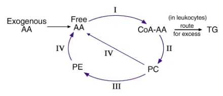
Figure 2
A simplified overview of arachidonate esterification and phospholipid remodeling in cells. The key enzymes are (I) Acyl Coenzyme A synthetase (or ligase), (II) PC/CoA-AA, phosphatidylcholine/CoA arachidonate transacylase, (III) CoA-IT, CoA-independent transacylase, and (IV) iPLA2,
calcium-independent PLA2. Not shown are distinct pools of free
arachi-donic acid that may be generated in subcellular compartments by the actions of other lipases (such as secreted PLA2[sPLA2], cytosolic PLA2
[cPLA2], phospholipases C or D [PLC, PLD]), e.g. on the nuclear
29. Badwey, J.A., Curnutte, J.T., and Karnovsky, M.L. 1981. cis-Polyunsat-urated fatty acids induce high levels of superoxide production by human neutrophils. J. Biol. Chem.256:12640–12643.
30. Dana, R., Leto, T.L., Malech, H.L., and Levy, R. 1998. Essential require-ment of cytosolic phospholipase A2 for activation of the phagocyte NADPH oxidase. J. Biol. Chem.273:441–445.
31. Henderson, L.M., Thomas, S., Banting, G., and Chappell, J.B. 1997. The arachidonate-activatable, NADPH oxidase-associated H+ channel is contained within the multi-membrane-spanning N-terminal region of gp91-phox. Biochem. J.325:701–705.
32. Henderson, L.M., and Meech, R.W. 1999. Evidence that the product of the human X-linked CGD gene, gp91-phox, is a voltage-gated H(+) pathway. J. Gen. Physiol.114:771–786.
33. Lowenthal, A., and Levy, R. 1999. Essential requirement of cytosolic phospholipase A2for activation of the H(+) channel in phagocyte-like
cells. J. Biol. Chem.274:21603–21608.
34. Shiose, A., and Sumimoto, H. 2000. Arachidonic acid and phosphory-lation synergistically induce a conformational change of p47phox to activate the phagocyte NADPH oxidase. J. Biol. Chem.
275:13793–13801.
35. Lesage, F., Maingret, F., and Lazdunski, M. 2000. Cloning and expres-sion of human TRAAK, a polyunsaturated fatty acids-activated and mechano-sensitive K(+) channel. FEBS Lett.471:137–140.
36. Lesage, F., Terrenoire, C., Romey, G., and Lazdunski, M. 2000. Human TREK2, a 2P domain mechano-sensitive K+ channel with multiple reg-ulations by polyunsaturated fatty acids, lysophospholipids, and Gs, Gi, and Gq protein-coupled receptors. J. Biol. Chem.275:28398–28405. 37. Mignen, O., and Shuttleworth, T.J. 2000. I(ARC), a novel
arachidonate-regulated, noncapacitative Ca(2+) entry channel. J. Biol. Chem.
275:9114–9119.
38. Miyachi, E., Kato, C., and Nakaki, T. 1994. Arachidonic acid blocks gap junctions between retinal horizontal cells. Neuroreport.5:485–488. 39. Ingram, S.L., and Amara, S.G. 2000. Arachidonic acid stimulates a
novel cocaine-sensitive cation conductance associated with the human dopamine transporter. J. Neurosci.20:550–557.
40. Kim, D., and Pleumsamran, A. 2000. Cytoplasmic unsaturated free fatty acids inhibit ATP-dependent gating of the G protein-gated K(+) channel. J. Gen. Physiol.115:287–304.
41. Finstad, H.S., et al. 1994. Effect of n-3 and n-6 fatty acids on prolifer-ation and differentiprolifer-ation of promyelocytic leukemic HL-60 cells. Blood.
84:3799–3809.
42. Chan, T.A., Morin, P.J., Vogelstein, B., and Kinzler, K.W. 1998. Mecha-nisms underlying nonsteroidal antiinflammatory drug-mediated apoptosis. Proc. Natl. Acad. Sci. USA.95:681–686.
43. Jayadev, S., Linardic, C.M., and Hannun, Y.A. 1994. Identification of arachidonic acid as a mediator of sphingomyelin hydrolysis in response to tumor necrosis factor alpha. J. Biol. Chem.269:5757–5763. 44. Surette, M.E., Winkler, J.D., Fonteh, A.N., and Chilton, F.H. 1996. Rela-tionship between arachidonate-phospholipid remodeling and apop-tosis. Biochemistry.35:9187–9196.
45. Surette, M.E., Fonteh, A.N., Bernatchez, C., and Chilton, F.H. 1999. Perturbations in the control of cellular arachidonic acid levels block cell growth and induce apoptosis in HL-60 cells. Carcinogenesis.
20:757–763.
46. Cao, Y., Pearman, A.T., Zimmerman, G.A., McIntyre, T.M., and Prescott, S.M. 2000. Intracellular unesterified arachidonic acid signals apoptosis. Proc. Natl. Acad. Sci. USA.97:11280–11285.
9. Kleinfeld, A.M. 2000. Lipid phase fatty acid flip-flop, is it fast enough for cellular transport? J. Membr. Biol.175:79–86.
10. DiRusso, C.C., and Black, P.N. 1999. Long-chain fatty acid transport in bacteria and yeast. Paradigms for defining the mechanism underly-ing this protein-mediated process. Mol. Cell. Biochem.192:41–52. 11. Cao, Y., Traer, E., Zimmerman, G.A., McIntyre, T.M., and Prescott, S.M.
1998. Cloning, expression, and chromosomal localization of human long-chain fatty acid-CoA ligase 4 (FACL4). Genomics.49:327–330. 12. Greenwalt, D.E., et al. 1992. Membrane glycoprotein CD36: a review of
its roles in adherence, signal transduction, and transfusion medicine.
Blood.80:1105–1115.
13. Isola, L.M., et al. 1995. 3T3 fibroblasts transfected with a cDNA for mitochondrial aspartate aminotransferase express plasma membrane fatty acid-binding protein and saturable fatty acid uptake. Proc. Natl. Acad. Sci. USA.92:9866–9870.
14. Brash, A.R., and Ingram, C.D. 1986. Lipoxygenase metabolism of endogenous and exogenous arachidonate in leukocytes: GC-MS analy-ses of incubations in H218O buffers. Prostaglandins Leukot. Med.
23:149–154.
15. Sautebin, L., Caruso, D., Galli, G., and Paoletti, R. 1983. Preferential utilization of endogenous arachidonate by cyclo-oxygenase in incuba-tions of human platelets. FEBS Lett.157:173–178.
16. Peters-Golden, M. 1998. Cell biology of the 5-lipoxygenase pathway.
Am. J. Respir. Crit. Care Med.157:S227–S231, discussion S231–S232, S247–S248.
17. Hwang, D.H. 1982. Characteristics of the formation of the platelet lipoxygenase product from endogenous arachidonic acid. Lipids.
17:845–847.
18. Murphy, R.C., and Clay, K.L. 1990. Preparation of labeled molecules by exchange with oxygen-18 water. Methods Enzymol.193:338–348. 19. Pollard, M., and Ohlrogge, J. 1999. Testing models of fatty acid
trans-fer and lipid synthesis in spinach leaf using in vivo oxygen-18 labeling.
Plant Physiol.121:1217–1226.
20. Neufeld, E.J., and Majerus, P.W. 1983. Arachidonate release and phos-phatidic acid turnover in stimulated human platelets. J. Biol. Chem.
258:2461–2467.
21. Hammarström, S., et al. 1975. Increased concentrations of nonesteri-fied arachidonic acid, 12L-hydroxy-5,8,10,14-eicosatetraenoic acid, prostaglandin E2, and prostaglandin F2αin epidermis of psoriasis. Proc. Natl. Acad. Sci. USA.72:5130–5134.
22. Chilton, F.H., Fonteh, A.N., Surette, M.E., Triggiani, M., and Winkler, J.D. 1996. Control of arachidonate levels within inflammatory cells.
Biochim. Biophys. Acta.1299:1–15.
23. Ramanadham, S., Gross, R., and Turk, J. 1992. Arachidonic acid induces an increase in the cytosolic calcium concentration in single pancreatic islet beta cells. Biochem. Biophys. Res. Commun.184:647–653. 24. Spector, A.A. 1999. Essentiality of fatty acids. Lipids.34(Suppl.):S1–S3. 25. Serhan, C.N., and Oliw, E. 2001. Unorthodox routes to prostanoid formation: new twists in cyclooxygenase-initiated pathways. J. Clin. Invest.In press.
26. Michalik, L., and Wahli, W. 1999. Peroxisome proliferator-activated receptors: three isotypes for a multitude of functions. Curr. Opin. Biotechnol.10:564–570.
27. Lin, Q., Ruuska, S.E., Shaw, N.S., Dong, D., and Noy, N. 1999. Ligand selectivity of the peroxisome proliferator-activated receptor alpha. Bio-chemistry.38:185–190.