Is the class A macrophage scavenger receptor
(SR-A) multifunctional? — The mouse’s tale
Nick Platt, Siamon Gordon
J Clin Invest.
2001;
108(5)
:649-654.
https://doi.org/10.1172/JCI13903
.
The class A macrophage scavenger receptor (SR-A) is the prototypic member of an
expanding family of membrane receptors collectively termed scavenger receptors (SRs) (1–
3). Receptors of this group recognize a number of ligands, including chemically modified or
altered molecules and, in particular, the modified lipoproteins that are pertinent to the
development of vascular disease. As shown in Table 1, all characterized SR-A ligands are
polyanionic, although many polyanions fail to bind SR-A. Here, we wish to summarize and
comment upon the evidence for SR-A being both a multiligand and multifunctional receptor,
considering both the available in vitro data and the results of several studies of SR-A–
deficient mice. Two other SRs, namely SR-B1 (Krieger, ref. 4) and LRP (Herz and
Strickland, ref. 5), are the subject of separate reviews in this Perspective series. Two
themes that can be drawn from studies of SRs as a whole are particularly relevant to this
discussion. First, because these receptors display broad and seemingly overlapping
ligand-binding properties, biological specificity is likely to be determined not only by ligand
structure and the signal transduced following ligand binding, but also by other
considerations. These include the distribution and availability of the various SRs, their
ability to interact with other receptors, and their relative affinities for the various ligands.
Second, the apparent redundancy of ligand binding is achieved […]
Perspective
Find the latest version:
PERSPECTIVE SERIES
Monty Krieger and David M. Stern, Series Editors
Multiligand receptors
The class A macrophage scavenger receptor (SR-A) is the prototypic member of an expanding family of mem-brane receptors collectively termed scavenger receptors (SRs) (1–3). Receptors of this group recognize a number of ligands, including chemically modified or altered mol-ecules and, in particular, the modified lipoproteins that are pertinent to the development of vascular disease. As shown in Table 1, all characterized SR-A ligands are polyanionic, although many polyanions fail to bind SR-A. Here, we wish to summarize and comment upon the evidence for SR-A being both a multiligand and mul-tifunctional receptor, considering both the available in vitro data and the results of several studies of SR-A–defi-cient mice. Two other SRs, namely SR-B1 (Krieger, ref. 4) and LRP (Herz and Strickland, ref. 5), are the subject of separate reviews in this Perspective series.
Two themes that can be drawn from studies of SRs as a whole are particularly relevant to this discussion. First, because these receptors display broad and seemingly over-lapping ligand-binding properties, biological specificity is likely to be determined not only by ligand structure and the signal transduced following ligand binding, but also by other considerations. These include the distribution and availability of the various SRs, their ability to inter-act with other receptors, and their relative affinities for the various ligands. Second, the apparent redundancy of ligand binding is achieved despite the absence of con-served protein sequences among the distinct classes of SRs. A mechanistic understanding of individual SRs’ broad yet specific ligand recognition and of the features shared by these unrelated molecules will require the res-olution of tertiary structures of multiple SRs.
The structural basis of ligand binding by SR-A
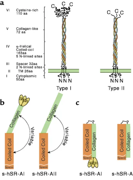
As shown in Figure 1, SR-A is a multidomain trimeric molecule composed of three identical protein chains. The Class A SRs comprise SR-A types I and II, the nonfunc-tional splice variant type III, and a distant receptor called MARCO (2). These molecules each contain a characteris-tic collagenous domain, and binding studies on a series of truncation and point mutant receptors show that the collagenous region of SR-A is required for ligand recog-nition, at least for binding modified lipoproteins such as
oxidized or acetylated LDL (6). This domain has therefore been the focus of mutagenesis approaches to attempt to define the structural basis of broad ligand binding.
The original structure-activity investigation demon-strated that a cluster of four lysines in the most C-ter-minal portion of the receptor is the ligand-binding site, and that within this motif, lysine-337 of bovine SR-A is essential for lipoprotein recognition. These results were interpreted in a computer model prediction of SR-A structure in which these residues formed a positively charged groove responsible for the binding of the nega-tive charges on ligands. A subsequent, more extensive mutagenesis study has shown the contribution of other residues spanning the entire collagen domain, which were revealed when binding assays were performed at 4°C (7). These studies suggested high affinity recogni-tion is dependent upon conformarecogni-tional interacrecogni-tions between distinct domains and the contact of residues on them. This is consistent with a model deduced from the demonstration of a possible hairpin configuration for the receptor, in which the collagen domain is folded back against the α-helical coiled-coil domain, rather than being fully extended (8). Given this structural model,
lig-Is the class A macrophage scavenger receptor (SR-A)
multifunctional? — The mouse’s tale
Nick Platt and Siamon Gordon
Sir William Dunn School of Pathology, University of Oxford, Oxford, United Kingdom
Address correspondence to: Nick Platt, Sir William Dunn School of Pathology, University of Oxford, South Parks Road, Oxford OX1 3RE, United Kingdom. Phone: 44-1865-275533; Fax: 44-1865-275515; E-mail: [email protected].
J. Clin. Invest.108:649–654 (2001). DOI:10.1172/JCI200113903.
Table 1
SR-A ligands
Ligand Non-ligand
Oxidized Low Density Lipoprotein (LDL) Native LDL Acetylated LDL
Oxidized High Density Lipoprotein (HDL) Native HDL Maleylated bovine serum albumin (BSA) BSA Malondialdehyde BSA
Fucoidan Heparin
Dextran Sulfate Chondroitan Sulfate Polyguanylic acid (poly G) Polycytidylic acid (poly C) Polyinosinic acid (poly I) Polyadenylic acid (poly A) Crocidolite asbestos
Silica
Lipopolysaccharide (LPS) Lipoteichoic acid (LTA) Gram negative bacteria Gram positive bacteria Apoptotic cells
and affinities may depend on the regulated folding of the receptor, which would influence the extent of con-tact between domains. The construction of suitable mutants that either promote or prevent flexible articu-lation of the receptor should permit testing of this hypothesis. It would be of interest to examine whether similar binding properties are observed with ligands other than AcLDL, or if configuration has a greater or lesser effect on their recognition. Interestingly, a natu-rally occurring mutation of SR-A in the C57/BL6 mouse strain has recently been described, which has been iden-tified as amino-acid changes in the α-helical coiled-coil domain that affect receptor immunoreactivity, but apparently not function (9, 10). Detailed kinetic studies of this or other sequence variants may reveal important clues to the relationship between ligand binding and the receptor’s conformational state.
SR-A shares with SR-B1 (see Krieger, this Perspective series, ref. 4) an unusual binding property referred to as nonreciprocal cross-competition of ligands (1). This term is used to describe a situation in which ligand A can completely compete for the binding of ligand B, but the latter cannot effectively displace the former. For example, although oxLDL inhibits the binding of acLDL by SR-A totally, acLDL can only block that of oxLDL partially. The structural basis of this pattern of ligand binding is unclear, although it has been inter-preted as reflecting the existence of two discrete but
overlapping ligand binding sites on the receptor. Non-reciprocal interactions have been observed so far only in experiments with cultured cells, but the prospect that they occur in vivo raises exciting possibilities. In partic-ular, such a situation would favor the hierarchical and sequential interactions of the receptor with different lig-ands and thus might account for some of the biological complexity of SR function. For instance, under some circumstances, SR-A might colocalize with a known lig-and but fail to bind it because another liglig-and is bound preferentially, even though the latter interaction may be of similar or lower affinity. In order to define the recep-tor-ligand events fully, it will be necessary to identify all the types of SRs and characterize all the ligands that are present in any given biological context.
With the cloning of the gene for the bovine SR-A, two transcripts were identified encoding different forms of the receptor (1). Identical isoforms have been shown in the subsequent cloning of the murine, human, and rab-bit homologues (11). Type I SR-A is distinguished by the presence of an additional C-terminal region, the SR cys-teine-rich (SRCR) domain. Not only has this domain been conserved in the SR-As of other mammals, it has been found in a number of proteins across different phyla (12). Because it has not so far been shown to influ-ence the binding of modified lipoprotein, the presinflu-ence of SRCR-like motifs is not predictive of SR activity. However, its conservation implies an unidentified func-tion. The potential identification of molecules that bind specifically to the SRCR domain and not the collage-nous region could reveal significant biological interac-tions, but to date, with a few relatively minor excepinterac-tions, the binding properties of type I and type II SR-A are cur-rently considered identical. The recent generation of a mouse lacking type I but retaining type II expression may clarify the role of the SRCR (M. de Winther and M. Hofker, personal communication).
Although other SRs have been identified in inverte-brates, no obvious homologues of SR-A are found in the fully sequenced genomes of Caenorhabditis elegans
and Drosophila melanogaster. Therefore, the evolutionary origin of the SR-A isoforms is uncertain. The gene appears to be common among the vertebrates, as relat-ed expressrelat-ed sequence tags have been reportrelat-ed for zebrafish and Xenopus, but SR-A homologues in more primitive chordate species have not yet been reported.
Analyses of SR-A–deficient mice
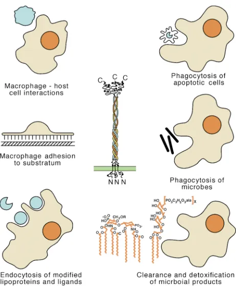
[image:3.576.61.285.50.352.2]Although its activities are well-defined in vitro, the con-tributions of SR-A in vivo have been difficult to deter-mine. The existence of other receptors that display very similar activities and the lack of reagents to effectively dis-tinguish between them, make defining specifics very chal-lenging. Most of the known ligands do not distinguish between receptors, so a detailed comparison of ligand profiles would be needed to infer the identity of a partic-ular SR. Fortunately, specific antibodies can now be used to define the cellular expression, distribution, and abun-dance of the known SRs, and even to suggest previously unrecognized activities for these molecules. The mAb 2F8, which is directed against the murine SR-A, revealed a role for this receptor in macrophage (Mφ) adhesion. We Figure 1
have used this mAb to follow SR-A expression, and we found it widespread among discrete Mφpopulations in vivo, including those in the spleen, thymus, liver, heart, and gut (13, 14). It is restricted to mature Mφand is not expressed by monocytes (N. Platt and S. Gordon, unpub-lished observations). These observations might therefore suggest either a function common to almost all Mφor multiple contributions unique to particular cells.
Unfortunately, because it apparently clears rapidly from the bloodstream, mAb 2F8 did not prove useful as an inhibitor of function in vivo (S. Gordon, et al., unpublished observations). Rather, as is now almost universal for investigations of biological function, the production of mice genetically deficient in SR-A has paved the way for whole-animal experimentation. Phe-notypic analysis of the SR-A–/–mouse currently allows
the most sophisticated investigations into the various roles of SR-A and it is timely to evaluate these studies.
Atherogenesis in SR-A–deficient mice
Despite broadening of the activities with which SR-A is now associated (Figure 2), studies of its involvement in vascular pathologies remain dominant because of the relationship to disease. To begin to evaluate the roles of SR-A, Suzuki and colleagues generated a mouse defi-cient in both isoforms of the receptor (15). Isolated peri-toneal Mφ from these animals showed a significant decrease in the endocytic degradation of acLDL (> 80%) and oxLDL (> 30%). This was in accordance with a series of previous in vitro data that collectively demonstrated SR-A–mediated uptake of modified lipoproteins by Mφ, leading to the formation of lipid-laden foam cells (1). To study the contribution of SR-A to atherosclerosis, Suzu-ki et al. crossed these mice with atherosclerosis-prone apoE-null animals. The resulting doubly deficient mice had moderately increased serum cholesterol and signif-icantly reduced lesion formation (on the order of 60%) compared with apoE-null controls, confirming that SR-A promotes atherosclerosis (15). However, the work in other models has raised doubt about the generality of this finding. On the LDL receptor-deficient back-ground, which promotes susceptibility to diet-induced disease, the absence of SR-A only results in a marginal lesion reduction (16); for mice on the apoE3Leiden transgenic background, which are hypercholesterolemic and susceptible to diet-related disease, atherosclerotic pathology is actually enhanced in the absence of SR-A (17). In a reciprocal experiment, crossing LDL recep-tor–null mice with a transgenic strain that overexpress-es SR-A lead to reduced atherosclerosis (17).
Is it possible to reconcile these apparent discrepancies? If, for the sake of argument, we discount the possibility that the pathologies differ mechanistically in the differ-ent experimdiffer-ental systems, the simplest explanation is that SR-A plays multiple roles in atherogenesis. Some activi-ties, such as the endocytosis of modified lipoproteins, may promote disease, whereas other, currently undefined activities may be antiatherogenic. Subtle differences in disease progression between the various models may therefore involve SR-A at different sites or in different biological contexts. SR-A may also function differential-ly depending on an animal’s genetic background, as has
been suggested from a recent study discussed by Maz-zone (18). Finally, a caveat that applies to knockout stud-ies in general is worth considering here. Since knockout mice lack the targeted gene product not only in the tissue studied, but globally and throughout development, they have ample opportunities to alter the expression of other functionally related genes. Thus, these studies may reveal more about molecular plasticity or the ability of mice to compensate developmentally for the absence of a specif-ic protein than about the role of the protein in normal adult tissues. If such compensatory changes vary among the different mutant and transgenic lines, the different phenotypes seen may not strictly reflect the lack of SR-A, but also the consequences of all the resulting changes in gene expression. Until these issues are resolved, the poten-tial utility of drugs that perturb SR-A activity to prevent vascular disease remains problematic.
Although Kupffer cells and the sinusoidal endotheli-um express SR-A, and isolated cells prepared from
SR-A–/–mice show reduced endocytic activity, the
[image:4.576.307.538.355.643.2]clear-ance of exogenous lipoprotein is identical in livers of wild-type and receptor-null animals (19). Because greater than 90% of the material is removed within 5 minutes of introduction, the capacity of the organ may be too great for detection of the SR-A–dependent component. This apparent excess of general SR activity is a theme that
Figure 2
occurs repeatedly, perhaps because current experimen-tal designs are inadequate. Alternatively, it may be that SR activities are so important, it is essential to have a sur-plus. The identification of new SRs and their manipula-tion in vivo should help resolve these possibilities.
Mφadhesion
Recruitment and retention at relevant tissue sites is a prerequisite for the appropriate Mφ function. This is true for both resident tissue populations and activated cells that migrate towards sites of infection and inflam-mation. SR-A–mediated Mφ adhesion was first demon-strated with the isolation of the 2F8 mAb, which inhibits a cation-independent, serum-dependent component of adhesion (13). SR-A–/– peritoneal cells also display
reduced adhesion and spreading over the first 24 hours after isolation (15), although they subsequently adhere normally. We have detected no obvious absence of any specific tissue Mφpopulations or altered retention of activated cells in either untreated animals or bacillus Cal-mette-Guerin (BCG) granulomatas of SR-A–null mice. van Velzen and colleagues have reported a reduction in Kupffer cell number compared with wild-type (20). How-ever, there is no evidence yet of affected adhesion in vivo or resultant altered liver physiology. Mato’s cells, which are granular epithelial cells of the glial limitans of neu-ral tissues, fail to extend and develop properly in SR-A–/–
mice, perhaps because of impaired adhesion (21). SR-A–dependent adhesion may only be of significance in certain pathological states, when normal molecules are suitably modified to become ligands for SR-A, thus promoting unwanted cell retention. In particular, gly-cated ECM components, which accumulate with age and in conditions including diabetes, serve as adhesive ligands for SR-A in vitro (22). A further link is suggest-ed by the observation of acceleratsuggest-ed atherosclerosis in diabetic individuals, but to date studies of an appropri-ate model formally demonstrating an interaction in vivo have not been reported.
Host defense and innate immunity
The tenet that SR-A is an element of the innate immune system developed principally from two lines of evidence. First, binding studies of SR-A demonstrated that mole-cules derived from certain pathogens, specifically, LPS of Gram negative and lipoteichoic acid of Gram positive bacteria compete for the binding of other known ligands (1, 2), suggesting that SR-A could function as a pattern recognition receptor, activating innate responses to con-served bacterial structures. Second, SR-A is expressed primarily by Mφ, which are among the first line of anti-microbial defense (14). Suzuki et al. originally reported that SR-A–/–mice have impaired protection against
infec-tion by Listeria monocytogenesand herpes simplex virus (15); subsequently, Thomas et al. (23) confirmed increased sensitivity to Staphylococcus aureusbecause of impaired clearance of bacteria. Recently, a defect in the uptake of Neisseria meningitidis in vitro has been shown in
SR-A–/–bone marrow culture–derived Mφ(S. Gordon et
al., unpublished observations). However, the nature of the protective mechanisms provided by SR-A against these very different pathogens is not obvious and could
vary with the microbe. More detailed analyses of the underlying process in infected SR-A–/–mice should be
informative with regard to the diversity of pathogens and surface components that are able to bind to SR-A. A receptor that could both recognize a number of differ-ent infectious agdiffer-ents and engage disparate anti-micro-bial effector systems would be a valuable component of protection against the evolving pressures of pathogens. Uncontrolled cellular activation, a dangerous conse-quence of disseminated infection and the release of microbial components, can lead to toxic shock and sep-ticemia. Haworth et al. (24) tested the in vivo relevance of the data of Hampton and coworkers, which showed that SR-A can mediate clearance and detoxification of plasma endotoxin (25). Following LPS challenge, BCG-primed SR-A deficient mice are more susceptible than similarly treated wild-type mice to endotoxic mortality caused by overproduction of TNF-αand IL-6. Consis-tent with Hampton’s in vitro work, these data suggest that SR-A–dependent uptake of LPS does not activate, and might even suppress, cytokine responses to this bac-terial component. However, recently, the opposite find-ings have been reported, namely that SR-A–/–mice are
more resistant to endotoxic shock (26). Although there are several possible explanations for these contradictory results, one attractive idea, drawn from the different experimental protocols used in the two series of experi-ments, is that the effect of LPS binding to SR-A is deter-mined by the extent of activation of the Mφ. Such sensi-tivity to cellular physiology might add a further dimension to the roles of this multifunctional receptor.
Adaptive responses
As with Mφand other cells of the myelomonocytic line-age, SR-A may function in both the innate and the adap-tive branches of the immune response. Evidence is now emerging for a role for SR-A in antigen presentation. It has been recognized for some time that the acquired immune system can effectively respond to modified anti-gens capable of interacting with SRs (2). Alteration of molecules into structures that can bind SRs can induce responses that may be enhanced in comparison with those elicited by the corresponding native antigen. How-ever, the precise identity of the SRs involved has remained obscure. Nicoletti et al. (27) have recently reported significantly diminished responses to maley-lated albumin in SR-A–/–mice, but at this time we can
only speculate as to the precise mechanism and the cells responsible. However, these findings are potentially use-ful therapeutically, as they may indicate a route for pro-moting antigen delivery within vaccination strategies.
Disorders of the nervous system
mediat-ed by SRs (30), and that microglial adhesion to amyloid fibrils (31) and the consequent production of neurotox-ins are attributable to SR-A. Therefore, SR-A may enhance pathogenesis by helping to establish or to main-tain the characteristic plaques and the deleterious pro-duction of reactive species. On the other hand, the poten-tial for SR-A–dependent endocytic clearance of Aβfrom the brain could slow plaque formation and thus be pro-tective. Lack of the receptor does not lead to any obvious defect in brain development or to signs of pathology in
SR-A–/–animals. Therefore, in order to examine the
con-sequences of the absence of SR-A, Huang et al. (32) bred receptor-deficient mice with a transgenic line that expresses human amyloid precursors (hAPP), in which amyloid is deposited and synaptic degeneration observed. In their experiments, hAPP-transgenic SR-A–/–
animals showed no clear reduction or promotion in plaque accumulation and concomitant degeneration, suggesting that SR-A does not play a unique pathologi-cal role in amyloidosis (32). These observations are more in line with the induction of SR-A by adverse conditions, as was true in a previous study in which only LPS-acti-vated microglia expressed the receptor. Because the hAPP-transgenic mouse model shows AD-associated pathological changes but no other disease symptoms, it may not provide an ideal model in which to evaluate all relevant effects of the absence of SR-A.
Phagocytosis of apoptotic cells
The burgeoning interest in the process of apoptosis has relatively recently extended to the fate of cells commit-ted to die. In vivo, apoptotic or senescent cells are rap-idly phagocytosed, frequently by Mφ, via mechanisms that do not provoke inflammation (33). As discussed by Fadok et al. (this Perspective series, ref. 34), several SRs accounted for the different phagocytic activities. In fact, apoptotic cell recognition may be a universal prop-erty of SRs, because it has been demonstrated for all those receptors that have been examined so far, includ-ing invertebrate ones (3). It is temptinclud-ing therefore, to propose that this is their ancestral function, because of the early evolutionary emergence of the process.
SR-A has been implicated by in vitro experimentation in the recognition of at least two populations of dying cells — apoptotic thymocytes and activated platelets (33). In both cases the anti–SR-A mAb 2F8 significantly inhibits ingestion by activated Mφ. Furthermore, the impaired recognition of apoptotic thymocytes seen in experiments with SR-A–/–thymic Mφis comparable to
that achieved through mAb blockade of wild-type cells. However, detailed scrutiny of thymocyte clearance under normal and enhanced apoptotic cell load failed to reveal an obvious phagocytic deficit in vivo (34). This observa-tion, like the other reports that do not shown pheno-typic alterations in the absence of SR-A, could be due to imprecise methodology that is unable to detect a small or transient difference, or to the ability of other receptors to compensate fully. SR-A may well not be uniquely important for this process in the thymus. More recent work, however, indicates that phagocytosis of apoptotic neutrophils in situ occurs inefficiently in SR-A–deficient mice (N. Platt et al., unpublished observations).
Is SR-A multifunctional?
Given the many disparities between in vitro and in vivo findings and even between different studies in SR-A–/–
mice, it remains likely but is still not proven that this receptor is multifunctional. Others have described knock-out mice generated by homologous recombination as both panacea and frustration, which might apply to
SR-A–/–animals. Although in most cases, studies of
knock-out mice have produced results consistent with what might have been predicted from in vitro activities, some-times overtly similar, but subtly different approaches have yielded conflicting data. Because separate research groups have not performed identical investigations it is not always easy to determine a consensus view. In the case of
SR-A–/–mice, such apparent discrepancies could be
inter-preted as reflecting the biological complexity and multi-ple functions of this receptor. The demonstration of a pro-tective role for SR-A in host defense, with which almost all of the reports are in agreement, offers up a separate dilem-ma: How does SR-A prevent infection by such an array of pathogens? Again, it could be explained by either a single, unifying mechanism or several separate activities.
Why multiple ligands for SR-A?
The most intriguing and defining property of SR-A is its unique pattern of ligand-binding activities. Together with its expression on the ubiquitous and highly versatile Mφ, the diversity of ligands would seem to predict a multitude of functions for this molecule (35). Conversely, the rea-son Mφcan perform multiple tasks is in part because of their expression of molecules such as SR-A. We might wonder whether this strategy of employing a single recep-tor to fulfil several requirements is advantageous com-pared with the situation of having many receptors, each performing more restricted functions. It is not obvious that there is a disadvantage to the latter, because the vast majority of plasma membrane receptors are very narrow in the range of molecules with which they can interact. Typically they have a limited number of ligands. There does not seem to be any sort of theoretical limit on the generation of receptor diversity. Even when inherited material becomes limiting, genetic recombination has facilitated greater variation in specific cases (e.g., immunoglobulins). Interestingly, other receptors that are part of the innate system, such as complement receptors, CD14, toll-like receptors, and the Mφmannose receptor may also interact with a relatively wide range of (nonover-lapping) ligands (2, 36). Therefore, evolutionary pressure may have favored the emergence of membrane molecules with this property, before subsequent surges in receptor diversity, after which they remained sufficiently impor-tant to ensure their conservation.
from the native molecule in part by charge. These mod-ifications, such as oxidation and glycation, may arise from either enzymatic or nonenzymatic reactions (1, 2). Recognition is restricted to specific modifications. While the amino-acid sequence of the native molecule may ultimately limit what can be modified, the extent and forms of modification are potentially quite variable. For instance, minimally and moderately oxidized species of LDL probably interact with separate receptors and induce different biological outcomes.
Since oxidation and the other events occur frequently in tissues, SR ligands are likely to be widespread, although they may be particularly prevalent under con-ditions such as cellular stress, damage, aging, and death. The chemical modifications that render a native mole-cule recognizable by SRs may therefore be seen as mark-ers of unwanted potentially dangerous molecules and cells and direct them for removal and disposal. This may be the common link. Thus, Krieger and colleagues have very entertainingly described SR-A as behaving as “molecular flypaper” (1). Although some of the ligands that have been identified are relevant to specific patholo-gies (or, in the case of microbial products, associated with disease-causing agents) the majority are nonphysi-ological, and there is a dearth of identified endogenous ligands. It is possible that a major function of SR-A has not been analyzed because of our lack of knowledge of the identity of naturally occurring ligands within hosts. The challenge remains to explain the occurrence of other receptors whose activities apparently overlap with SR-A to such a large extent. This may be due to volume of biological need and the requirement for excess capac-ity to avoid potentially harmful situations. We current-ly have much greater knowledge of the shared proper-ties of SRs than of those that are specific to distinct receptors. Continued study of SR-A–/–mice will help us
understand not only what this particular receptor does, but also what it does not do.
Acknowledgments
The authors would like to thank Rosangela da Silva and Hugh Perry for comments and ideas on the manu-script. We apologize to those authors whose work could not be cited as the original publication, owing to space constraints. The authors would like to thank the British Heart Foundation and the Medical Research Council for financial support.
1. Krieger, M., and Herz, J. 1994. Structures and functions of multiligand lipoprotein receptors: macrophage scavenger receptors and LDL receptor-related protein (LRP). Annu. Rev. Biochem. 63:601–637.
2. Pearson, A.M. 1996. Scavenger receptors in innate immunity. Curr. Opin. Immunol. 8:20–28.
3. Platt, N., and Gordon, S. 1998. Scavenger receptors: diverse activities and promiscuous binding of polyanionic ligands. Chem. Biol. 5:R193–R203. 4. Krieger, M. 2001. Scavenger receptor class B type I (SR-BI) is a multiligand
high density lipoprotein receptor that influences diverse physiologic sys-tems. J. Clin. Invest.In press.
5. Herz, J., and Strickland, D.K. 2001. LRP: a multifunctional scavenger and signaling receptor. J. Clin. Invest.In press.
6. Doi, T., et al. 1993. Charged collagen structure mediates the recognition of negatively charged macromolecules by macrophage scavenger receptors. J. Biol. Chem. 268:2126–2133.
7. Andersson, L., and Freeman, M.W. 1998. Functional changes in scavenger receptor binding conformation are induced by charge mutants spanning the entire collagen domain. J. Biol. Chem. 273:19592–19601.
8. Resnick, D., Chatterton, J.E., Schwartz, K., Slayter, H., and Krieger, M. 1996.
Structures of class A macrophage scavenger receptors. Electron microscop-ic study of flexible, multidomain, fibrous proteins and determination of the disulfide bond pattern of the scavenger receptor cysteine-rich domain. J. Biol. Chem. 271:26924–26930.
9. Fortin, A., Penman, M., Stevenson, M.M., Krieger, M., and Gros, P. 2000. Identification and characterisation of naturally occurring variants of the macrophage scavenger receptor (SR-A). Mamm. Genome. 11:779–785. 10. Daugherty, A., Whitman, S.C., Block, A.E., and Rateri, D.L. 2000.
Polymor-phism of class A scavenger receptors in C57BL/6 mice. J. Lipid Res.
41:1568–1577.
11. Freeman, M., et al. 1990. An ancient, highly conserved family of cysteine-rich protein domains revealed by cloning type I and type II murine macrophage scavenger receptors. Proc. Natl. Acad. Sci. USA. 87:8810–8814.
12. Resnick, D., Pearson, A., and Krieger, M. 1994. The SRCR superfamily: a fam-ily reminiscent of the Ig superfamfam-ily. Trends Biochem. Sci. 19:5–8. 13. Fraser, I., Hughes, D., and Gordon, S. 1993. Divalent cation-independent
macrophage adhesion inhibited by monoclonal antibody to murine scav-enger receptor. Nature. 364:343–346.
14. Hughes, D.A., Fraser, I.P., and Gordon, S. 1995. Murine macrophage scav-enger receptor: in vivo expression and function as receptor for macrophage adhesion in lymphoid and non-lymphoid organs. Eur. J. Immunol.
25:466–473.
15. Suzuki, H., et al. 1997. A role for macrophage scavenger receptors in ather-osclerosis and susceptibility to infection. Nature. 386:292–296.
16. Sakaguchi, H., et al. 1998. Role of macrophage scavenger receptors in diet-induced atherosclerosis in mice. Lab. Invest. 78:423–434.
17. de Winther, M.P., van Dijk, K.W., Havekes, L.M., and Hofker, M.H. 2000. Macrophage scavenger receptor class A: a multifunctional receptor in ath-erosclerosis. Arterioscler. Thromb. Vasc. Biol. 20:290–297.
18. Mazzone, T. 2000. Scavenger receptors in atherosclerosis. New answers, new questions. Arterioscler. Thromb. Vasc. Biol.20:2506–2508.
19. Ling, W., et al. 1997. Oxidized or acetylated low density lipoproteins are rap-idly cleared by the liver in mice with disruption of the scavenger receptor class A type I/II gene. J. Clin. Invest. 100:244–252.
20. van Velzen, A.G., Suzuki, H., Kodama, T., and van Berkel, T.J. 1999. The role of scavenger receptor class A in the adhesion of cells is dependent on cell type and cellular activation state. Exp. Cell Res. 250:264–271.
21. Mato, M., Ookawara, S., and Sakamoto, A. 1997. Growth retardation of Mato’s fluorescent granular perithelial (FGP) cells in scavenger receptor knockout (SRKO) mice. Anat. Rec. 247:307–316.
22. El Khoury, J., et al. 1994. Macrophages adhere to glucose-modified basement membrane collagen IV via their scavenger receptors. J. Biol. Chem.
269:10197–10200.
23. Thomas, C.A., et al. 2000. Protection from lethal gram-positive infection by macrophage scavenger receptor-dependent phagocytosis. J. Exp. Med.
191:147–156.
24. Haworth, R., et al. 1997. The macrophage scavenger receptor type A is expressed by activated macrophages and protects the host against lethal endotoxic shock. J. Exp. Med. 186:1431–1439.
25. Hampton, R.Y., Golenbock, D.T., Penman, M., Krieger, M., and Raetz, C.R. 1991. Recognition and plasma clearance of endotoxin by scavenger recep-tors. Nature. 352:342–344.
26. Kobayashi, Y., et al. 2000. Role of macrophage scavenger receptor in endo-toxin shock. J. Pathol.192:263–272.
27. Nicoletti, A., et al. 1999. The macrophage scavenger receptor type A directs modified proteins to antigen presentation. Eur. J. Immunol. 29:512–521. 28. Bell, M.D., et al. 1994. Upregulation of the macrophage scavenger receptor
in response to different forms of injury in the CNS. J. Neurocytol. 23:605–613. 29. Christie, R.H., Freeman, M., and Hyman, B.T. 1996. Expression of the macrophage scavenger receptor, a multifunctional lipoprotein receptor, in microglia associated with senile plaques in Alzheimer’s disease. Am. J. Pathol.
148:399–403.
30. Paresce, D.M., Ghosh, R.N., and Maxfield, F.R. 1996. Microglial cells inter-nalize aggregates of the Alzheimer’s disease amyloid beta-protein via a scav-enger receptor. Neuron. 17:553–565.
31. El Khoury, J., et al. 1996. Scavenger receptor-mediated adhesion of microglia to beta-amyloid fibrils. Nature. 382:716–719.
32. Huang, F., et al. 1999. Elimination of the class A scavenger receptor does not affect amyloid plaque formation or neurodegeneration in transgenic mice expressing human amyloid protein precursors. Am. J. Pathol. 155:1741–1747. 33. Platt, N., da Silva, R.P., and Gordon, S. 1998. Recognizing death: the
phago-cytosis of apoptotic cells. Trends Cell Biol. 8:365–372.
34. Fadok, V.A., Bratton, D.L., and Henson, P.M. 2001. Phagocyte receptors for apoptotic cells: recognition, uptake, and consequences. J. Clin. Invest.In press. 35. Platt, N., Suzuki, H., Kodama, T., and Gordon, S. 2000. Apoptotic thymo-cyte clearance in scavenger receptor class A-deficient mice is apparently nor-mal. J. Immunol. 164:4861–4867.
36. Aderem, A., and Ulevitch, R.J. 2000. Toll-like receptors in the induction of the innate immune response. Nature. 406:782–787.