CD28 costimulation independence of target
organ versus circulating memory
antigen-specific CD4
+
T cells
Andrew P. Fontenot, … , Lee S. Newman, Brian L. Kotzin
J Clin Invest.
2003;
112(5)
:776-784.
https://doi.org/10.1172/JCI18317
.
T cell receptor engagement with CD28 costimulation is generally required for naive T cell
activation, whereas reactivation of memory cells is less dependent on CD28 costimulation.
We studied this process in chronic beryllium disease, in which the frequency of
antigen-specific CD4
+T cells in the lung is large and circulating antigen-specific cells are also
detectable. In the lung, a large fraction of CD4
+T cells stopped expressing CD28 mRNA
and protein, and this change in phenotype correlated with lung inflammation. In the
presence of concentrations of CTLA-4Ig that inhibited the CD28-B7 interaction,
beryllium-specific CD4
+T cells in lung were still able to proliferate and secrete IFN-
g
in response to
beryllium in culture. This functional independence of CD28 costimulation included lung
CD28
+effector cells. Although lung CD4
+CD28
–cells retained the ability to secrete
Th1-type cytokines in response to beryllium, they showed less proliferative capacity and were
more susceptible to cell death compared with CD28
+T cells. In contrast to lung cells,
inhibition of the CD28-B7 interaction markedly reduced responses of beryllium-specific T
cells in blood. Taken together, these findings suggest transition within memory CD4
+T cells
from CD28 dependence in central memory cells to functional independence and then loss
of CD28 expression in effector cells.
Article
Aging
Find the latest version:
Introduction
The differentiation of naive T cells into memory T cells is a crucial step in the evolution of an immune response. Optimal activation of naive T cells requires two signaling events (1, 2). Signal one is mediated by T cell receptor (TCR) engagement with antigen and MHC molecule on APCs. Signal two, termed costimulation, is most frequently mediated by the engagement of the CD28 molecule on the T cell with its ligands, B7-1 or B7-2, on the APC. In the absence of costimulation, engagement of TCR on naive T cells can result in an unresponsive state of clonal anergy (3, 4), and this clon-al inactivation is believed to play an important role in the maintenance of peripheral T cell tolerance (5). In contrast to naive T cells, memory T cells can proliferate and secrete cytokines after TCR engagement without CD28-mediated costimulation (6–8).
Previous studies in our laboratory have demonstrated that patients with chronic beryllium disease (CBD) accu-mulate large numbers of beryllium-specific CD4+T cells in the lung (9), and this disease provides an opportunity to study the scope of antigen-specific memory cells in a human disease. Because of its desirable chemical and physical properties, beryllium continues to be used in many industries, and exposure to beryllium in the work-place remains an important public health concern (10, 11). Of the greater than one million individuals at risk for developing CBD, the disorder will develop in 1–16%, depending on the nature of the exposure and the genetic susceptibility of the individual (10–16). CBD is charac-terized by the presence of granulomatous inflammation in the lung, and this disease is clinically and pathologi-cally similar to a more common idiopathic disease, sar-coidosis (17, 18). Considerable evidence indicates that the accumulation and activation of beryllium-specific CD4+ T cells in the lung is central to the pathogenesis of disease (9, 10, 19, 20). The ability of peripheral blood and/or bronchoalveolar lavage (BAL) CD4+T cells to proliferate in the presence of beryllium salts in vitro forms part of the current disease definition of CBD (11, 13, 14, 19, 21, 22). In the lung, the large majority of beryllium-specific CD4+T cells express an effector-memory phenotype and demonstrate immediate release of Th1-type cytokines when stimulated with beryllium in culture (9, 23). The known identity of antigen and high frequency of antigen-specific cells distinguish CBD from other human condi-tions in terms of ability to study T cell responses.
CD28 costimulation independence of target organ versus
circulating memory antigen-specific CD4
+T cells
Andrew P. Fontenot,
1,2Laia Gharavi,
1Sean R. Bennett,
1,2Scott J. Canavera,
1Lee S. Newman,
1,3,4and Brian L. Kotzin
1,2,41Department of Medicine, 2Department of Immunology, and
3Department of Preventive Medicine and Biometrics, University of Colorado Health Sciences Center, Denver, Colorado, USA 4Department of Medicine, National Jewish Medical and Research Center, Denver, Colorado, USA
T cell receptor engagement with CD28 costimulation is generally required for naive T cell activation, whereas reactivation of memory cells is less dependent on CD28 costimulation. We studied this process in chronic beryllium disease, in which the frequency of antigen-specific CD4+T cells in the
lung is large and circulating antigen-specific cells are also detectable. In the lung, a large fraction of CD4+T cells stopped expressing CD28 mRNA and protein, and this change in phenotype correlated
with lung inflammation. In the presence of concentrations of CTLA-4Ig that inhibited the CD28-B7 interaction, beryllium-specific CD4+T cells in lung were still able to proliferate and secrete IFN-γin
response to beryllium in culture. This functional independence of CD28 costimulation included lung CD28+effector cells. Although lung CD4+CD28–cells retained the ability to secrete Th1-type cytokines
in response to beryllium, they showed less proliferative capacity and were more susceptible to cell death compared with CD28+T cells. In contrast to lung cells, inhibition of the CD28-B7 interaction
markedly reduced responses of beryllium-specific T cells in blood. Taken together, these findings suggest transition within memory CD4+T cells from CD28 dependence in central memory cells to
functional independence and then loss of CD28 expression in effector cells.
J. Clin. Invest.112:776–784 (2003). doi:10.1172/JCI200318317.
Received for publication March 10, 2003, and accepted in revised form June 10, 2003.
Address correspondence to: Andrew P. Fontenot, Division of Clinical Immunology (B164), University of Colorado Health Sciences Center, 4200 East Ninth Avenue, Denver, Colorado 80262, USA. Phone: (303) 315-7601; Fax: (303) 315-7642; E-mail: [email protected].
Conflict of interest:The authors have declared that no conflict of interest exists.
Here, we examined the role of CD28-mediated costim-ulation in antigen-specific T cell activation and survival. The results demonstrate an apparent evolution of inde-pendence from CD28-mediated costimulation that cor-relates with memory cell differentiation. Memory CD4+T cells in blood continued to require CD28 costimulation for proliferative and cytokine responses to beryllium. In the lung, proliferation and secretion of Th1-type cytokines by effector memory cells were functionally independent of CD28 costimulation, and a proportion of these cells stopped expressing CD28. These CD4+CD28– T cells showed decreased proliferative capacity and an increased rate of apoptosis after stimulation with antigen, suggest-ing transition to a presenescent state.
Methods
Study population. Twelve patients with a diagnosis of CBD were enrolled in this study. The diagnosis of CBD was established using the following criteria: a history of beryl-lium exposure, the presence of granulomatous inflam-mation found as a result of a lung biopsy, and a positive proliferative response of blood and/or BAL T cells to BeSO4in vitro (10, 11). A group of four sarcoidosis pa-tients and nine healthy subjects served as negative control subjects. Informed consent was obtained from each CBD patient and control subject, and the protocol was approved by the Human Subject Institutional Review Boards at the University of Colorado Health Sciences Cen-ter and National Jewish Medical and Research CenCen-ter.
Immunofluorescence staining and analysis of CD28 gene expression. PBMCs were isolated from heparinized blood by Ficoll-Hypaque (Amersham Pharmacia Biotech, Piscataway, New Jersey, USA) density-gradient separation, and BAL was performed as described previ-ously (12, 24–26). PBMCs and BAL cells were stained with mAb’s to CD4 and CD28 (both from Becton Dick-inson Immunocytometry Systems, San Jose, California, USA). A subset of the BAL cells was labeled with CFSE as described (27). The lymphocyte population was iden-tified using forward and 90-degree light-scatter pat-terns, and fluorescence intensity was analyzed using an FACScalibur cytometer (Becton Dickinson Immuno-cytometry Systems) as described previously (9).
In some experiments, CD28 gene expression was also analyzed by RT-PCR. BAL cells were stained for CD4 and CD28, and CD4+CD28+and CD4+CD28–cells were sort-ed using a MoFlo cell sorter (Cytomation Inc., Fort Collins, Colorado, USA). Equal numbers of cells of each subset were isolated and immediately frozen prior to preparation of total cellular RNA and cDNA. Sterile water and cDNA from a normal subject served as nega-tive and posinega-tive controls for the RT-PCR, respecnega-tively. PCR amplification of a CD28 gene fragment was accom-plished with the forward primer, 5′- AACAAGATTTTGGT-GAAGCAGTCGCCC-3′, and the reverse primer, 5′- GTTCAT-GTAGTCACTGTGCAGGAGCCT-3′, as described (28). As a control for an equal number of cells, a TCR BV3S1gene segment was also amplified as described (28). PCR prod-ucts were visualized electrophoretically on agarose gels.
Cell culture and immunofluorescence analysis for intracel-lular cytokine expression. Proliferation assays were per-formed as described previously (9, 12). In brief, BAL cells (105cells/well) and PBMCs (2.5 ×10⁵cells/well) were cultured in 96-well flat-bottom microtiter plates in the presence of 5 µg/ml PHA for 48 hours or 10–5M BeSO4with or without the addition of various con-centrations of CTLA-4Ig (R&D Systems Inc., Min-neapolis, Minnesota, USA) for 4 days. During the last 18 hours, the wells were pulsed with 1 µCi of [3H] thymidine, and incorporation of radioactivity was determined by β-emission spectroscopy. Proliferation assays were performed in triplicate.
For stimulation of cytokine secretion, BAL cells (2.5 ×10⁵ to 5 ×10⁵cells) or PBMCs (106cells) were placed in 12 ×75-mm polypropylene tubes (Fisher Scientific Co., Pittsburgh, Pennsylvania, USA) containing 1 ml of RPMI-1640 supplemented with 10% heat-inactivated human serum (Gemini Biological Products, Woodland, California, USA) and one of the following experimental conditions: medium alone, 10 ng/ml staphylococcal enterotoxin B (SEB), or 10–5M BeSO4 with or without CTLA-4Ig. Cells were incubated for a total of 6 hours with 10 µg/ml brefeldin A added after the first hour of stimulation (9). After stimulation, cells were washed and stained with mAb’s directed against CD4, CD8, and CD28 (all from Becton Dickinson Immunocytometry Systems). Cells were then washed with PBS containing 1% BSA and placed in fixation medium (Caltag Labora-tories Inc., Burlingame, California, USA) for 15 minutes at room temperature. Following washing with PBS con-taining 1% BSA, cells were added to permeabilization medium (Caltag Laboratories Inc.) and stained with a mAb directed against IFN-γor IL-2 (both from Caltag Laboratories Inc.) for 30 minutes at 4°C (9). Flow-cyto-metric analysis was performed as described above.
The generation of the BAL T cell clones was performed as described previously (25). In brief, variable numbers of sorted CD4+T cells and 104irradiated autologous EBV-transformed lymphoblastoid cells were cultured in 96-well microtiter plates (Falcon; Becton Dickinson Lab-ware, Franklin Lakes, New Jersey, USA) in RPMI-1640 media (BioWhitaker Inc., Walkersville, Maryland, USA) supplemented with 10% heat-inactivated human serum (Gemini Biological Products), 20 mM HEPES, 100 U/ml penicillin, 100 µg/ml streptomycin, 2 mM L-glutamine
(all from Life Technologies Inc., Gaithersburg, Mary-land, USA), and 10–5M BeSO4. After 12–14 days, T cell colonies were transferred to 1-ml cultures supplement-ed with 20 U/ml recombinant human IL-2 (R&D Sys-tems Inc.). The T cell clones were maintained in culture by cycles of restimulation every 2–4 weeks.
Results
CD28 expression on blood and BAL CD4+T cells. Since a
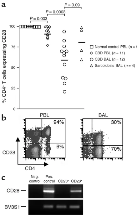
sub-set of CD4+T cells has been reported to lose CD28 expres-sion in certain other chronic inflammatory conditions (29–32), we initially analyzed CD28 expression on blood and BAL CD4+T cells from CBD patients. Freshly isolat-ed PBMCs and BAL cells were obtainisolat-ed from CBD patients and control subjects, and the percentage of CD4+ T cells expressing CD28 was determined by immunoflu-orescence staining and cytofluorographic analysis. As shown in Figure 1a, nearly all CD4+T cells from the blood of healthy control subjects express CD28 (mean ± SE, 99.8% ± 0.07%). This frequency was significantly decreased in CBD patients (92% ± 2.2%; P = 0.003). The loss of CD28
expression was even more marked in the BAL, and the per-centage of CD4+T cells expressing CD28 ranged from 21% to 94% (mean ± SE, 59% ± 6.5%; P = 0.0003, compared with PBMCs). This loss was not specific to CBD since decreases were also noted in the BAL of sarcoidosis patients (P = 0.09 compared with CBD). Analysis of the level of CD28 expression on negative cells by fluorescence intensity (Figure 1b) suggested that CD28 expression was absent, as opposed to partially downregulated. The essen-tially complete loss of CD28 expression was confirmed by analysis of CD28 mRNA in sorted CD4+CD28+ and CD4+CD28–populations (Figure 1c).
Correlation between loss of CD28 and degree of inflammation in the lung. Based on the observation that the frequency of CD4+CD28–T cells in patients with rheumatoid arthritis was associated with the severity of disease (31, 33), we cor-related the percentage of BAL CD4+T cells lacking CD28 expression with total BAL white blood cell (WBC), lym-phocyte and macrophage counts in patients with CBD (Figure 2). Significant positive correlations were seen for both total BAL WBC count (r2= 0.495; P = 0.012) and lymphocyte count (r2= 0.482; P = 0.015). On the other hand, no correlation was observed between the frequen-cy of BAL CD4+CD28–T cells and total BAL macrophage count (r2= 0.031; P = 0.52). Thus, the severity of the CD4+ T cell alveolitis appears to be tied to the loss of CD28 expression on CD4+T cells in the BAL.
IFN-γand IL-2 expression by BAL and peripheral blood CD4+T cells after stimulation with BeSO
4. Using
intracellu-lar IFN-γexpression as a measure of response (9), BAL cells and PBMCs were cultured with BeSO4at the opti-mal concentration of 10–5M for a total of 6 hours. Pre-vious studies have shown that PBMCs from healthy individuals and disease controls, as well as BAL cells from sarcoidosis patients, demonstrate no response to BeSO4in these assays, but do have the ability to be stim-ulated by SEB (9). In contrast, cells from CBD patients can show marked responses after the addition of BeSO4. A representative example of intracellular cytokine expression of BAL CD4+T cells from CBD patient 1 is shown in Figure 3a. We observed minimal background IFN-γstaining in the absence of stimulation (medium alone). Following BeSO4stimulation of fresh BAL cells from patient 1, a greater frequency of CD4+CD28+cells express IFN-γafter BeSO4exposure as compared with their CD28–counterparts (21% versus 12%) (Figure 3a). Among the 12 patients studied, the frequency of IFN-γ– producing cells in the CD4+CD28–and CD4+CD28+ subsets was 11.3% ± 3.3% and 18.9% ± 3.6% (P = 0.08), respectively (Figure 3c). Overall, only CBD patients 3 and 10 had a greater percentage of IFN-γ–producing cells in the CD4+CD28–population. On the other hand, SEB stimulated a larger number of cells to secrete IFN-γ, and the CD4+CD28–and CD4+CD28+subsets each had about the same proportion (mean ± SEM; 51.3 ± 3.0 ver-sus 49.2 ± 2.8) of responsive cells (Figure 3a).
[image:4.576.58.277.52.396.2]Previous studies have shown that a subset of the IFN-γ– expressing cells also produce IL-2 when stimulated by BeSO4(9). Despite the importance of CD28 signaling
Figure 1
Expression of CD28 on the surface of CD4+T cells from peripheral
blood (PBL) and BAL. (a) The percentage of CD4+T cells expressing
CD28 in blood of normal control subjects, blood of CBD patients, BAL of CBD patients, and BAL of sarcoidosis patients is shown. The black bar represents the mean value for each disease group. (b) Rep-resentative flow-cytometric staining of blood and BAL CD4+T cells
from CBD patients for expression of CD28. The numbers in the upper- and lower-right quadrants are the percentages of CD4+T cells
staining positively and negatively for CD28 expression, respectively. (c) Expression of CD28 mRNA in BAL cells sorted into CD4+CD28+
and CD4+CD28–subsets. RNA was obtained from the sorted
in IL-2 production (34, 35), a significant fraction of CD4+CD28–T cells expressed IL-2, and levels appeared to be similar compared with CD28+-secreting cells (Fig-ure 3b). Among the 10 BAL samples studied for IL-2 production, however, the percentage of CD4+CD28– BAL cells that expressed IL-2 was significantly lower compared with the CD4+CD28+cells (4.3% ± 1.1% ver-sus 11.8% ± 2.7%; P = 0.02) (Figure 3c), and the ratio to IFN-γ–secreting cells was also lower in this subset (0.46 ± 0.04 versus 0.68 ± 0.04; P = 0.001).
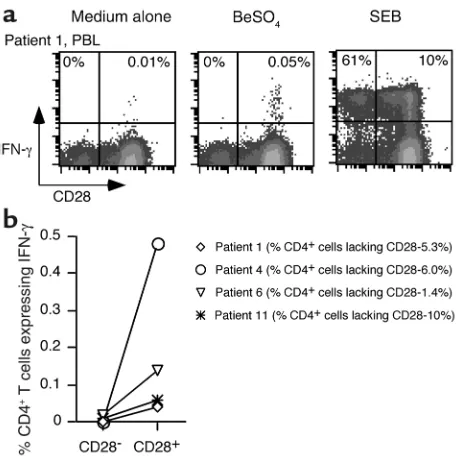
We also analyzed the CD28 phenotype of BeSO4-responsive CD4+ T cells in the peripheral blood of patients with CBD. Previous studies showed that the fre-quency of peripheral blood IFN-γexpressing CD4+T cells after stimulation with BeSO4ranged from undetectable to approximately 1 in 500 cells in CBD patients (9). For the current studies, we selected PBMC samples that pro-liferated in the presence of BeSO4and had detectable cytokine-producing cells. One example is shown in Fig-ure 4a. Following short-term cultFig-ure with BeSO4, approx-imately 1 in 2,000 CD4+T cells from patient 1 expressed IFN-γ. The beryllium-responsive cells were localized to the CD28+T cell population, although this individual had a sizable percentage (5.3%) of CD4+CD28–T cells in the blood. In the example shown, 61% of the CD4+CD28–
cells expressed IFN-γafter SEB stimulation compared with 10% of the CD4+CD28+T cells, yet essentially none (detection limit less than 1:2,000) of the CD4+CD28–cells expressed IFN-γfollowing BeSO4stimulation. Few, if any, IFN-γ–producing cells were detected in the CD4+CD28– population of any of the four blood samples from CBD patients (Figure 4b), and the results indicated that this population was not greatly enriched for beryllium-spe-cific effector cells. In these four CBD patients, the fre-quency of CD4+CD28–T cells ranged from 1.4% to 10% (Figure 4b). Thus, it is possible that the small size of the CD28–population in blood might have prevented the detection of rare responding cells.
[image:5.576.59.533.56.191.2]Functional dependence on CD28 costimulation. We used inhibitory concentrations of CTLA-4Ig to test for the
Figure 2
Correlation between the percentage of CD4+T cells lacking CD28 expression and total BAL WBC, lymphocyte, and macrophage cell
counts in 12 CBD patients.
Figure 3
Intracellular staining for IFN-γand IL-2 in stimulated BAL T cells from CBD patients. Representative experiments are shown for the flow-cytometric analysis of BAL CD4+T cells from CBD patient 1 (a)
and patient 3 (b) stimulated with either medium alone, BeSO4, or
SEB and subsequently stained for surface CD4 and CD28 and intra-cellular IFN-γ or IL-2. BAL cells were gated on CD4 expression. The numbers in the upper-left and -right quadrants of each density plot are the percentages of CD4+CD28–and CD4+CD28+cells,
respec-tively, that express IFN-γor IL-2. (c) Percentage of BAL CD4+CD28–
and CD4+CD28+cells that express intracellular IFN-γand IL-2 after
short-term stimulation with BeSO4. The mean percentage of Th1
cytokine produced by CD4+CD28–and CD4+CD28+cells from CBD
patients is shown as a solid line: mean ± SEM for IFN-γ (n = 12), 11.3% ± 3.3% for CD4+CD28– cells and 18.9% ± 3.6% for
CD4+CD28+cells; IL-2 (n = 10), 4.3% ± 1.1% for CD4+CD28–cells
[image:5.576.301.527.449.731.2]requirement of CD28-mediated costimulation when cells from CBD patients are stimulated with beryllium in culture. PBMCs from all CBD patients analyzed demonstrated a positive proliferative response (defined as a stimulation index of 2.5) in the presence of 10–5M
BeSO4(Figure 5a). For example, PBMCs from CBD
patient 1 proliferated in response to BeSO4, with peak thymidine incorporation of 65,772 ± 2173 cpm com-pared with background proliferation of 431 ± 165 cpm (Figure 5a). Following the addition of 30 µg/ml CTLA-4Ig, beryllium-induced proliferation decreased 93%. Among the seven patients studied, six demon-strated greater than 90% inhibition with this
concen-tration of CTLA-4Ig (Figure 5a). The one exception, patient 7, showed the lowest proliferative response, which was inhibited 35% with CTLA-4Ig.
BAL cells from five CBD patients were selected based on positive proliferative responses to 10–5M BeSO4in culture (Figure 5b). In contrast to the effects seen in peripheral blood, CTLA-4Ig had minimal, if any, inhibitory effects on the beryllium-induced prolifera-tive response of BAL T cells. Only cells from patient 3 showed a 37% reduction in the proliferative response following the addition of 30 µg/ml CTLA-4Ig (Figure 5b). In the remaining four patients, there was either no effect or enhancement of the proliferative response of BAL T cells when CTLA-4Ig was added to the culture. Figure 5c shows the effects of varying concentrations of CTLA-4Ig on the proliferative responses of PBMCs and BAL cells. These studies again used optimal stimu-latory concentrations of BeSO4(10–5M). Proliferation of PBMCs was reproducibly inhibited with concentra-tions of CTLA-4Ig ranging from 0.3 to 30 µg/ml (Fig-ures 5, a and c). As shown in Figure 5c, 0.3 µg/ml, 3 µg/ml, and 30 µg/ml CTLA-4Ig inhibited the
beryllium-Figure 4
Intracellular staining for IFN-γin stimulated PBMCs from CBD patients. A representative experiment is shown for the flow-cytomet-ric analysis of blood CD4+T cells from CBD patient 1 (a) stimulated
with either medium alone, BeSO4, or SEB, and stained for surface
CD4 and CD28 and intracellular IFN-γ. Peripheral blood cells were gated for CD4 expression, and the numbers in the upper quadrants of each density plot are the percentages of CD4+CD28–and
CD4+CD28+cells that express IFN-γ. (b) Comparison of the
fre-quency of CD4+CD28–and CD4+CD28+cells that express IFN-γ in
[image:6.576.55.283.52.280.2]fresh PBMCs from four CBD patients. The symbols for each CBD patient correspond to those shown in Figure 3.
Figure 5
Proliferative responses of PBMCs (a) and BAL cells (b) from CBD patients. Proliferation of the cells in medium alone and after the addi-tion of BeSO4with and without 30 µg/ml CTLA-4Ig is shown. The data are expressed as the mean counts per minute ± SEM. (c) Inhibition
of the T cell proliferative responses of PBMCs and BAL cells to beryllium with various concentrations of CTLA-4Ig. PBMCs (n = 7) and BAL cells (n = 5) from CBD patients were cultured with 10–5M BeSO
4and increasing concentrations of CTLA-4Ig. The data are expressed as
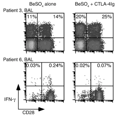
[image:6.576.56.529.472.670.2]induced proliferative response 69% ± 12.9%, 85% ± 4.7%, and 92% ± 5.4%, respectively. On the other hand, CTLA-4Ig had little effect on proliferation of BAL T cells, with 0% inhibition when 0.3 µg/ml of CTLA-4Ig was added to the culture conditions. Studies described below show that the BAL T cell proliferative responses occurred in both the CD4+CD28+and CD4+CD28–populations; therefore, both populations were functionally inde-pendent of CD28 costimulation. These studies indicate that the beryllium-induced proliferative response in PBMCs occurs in a CD28-dependent manner, while in the lung, CD28-mediated costimulation is unnecessary. We also determined what proportion of induced cytokine secretion in BAL CD4+cells was independent of CD28 costimulation. The addition of 30 µg/ml CTLA-4Ig had no inhibitory effect on IFN-γ produc-tion induced in BAL CD4+T cells following short-term culture with BeSO4 (Figure 6). The lack of inhibition was equally apparent for both the CD4+CD28+and CD4+CD28–populations with an enhancement of IFN-γ expression with CTLA-4Ig in both populations. In addition to the findings shown for patient 3 in Figure 6, BAL CD4+CD28+and CD4+CD28–T cells from pa-tient 8 showed a 21% and 9.6% increase in IFN-γ expres-sion after the addition of CTLA-4Ig. Similar findings were seen with 0.3 and 3 µg/ml CTLA-4Ig. Blockade of B7 molecules with CTLA-4Ig also resulted in enhanced beryllium-induced TNF-αproduction in both CD28+ and CD28–populations (data not shown).
[image:7.576.65.261.53.248.2]Inhibition of cytokine secretion in stimulated PBMCs was more difficult to test because of the low frequency of antigen-specific cells in blood (9). In the peripheral blood of patient 6, the frequency of beryllium-responsive CD4+ T cells was approximately 1 in 415 (0.24%), and the addi-tion of 30 µg/ml CTLA-4Ig decreased IFN-γexpression after BeSO4stimulation to background levels (0.07% in the experiment shown in Figure 6). These studies there-fore suggest that activation of beryllium-specific CD4+T cells in blood, whether quantified by cytokine secretion or proliferation, is dependent on CD28 costimulation. Proliferative capacity of BAL CD4+T cells in relation to CD28 expression. In preliminary studies, we noted an increased percentage of CD4+T cells that expressed CD28 follow-ing 14 days of beryllium stimulation of BAL T cells (data not shown). These findings suggested that the CD28+ BAL T cells possess either a proliferative and/or a sur-vival advantage following antigen-specific T cell activa-tion. Using CFSE to track T cell division (27), we com-pared the proliferative capacity of CD4+CD28+ and CD4+CD28–BAL T cells. After 5 days of beryllium expo-sure, 41% of the CD4+CD28+T cells underwent at least one cell division compared with 29% of the CD4+CD28– T cells (Figure 7a). Prior to culture, 30% of the CD4+T
Figure 6
Intracellular expression of IFN-γafter BeSO4stimulation of BAL T
cells and PBMCs from CBD patients with and without the addition of CTLA-4Ig. Representative experiments are shown for the flow-cytometric analysis of BAL and blood CD4+ T cells from CBD
patients 3 and 6 stimulated with 10–5M BeSO
4and with or without
30 µg/ml CTLA-4Ig. Cells were stained for surface CD4 and CD28 and intracellular IFN-γand then gated for CD experssion. The expres-sion of IFN-γafter BeSO4and CTLA-4Ig exposure in the blood of
[image:7.576.310.523.349.597.2]patient 6 was no greater than background staining (0.06%). The numbers in the upper quadrants of each density plot are the per-centages of CD4+CD28–and CD4+CD28+cells that express IFN-γ.
Figure 7
Differences in beryllium-specific BAL CD4+T cell proliferation between
cells with and without CD28 expression. (a) A representative example of the proliferation in CFSE-labeled BAL CD4+T cells after 5 days of
culture in the presence of 10–5M BeSO
4is shown. Proliferation is
meas-ured by a 50% decrease in fluorescence intensity of CFSE-labeled cells with each division. BAL cells were gated for CD4 expression, and the number in each quadrant of the density plot represents the percentage of CD4+ T cells. (b) Proliferation of sorted CD4+CD28+ and
CD4+CD28–cells from CBD patient 3 in medium alone and after the
addition of 10–5M BeSO
4is shown. The data are expressed as the
cells expressed CD28, compared with 34% 5 days after beryllium stimulation. Based on the similar frequencies of CD28+and CD28–T cells before and after T cell stim-ulation, it seemed unlikely that beryllium-specific CD28+T cells lost CD28 expression during culture. In separate experiments, BAL T cells were sorted into two populations, CD4+CD28+and CD4+CD28–, prior to cul-ture with autologous APCs and BeSO4. A strong prolif-erative response was induced in both populations of T cells (representative example shown in Figure 7b). The proliferative response (stimulated minus background proliferation), however, of the CD4+CD28–cells was 58% of the level of CD4+CD28+T cells.
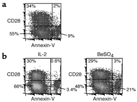
Activation-induced apoptosis of BAL CD4+T cells in relation to CD28 expression. We also studied whether the beryllium-specific CD4+CD28–T cells in lung may be more suscep-tible to activation-induced cell death compared with their CD28+counterparts. Fresh BAL cells and BAL-derived T cell clones were stimulated with BeSO4for 24 hours, and the CD4+CD28+and CD4+CD28–subsets were stained with annexin V and propidium iodide to determine the fraction of apoptotic cells (representative experiments are shown in Figure 8). Minimal annexin V staining was seen on fresh BAL cells after culture with 20 U/ml of IL-2 for 24 hours (data not shown). After 24-hour stimulation with beryllium, 5.5% and 14% of the CD4+CD28+and CD4+CD28–subsets, respectively, expressed annexin V (Figure 8a). Using a beryllium-specific T cell clone derived from CBD patient 12, the fraction of apoptotic cells in the CD4+CD28+and the CD4+CD28–subsets were 2% and 4.9%, respectively, in cultures with IL-2. We observed a dramatic increase in apoptotic cells to 30% of the CD4+CD28–subset after culture with BeSO4, compared with 9.4% of the CD4+CD28+subset (Figure 8b). The
increased frequency of apoptotic cells in the CD4+CD28– subset was observed in all three experiments in which it was studied and indicated the increased susceptibility of this subset to activation-induced cell death.
Discussion
We used patients with CBD to investigate the role of CD28-mediated costimulation in human memory CD4+ T cell subsets. Large numbers of beryllium-specific CD4+ T cells are compartmentalized within the lungs of these subjects (9). These cells proliferate and rapidly secrete Th1-type cytokines following recognition of beryllium bound to HLA-DP molecules on the surface of APCs (9, 12, 23, 36). Previous experiments have also shown that these cells have the phenotype of effector-memory cells, including the lack of surface CCR7 and CD62L expression (9). The current studies indicate that activation of these lung cells is independent of CD28-mediated costimulation. Indeed, a large proportion of BAL antigen-specific cells lost expression of CD28 at both the protein and mRNA levels, but continued to respond in culture. In contrast, the pro-liferative and cytokine responses of beryllium-specific T cells in blood were markedly inhibited by CTLA4-Ig, strongly suggesting a need for CD28-mediated costimu-lation. Although some CD28–cells were present in blood, few, if any, beryllium-specific cells could be demonstrated in this subset. Taken together, these studies suggest that a decreased reliance on CD28-mediated costimulation is not a universal phenomenon among all memory CD4+T cells and that functionally distinct memory subsets may be distinguished by CD28 function and expression.
[image:8.576.62.281.52.215.2]The loss of CD28 on a subset of activated CD4+T cells has been described in several chronic inflammatory dis-orders, such as sarcoidosis, rheumatoid arthritis, multi-ple sclerosis, type 1 diabetes, and chronic infection (29–32, 37). It has also been observed in elderly individ-uals (38). Although most of these studies have focused on peripheral blood cells, downregulation of CD28 expression has also been observed on BAL cells from patients with sarcoidosis, a pathologically similar lung disease (37). In studies of CD28–cells in rheumatoid arthritis, the decreased surface CD28 was similarly asso-ciated with a loss of gene expression (31, 38, 39). What signals the cell to alter its CD28 gene expression is not known. The lungs in both CBD and sarcoidosis are char-acterized by an accumulation of activated CD4+T cells that secrete Th1-type cytokines (9, 23, 37). The positive correlation between the loss of CD28 expression on CD4+T cells and the magnitude of the T cell alveolitis suggests that prolonged Th1-type cytokine exposure might be the underlying mechanism. Support for this idea includes the observation that exposure of CD4+T cells to increasing concentrations of TNF-αresulted in decreased CD28 expression and even CD28nullcells after 8 weeks of continuous TNF-αexposure in culture (40). Furthermore, in the present studies, the loss of CD28 extended to cells that are not responsive to beryllium in our assay systems. Indeed, beryllium-specific cells were only a relatively small fraction of CD28–cells in the lung
Figure 8
BAL CD4+CD28+T cells are relatively resistant to beryllium-induced
cell death. Data are shown for representative experiments using fresh BAL cells (a) and T cell clones derived from the lung of CBD patient 12 (b) stimulated with either 20 U/ml IL-2 or BeSO4. Following
and were absent or rarely present in the blood CD28– cells. On the other hand, the blood CD28–cells were capable of secreting Th1-type cytokines following super-antigen stimulation, indicating that they are not anergic. The loss of CD28 expression has been extensively described in CD8+cells, especially in chronic infection and in elderly individuals that harbor clonal expansions (28, 41–45). In these studies, the loss of CD28 more clear-ly has been shown to be associated with differentiation to effector memory CD8+cells. For example, in one study of functional subsets within individually expanded CD8+clones, loss of CD28 was associated with increased perforin expression and loss of proliferative potential (28). We were unable to detect increased perforin expres-sion in the BAL CD4+CD28–cells of CBD patients (data not shown). Studies of CD8+clonal expansions have also clearly demonstrated that the CD28–cells derive from CD28+cells (28), and it seems likely that the lung beryl-lium-specific CD28–cells in CBD patients also arise from CD28+precursors, especially based on the mechanistic studies described above (40). Interestingly, loss of CD28 expression is apparently not a feature of CD4+or CD8+ memory T cell differentiation in mice (P. Marrack, per-sonal communication), thus this phenomenon has not been studied in nonhuman systems.
Similar to studies of CD8+ T cells, we also noted decreased proliferation of CD4+CD28–compared with CD4+CD28+cells after stimulation with antigen, despite maintained effector function. Consistent with this find-ing, CD28-mediated costimulation lowers the anti-CD3 concentration necessary to induce T cell proliferation in vitro (34) and promotes cell cycle progression and IL-2 production, both of which are required for a sustained T cell response (2). The current studies are the first to sug-gest that loss of CD28 on human memory CD4+cells is associated with susceptibility to activation-induced cell death. Our results are, however, consistent with previous studies that have implicated CD28 costimulation in enhanced T cell survival. This effect may be mediated through the enhanced production of IL-2 and increased expression of the antiapoptotic molecule, Bcl-xL (46). CD28-costimulated cells have also been shown to be less dependent on extrinsic growth factor (e.g., IL-2) for their ability to survive in culture (46). Recent studies also sug-gest that the CD28 signaling pathway affects the regula-tion of glucose metabolism (47), which also may be important in the prolonged survival of activated T cells. In a target organ characterized by persistent antigen exposure and continuous T cell activation, it is conceiv-able that the loss of CD28 on a metabolically active T cell limits access to nutrients and marks the differentiation of these cells into presenescent state.
We also noted that the addition of CTLA-4Ig actually enhanced proliferation and cytokine production of beryllium-specific cells in lung in a number of different experiments. In studies quantifying intracellular cytokine expression, this enhancement occurred in both the CD28–and CD28+populations. These results seem most likely related to the ability of CTLA-4Ig to block
the interaction of CTLA-4 (induced on the T cell after acti-vation) with B7 on the APC, an interaction that usually leads to a downregulatory signal (2, 48, 49). In preliminary experiments, CTLA-4 is upregulated following beryllium stimulation in both CD4+CD28+and CD4+CD28–BAL T cells (A. Fontenot and B. Kotzin, unpublished data). It is noteworthy that our studies show that CTLA-4 may modulate the immediate cytokine production that occurs in these reactions.
Remarkable in the current study was the continued dependence of peripheral blood CD4+cells on CD28 cos-timulation for both proliferative and cytokine responses to beryllium in culture. These findings contrast with stud-ies of one group of investigators that have studied responses to the recall antigen, tetanus toxoid, and to cer-tain autoantigens in multiple sclerosis (29) and type 1 dia-betes (50). For example, in multiple sclerosis, myelin basic protein–reactive (MBP-reactive) T cells in the blood were found to be less dependent on CD28 costimulation for T cell proliferation as compared with MBP-specific T cells from healthy control subjects (29). Similar findings with respect to glutamic acid decarboxylase 65-reactive T cells in type 1 diabetes have also been described (50). The expla-nation for the difference in findings is not known. Although Th2- versus Th1-type cells have been described to be more dependent on CD28 costimulation, our cells are almost all Th1 type, as in the above studies. Further-more, although different reagents were used to test for CD28 dependence, we believe that it is unlikely that the CTLA-4Ig used in our studies could have inhibited through a mechanism other than blocking the CD28-B7 interaction (49). The different findings may relate to the recirculation of effector cells in blood in the different dis-eases being studied. Our previous studies have shown that the lung effector cells in CBD patients do not circulate in large numbers (9, 25). The remarkable compartmental-ization of beryllium-specific cells in the lung may be relat-ed to the fact that antigen stimulation is only occurring in the lung. It is possible that differences in where antigen stimulation takes place compared with CBD, or the tim-ing of systemic antigen stimulation in relation to sam-pling, is key to whether circulating antigen-specific cells are dependent on CD28 costimulation. In this regard, we have noted that patch testing with beryllium leads to a large influx of antigen-specific cells into the blood within a few weeks, and these cells more closely resemble the lung CD4+ effector cells in terms of TCR expression (26). Whether these cells might also be independent of CD28 costimulation is currently unknown.
emphasizes that the accumulation of effector cells in the target organ may preclude therapeutic efficacy with these agents. Furthermore, the ability to target circu-lating memory cells may be extremely important for this therapeutic strategy to work. Finally, one may need to be cautious when using this therapy in diseases where large numbers of effector cells have accumulat-ed in the target organ, since enhancement of the patho-logic response is possible before benefit may take place.
Acknowledgments
This work was supported by the following NIH grants: HL62410, HL03982, ES06358, and P01ES011810, and the General Clinical Research Center (M01-RR-0051) from the Division of Research Resources.
1. Bretscher, P., and Cohn, M. 1970. A theory of self-nonself discrimination.
Science.169:1042–1049.
2. Bluestone, J.A. 1995. New perspectives of CD28-B7–mediated T cell cos-timulation. Immunity.2:555–559.
3. Harding, F.A., McArthur, J.G., Gross, J.A., Raulet, D.H., and Allison, J.P. 1992. CD28-mediated signaling co-stimulates murine T cells and prevents induction of anergy in T-cell clones. Nature.356:607–609.
4. Schwartz, R.H. 1990. A cell culture model for T lymphocyte clonal anergy.
Science.248:1349–1356.
5. Liu, Y., and Janeway, C.A., Jr. 1992. Cells that present both specific ligand and costimulatory activity are the most efficient inducers of clonal expan-sion of normal CD4 T cells. Proc. Natl. Acad. Sci. U. S. A.89:3845–3849. 6. Croft, M., Bradley, L.M., and Swain, S.L. 1994. Naive versus memory CD4
T cell response to antigen. Memory cells are less dependent on accessory cell costimulation and can respond to many antigen-presenting cell types including resting B cells. J. Immunol.152:2675–2685.
7. Garcia, S., DiSanto, J., and Stockinger, B. 1999. Following the development of a CD4 T cell response in vivo: from activation to memory formation.
Immunity.11:163–171.
8. London, C.A., Lodge, M.P., and Abbas, A.K. 2000. Functional responses and costimulator dependence of memory CD4+ T cells. J. Immunol.
164:265–272.
9. Fontenot, A.P., Canavera, S.J., Gharavi, L., Newman, L.S., and Kotzin, B.L. 2002. Target organ localization of memory CD4+T cells in patients with
chronic beryllium disease. J. Clin. Invest.110:1473–1482. doi:10.1172/ JCI200215846.
10. Fontenot, A.P., Newman, L.S., and Kotzin, B.L. 2001. Chronic beryllium disease: T cell recognition of a metal presented by HLA-DP. Clin. Immunol.
100:4–14.
11. Newman, L.S., Maier, L.A., and Nemery, B. 1998. Interstitial lung disorders due to beryllium and cobalt. In Interstitial lung disease. 3rd edition. M.I. Schwarz and T.E. King, Jr., editors. B.C. Decker Inc. Hamilton, Ontario, Canada. 367–392.
12. Fontenot, A.P., Torres, M., Marshall, W.H., Newman, L.S., and Kotzin, B.L. 2000. Beryllium presentation to CD4+T cells underlies disease
suscepti-bility HLA-DP alleles in chronic beryllium disease. Proc. Natl. Acad. Sci. U. S. A.97:12717–12722.
13. Kreiss, K., Wasserman, S., Mroz, M.M., and Newman, L.S. 1993. Beryllium disease screening in the ceramics industry: blood test performance and exposure-disease relations. J. Occup. Med.35:267–274.
14. Kreiss, K., Mroz, M.M., Zhen, B., Martyny, J.W., and Newman, L.S. 1993. Epidemiology of beryllium sensitization and disease in nuclear workers.
Am. Rev. Respir. Dis.148:985–991.
15. Richeldi, L., et al. 1997. Interaction of genetic and exposure factors in the prevalence of berylliosis. Am. J. Ind. Med.32:337–340.
16. Wang, Z., et al. 1999. Differential susceptibilities to chronic beryllium dis-ease contributed by different Glu69 HLA-DPB1 and -DPA1 alleles.
J. Immunol.163:1647–1653.
17. Newman, L.S., and Kreiss, K. 1992. Nonoccupational beryllium disease masquerading as sarcoidosis: identification by blood lymphocyte prolif-eration response to beryllium. Am. Rev. Respir. Dis.145:1212–1214. 18. Newman, L.S., Rose, C.S., and Maier, L.A. 1997. Sarcoidosis. N. Engl. J. Med.
336:1224–1234.
19. Rossman, M.D., et al. 1988. Proliferative response of bronchoalveolar lym-phocytes to beryllium. Ann. Intern. Med.108:687–693.
20. Saltini, C., Winestock, K., Kirby, M., Pinkston, P., and Crystal, R.G. 1989. Maintenance of alveolitis in patients with chronic beryllium disease by beryllium-specific helper T cells. N. Engl. J. Med.320:1103–1109. 21. Kreiss, K., Newman, L.S., Mroz, M., and Campbell, P.A. 1989. Screening
blood test identifies subclinical beryllium disease. J. Occup. Med.31:603–608.
22. Mroz, M.M., Kreiss, K., Lezotte, D.C., Campbell, P.A., and Newman, L.S. 1991. Reexamination of the blood lymphocyte transformation test in the diagnosis of chronic beryllium disease. J. Allergy Clin. Immunol.
88:54–60.
23. Tinkle, S.S., Kittle, L.A., Schumacher, B.A., and Newman, L.S. 1997. Beryl-lium induces IL-2 and IFN-γin berylliosis. J. Immunol.158:518–526. 24. Fontenot, A.P., Kotzin, B.L., Comment, C., and Newman, L.S. 1998.
Expan-sions of T-cell subsets expressing particular T cell receptor variable regions in chronic beryllium disease. Am. J. Respir. Cell Mol. Biol.18:581–589. 25. Fontenot, A.P., Falta, M.T., Freed, B.M., Newman, L.S., and Kotzin, B.L.
1999. Identification of pathogenic T cells in patients with beryllium-induced lung disease. J. Immunol.163:1019–1026.
26. Fontenot, A.P., et al. 2002. Beryllium skin patch testing to analyze T cell stimulation and granulomatous inflammation in the lung. J. Immunol.
168:3627–3634.
27. Lyons, A.B., and Parish, C.R. 1994. Determination of lymphocyte division by flow cytometry. J. Immunol. Methods.171:131–137.
28. Chamberlain, W.D., Falta, M.T., and Kotzin, B.L. 2000. Functional subsets within clonally expanded CD8(+) memory T cells in elderly humans. Clin. Immunol.94:160–172.
29. Lovett-Racke, A.E., et al. 1998. Decreased dependence of myelin basic pro-tein-reactive T cells on CD28-mediated costimulation in multiple sclero-sis patients. A marker of activated/memory T cells. J. Clin. Invest.
101:725–730.
30. Markovic-Plese, S., Cortese, I., Wandinger, K.P., McFarland, H.F., and Mar-tin, R. 2001. CD4+CD28– costimulation-independent T cells in multiple sclerosis. J. Clin. Invest.108:1185–1194. doi:10.1172/JCI200112516. 31. Schmidt, D., Goronzy, J.J., and Weyand, C.M. 1996. CD4+ CD7– CD28– T
cells are expanded in rheumatoid arthritis and are characterized by autore-activity. J. Clin. Invest.97:2027–2037.
32. Haffar, O.K., et al. 1993. Costimulation of T-cell activation and virus pro-duction by B7 antigen on activated CD4+ T cells from human immunod-eficiency virus type 1-infected donors. Proc. Natl. Acad. Sci. U. S. A.
90:11094–11098.
33. Martens, P.B., Goronzy, J.J., Schaid, D., and Weyand, C.M. 1997. Expansion of unusual CD4+ T cells in severe rheumatoid arthritis. Arthritis Rheum.
40:1106–1114.
34. Gimmi, C.D., et al. 1991. B-cell surface antigen B7 provides a costimula-tory signal that induces T cells to proliferate and secrete interleukin-2.
Proc. Natl. Acad. Sci. U. S. A.88:6575–6579.
35. Jenkins, M.K., Taylor, P.S., Norton, S.D., and Urdahl, K.B. 1991. CD28 delivers a costimulatory signal involved in antigen-specific IL-2 produc-tion by human T cells. J. Immunol.147:2461–2466.
36. Lombardi, G., et al. 2001. HLA-DP allele-specific T cell responses to beryl-lium account for DP-associated susceptibility to chronic berylberyl-lium disease.
J. Immunol.166:3549–3555.
37. Katchar, K., Wahlstrom, J., Eklund, A., and Grunewald, J. 2001. Highly acti-vated T-cell receptor AV2S3(+) CD4(+) lung T-cell expansions in pul-monary sarcoidosis. Am. J. Respir. Crit. Care Med.163:1540–1545. 38. Vallejo, A.N., Nestel, A.R., Schirmer, M., Weyand, C.M., and Goronzy, J.J.
1998. Aging-related deficiency of CD28 expression in CD4+ T cells is asso-ciated with the loss of gene-specific nuclear factor binding activity. J. Biol. Chem.273:8119–8129.
39. Vallejo, A.N., et al. 2002. Molecular basis for the loss of CD28 expression in senescent cells. J. Biol. Chem.277:46940–46949.
40. Bryl, E., Vallejo, A.N., Weyand, C.M., and Goronzy, J.J. 2001. Down-regu-lation of CD28 expression by TNF-alpha. J. Immunol.167:3231–3238. 41. Posnett, D.N., Sinha, R., Kabak, S., and Russo, C. 1994. Clonal populations
of T cells in normal elderly humans: the T cell equivalent to “benign mon-oclonal gammapathy”. J. Exp. Med.179:609–618.
42. Azuma, M., Phillips, J.H., and Lanier, L.L. 1993. CD28– T lymphocytes. Antigenic and functional properties. J. Immunol.150:1147–1159. 43. Fitzgerald, J.E., et al. 1995. Analysis of clonal CD8+T cell expansions in
normal individuals and patients with rheumatoid arthritis. J. Immunol.
154:3538–3547.
44. Weekes, M.P., Carmichael, A.J., Wills, M.R., Mynard, K., and Sissons, J.G. 1999. Human CD28–CD8+ T cells contain greatly expanded functional virus-specific memory CTL clones. J. Immunol.162:7569–7577. 45. Appay, V., et al. 2002. Memory CD8+T cells vary in differentiation
pheno-type in different persistent virus infections. Nat. Med.8:379–385. 46. Boise, L.H., et al. 1995. CD28 costimulation can promote T cell survival by
enhancing the expression of Bcl-XL. Immunity.3:87–98.
47. Frauwirth, K.A., et al. 2002. The CD28 signaling pathway regulates glu-cose metabolism. Immunity.16:769–777.
48. Lenschow, D.J., Walunas, T.L., and Bluestone, J.A. 1996. CD28/B7 system of T cell costimulation. Annu. Rev. Immunol.14:233–258.
49. Salomon, B., and Bluestone, J.A. 2001. Complexities of CD28/B7: CTLA-4 costimulatory pathways in autoimmunity and transplantation. Annu. Rev. Immunol.19:225–252.