comment
reviews
reports
deposited research
interactions
information
refereed research
Research
Phylogenetic analysis of 277 human G-protein-coupled receptors
as a tool for the prediction of orphan receptor ligands
Patrick Joost and Axel Methner
Address: Research Group Protective Signaling, Zentrum für Molekulare Neurobiologie Hamburg and Klinik für Neurologie, Universitätskrankenhaus Eppendorf, Martinistrasse 52, D-20251 Hamburg, Germany.
Correspondence: Axel Methner. E-mail: methner@uke.uni-hamburg.de
Abstract
Background:G-protein-coupled receptors (GPCRs) are the largest and most diverse family of
transmembrane receptors. They respond to a wide range of stimuli, including small peptides, lipid analogs, amino-acid derivatives, and sensory stimuli such as light, taste and odor, and transmit signals to the interior of the cell through interaction with heterotrimeric G proteins. A large number of putative GPCRs have no identified natural ligand. We hypothesized that a more complete knowledge of the phylogenetic relationship of these orphan receptors to receptors with known ligands could facilitate ligand identification, as related receptors often have ligands with similar structural features.
Results: A database search excluding olfactory and gustatory receptors was used to compile a
list of accession numbers and synonyms of 81 orphan and 196 human GPCRs with known ligands. Of these, 241 sequences belonging to the rhodopsin receptor-like family A were aligned and a tentative phylogenetic tree constructed by neighbor joining. This tree and local alignment tools were used to define 19 subgroups of family A small enough for more accurate maximum-likelihood analyses. The secretin receptor-like family B and metabotropic glutamate receptor-like family C were directly subjected to these methods.
Conclusions:Our trees show the overall relationship of 277 GPCRs with emphasis on orphan
receptors. Support values are given for each branch. This approach may prove valuable for identification of the natural ligands of orphan receptors as their relation to receptors with known ligands becomes more evident.
Published: 17 October 2002
GenomeBiology2002, 3(11):research0063.1–0063.16
The electronic version of this article is the complete one and can be found online at http://genomebiology.com/2002/3/11/research/0063 © 2002 Joost and Methner, licensee BioMed Central Ltd
(Print ISSN 1465-6906; Online ISSN 1465-6914)
Received: 19 June 2002 Revised: 7 August 2002 Accepted: 18 September 2002
Background
G-protein-coupled receptors (GPCRs) are the largest and most diverse family of transmembrane receptors. They respond to a wide range of stimuli including small peptides, lipid analogs, amino-acid derivatives, and sensory stimuli such as light, taste and odor [1], and transmit signals to the interior of the cell through interaction with heterotrimeric G
orphan receptors uses changes in second-messenger activation in cells stably expressing the receptor in response to tissue extracts expected to contain the natural ligand [5]. In a second step, these extracts are tested and fractionated to purity, before being analyzed by mass spectrometry. This strategy led to the identification of several novel bioactive peptides or peptide families (for review see [6]). The identifi-cation of these natural ligands is likely to give further insight into the physiological role of these receptors and advance the design of pharmacologically active receptor agonists or antagonists. This is of particular interest, as GPCRs are the most targeted protein superfamily in pharmaceutical research [7]. Better prediction of the presumed chemical class or structure of the ligand facilitates the identification of orphan receptors by the strategy described above, as the ligand purification process can be tailored more specifically to the assumed class of substances.
Phylogenetic analysis of receptor relationships has already been used to elucidate the chemical nature of receptor ligands. The identification of sphingosine 1-phosphate as the ligand for the GPCR EDG-1 led to the prediction that EDG-3, EDG-5, EDG-6 and EDG-8 have the same ligand [8-11]. In contrast, phylogenetically distinct members of the EDG cluster - EDG-2, EDG-4 and EDG-7 - are receptors for the similar but distinct ligand lysophosphatidic acid (LPA) [12-14]. Neuromedin U, a potent neuropeptide that causes contraction of smooth muscle, was correctly predicted phylogenetically to be the ligand of the orphan GPCR FM3 (NMUR) [15]. Not only the ligand, but also the pharmacol-ogy of a novel receptor for histamine, was predicted and con-firmed through phylogeny [16]. GPR86, related to the ADP receptor P2Y12, was similarly recently shown to bind ADP [17], and UDP-glucose, a molecule involved in carbohydrate biosynthesis, was shown to be the ligand for the related receptor KIAA0001 [18].
Mammalian GPCRs were previously classified by phylogeny into three families [19,20]: the rhodopsin receptor-like family (A), the secretin receptor-like receptor family (B) and the metabotropic glutamate receptor family (C). These results were generated by neighbor joining, a fast distance-based method suited for large datasets, but influenced by methodological flaws that can in part be overcome by methods not generally applied previously.
In this work, we compiled an exhaustive list that includes all available synonyms and accession numbers of 196 human GPCRs with known ligands and 84 human orphan receptors. The 241 sequences belonging to family A were aligned, and a tentative tree constructed by neighbor joining with 1,000 bootstrap steps. Subgroups of family A defined by this tree and sequences from families B and C were then used for more accurate phylogenetic analysis by state-of-the-art tech-niques. From this analysis, we tried to predict possible ligands for orphan receptors.
Results and discussion
We set out to define the phylogenetic relationship of human GPCRs by state-of-the-art tools, assuming that the identifi-cation of cognate ligands of orphan receptors will be facili-tated by a more complete knowledge of their relationship within the large and diverse superfamily.
Database mining and multiple sequence alignment
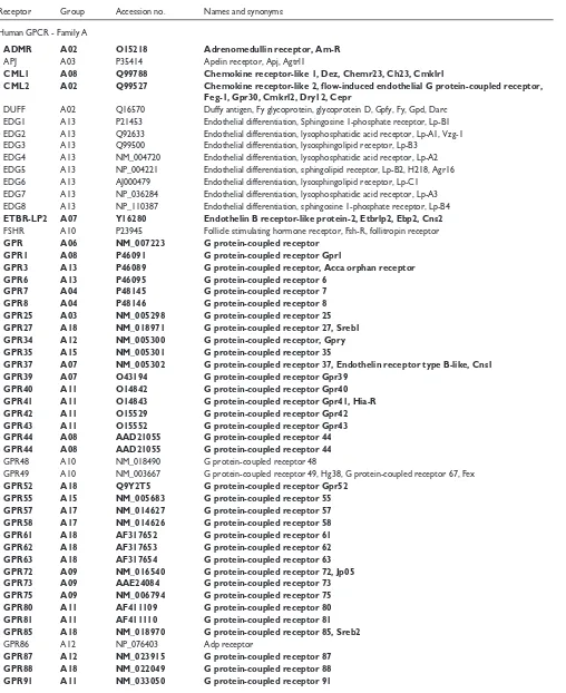
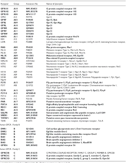
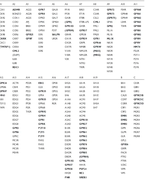
Most receptors were identified by different groups; there-fore, many confusing names and synonyms exist. We adhered to SWISS-PROT names where possible, and com-piled a list including all available synonyms and accession numbers of 196 human GPCRs with known ligands and 84 human orphan receptors (Table 1 shows all receptors men-tioned in this work; the complete list is supplied as an addi-tional data file with the online version of this paper). Gustatory and olfactory receptors were omitted. Multiple protein sequences were aligned and the extremely variable amino termini upstream of the first transmembrane domain and carboxyl termini downstream of the seventh transmem-brane domain were deleted to avoid length heterogeneity (see Figure 1). The deleted regions contained no significant sequence conservation.
Phylogenetic analysis
Because of the large number of sequences in family A, we had to use a combination of computational methods to accomplish the best possible description of their phyloge-netic relationship. In a first step we used the distance-based neighbor-joining method as the only one computationally feasible. Neighbor joining has been shown to be efficient at recovering the correct tree topology [21], but is greatly influ-enced by methodological errors, for example, the sampling error [22]. This can in part be overcome by bootstrapping, a method of testing the reliability of a dataset by the creation of pseudoreplicate datasets by resampling. Bootstrapping assesses whether stochastic effects have influenced the dis-tribution of amino acids [23]. In previous publications on this topic, bootstrapping has not been generally used.
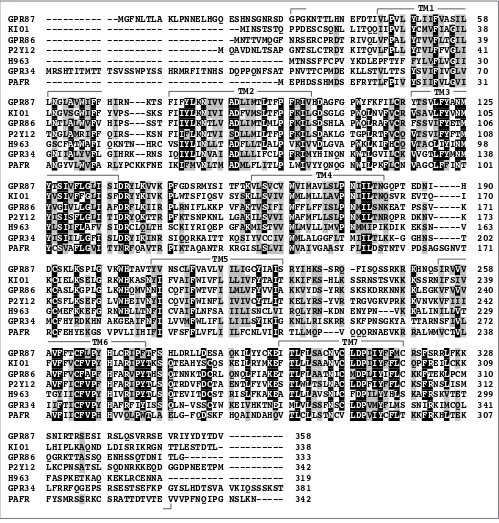
We generated a neighbor-joining tree of family-A sequences, and considered tree branches to be confirmed if they were found in more than 500 of 1,000 bootstrap steps (Figure 2). The same branching pattern was found by least squares (data not shown) as implemented in FITCH [24], but it was not possible to compute enough bootstrap steps with the equipment used. The remaining sequences of unconfirmed branches were then assigned to existing branches according to results obtained with the local alignment tool BLASTP (see Additional data files) [25] to account for similarities in parts of the sequences not sufficient for repeated global alignment. The p-value was used as a measure of similarity.
comment
reviews
reports
deposited research
interactions
information
[image:3.609.58.566.118.740.2]refereed research
Table 1
List of example receptor names, accession numbers and abbreviations
Receptor Group Accession no. Names and synonyms Human GPCR - Family A
ADMR A02 O15218 Adrenomedullin receptor, Am-R
APJ A03 P35414 Apelin receptor, Apj, Agtrl1
CML1 A08 Q99788 Chemokine receptor-like 1, Dez, Chemr23, Ch23, Cmklr1
CML2 A02 Q99527 Chemokine receptor-like 2, flow-induced endothelial G protein-coupled receptor, Feg-1, Gpr30, Cmkrl2, Dry12, Cepr
DUFF A02 Q16570 Duffy antigen, Fy glycoprotein, glycoprotein D, Gpfy, Fy, Gpd, Darc EDG1 A13 P21453 Endothelial differentiation, Sphingosine 1-phosphate receptor, Lp-B1 EDG2 A13 Q92633 Endothelial differentiation, lysophosphatidic acid receptor, Lp-A1, Vzg-1 EDG3 A13 Q99500 Endothelial differentiation, lysosphingolipid receptor, Lp-B3
EDG4 A13 NM_004720 Endothelial differentiation, lysophosphatidic acid receptor, Lp-A2 EDG5 A13 NP_004221 Endothelial differentiation, sphingolipid receptor, Lp-B2, H218, Agr16 EDG6 A13 AJ000479 Endothelial differentiation, lysosphingolipid receptor, Lp-C1 EDG7 A13 NP_036284 Endothelial differentiation, lysophosphatidic acid receptor, Lp-A3 EDG8 A13 NP_110387 Endothelial differentiation, sphingosine 1-phosphate receptor, Lp-B4
ETBR-LP2 A07 Y16280 Endothelin B receptor-like protein-2, Etbrlp2, Ebp2, Cns2
FSHR A10 P23945 Follicle stimulating hormone receptor, Fsh-R, follitropin receptor
GPR A06 NM_007223 G protein-coupled receptor
GPR1 A08 P46091 G protein-coupled receptor Gpr1
GPR3 A13 P46089 G protein-coupled receptor, Acca orphan receptor
GPR6 A13 P46095 G protein-coupled receptor 6
GPR7 A04 P48145 G protein-coupled receptor 7
GPR8 A04 P48146 G protein-coupled receptor 8
GPR25 A03 NM_005298 G protein-coupled receptor 25
GPR27 A18 NM_018971 G protein-coupled receptor 27, Sreb1 GPR34 A12 NM_005300 G protein-coupled receptor, Gpry
GPR35 A15 NM_005301 G protein-coupled receptor 35
GPR37 A07 NM_005302 G protein-coupled receptor 37, Endothelin receptor type B-like, Cns1
GPR39 A07 O43194 G protein-coupled receptor Gpr39
GPR40 A11 O14842 G protein-coupled receptor Gpr40
GPR41 A11 O14843 G protein-coupled receptor Gpr41, Hia-R
GPR42 A11 O15529 G protein-coupled receptor Gpr42
GPR43 A11 O15552 G protein-coupled receptor Gpr43
GPR44 A08 AAD21055 G protein-coupled receptor 44
GPR44 A08 AAD21055 G protein-coupled receptor 44
GPR48 A10 NM_018490 G protein-coupled receptor 48
GPR49 A10 NM_003667 G protein-coupled receptor 49, Hg38, G protein-coupled receptor 67, Fex
GPR52 A18 Q9Y2T5 G protein-coupled receptor Gpr52
GPR55 A15 NM_005683 G protein-coupled receptor 55
GPR57 A17 NM_014627 G protein-coupled receptor 57
GPR58 A17 NM_014626 G protein-coupled receptor 58
GPR61 A18 AF317652 G protein-coupled receptor 61
GPR62 A18 AF317653 G protein-coupled receptor 62
GPR63 A18 AF317654 G protein-coupled receptor 63
GPR72 A09 NM_016540 G protein-coupled receptor 72, Jp05
GPR73 A09 AAE24084 G protein-coupled receptor 73
GPR75 A09 NM_006794 G protein-coupled receptor 75
GPR80 A11 AF411109 G protein-coupled receptor 80
GPR81 A11 AF411110 G protein-coupled receptor 81
GPR85 A18 NM_018970 G protein-coupled receptor 85, Sreb2
GPR86 A12 NP_076403 Adp receptor
GPR87 A12 NM_023915 G protein-coupled receptor 87
GPR88 A18 NM_022049 G protein-coupled receptor 88
Table 1(continued)
Receptor Group Accession No. Names & Synonyms
GPR101 A18 NM_054021 G protein-coupled receptor 101 GPR102 A17 NM_053278 G protein-coupled receptor 102
GPR103 A06 AF411117 G protein-coupled receptor 103
GPRC A13 P47775 Gpr12
GPRF A03 P49685 Gpr15, Bob
GPRJ A09 Q15760 Gpr19, Gpr-Nga
GPRL A18 Q99679 Gpr21
GPRM A06 Q99680 Gpr22
GPRV A11 O00270 Gpr31
GPRW A08 O75388 Gpr32
HM74 A11 P49019 G protein-coupled receptor Hm74
KI01 A12 Q15391 Udp-Glucose receptor, Kiaa0001
LSHR A10 P22888 Lutropin-choriogonadotropic hormone receptor, Lh/Cg-R, Lsh-R, luteinizing hormone receptor, Lhcgr, Lhrhr, Lcgr
MAS A08 P04201 Mas proto-oncogene, Mas1
ML1A A09 P48039 Melatonin receptor Type 1a, Mel-1a-R, Mtnr1a ML1B A09 P49286 Melatonin receptor Type 1b, Mel-1b-R, Mtnr1b
ML1X A09 Q13585 Melatonin-related receptor, H9, Gpr50
MRG A08 P35410 Mas-related G protein-coupled receptor
NMU1R A07 AF272362 Neuromedin U receptor 1, Nmur1, Gpr66, Fm-3 NTR1 A07 P30989 Neurotensin receptor Type 1, Nt-R-1, Ntsr1, Ntrr
NTR2 A07 O95665 Neurotensin receptor Type 2, Nt-R-2, levocabastine-sensitive neurotensin receptor, Ntr2 receptor, Ntsr2
NY1R A09 P25929 Neuropeptide Y receptor Type 1, Npy1-R, Npy1r, Npyr, Npyy1 NY2R A09 P49146 Neuropeptide Y receptor Type 2, Npy2-R, Npy2r
NY4R A09 P50391 Neuropeptide Y receptor Type 4, Npy4-R, Pancreatic Polypeptide receptor 1, Pp1, Ppyr1, Npy4r
P2Y5 A15 P43657 P2y purinoceptor 5, P2y5, purinergic receptor 5, P2ry5, 6h1
P2Y7 A05 Q15722 P2y purinoceptor 7, P2y7, Leukotriene B4 receptor, Chemoattractant receptor-like 1, P2ry7, P2y7, Gpr16, Cmkrl1, Ltb4r
P2Y9 A15 Q99677 P2y purinoceptor 9, P2y9, purinergic receptor 9, Gpr23, P2ry9 P2Y10 A15 AF000545 Putative purinergic receptor P2y10
P2Y12 A12 AF313449 Adp receptor, Sp1999
PAFR A12 P25105 Platelet Activating Factor receptor, Paf-R, Ptafr
PNR A17 AF021818 Putative neurotransmitter receptor
PSP24 A18 U92642 High-affinity lysophosphatidic acid receptor homolog, Gpr45
RDC1 A02 P25106 G protein-coupled receptor Rdc1 homolog
RE2 A18 AF091890 G protein-coupled receptor Re2
SALPR A05 NM_016568 Somatostatin and angiotensin-like peptide receptor, Loc51289 SREB3 A18 NM_018969 Super conserved receptor expressed in brain 3
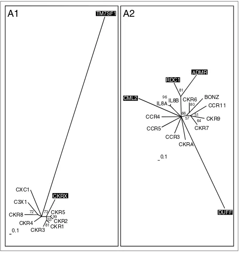
TM7SF1 A01 AF027826 Putative seven pass transmembrane protein
TSHR A10 P16473 Thyroid stimulating hormone receptor, thyrotropin receptor , Tsh-R Human GPCR - Family B
EMR1 B Q14246 Cell surface glycoprotein emr1, Emr1 hormone receptor
EMR2 B AF114491 Egf-like module Emr2
EMR3 B AF239764 Egf-like module-containing mucin-like receptor Emr3
BAI1 B O14514 Brain-specific angiogenesis inhibitor 1
BAI2 B O60241 Brain-specific angiogenesis inhibitor 2
BAI3 B O60242 Brain-specific angiogenesis inhibitor 3, Kiaa0550
GPR56 B NM_005682 G protein-coupled receptor 56
Human GPCR - Family C
GPRC5B C NM_016235 G PROTEIN-COUPLED RECEPTOR, FAMILY C, GROUP 5, MEMBER B, GPRC5B
GPRC5C C NM_018653 G protein-coupled receptor, family C, group 5, member C, Gprc5c GPRC5D C NM_018654 G protein-coupled receptor, family C, group 5, member D, Gprc5d
subgroups A1 and 2, A4 and 5, A11 and 15 and A17 and 18. This approach finally resulted in 19 differently sized sub-groups of family A (Table 2) that were further subjected to the more reliable maximum-likelihood and quartet-puzzling
algorithms. Maximum-likelihood approaches calculate the probability of the observed data assuming that it has evolved in accordance with a chosen evolutionary model. Phyloge-nies are then inferred by finding trees and parameters that
comment
reviews
reports
deposited research
interactions
information
[image:5.609.56.555.160.683.2]refereed research
Figure 1
An example multiple sequence alignment of seven receptors. Protein sequences of GPR87, KI01, GPR86, P2Y12, H963, GPR34 and PAFR belonging to subgroup 12 were aligned with ClustalX and modified by deleting the extremely variable amino termini upstream of the first transmembrane domain and carboxyl termini downstream of the seventh transmembrane domain as indicated. Identical amino-acid residues in all aligned sequences are shaded in black and similar residues in gray. Transmembrane (TM) domains identified by the TMpred program are indicated.
GPR87 --- --MGFNLTLA KLPNNELHGQ ESHNSGNRSD GPGKNTTLHN EFDTIVLPVL YLIIFVASIL 58 KI01 --- --- --- --MINSTSTQ PPDESCSQNL LITQQIIPVL YCMVFIAGIL 38 GPR86 --- --- --- -MNTTVMQGF NRSERCPRDT RIVQLVFPAL YTVVFLTGIL 39 P2Y12 --- --- ---M QAVDNLTSAP GNTSLCTRDY KITQVLFPLL YTVLFFVGLI 41 H963 --- --- --- --- MTNSSFFCPV YKDLEPFTYF FYLVFLVGII 30 GPR34 MRSHTITMTT TSVSSWPYSS HRMRFITNHS DQPPQNFSAT PNVTTCPMDE KLLSTVLTTS YSVIFIVGLV 70 PAFR --- --- --- ---M EPHDSSHMDS EFRYTLFPIV YSIIFVLGVI 31
GPR87 LNGLAVWIFF HIRN---KTS FIFYLKNIVV ADLIMTLTFP FRIVHDAGFG PWYFKFILCR YTSVLFYANM 125 KI01 LNGVSGWIFF YVPS---SKS FIIYLKNIVI ADFVMSLTFP FKILGDSGLG PWQLNVFVCR VSAVLFYVNM 105 GPR86 LNTLALWVFV HIPS---SST FIIYLKNTLV ADLIMTLMLP FKILSDSHLA PWQLRAFVCR FSSVIFYETM 106 P2Y12 TNGLAMRIFF QIRS---KSN FIIFLKNTVI SDLLMILTFP FKILSDAKLG TGPLRTFVCQ VTSVIFYFTM 108 H963 GSCFATWAFI QKNTN--HRC VSIYLINLLT ADFLLTLALP VKIVVDLGVA PWKLKIFHCQ VTACLIYINM 98 GPR34 GNIIALYVFL GIHRK--RNS IQIYLLNVAI ADLLLIFCLP FRIMYHINQN KWTLGVILCK VVGTLFYMNM 138 PAFR ANGYVLWVFA RLYPCKKFNE IKIFMVNLTM ADMLFLITLP LWIVYYQNQG NWILPKFLCN VAGCLFFINT 101
GPR87 YTSIVFLGLI SIDRYLKVVK PFGDSRMYSI TFTKVLSVCV WVIMAVLSLP NIILTNGQPT EDNI---H 190 KI01 YVSIVFFGLI SFDRYYKIVK PLWTSFIQSV SYSKLLSVIV WMLMLLLAVP NIILTNQSVR EVTQ---I 170 GPR86 YVGIVLLGLI AFDRFLKIIR PLRNIFLKKP VFAKTVSIFI WFFLFFISLP NMILSNKEAT PSSV---K 171 P2Y12 YISISFLGLI TIDRYQKTTR PFKTSNPKNL LGAKILSVVI WAFMFLLSLP NMILTNRQPR DKNV---K 173 H963 YLSIIFLAFV SIDRCLQLTH SCKIYRIQEP GFAKMISTVV WLMVLLIMVP NMMIPIKDIK EKSN---V 163 GPR34 YISIILLGFI SLDRYIKINR SIQQRKAITT KQSIYVCCIV WMLALGGFLT MIILTLKK-G GHNS---T 202 PAFR YCSVAFLGVI TYNRFQAVTR PIKTAQANTR KRGISLSLVI WVAIVGAASY FLILDSTNTV PDSAGSGNVT 171
GPR87 DCSKLKSPLG VKWHTAVTYV NSCLFVAVLV ILIGCYIAIS RYIHKS-SRQ -FISQSSRKR KHNQSIRVVV 258 KI01 KCIELKSELG RKWHKASNYI FVAIFWIVFL LLIVFYTAIT KKIFKS-HLK SSRNSTSVKK KSSRNIFSIV 239 GPR86 KCASLKGPLG LKWHQMVNNI CQFIFWTVFI LMLVFYVVIA KKVYDS-YRK SKSKDRKNNK KLEGKVFVVV 240 P2Y12 KCSFLKSEFG LVWHEIVNYI CQVIFWINFL IVIVCYTLIT KELYRS-YVR TRGVGKVPRK KVNVKVFIII 242 H963 GCMEFKKEFG RNWHLLTNFI CVAIFLNFSA IILISNCLVI RQLYRN-KDN ENYPN---VK KALINILLVT 229 GPR34 MCFHYRDKHN AKGEAIFNFI LVVMFWLIFL LIILSYIKIG KNLLRISKRR SKFPNSGKYA TTARNSFIVL 272 PAFR RCFEHYEKGS VPVLIIHIFI VFSFFLVFLI ILFCNLVIIR TLLMQP---V QQQRNAEVKR RALWMVCTVL 238
GPR87 AVFFTCFLPY HLCRIPFTFS HLDRLLDESA QKILYYCKEI TLFLSACNVC LDPIIYFFMC RSFSRRLFKK 328 KI01 FVFFVCFVPY HIARIPYTKS QTEAHYSCQS KEILRYMKEF TLLLSAANVC LDPIIYFFLC QPFREILCKK 309 GPR86 AVFFVCFAPF HFARVPYTHS QTNNKTDCRL QNQLFIAKET TLFLAATNIC MDPLIYIFLC KKFTEKLPCM 310 P2Y12 AVFFICFVPF HFARIPYTLS QTRDVFDCTA ENTLFYVKES TLWLTSLNAC LDPFIYFFLC KSFRNSLISM 312 H963 TGYIICFVPY HIVRIPYTLS QTEVITDCST RISLFKAKEA TLLLAVSNLC FDPILYYHLS KAFRSKVTET 299 GPR34 IIFTICFVPY HAFRFIYISS QLN-VSSCYW KEIVHKTNEI MLVLSSFNSC LDPVMYFLMS SNIRKIMCQL 341 PAFR AVFIICFVPH HVVQLPWTLA ELG-FQDSKF HQAINDAHQV TLCLLSTNCV LDPVIYCFLT KKFRKHLTEK 307
GPR87 SNIRTRSESI RSLQSVRRSE VRIYYDYTDV --- 358
KI01 LHIPLKAQND LDISRIKRGN TTLESTDTL- --- 338
GPR86 QGRKTTASSQ ENHSSQTDNI TLG--- --- 333
P2Y12 LKCPNSATSL SQDNRKKEQD GGDPNEETPM --- 342
H963 FASPKETKAQ KEKLRCENNA --- --- 319
GPR34 LFRRFQGEPS RSESTSEFKP GYSLHDTSVA VKIQSSSKST 381 PAFR FYSMRSSRKC SRATTDTVTE VVVPFNQIPG NSLKN--- 342
TM2 TM3
TM1
TM4
TM5
Figure 2
Neighbor-joining tree of the rhodopsin receptor-like family A inferred from the multiple sequence alignment using PHYLIP 3.6. Support values for each internal branch were obtained by 1,000 bootstrap steps, and are indicated. Pairwise distances were determined with PROTDIST and the JTT substitution frequency matrix. The tree was calculated with NEIGHBOR using standard parameters and rooted with the distant, though related, family-B receptor GPRC5B as the outgroup. The consensus tree of all bootstrapped sequences was obtained with CONSENSE. Orphan receptors are shown in bold. Scale bar indicates the branch length of 100 substitutions per site.
comment
reviews
reports
deposited research
interactions
information
[image:7.609.58.557.114.730.2]refereed research
Table 2
Receptor subgroups derived from a combination of neighbor-joining and BLASTP results
A1 A2 A3 A4 A5 A6 A7 A8 A9 A10 A11
C3X1 ADMR AG22 GPR7 GALR FF1R BRS3 C3AR GPR72 FSHR GPR40
CKR1 BONZO AG2R GPR8 GALS FF2R ET1R C5AR GPR73 GPR48 GPR41
CKR2 CCR11 AG2S OPRD GALT GASR ETBR C5L2 (GPR75) GPR49 GPR42
CKR3 CCR3 APJ OPRK GPR54 (GPR) ETBR-LP2 CML1 GPRA LSHR GPR43
CKR4 CCR4 BRB1 OPRM GPRO GPR103 GHSR FML1 GPRJ TSHR GPR80
CKR5 CCR5 BRB2 OPRX P2Y7 (GPRM) GPR37 FML2 ML1A GPR81
CKR8 CKR6 GPR25 SSR1 SALPR GRHR GPR38 FMLR ML1B GPR82
CKRX CKR7 GPRF SSR2 UR2R OX1R GPR39 GPR1 ML1X GPR91
CXC1 CKR9 SSR3 OX2R GRPR GPR44 NK1R GPRV
(TM7SF1) CKRA SSR4 OXYR NMBR GPRW NK2R HM74
CML2 SSR5 V1AR NMU1R (MAS) NK3R P2UR
(DUFF) V1BR NMU2R (MRG) NK4R P2Y11
IL8A V2R NTR1 NY1R P2Y4
IL8B NTR2 NY2R P2Y6
RDC1 TRFR NY4R P2YR
NY5R
A12 A13 A14 A15 A16 A17 A18 A19 B C
GPR34 ACTR PD2R EBI2 OPSB 5H2A AA1R 5H1A BAI1 CASR
GPR86 CB1R PE21 G2A OPSD 5H2B AA2A 5H1B BAI2 GBR1
GPR87 CB2R PE22 GPR35 OPSG 5H2C AA2B 5H1D BAI3 GBR2
H963 EDG1 PE23 GPR4 OPSR 5H6 AA3R 5H1E CALR GPRC5B
KI01 EDG2 PE24 GPR55 OPSX A1AA ACM1 5H1F CD97 GPRC5C
P2Y12 EDG3 PF2R GPR65 RGR A1AB ACM2 5H5A CGRR GPRC5D
PAFR EDG4 PI2R GPR68 A1AD ACM3 5H7 CRF1 MGR1
EDG5 TA2R GPR92 A2AA ACM4 CRF2 MGR2
EDG6 GPRH A2AB ACM5 EMR1 MGR3
EDG7 GPRI A2AC GPR101 EMR2 MGR4
EDG8 GPRK A2AD GPR27 EMR3 MGR5
GPR3 P2Y10 B1AR GPR52 GIPR MGR6
GPR6 P2Y5 B2AR GPR61 GLPR MGR7
GPRC P2Y9 B3AR GPR62 GLR MGR8
MC3R PAR2 D2DR GPR63 GPL2
MC4R PAR3 D3DR GPR78 GPR56
MC5R THRR D4DR GPR84 GRFR
MSHR DADR GPR85 PACR
DBDR (GPR88) PTR2
GPR102 GPRL PTRR
GPR57 HH1R SCRC
GPR58 PSP24 VIPR
HH2R RE2 VIPS
PNR SREB3
yield the highest likelihood. Maximum-likelihood approaches tend to outperform alternative methods such as parsimony or distance-based methods. The main advantage is the appli-cation of a well defined model of sequence evolution to a given dataset [26]. Maximum likelihood is the estimation method least affected by sampling error and tends to be robust to many violations of the assumptions in the evolu-tionary model. The methods are statistically well founded, evaluate different tree topologies and use all sequence infor-mation available [27,28]. Because of their smaller size, fami-lies B and C could be subjected to these methods without prior subgrouping. This resulted in 19 phylogenetic trees, comprising 241 receptors for family A (Figures 3-6), one tree from 23 sequences for family B and one tree from 14 sequences for family C (Figure 7). Family-A trees were rooted with the human family-B receptor GPRC5B and fam-ilies B and C with family-A receptor 5H1A. The sequence used to root the tree (the outgroup) is supposed to be a distant, though related, sequence. In some of our groups, the phylogenetic trees could not be fully resolved. This could be due to either very similar or very distant sequences. In both cases the phylogenetic signal is too weak to resolve the tree [29]. Several receptors (for example, TM7SF1, DUFF, GPR, GPRM, GPR75, GPR88, MAS and MRG) were found to be only distantly related to other known receptors used in
our analysis. A possible explanation could be the previously proposed convergent evolution of this large protein family, meaning that these receptors have acquired the compelling similarity in their overall structures as a result of functional need, not phylogenetic relationship. The lack of significant sequence similarity among the different GPCR families favors this assumption [30-32]. Other explanations for the lack of significant sequence similarities might be an extra-ordinary divergence (genetic drift) or technical problems of the sequence-analysis methods used in analyzing polytopic membrane proteins or large protein families [33].
Receptor family A subgroups
In contrast to the subfamilies presented in GPCRDB [34], a database widely used in the field, our grouping shows the orphan receptors within their respective subgroup and their relationship to receptors with known ligands. In addition, our method sometimes resulted in subgroups with members whose ligands belong to different substance classes. These results are discussed in more detail below.
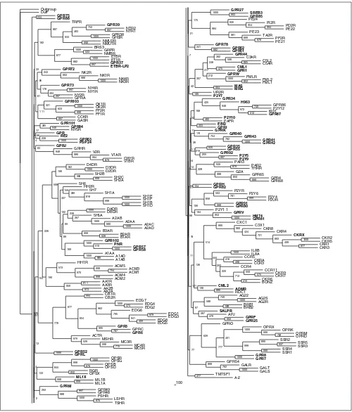
Chemokine receptors
[image:8.609.55.298.405.664.2]Groups A1 and A2 comprise the chemokine receptors (Figure 3). The chemokine ligand superfamily is defined by four conserved cysteines that form two disulfide bonds, and can be structurally subdivided into two major branches based on the spacing of the first cysteine pair. Chemokines in which these residues are adjacent form the CC subfamily (corresponding to the SWISS-PROT CKR nomenclature used here), and those separated by a single amino acid com-prise the CXC subfamily (here CCR and IL8R; for a review see [35]). We had to divide the whole subfamily into two groups to perform a detailed phylogenetic analysis. This sub-grouping produced the same dichotomy, as suggested by the two-ligand motifs, as another example of the parallel evolu-tion of receptors and ligands. Similar results describing this parallel evolution were found previously using a different computational approach [36].
Group A1 mainly comprises the CC family. We hypothesize that the orphan receptor CKRX, which constitutes a separate branch related to CKR1, 2, 3 and 5, might also bind a CC ligand. In contrast, TM7SF1 in this group seems to be only distantly, if at all, related to family-A receptors. It was grouped according to BLASTP results, where a misleading local alignment of approximately 20 amino acids placed it in the vicinity of the chemokine receptors. Group A2 is more heterogeneous and comprises receptors for CC and CXC ligands, as well as an orphan receptor (ADMR) previously thought to bind the peptide adrenomedullin. Adrenomedullin has now been shown to bind a family-B receptor and is discussed further below. The orphan receptor RDC1 in group A2 was first believed to be a receptor for vasointestinal peptide VIP [37], a notion not supported by phylogeny and later dismissed by experimental data [38]. Our results place it closer to the ADMR receptor than to the Figure 3
Chemokine receptors (subgroups A1 and A2). Phylogenetic trees of the subgroups were inferred using Puzzle 5.0 corrected by the JTT substitution frequency matrix. Quartet-puzzling support percentage values from 10,000 puzzling steps are shown. The scale bars indicate a maximum likelihood branch length of 0.1 inferred substitutions per site. Orphan receptors are shaded.
A2
57 66
IL8B IL8A
DUFF
CCR3 CCR5 CCR4
CML2 96
RDC1
81
CKR6 BONZ
80 CCR1 1
CKR9
CKR7
64 51
0.1 CKRA
A1
CKR5
53
CKRX
CKR2
99
CKR1 CKR3
81
CKR4 CKR8
C3X1
72
CXC1
73
0.1
TM7SF1
CKRX
comment
reviews
reports
deposited research
interactions
information
[image:9.609.57.553.84.707.2]refereed research
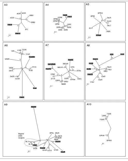
Figure 4
Peptide receptors (subgroups A3-A10). The scale bar indicates a maximum likelihood branch length of 0.1 inferred substitutions per site. Orphan receptors are shaded. For method see Figure 2.
A3 A4 A5
A6
GPRM
CCKR GASR 98
OX1R OX2R
95
GRHR
V2R V1BR OXYR V1AR
94 100
94
GPR103
FF1R
FF2R
75 57 55
0.1
A7 A8
67 98
FMLR FML1 FML2
C3AR
C5AR C5L2
76 75
GPR44 CML1
GPR1
70 62
GPRW
MAS
83
71
0.1
64
A10 AG2S
97
APJ
GPR25 GPRF
75
AG22 AG2R
BRB1
BRB2
83 63
0.1
SSR1 SSR4
97
SSR2 SSR3 SSR596 96
100
OPRX OPRD
OPRMOPRK73 99 100
GPR7 GPR8
85
0.1 UR2R
SALPR
P2Y7 GPRO
GPR54
GALR
GALS
GAL T
100 70 94
0.1
GPR
TRFR GPR38 GHSR
97
BRS3 NMBR GRPR
94 99
ET1R ETBR
98
GPR37 ETBR-LP2
98
83 92
NMU1R NMU2R
56
GPR39
NTR1
NTR2
67 55
0.1
MRG
A9
92 93
GPRA NK2R
NK1R NK3R NK4R
99
GPR72
GPRJ
87
NY5R NY1R NY4R
92 61
NY2R GPR73
57
ML1X
ML1A ML1B
ML1X
98 95
GPR75
92 61
0.1
100
FSHR LSHR
TSHR
59 100
0.1 Biogenic
amine receptors
GPR49
typical chemokine receptors. CML2 is a typical, but distant, member of the chemokine receptor family. The DUFF recep-tor (the Duffy antigen) is also very distantly related and was only grouped into A2 by BLASTP results.
Peptide receptors
Group A3 consists of receptors for the small peptides angiotensin (8 amino acids), bradykinin (9 amino acids) and apelin (Figure 4). Four forms of apelin (12, 13, 17 and 36 amino acids) have been described, but only those of 12 and 13 amino acids bind in nanomolar concentrations [39]. The orphan receptors GPRF and GPR25 in this group are related as closely to the apelin receptor APJ as to the angiotensin or bradykinin receptors, and might also bind small peptides. GPRF acts as a co-receptor for the human immunodeficiency virus (HIV) [40], like the APJ receptor [41], which further hints at structural homology of the two ligands. Opioid and somatostatin receptors make up group A4. Both somatostatin and opioid peptides are derived from the processing of larger precursors. The somatostatins are cyclic peptides of 14 and
28 amino acids. The opioid precursors preproenkephalin, preprodynorphin, prepro-opiomelanocortin and prepronoci-ceptin display a strikingly similar general organization and a conserved amino-terminal region that contains six cysteines, probably involved in disulfide bond formation.
The processed neuropeptides, in contrast, are less similar to each other. It could be speculated that the receptors first bound the precursors themselves, and that the diversity derived from processing is evolutionarily new. Processing prepronociceptin gives rise to two evolutionarily conserved peptides besides orphanin FQ, the ligand for OPRX. It has not been reported whether these peptides bind to the orphan receptors GPR7 and GPR8, which constitute a new branch related to the opioid receptors.
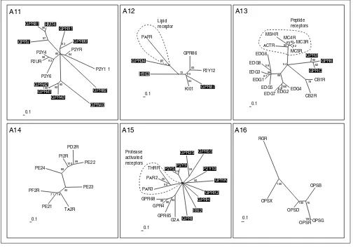
[image:10.609.54.558.88.442.2]In group A5 we find three receptors that bind the 30-amino-acid peptide galanin, and related to these the GPR54 recep-tor, which is activated by the 54-, 14-, and 13-amino-acid peptides derived from the product of KiSS-1, a metastasis Figure 5
Nucleotide and lipid receptors (subgroups A11-A16). The scale bar indicates a maximum-likelihood branch length of 0.1 inferred substitutions per site. Orphan receptors are shaded. For method see Figure 2.
A14
PE23
TA2R PE21 PF2R
71 76 93
PE24 PI2R
PD2R
PE22
99 100 68
0.1
A15 A12 A11
GPR82
GPR40 GPR43
GPR41 GPR42
100 92 92
P2Y6 P2UR
P2Y4
98
92
GPRV
GPR81 HM74
96 87
GPR91
GPR80
83
P2YR
P2Y1 1
64 85
0.1
GPR86
P2Y12
83
GPR87
KI01
64
H963 GPR34
PAFR
63 72
0.1
95
Protease activated receptors
Lipid receptor
A13
99
CB1R
CB2R
78
EDG4 EDG2 EDG7
62100
EDG5 EDG1 EDG3
87
EDG8 EDG6
56 98
ACTR MSHR
MC4R MC3R
MC5R
62 83 67 93
GPR3 GPR6
GPRC
62 92 54
0.1
A16
OPSB
OPSG OPSR
100
OPSD OPSX
RGR
100
100 70
0.1
Peptide receptors
GPR92
GPRH
EBI2
GPRI
G2A GPR65 GPR4 GPR68
99 98 93
PAR3 PAR2
THRR
55 90
P2Y5P2Y9
85
GPR35 GPR55
59 P2Y10 GPRK
50
suppressor gene for melanoma cells. These kisspeptins all share a common RF-amide caboxyl terminus. Although only distantly related to each other, both GPRO (melanin-concen-trating hormone) and UR2R (urotensin II peptide) bind cyclic peptides originally isolated from fish. Similarly distant is the orphan receptor SALPR, which shares sequence simi-larity with somatostatin (A4) and angiotensin (A3) recep-tors, but subgrouping of groups A4 and 5 by neighbor joining led to its placement in group 5. SALPR does not bind somatostatin or angiotensin ligands [42], but could bind another cyclic peptide. The P2Y7 receptor in group A5 does not bind nucleotides [43], as suggested by the name, but was published as a receptor for the lipid leukotriene B4 [44], a notion not supported by phylogeny. In addition, two new leukotriene receptors - CLT1 and CLT2 - have been cloned and characterized during the preparation of this manuscript [45,46] and were found to be unrelated to P2Y7.
Group A6 is again composed solely of receptors for peptide ligands. The orphan receptor GPR103 is related to the neuropeptide FF receptors that bind two amidated mam-malian neuropeptides - NPAF (A-18-F-amide) and NPFF (F-8-F-amide), also known as morphine-modulating peptides. These peptides, which may also be the ligand for GPR103, are members of a large family of neuropeptides related to the molluscan cardioexcitatory neuropeptide (FMRF-amide, Phe-Met-Arg-Phe-amide). The orphan receptors GPRM and GPR in group A6 are most probably also peptide receptors, but are only very distantly related to the others and show no
relationship to receptors with known ligands. Group A7 is also composed of receptors for peptide ligands: neuromedin, neurotensin, motilin, endothelin, bombesin and the releas-ing hormones for growth hormone and thyrotropin. GPR39 might bind a small peptide ligand like the closely related neurotensin receptors NTR1 and 2, which binds a 13-amino-acid peptide derived from a larger precursor protein. GPR37 and ETBR-LP2 are related to each other and branch off the endothelin receptors that bind characteristic bicyclic pep-tides of 21 amino acids containing four cysteines linked by two disulfide bonds.
Group A8 has two branches with receptors with known ligands. These receptors bind the structurally diverse but functionally related chemotactic substances N -formyl-methionyl and the anaphylatoxic complement factors. The N-formylmethionyl ligands are small hydrophilic peptides of bacterial origin, but recently a number of new peptide ago-nists have been identified that selectively activate the high-affinity fMLF receptor FPR and/or its low-high-affinity variant FPRL1. These agonists include peptide domains derived from the envelope proteins of HIV type 1 and at least three amyloidogenic polypeptides, the human acute-phase protein serum amyloid A, the 42-amino-acid form of beta-amyloid peptide and a 21-amino-acid fragment of the human prion protein. Furthermore, a cleavage fragment of neutrophil granule-derived bactericidal cathelicidin, LL-37, is also a chemotactic agonist for FPRL1 (for a review see [47]). The complement factors C3a and C5a are large but highly
comment
reviews
reports
deposited research
interactions
information
[image:11.609.53.557.87.341.2]refereed research
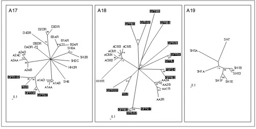
Figure 6
Biogenic amine receptors (subgroups A17-A19). The scale bar indicates a maximum-likelihood branch length of 0.1 inferred substitutions per site. Orphan receptors are shaded. For method see Figure 2.
A17 A19
5H1B 5H1D
100
5H1E 5H1F
100
5H1A 5H5A
5H7
74
0.1
98
5H2C
HH2R
5H6 A1AB A1AA A1AD
57 95
GPR57 GPR58
98
PNR GPR102
96 90
A2AB A2AA
A2ACA2AD97 59
88
DADR DBDR
79
D4DR D2DR
D3DR
66
74
B3AR B1AR
B2AR
80 69
5H2A
5H2B
54
0.1
A18
HH1R
GPR78
GPR52
GPRL
97
PSP24
GPR63
96
AA2A AA2B
99
AA1R
AA3R
95 94
GPR27 SREB3 GPR8555 86
RE2 GPR101
83
ACM2 ACM4
86
ACM1 ACM3 ACM5
66 72
74
GPR61 GPR62
61
GPR88
GPR84
51
hydrophilic proteins with a mainly alpha-helical structure held together by three disulfide bridges. C5a is rapidly desarginated to the less potent derivative C5adR74, which is the ligand for the C5L2 receptor. The orphan receptors GPR1, CML1 and GPR44 all cluster, and constitute a sepa-rate branch as distant as the other two branches. No predic-tion of the possible structure of the ligands for these receptors can be derived from this tree, but maybe they will function as chemotactic peptides. This could at least hint at leukocytes or inflamed tissue as a possible source for these ligands. The receptor GPRW constitutes its own branch, not as distant to the main group as the MAS oncogene product and the related receptor MRG, which are only very distantly related to the group.
All receptors in group A9 with known ligands bind peptides, except for a side branch consisting of receptors for the bio-genic amine melatonin. The orphan receptor ML1X is closely related to melatonin receptors ML1A and B, but apparently does not bind melatonin [48]. GPR73 is related to the neuro-peptide Y (NPY) receptor NY2R which mainly binds the pan-creatic peptide YY of 36 amino acids, and these two are placed together on a branch distinct from the NPY receptors NY4R and NY1R. GPR73 does not bind the NPY ligand family [49], but possibly a similar large peptide ligand. The orphan receptors GPR72 and GPRJ constitute a new
subgroup that most probably bind related peptide ligands. GPR72 does not bind a NPY ligand [49]. GPR75 is only very distantly related to the whole A9 group. The receptors for the glycoprotein hormones thyroid-stimulating hormone (TSH), luteinizing hormone (LSH) and follicle-stimulating hormone (FSH) make up Group A10. GPR48 and 49 are very similar in their overall structure, with long amino termini, but their relationship is also evident in the neighbor-joining tree con-structed from alignments without amino and carboxyl termini. It has been recently shown that these receptors mediate the action of relaxin, a peptide hormone of the insulin-like growth factor family secreted by the corpus luteum during pregnancy [50].
Nucleotide and lipid receptors
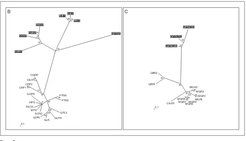
[image:12.609.55.557.85.373.2]The receptors with known ligands in group A11 are the P2Y receptors, which bind pyrimidine as well as purine nucleotides (Figure 5). Several orphan receptors constitute new clusters. GPR80 and GPR91 are distantly related to each other and rel-atively close to the P2Y receptors. GPR80 is the closest relative of the newly identified CLT2 receptor for leukotrienes as judged by BLASTP results. GPR81, HM74 and GPRV and GPR 40-43 belong to branches only distantly related to P2Y recep-tors. Within these potential new subfamilies, GPR41-43, GPR81 and HM74 are more closely related to each other than to GPR40 (for GPR41-43) and GPRV (for GPR81 and HM74). Figure 7
Families B and C of the G-protein-coupled receptors (GPRCs). Phylogenetic trees of families B and C were inferred using Puzzle 5.0 corrected by the JTT substitution frequency matrix. Quartet-puzzling support percentage values from 10,000 puzzling steps are shown. The scale bar indicates a maximum likelihood branch length of 0.1 inferred substitutions per site. Orphan receptors are shaded.
B C
PTRR PTR2
98
GPL2 GLPR GLR GIPR 68
60 89
SCRC VIPR VIPS PACR
GRFR
67 92
CRF1 CRF2
88
CALR
CGRR
87
74 EMR1
CD97 EMR2
EMR3
78
73
92
BAI1 BAI2
BAI3 90 86
GPR56
53
98
90
MGR2 MGR3
89
MGR1 MGR5
95
MGR4 MGR6 MGR7 MGR8 93
63
CASR GBR1
GBR2
99 GPRC5B
GPRC5C GPRC5D
99
97
86
86
0.1
In group A12, the platelet-activated receptor, a lipid receptor and receptors activated by nucleotides mingle, but are found on different side branches. The orphan receptor GPR87 is closely related to the receptor for UDP-glucose KI01 and to the ADP-binding receptors P2Y12 and GPR86. We assume that this receptor might also bind UDP-glucose or another modified nucleotide. GPR34 is distantly related to the platelet-activating factor (PAF) receptor; it was not activated by available lipid ligands [51], but might nevertheless bind a lipid ligand. Group A13 contains both peptide and lipid receptors but they make up different branches. The peptide branch binds peptides derived from the processing of pro-opiomelanocortin that gives rise to peptides of between 12 and 36 amino acids. The EDG and cannabinoid receptors constitute clusters, and one cluster distinct from the other three consists of the orphan receptors GPR3, GPR6 and GPRC, which have been grouped closer to the lipid EDG receptors in the overall neighbor-joining tree (Figure 2). This information helped to identify a phospholipid ligand for GPRC (H. Chica Schaller, personal communication).
The receptors in group A14 all bind ligands derived from arachidonic acid by the action of cyclooxygenase. These receptors for lipid-derived autacoids or prostanoids com-prise receptors for the prostaglandins and thromboxanes. There are no orphan receptors in this group. Group A15 is a very heterogenous group composed of receptors for the lipids sphingosylphosphorylcholine (SPC), lysophos-phatidylcholine (LPC) and psychosine, and receptors acti-vated by proteases. GPR4 and GPR68 both bind SPC, like the EDG receptor branch consisting of the EDG1, 3, 6 and 8 receptors in A13, but are not closely related. Protease-activated receptors become Protease-activated by a part of the former amino terminus cleaved by the protease. The new amino terminus then functions as a tethered ligand and activates the receptor. This can be mimicked by very small peptides derived from this ligand; such receptors should therefore rather resemble peptide receptors. The orphans P2Y5, P2Y9 and P2Y10 receptors were not placed in group 11 and 12 like most P2Y receptors, but in group A15, sup-porting the fact that they were misnamed. P2Y5 and P2Y9 do not bind nucleotides [52,53], but this has not been shown yet for P2Y10. All other orphan receptors in this group, with the exception of GPR35 and GPR55 which cluster together, are as distantly related to each other as to the receptors with known ligands. Group A16 contains the opsins, receptors that are activated by isoprenoid ligands, and no orphan receptors.
Biogenic amine receptors
Some serotonin receptors and receptors for the biogenic amines adrenaline, dopamine and histamine are all placed on different branches in group A17 (Figure 6). An additional branch consists of the orphan receptors GPR102, PNR, GPR57 and GPR58, which are as distantly related to the others as, for example, is the alpha-adrenergic receptor
branch. PNR and GPR58 expressed in COS cells did not bind various serotonin receptor-specific ligands [54]. Their ligands might be small molecules with similar properties. Group A18 is very heterogeneous and consists of receptors for the biogenic amines acetylcholine and adenosine, and the HH1R receptor for histamine, as well as many distantly related orphan GPCRs. GPR63 is closely related to the orphan receptor PSP24. The Xenopus laevishomolog of this receptor binds LPA [55]. GPR101 and RE2, GPRL and GPR52, and GPR61 and GPR62 constitute their own sub-groups. In particular, the SREB1-3 cluster (GPR85, GPR27 and SREB3) makes up its own family, with only a distant relationship to other GPCRs in this group. No orphan recep-tors are found in group A19, which consists entirely of sero-tonin receptors distinct from those in A17.
During the preparation of this manuscript several new family-A receptors that could not be fitted into our analysis were identified. These comprise 15 new receptors distinct from the classical biogenic amine receptors that apparently bind the trace amines tyramine, -phenylethylamine, trypta-mine and octopatrypta-mine [56]. In addition, a new subfamily of GPCRs related to the masoncogene and uniquely expressed in small nociceptive sensory neurons were shown to be the receptors for a number of enkephalin fragments [57].
Receptor families B and C
Family B (Figure 7) was named after the secretin receptor. Yet proteins showing homology to this receptor make up only one of four distantly related subgroups. The receptors EMR1, EMR2 and EMR3, and the CD97 surface antigen, all have several epidermal growth factor (EGF)-like domains in the extracellular amino terminus. They constitute their own cluster only distantly related to the rest of the family. The same applies to the brain-specific angiogenesis inhibitor family BAI1-3. GPR56 was assigned to family B because it shows the typical signature [58], but is so far the only one of its kind. So far no non-protein ligand has been identified as a ligand for family-B receptors. Astonishingly, one family-B receptor, namely the CGRP receptor, requires coexpression with single transmembrane receptor activity-modifying proteins (RAMP1-3) for ligand binding and signal transduc-tion [59]. Coexpression of different RAMPs results in binding of different cyclic peptide ligands such as adrenomedullin, amylin or the calcitonin gene-related peptide (for a review see [60]). This could further compli-cate the identification of the cognate ligands for these family-B orphan receptors, but we assume that they will also bind large peptide ligands. In family C (Figure 7), the metabotropic glutamate receptors MGR1-8 bind the small molecule glutamate, the CASR receptor senses extracellular calcium concentration, and receptors GBR1-2 bind the small molecule gamma-amino butyric acid (GABA). GPRC5B, C and D constitute their own subgroup with no closer relationship to the other members, but might also bind small molecules.
comment
reviews
reports
deposited research
interactions
information
Conclusions
In this work, we calculated the phylogenetic distances of 277 human GPCRs and show the relationship of orphan recep-tors to receprecep-tors for known ligands with support values for each branch. We then grouped orphan receptors and recep-tors with known ligands into 19 subgroups that sometimes differ from previous classifications. Three subgroups are composed of receptors for ligands that belong to different substance classes; for example, in group A12, lipid receptors and receptors activated by nucleotides mingle, and in groups A13 and A15, peptide and lipid receptors. In both subgroups the receptors binding ligands of different substance classes make up different branches. We hope that this approach proves valuable for identifying the natural ligands of orphan receptors, as related receptors have previously been shown to have ligands with similar structural features.
Materials and methods
Sequence database mining
A database search excluding olfactory and gustatory recep-tors identified the amino-acid sequences of 281 human GPCRs. Only sequences annotated as GPCRs in the following databases were used: NCBI [61], SWISS-PROT [62], EMBL [63] and GPCRDB [34,64]. Receptors without published ligands in PubMed [65] were defined as orphan GPCRs.
Multiple sequence alignments
Multiple protein sequences were aligned with ClustalX 1.81 [66]. Pairwise alignment parameters were set as: slow/accu-rate alignment; gap opening penalty 10; gap extension penalty 0.10; protein weight matrix BLOSUM 30. Multiple alignment parameters were set as: gap opening penalty 10; gap extension penalty 0.05; delay divergent sequences 35%; protein weight matrix BLOSUM series [67]. The alignments were modified by deleting the extremely variable amino termini upstream of the first transmembrane domain and carboxyl termini downstream of the seventh transmembrane domain. Alignment editing and shading was done using BioEdit Sequence Alignment Editor [68] and GeneDoc Mul-tiple Sequence Alignment Editor [69]. Transmembrane domains were identified using the TMpred program [70] and, where available, data from the original publication [71].
Clustering of subgroups
An overall phylogenetic tree of family A was inferred from the multiple sequence alignment with PHYLIP 3.6 [72]. Bootstrapping was performed 1,000 times using SEQBOOT to obtain support values for each internal branch. Pairwise distances were determined with PROTDIST and the JTT substitution frequency matrix [73]. Neighbor-joining phylo-genetic trees [21] were calculated with NEIGHBOR using standard parameters. The human GPRC5B receptor belong-ing to family B was used as outgroup for family A. The out-group sequence is supposed to be a distant, though related, sequence and is used to root the tree. The majority-rule
consensus trees of all bootstrapped sequences were obtained with the program CONSENSE. Representations of the calcu-lated trees were constructed with TreeView [74]. Clusters with bootstrap values greater than 50% were defined as confirmed subgroups, and sequences with lower values added to these subgroups according to their sequence similarity in the align-ment as judged by visual inspection and the results of pairwise local alignments with all other sequences by BLASTP [25]. The p-value was used as a measure of similarity.
Quartet-puzzling trees
Multiple protein sequence alignments of these new subgroups were created as described above. Phylogenetic trees were inferred from these alignments using Puzzle 5.0 [75] to calcu-late maximum-likelihood distances corrected by the JTT sub-stitution-frequency matrix [73] with amino-acid usage estimated from the data, site-to-site rate variation modeled on a gamma distribution with eight rate categories plus invariant sites, and the shape parameter estimated from the data. The human GPRC5B receptor of family B was used as an outgroup for family A. The human 5H1A receptor of family A was used as an outgroup for families B and C (the outgroups are not shown in the figures here). Quartet-puzzling (QP) trees were constructed with the described settings and 10,000 puzzling steps to obtain support values (QP reliability) for each internal branch. The program Puzzle 5.0 was used in a parallelized version (ppuzzle) with a message-passing inter-face (MPI) implementation on a HP 9000 N-Class Enterprise Server Cluster consisting of five HP 9000 N-Class shared-memory multiprocessor systems with eight PA-RISC 8600 (552 MHz) processors each. Representations of the quartet-puzzling trees were constructed with TreeView [74].
Additional data files
Additional data files available with the online verson of this paper include a data table with names, synonyms and acces-sion numbers of all GPCRs, and the BLASTP results of all GPCRs (full-length sequences and sequences without amino or carboxyl termini).
Acknowledgements
The DFG Graduiertenkolleg 255, the Dr Kurt und Irmgard Meister-Stiftung and the Hamburgische Wissenschaftliche Gesellschaft, supported this study. We appreciate the help of Chica Schaller in finding additional sequences and of Andreas Schuldei in reconfiguring ppuzzle and using MPI. Klaus Martens and his colleagues at the computing center of the Technical University Hamburg-Harburg provided an account at the HP N-Class Enterprise Server Cluster and helped us to use the software environment.
References
1. Gether U: Uncovering molecular mechanisms involved in activation of G protein-coupled receptors. Endocr Rev 2000,
21:90-113.
3. Methner A, Hermey G, Schinke B, Hermans-Borgmeyer I: A novel G protein-coupled receptor with homology to neuropeptide and chemoattractant receptors expressed during bone development. Biochem Biophys Res Commun 1997, 233:336-342. 4. Lee D, George S, Evans J, Lynch K, O’Dowd B: Orphan G
protein-coupled receptors in the CNS.Curr Opin Pharmacol 2001, 1: 31-39.
5. Reinscheid RK, Nothacker HP, Bourson A, Ardati A, Henningsen RA, Bunzow JR, Grandy DK, Langen H, Monsma FJ Jr, Civelli O:
Orphanin FQ: a neuropeptide that activates an opioidlike G protein-coupled receptor. Science 1995, 270:792-794.
6. Civelli O, Nothacker HP, Saito Y, Wang Z, Lin SH, Reinscheid RK:
Novel neurotransmitters as natural ligands of orphan G-protein-coupled receptors.Trends Neurosci 2001, 24:230-237. 7. Drews II: Drug discovery today - and tomorrow. Drug Discov
Today 2000, 5:2-4.
8. Lee MJ, Van Brocklyn JR, Thangada S, Liu CH, Hand AR, Menzeleev R, Spiegel S, Hla T: Sphingosine-1-phosphate as a ligand for the G protein-coupled receptor EDG-1. Science 1998, 279: 1552-1555.
9. Van Brocklyn JR, Tu Z, Edsall LC, Schmidt RR, Spiegel S: Sphingo-sine 1-phosphate-induced cell rounding and neurite retrac-tion are mediated by the G protein-coupled receptor H218.
J Biol Chem 1999, 274:4626-4632.
10. Van Brocklyn JR, Graler MH, Bernhardt G, Hobson JP, Lipp M, Spiegel S: Sphingosine-1-phosphate is a ligand for the G protein-coupled receptor EDG-6.Blood 2000, 95:2624-2629. 11. Im DS, Heise CE, Ancellin N, O’Dowd BF, Shei GJ, Heavens RP,
Rigby MR, Hla T, Mandala S, McAllister G, et al.: Characterization of a novel sphingosine 1-phosphate receptor, Edg-8. J Biol Chem 2000, 275:14281-14286.
12. Hecht JH, Weiner JA, Post SR, Chun J: Ventricular zone gene-1 (vzg-1) encodes a lysophosphatidic acid receptor expressed in neurogenic regions of the developing cerebral cortex.J Cell Biol 1996, 135:1071-1083.
13. An S, Bleu T, Hallmark OG, Goetzl EJ: Characterization of a novel subtype of human G protein-coupled receptor for lysophosphatidic acid.J Biol Chem 1998, 273:7906-7910. 14. Im DS, Heise CE, Harding MA, George SR, O’Dowd BF,
Theodor-escu D, Lynch KR: Molecular cloning and characterization of a lysophosphatidic acid receptor, Edg-7, expressed in prostate.Mol Pharmacol 2000, 57:753-759.
15. Szekeres PG, Muir AI, Spinage LD, Miller JE, Butler SI, Smith A, Rennie GI, Murdock PR, Fitzgerald LR, Wu H, et al.: Neuromedin U is a potent agonist at the orphan G protein-coupled receptor FM3.J Biol Chem 2000, 275:20247-20250.
16. Zhu Y, Michalovich D, Wu H, Tan KB, Dytko GM, Mannan IJ, Boyce R, Alston J, Tierney LA, Li X, et al.: Cloning, expression, and pharmacological characterization of a novel human hista-mine receptor.Mol Pharmacol 2001, 59:434-441.
17. Communi D, Gonzalez NS, Detheux M, Brezillon S, Lannoy V, Par-mentier M, Boeynaems JM: Identification of a novel human ADP receptor coupled to G(i).J Biol Chem 2001, 276:41479-41485. 18. Chambers JK, Macdonald LE, Sarau HM, Ames RS, Freeman K, Foley
JJ, Zhu Y, McLaughlin MM, Murdock P, McMillan L, et al.: A G protein-coupled receptor for UDP-glucose.J Biol Chem 2000,
275:10767-10771.
19. Attwood TK, Findlay JB: Fingerprinting G-protein-coupled receptors.Protein Eng 1994, 7:195-203.
20. Kolakowski LF Jr: GCRDb: a G-protein-coupled receptor data-base.Receptors Channels 1994, 2:1-7.
21. Saitou N, Nei M: The neighbor-joining method: a new method for reconstructing phylogenetic trees. Mol Biol Evol 1987,
4:406-425.
22. Page RD, Holmes EC: Molecular Evolution: A Phylogenetic Approach. Oxford: Blackwell Science; 1999: 218-225.
23. Kumar S, Gadagkar SR: Efficiency of the neighbor-joining method in reconstructing deep and shallow evolutionary relationships in large phylogenies.J Mol Evol 2000, 51:544-553. 24. Fitch WM, Margoliash E: Construction of phylogenetic trees.
Science 1967, 155:279-284.
25. Altschul SF, Gish W, Miller W, Myers EW, Lipman DJ: Basic local alignment search tool. J Mol Biol 1990, 215:403-410.
26. Felsenstein J: Phylogenies from molecular sequences: infer-ence and reliability.Annu Rev Genet 1988, 22:521-565.
27. Strimmer K, Goldman N, von Haeseler A: Bayesian Probabilities and Quartet Puzzling. Mol Biol Evol 1997, 14:210-211.
28. Strimmer K, von Haeseler A: Quartet puzzling: a quartet maximum-likelihood method for reconstructing tree topologies.Mol Biol Evol 1996, 13:964-211.
29. Strimmer K, Robertson DL: Inference and applications of mole-cular phylogenies: an introductory guide. In The Internet for Molecular Biologists (Practical Approach Series), edited by Sansom C, Horton RM. Oxford: Oxford University Press; 2001.
30. Pin JP, Joly C, Heinemann SF, Bockaert J: Domains involved in the specificity of G protein activation in phospholipase C-coupled metabotropic glutamate receptors. EMBO J 1994,
13:342-348.
31. Probst WC, Snyder LA, Schuster DI, Brosius J, Sealfon SC:
Sequence alignment of the G-protein coupled receptor superfamily.DNA Cell Biol 1992, 11:1-20.
32. Burbach JP, Meijer OC: The structure of neuropeptide recep-tors.Eur J Pharmacol 1992, 227:1-18.
33. Graul R, Sadee W: Evolutionary relationships among G protein-coupled receptors using a clustered database approach.AAPS PharmSci 2001, 3:E12.
34. Horn F, Weare J, Beukers MW, Horsch S, Bairoch A, Chen W, Edvardsen O, Campagne F, Vriend G: GPCRDB: an information system for G protein-coupled receptors.Nucleic Acids Res 1998,
26:275-279.
35. Cascieri MA, Springer MS: The chemokine/chemokine-receptor family: potential and progress for therapeutic intervention.
Curr Opin Chem Biol 2000, 4:420-427.
36. Goh C, Bogan A, Joachimiak M, Walther D, Cohen F: Co-evolution of proteins with their interaction partners. J Mol Biol 2000,
299:283-293.
37. Sreedharan SP, Robichon A, Peterson KE, Goetzl EJ: Cloning and expression of the human vasoactive intestinal peptide receptor. Proc Natl Acad Sci USA 1991, 88:4986-4990.
38. Nagata S, Ishihara T, Robberecht P, Libert F, Parmentier M, Christophe J, Vassart G: RDC1 may not be VIP receptor.Trends Pharmacol Sci 1992, 13:102-103.
39. Tatemoto K, Hosoya M, Habata Y, Fujii R, Kakegawa T, Zou MX, Kawamata Y, Fukusumi S, Hinuma S, Kitada C, et al.: Isolation and characterization of a novel endogenous peptide ligand for the human APJ receptor. Biochem Biophys Res Commun 1998,
251:471-476.
40. Farzan M, Choe H, Martin K, Marcon L, Hofmann W, Karlsson G, Sun Y, Barrett P, Marchand N, Sullivan N, et al.: Two orphan seven-transmembrane segment receptors which are expressed in CD4-positive cells support simian immunodefi-ciency virus infection.J Exp Med 1997, 186:405-411.
41. Choe H, Farzan M, Konkel M, Martin K, Sun Y, Marcon L, Cayabyab M, Berman M, Dorf ME, Gerard N, et al.: The orphan seven-transmembrane receptor apj supports the entry of primary T-cell-line-tropic and dualtropic human immunodeficiency virus type 1.J Virol 1998, 72:6113-6118.
42. Matsumoto M, Kamohara M, Sugimoto T, Hidaka K, Takasaki J, Saito T, Okada M, Yamaguchi T, Furuichi K: The novel G-protein coupled receptor SALPR shares sequence similarity with somatostatin and angiotensin receptors.Gene 2000, 248: 183-189.
43. Herold CL, Li Q, Schachter JB, Harden TK, Nicholas RA: Lack of nucleotide-promoted second messenger signaling responses in 1321N1 cells expressing the proposed P2Y receptor, p2y7. Biochem Biophys Res Commun 1997, 235:717-721.
44. Yokomizo T, Izumi T, Chang K, Takuwa Y, Shimizu T: A G-protein-coupled receptor for leukotriene B4 that mediates chemo-taxis. Nature 1997, 387:620-624.
45. Lynch KR, O’Neill GP, Liu Q, Im DS, Sawyer N, Metters KM, Coulombe N, Abramovitz M, Figueroa DJ, Zeng Z, et al.: Charac-terization of the human cysteinyl leukotriene CysLT1 receptor. Nature 1999, 399:789-793.
46. Takasaki J, Kamohara M, Matsumoto M, Saito T, Sugimoto T, Ohishi T, Ishii H, Ota T, Nishikawa T, Kawai Y, et al: The molecular char-acterization and tissue distribution of the human cysteinyl leukotriene CysLT(2) receptor. Biochem Biophys Res Commun 2000, 274:316-322.
47. Le Y, Yang Y, Cui Y, Yazawa H, Gong W, Qiu C, Wang JM: Recep-tors for chemotactic formyl peptides as pharmacological targets. Int Immunopharmacol 2002, 2:1-13.
48. Reppert SM, Weaver DR, Ebisawa T, Mahle CD, Kolakowski LF Jr: Cloning of a melatonin-related receptor from human pitu-itary. FEBS Lett 1996, 386:219-224.
comment
reviews
reports
deposited research
interactions
information
49. Parker R, Liu M, Eyre HJ, Copeland NG, Gilbert DJ, Crawford J, Sutherland GR, Jenkins NA, Herzog H: Y-receptor-like genes GPR72 and GPR73: molecular cloning, genomic organisa-tion and assignment to human chromosome 11q21.1 and 2p14 and mouse chromosome 9 and 6. Biochim Biophys Acta 2000, 1491:369-375.
50. Hsu S, Nakabayashi K, Nishi S, Kumagai J, Kudo M, Sherwood O, Hsueh A: Activation of orphan receptors by the hormone relaxin.Science 2002, 295:671-674.
51. Schoneberg T, Schulz A, Grosse R, Schade R, Henklein P, Schultz G, Gudermann T: A novel subgroup of class I G-protein-coupled receptors.Biochim Biophys Acta 1999, 1446:57-70.
52. Li Q, Schachter JB, Harden TK, Nicholas RA: The 6H1 orphan receptor, claimed to be the p2y5 receptor, does not mediate nucleotide-promoted second messenger responses.
Biochem Biophys Res Commun 1997, 236:455-460.
53. Janssens R, Boeynaems JM, Godart M, Communi D: Cloning of a human heptahelical receptor closely related to the P2Y5 receptor. Biochem Biophys Res Commun 1997, 236:106-112. 54. Lee DK, Lynch KR, Nguyen T, Im DS, Cheng R, Saldivia VR, Liu Y,
Liu IS, Heng HH, Seeman P, et al.: Cloning and characterization of additional members of the G protein- coupled receptor family.Biochim Biophys Acta 2000, 1490:311-323.
55. Guo Z, Liliom K, Fischer DJ, Bathurst IC, Tomei LD, Kiefer MC, Tigyi G: Molecular cloning of a high-affinity receptor for the growth factor-like lipid mediator lysophosphatidic acid from
Xenopusoocytes. Proc Natl Acad Sci USA 1996, 93:14367-14372. 56. Borowsky B, Adham N, Jones K, Raddatz R, Artymyshyn R, Ogozalek
K, Durkin M, Lakhlani PP, Bonini JA, Pathirana S, et al.: Trace amines: identification of a family of mammalian G protein-coupled receptors. Proc Natl Acad Sci USA 2001, 98:8966-8971. 57. Lembo P, Grazzini E, Groblewski T, O’Donnell D, Roy M, Zhang J,
Hoffert C, Cao J, Schmidt R, Pelletier M, et al.: Proenkephalin A gene products activate a new family of sensory neuron-spe-cific GPCRs.Nat Neurosci 2002, 5:201-209.
58. Liu M, Parker RM, Darby K, Eyre HJ, Copeland NG, Crawford J, Gilbert DJ, Sutherland GR, Jenkins NA, Herzog H: GPR56, a novel secretin-like human G-protein-coupled receptor gene.
Genomics 1999, 55:296-305.
59. McLatchie LM, Fraser NJ, Main MJ, Wise A, Brown J, Thompson N, Solari R, Lee MG, Foord SM: RAMPs regulate the transport and ligand specificity of the calcitonin- receptor-like receptor.
Nature 1998, 393:333-339.
60. Muff R, Born W, Fischer J: Adrenomedullin and related peptides: receptors and accessory proteins. Peptides 2001, 22:1765-1772. 61. National Center for Biotechnology Information
[http://www.ncbi.nlm.nih.gov]
62. The Swiss 7TM Search Tool [http://www.expasy.ch/cgi-bin/search-7tm]
63. European Bioinformatics Institute [http://www.ebi.ac.uk] 64. GPCRDB: Information system for G protein-coupled
recep-tors (GPCRs)
[http://www.gpcr.org] 65. PubMed
[http://www.ncbi.nlm.nih.gov/entrez/query.fcgi?db=PubMed] 66. Thompson JD, Gibson TJ, Plewniak F, Jeanmougin F, Higgins DG:
The CLUSTAL_X windows interface: flexible strategies for multiple sequence alignment aided by quality analysis tools.
Nucleic Acids Res 1997, 25:4876-4882.
67. Henikoff S, Henikoff JG: Amino acid substitution matrices from protein blocks.Proc Natl Acad Sci USA 1992, 89:10915-10919. 68. Hall TA: BioEdit: a user-friendly biological sequence
align-ment editor and analysis program for Windows 95/98/NT.
Nucleic Acids Symp Ser 1999:95-98.
69. GeneDoc [http://www.psc.edu/biomed/genedoc/]
70. Hofmann K, Stoffel W: TMbase - A database of membrane span-ning proteins segments. Biol Chem Hoppe-Seyler 1993, 374:166. 71. TMpred - Prediction of Transmembrane Regions and
Orien-tation [http://www.ch.embnet.org/software/TMPRED_form.html] 72. PHYLIP [http://evolution.genetics.washington.edu/phylip.html] 73. Jones DT, Taylor WR, Thornton JM: The rapid generation of
mutation data matrices from protein sequences. Comput Appl Biosci 1992, 8:275-282.