REVIEW
Genomic imprinting in development, growth, behavior and
stem cells
Robert N. Plasschaert and Marisa S. Bartolomei*,‡
ABSTRACT
Genes that are subject to genomic imprinting in mammals are preferentially expressed from a single parental allele. This imprinted expression of a small number of genes is crucial for normal development, as these genes often directly regulate fetal growth. Recent work has also demonstrated intricate roles for imprinted genes in the brain, with important consequences on behavior and neuronal function. Finally, new studies have revealed the importance of proper expression of specific imprinted genes in induced pluripotent stem cells and in adult stem cells. As we review here, these findings highlight the complex nature and developmental importance of imprinted genes.
KEY WORDS: Imprinted genes, Fetal growth, DNA methylation, Induced pluripotency, Behavior, Neuronal development
Introduction
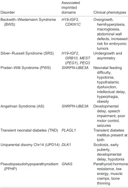
Mammals inherit two sets of chromosomes, one from each parent, and therefore possess two copies of each gene. For the majority of these genes, both alleles are expressed or repressed, depending upon the cell type. However, a small number of genes, designated imprinted genes, are monoallelically expressed in a parent-of-origin-specific manner. The murine genome contains∼150 such imprinted genes, although this number is likely to increase as more tissue-specific imprinting is described. Importantly, imprinting is well-conserved across mammals, with many, but not all, imprinted genes and imprinting mechanisms being conserved between mouse and human (Lee and Bartolomei, 2013). This conservation has greatly facilitated the study of imprinting, as researchers have used both experimental mouse models and human genetic disorders to expand our knowledge of imprinting. A significant consequence of imprinting is that mammalian development requires genetic contributions from both a mother and a father. Moreover, a number of rare congenital disorders (Table 1) are caused by parental-allele-specific mutation or misregulation of one or more imprinted genes (Butler, 2009).
Imprinted genes are typically located in clusters of 3-12 genes that are spread over 20 kb-3.7 Mb of DNA, although examples of single imprinted genes do exist (Edwards and Ferguson-Smith, 2007). The clusters harbor maternally and paternally expressed imprinted genes that encode both protein-coding and non-coding (nc) RNAs. Each well-studied cluster has a discrete imprinting control region (ICR) that governs imprinted expression and exhibits parent-of-origin-specific epigenetic marks, such as DNA methylation and post-translational histone modifications. Much
work over the past 25 years has centered on the identification of imprinted genes and understanding the mechanisms that underlie imprinted expression. For example, how imprints are acquired and reset in the germline and subsequently maintained and read after fertilization has been the subject of intense study. Although less effort has been focused on elucidating the function of imprinted genes, recent experiments have revealed broad roles for imprinted gene expression in mammals. These studies have shown that the small number of genes that exhibit imprinted expression have a large influence on mammalian development. As more imprinted genes have been discovered, it has become clear that they exert their effects on numerous and varied processes, including fetal growth, pluripotency, differentiation and behavior. Here, we review the mechanisms of imprinted gene regulation and the diverse roles of imprinted genes during development, highlighting recent work that expounds on their functional importance.
Establishing, maintaining and erasing imprints
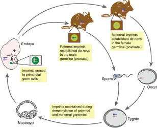
The identification of the first imprinted genes (Igf2r,Igf2andH19) in 1991 (Barlow et al., 1991; Bartolomei et al., 1991; DeChiara et al., 1991) sparked initial efforts towards elucidating the mechanisms of imprint establishment, maintenance and erasure, which together control the timing and placement of genomic imprinting (Fig. 1). Allele-specific DNA methylation of ICRs has been pursued as the best candidate mechanism for conferring parental-specific imprints following fertilization. Moreover, the hypothesis that parental-specific imprints are imposed when the parental genomes can be distinguished prompted investigators to assay methylation acquisition during gametogenesis, when the maternal and paternal genomes are in separate compartments and can be independently modified. It was initially shown that paternal-specific methylation of theH19/Igf2ICR is acquired prenatally in prospermatogonia prior to the onset of meiosis in the male germline (Davis et al., 2000). By contrast, maternal-specific ICR methylation occurs postnatally in growing oocytes, with different ICRs being methylated at a slightly different time during oocyte growth (Lucifero et al., 2004). In both germlines, DNA methylation is established through the action of thede novoDNA methyltransferase 3a (DNMT3A) and the accessory protein DNMT3L (Bourc’his et al., 2001; Hata et al., 2002; Kaneda et al., 2004; Okano et al., 1999). Although it remains poorly understood how specific sequences are chosen for allele-specific DNA methylation in the germline, recent work has indicated that transcription through ICR sequences provides a key instructive step for the DNA methyltransferase proteins (Chotalia et al., 2009; Henckel et al., 2012).
Following fertilization, the parental-specific imprints must be maintained despite the extensive genome reprogramming and DNA demethylation that occurs at this time (Bartolomei and Ferguson-Smith, 2011). In addition to the action of the maintenance DNA methytransferase DNMT1, which methylates the newly synthesized strand of DNA, it is likely that the recognition of uniquecis-acting
Department of Cell & Developmental Biology, Perelman School of Medicine at the University of Pennsylvania, Philadelphia, PA 19104, USA.
*Present address: 9-123 SCTR, 3400 Civic Center Blvd, Philadelphia, PA 19104, USA.
‡
Author for correspondence ([email protected])
DEVEL
O
sequences by trans-acting factors provides protection from post-fertilization reprogramming. For example, the maternal factor PGC7 (also known as STELLA or DPPA3) plays a general role in maintaining DNA methylation in the early mouse embryo, acting via interactions with dimethylated histone 3, lysine 9 residues (Nakamura et al., 2012). In addition, zinc finger protein homolog 57 (ZFP57) appears to play a more specific role in regulating imprinted genes. ZFP57mutations have been identified in transient neonatal diabetes patients and are associated with defective DNA methylation at several imprinted loci (Mackay et al., 2008). In line with this,Zfp57null mice show loss of imprinting at many, but not all, loci (Li et al., 2008). It is possible that other yet-to-be-identified proteins also maintain DNA methylation at ICRs in the early embryo.
Finally, to complete the imprinting cycle, the somatic pattern of biparental imprints is erased in primordial germ cells (PGCs), which are recruited from somatic cells in the early embryo. Although this process of erasure is poorly understood, it appears that imprints are lost through a series of active and passive events, including those involving the action of the ten-eleven translocation (Tet) family of methylcytosine dioxygenases, which catalyze the oxidation of 5-methylcytosine to 5-hydroxymethylcytosine (Dawlaty et al., 2013; Hackett et al., 2013; Yamaguchi et al., 2013), as well as the
action of DNA repair machinery (Pastor et al., 2013). Furthermore, the methylation of newly replicated DNA by DNMT1 is repressed in PGCs, probably by repression of Uhrf1, a factor essential for recruiting DNMT1 to the replication fork (Kagiwada et al., 2012).
Mechanisms of imprinted gene regulation
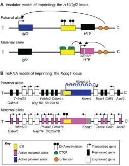
Two well-defined mechanisms of imprinted gene regulation have been described: the insulator model and the ncRNA model (Lee and Bartolomei, 2013). The insulator model (Fig. 2A) is best illustrated at theH19/Igf2locus. In this example, the ICR on the maternal allele is unmethylated and is bound by the insulator protein CCCTC binding factor (CTCF). This binding prevents the downstream enhancers that are shared by bothH19andIgf2from engaging the Igf2promoter, but allows the enhancers to accessH19and activate its expression. On the paternal allele, the ICR is hypermethylated, which prevents CTCF from binding and the insulator from forming. Consequently, theIgf2gene is activated by the shared downstream enhancers. Thus, in this model, the epigenetic state of the ICR determines the expression pattern of the locus.
The other major imprinting model is the ncRNA model (Fig. 2B), which is employed at a number of loci, including Igf2r/Airn and Kcnq1/Kcnq1ot1. In this case, the promoter of a long ncRNA is located within the ICR. On the paternal allele, the ICR is unmethylated thus allowing expression of the ncRNA, which in turn silences the rest of the genes in the domain incis. This occurs by either attracting machinery that lay down repressive chromatin marks (Nagano and Fraser, 2009) or by preventing RNA polymerase II recruitment at promoters (Latos et al., 2012), although these mechanisms are not fully understood. Methylation of the ICR on the opposing maternal allele results in silencing of the ncRNA, thereby allowing activation of proximal genes. Thus, in this example, the function of long ncRNAs is to facilitate silencing of adjacent genes.
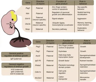
Imprinted genes and embryonic development and growth The conclusion that imprinted genes are essential for proper development was made after studies demonstrating the developmental arrest of uniparental mouse embryos. In mice, uniparental embryos can be experimentally produced through nuclear transfer of zygotic pronuclei. Androgenetic embryos (derived from two paternal pronuclei) and gynogenetic embryos (derived from two maternal pronuclei) lacked embryonic and extraembryonic tissues, respectively (Barton et al., 1984; McGrath and Solter, 1984), suggesting a central role for imprinted genes in early lineage commitment and growth. Consistently, the first identified imprinted genes were shown to be essential for normal fetal growth but, as we discuss below, roles for imprinted genes in placental growth and behavior have since been discovered (summarized in Fig. 3).
Fetal growth and development
[image:2.612.47.300.71.442.2]The most well-studied example of an imprinted gene that regulates growth is the paternally expressedIgf2 gene, which is a positive regulator of fetal growth. Inappropriate biallelic expression ofIgf2 results in broad embryonic overgrowth, whereas its reduction leads to growth restriction (DeChiara et al., 1991; Ferguson-Smith et al., 1991; Leighton et al., 1995). Interestingly, the effect of IGF2 on fetal growth is neutralized by a maternally expressed imprinted gene, Igf2r.Igf2r mutations are associated with overgrowth and embryonic death, but both phenotypes are rescued in anIgf2null background (Ludwig et al., 1996). Accordingly, IGF2R acts as an antagonistic receptor that binds IGF2 and targets it for lysosomal degradation (Foulstone et al., 2005). The H19 gene, which Table 1. Human genetic disorders associated with imprinted genes
Disorder
Associated imprinted
domains Clinical phenotypes
Beckwith–Wiedemann Syndrome (BWS)
H19-IGF2,
CDKN1C
Overgrowth, hemihyperplasia, macroglossia, abdominal wall defects, increased risk for embryonic tumors
Silver–Russell Syndrome (SRS) H19-IGF2,
GRB10,MEST
(PEG1),PEG3
Undergrowth and asymmetry
Prader–Willi Syndrome (PWS) SNRPN-UBE3A Neonatal feeding difficulty, hypotonia, hypothalamic dysfunction, intellectual delay, hyperphagia, obesity Angelman Syndrome (AS) SNRPN-UBE3A Developmental
delay, speech impairment, poor motor control, seizures Transient neonatal diabetes (TND) PLAGL1 Transient diabetes
mellitus present at birth
Uniparental disomy Chr14 (UPD14) DLK1 Scoliosis, early puberty, developmental delay, hypotonia Pseudopseudohypoparathyroidism
(PPHP)
GNAS Parathyroid hormone resistance, low energy, muscle cramps, bone thinning
Shown are some of the major congenital disorders associated within misexpression of imprinted genes, the clinical phenotype of these disorders and the associated imprinting domains (named for specific genes in that imprinted gene cluster).
DEVEL
O
expresses both a 2.3 kb ncRNA and a microRNA (miR-675) in a manner linked to the expression of Igf2 (Fig. 2A), has been proposed to be a growth repressor (Hao et al., 1993). However, it is unclear whether the longer ncRNA, the miRNA, or both exhibit growth repressive properties.
Another imprinted gene with a broad developmental effect on embryonic growth is Grb10. Maternally expressed in most murine tissues,Grb10acts as a crucial growth restrictor. Maternal Grb10 knockout embryos exhibit overgrowth, and deletion of the Grb10 ICR results in biallelic expression and significant undergrowth (Charalambous et al., 2003; Shiura et al., 2009). Like IGF2, GRB10 probably exerts its effect on growth through the insulin pathway, binding the insulin receptor and both insulin-like receptors IGF1R and IGF2R (Holt and Siddle, 2005). Recent work has also shown that GRB10 is a substrate for mammalian target of rapamycin (mTOR), with mTOR-mediated phosphorylation and stabilization of GRB10 leading to reduced insulin signaling (Yu et al., 2011).
AlthoughIgf2,Igf2r,H19andGrb10are important for modulating growth control pathways, many other imprinted genes affect embryonic growth through other mechanisms. It has been proposed that a number of these imprinted genes, includingPeg1(Mest),Gtl2 (Meg3), Cdkn1c, Plagl1 and Dlk1, are coordinately regulated in multiple tissues along withIgf2andGrb10to regulate growth in a proposed ‘imprinted gene network’ (Arima et al., 2005; Varrault et al., 2006). The full extent of the co-regulation of these genes and the mechanism of co-regulation are still unknown. The transcription factorZac1(Plagl1), which is a paternally expressed imprinted gene, has been suggested to alter the expression of genes in the imprinted gene network (Varrault et al., 2006). Deletion of this gene in mice results in intrauterine growth restriction and neonatal lethality. Moreover ZAC1 alters the expression of several imprinted genes, including Cdkn1cand Dlk1, and it directly regulates theH19/Igf2 locus through binding of its shared enhancer (Varrault et al., 2006). Additionally,H19has been proposed as a possible regulator of the imprinted gene network intransby recruiting MBD1 (Gabory et al., 2009; Monnier et al., 2013). Finally, BMI1, a member of the Polycomb Repressive Complex 1 (PRC1) has also been implicated in
the coordinated expression of multiple imprinted genes within this network (Zacharek et al., 2011).
Imprinting and placental development
Some imprinted genes also have key functions in placental development (Fig. 3). These genes control embryonic growth, as the placenta acts as the singular point of regulation between maternal and embryonic tissues, and is the source of many hormones and growth factors (Abu-Amero et al., 2006). A large number of imprinted genes (∼80) are reported to be highly expressed in the placenta, although recent work has highlighted the prevalence of confounding maternal contamination and has questioned whether some of these genes are in fact imprinted (Okae et al., 2012). The deletion of some imprinted genes, including the paternally expressed Peg3andPeg1, in mouse knockout models causes growth restriction of the entire placenta (Curley et al., 2004; Lefebvre et al., 1998). miR-675, which is processed from the first exon of H19, is also highly expressed in the placenta and is important for signaling the end of placental growth by downregulatingIgf1r(Keniry et al., 2012).
[image:3.612.51.359.59.309.2]Other imprinted genes are essential for the proper development of placental tissues. For example,Ascl2andPeg10are required for the development of the spongiotrophoblast, one of the major endocrine factor-producing regions of the placenta (Guillemot et al., 1994; Ono et al., 2006). Conversely,Phlda2and Cdkn1care maternally expressed imprinted genes that, when deleted, cause improper spongiotrophoblast expansion (Frank et al., 2002; Zhang et al., 1998). Rtl1, a paternally expressed imprinted gene, is crucial for the maintenance of placental capillaries (Sekita et al., 2008). Other imprinted genes are central to placental function; mono-amine uptake to the embryo through the placenta is inhibited by deletion ofSlc22a3 and nutrient uptake is inhibited by the deletion of the placental-specific isoform ofIgf2,Igf2 P0(Constância et al., 2002; Zwart et al., 2001). Given the roles of imprinted genes in the placenta, it is unsurprising that deletion of Dnmt3l, a member of the DNA methyltransferase family that acts with DNMT3A to establish DNA methylation imprints in germ cells, results in extensive abnormalities in placental development (Arima et al., 2006).
Fig. 1. Establishment, maintenance and erasure of genomic imprints during mouse development.
Imprints are acquired in a sex-specific manner in the mature germline (light green circles) during development, with paternal imprints (blue chromosomes) being established prenatally and maternal imprints (pink chromosomes) established postnatally. These imprints are retained despite the global changes in DNA methylation that occur after fertilization, which include active demethylation of the paternal genome and passive demethylation of the maternal genome. These imprints are maintained in somatic tissues throughout adulthood. In primordial germ cells (PGCs, dark green circles), imprints are erased (gray chromosomes) and reset for the next generation.
DEVEL
O
Imprinting and human growth disorders
Although the previously described work on imprinted genes was performed using mouse models, the consequence of misregulated imprinting on embryonic growth is obvious in humans where it manifests in multiple clinical phenotypes (Table 1). For example, Beckwith–Wiedemann syndrome (BWS) is an overgrowth disorder associated with genetic defects in two adjacent clusters of imprinted genes on chromosome 11. Individuals with BWS often present as large for gestational age at birth, with large tongues (macroglossia), large bodies and placental overgrowth. This syndrome most commonly arises owing to the loss of methylation at the ICR for the long ncRNA gene KCNQ1OT1 (Higashimoto et al., 2006). This loss of methylation results in biallelic expression of the ncRNA and consequently cis-acting repression of the protein-coding genes regulated by KCNQ1OT1 (Fig. 2B). One protein-coding gene from this cluster that drives the BWS phenotype isCDKN1C, which makes a protein product that
acts as a cell cycle inhibitor and growth restrictor; absence or mutation of CDKN1C promotes overgrowth (Andrews et al., 2007; Matsuoka et al., 1996). BWS is less frequently caused by activation of IGF2 and reduced H19 expression (typically through ICR deletions and an increase in methylation at theH19 promoter), although in these cases it is often accompanied by Wilms and other tumors (Choufani et al., 2013). In contrast to BWS, Silver–Russell syndrome (SRS) is a genetic disorder in which babies are born small for their gestational age and later exhibit dwarfism. SRS is highly associated with hypomethylation at the H19/IGF2 ICR (designated IC1 at the human locus), resulting in biallelic expression ofH19and biallelic repression of IGF2 (Gicquel et al., 2005). Both BWS and SRS are also associated with asymmetrical growth and a variety of deleterious phenotypes, suggesting that the role these imprinted genes play in growth has fundamental importance in many biological processes. Mouse mutants in the BWS and SRS orthologous regions have been instrumental in uncovering the mechanism of imprinting regulation at theH19/Igf2andKcnq1/Kcnq1ot1loci. Most of these loss-of-imprinting (LOI) mouse models for BWS and SRS mimic patient cases, although curiously they do not fully recapitulate the phenotypes observed in these human syndromes. Mice in which Igf2is overexpressed andCdkn1corIgf2ris deleted display some but not all of the BWS phenotypes (Caspary et al., 1999; Eggenschwiler et al., 1997). It has been suggested that mice cannot fully recapitulate the BWS phenotype at least in part because of differences in proliferation rates between mouse and human (Caspary et al., 1999). With respect to SRS, an engineered mouse strain in which CpG mutations prevented maintenance methylation of the paternally transmitted H19/Igf2 ICR exhibited diminished Igf2 expression and overexpression ofH19 (Engel et al., 2004). Although these mice are small, they do not appear to exhibit the other features characteristic of SRS, such as asymmetry.
Imprinted genes and neural development and function In addition to their roles in the general growth and health of the embryo, imprinted genes also play numerous, highly specialized and cell type-specific functions during development. Although this is true in a variety of tissues and contexts, imprinted genes have a particularly important and complex role in the development of the mammalian brain. This was first highlighted by foundational work examining the contribution of parthenogenetic (PG, similar to gynogenetic embryos but exclusively derived from an egg) and androgenetic (AG, paternal only) cells to the nervous system in a developing chimeric embryo (Keverne et al., 1996). Although both PG and AG cells exhibited low levels of contribution to the developing brain in chimeras, PG chimeras had a larger brain and a smaller body, whereas AG chimeras had a smaller brain, but a larger body. Furthermore, PG and AG cells contributed to distinct sub-regions of the brain, with PG cells being more prevalent in the neocortex and AG cells more prevalent in the pre-optic area and hypothalamus. This work was the first to suggest that the maternal and paternal genomes may have distinct roles in neuronal development, and has subsequently been tied to several key imprinted loci and their proper regulation.
Imprinted gene expression in the brain
[image:4.612.49.302.53.378.2]It is interesting to note that a number of imprinted genes have expression patterns and functions in the brain that are distinct from those seen in other tissues (Fig. 3).Ube3ais the most well studied example, being biallelically expressed in most tissues but maternally expressed within certain neuronal subtypes (Albrecht et al., 1997). Fig. 2. Imprinting mechanisms.(A) The insulator model is best
represented by theH19/Igf2locus. The ICR on the paternal allele of this locus is DNA methylated. By contrast, the ICR on the maternal allele is unmethylated, which allows binding of the insulator protein CTCF and prevents enhancers from interacting with the insulin-like growth factor 2 (Igf2) promoter. Instead, the enhancers activateH19expression. On the paternal allele, DNA methylation prevents CTCF from binding to the ICR, allowing the enhancers to activateIgf2expression. (B) The ncRNA model is best illustrated by theKcnq1locus. Here, the ICR contains the promoter of theKcnq1ot1long ncRNA. On the paternal allele, the ICR is unmethylated, allowing the expression ofKcnq1ot1, which in turn silences the paternal alleles of the adjacent genes. On the maternal allele,Kcnq1ot1is not expressed owing to DNA methylation of the ICR, and the adjacent imprinted genes are expressed. All imprinted domains are depicted for the mouse, although the human regions are largely conserved. T refers to the telomeric end of the cluster and C the proximal end of the chromosome. Not drawn to scale.
DEVEL
O
Maternal deficiency ofUBE3A causes Angelman syndrome (AS; Table 1), a neurodevelopmental disorder associated with cognitive and motor impairment (Kishino et al., 1997). UBE3A is an ubiquitin ligase that can target proteins for degradation. In vitro, UBE3A has been shown to ubiquitylate proteins important for cell cycle regulation, such as p53 (TRP53), HHR23A (RAD23A) and MGMT (Kumar et al., 1999; Scheffner et al., 1993; Srivenugopal et al., 1996). UBE3A has similarly been shown to ubiquitylate ARC, a protein localized to neuronal synapses that promotes internalization of specific glutamate receptors important for neural plasticity (Greer et al., 2010). Such results should be taken with caution, however, as ubiquitylation of these proteins by UBE3A in vivois largely unproven, or, in the case of ARC, has recently been contested (Kühnle et al., 2013). Nevertheless,Ube3aknockout in the mouse is associated with increased levels of ARC and p53 in brain lysates, suggesting at least a genetic role for Ube3a in regulating protein levels in neurons.In addition, Ube3a knockdown in primary hippocampal neurons results in decreased synaptic localization of AMPA receptors, which are important for the plasticity of neuronal connections and which can be sequestered by ARC (Greer et al., 2010). As mentioned above, very recent work argues that UBE3A does not directly ubiquitylate ARC, but rather that UBE3A acts as a negative regulator of estrogen-mediatedArc transcription (Kühnle et al., 2013). Consistent with this model, UBE3A has been shown to act as a transcriptional co-regulator by interacting with steroid hormone receptors, including the estrogen receptor (Ramamoorthy and Nawaz, 2008). However, biochemical analysis of the UBE3A point mutations in found in AS patients shows very frequent loss of ubiquitin ligase activity and a general preservation of co-activator activity (Cooper et al., 2004). Taken together, it is possible that defects in both mechanisms (transcription and ubiquitylation) may contribute to the AS phenotype.
Other imprinted genes that show brain-specific expression patterns contribute to the proper establishment of the highly complex cell types
that make up the brain. Peg3 is a paternally expressed gene that encodes a zinc finger protein that exhibits high expression levels in certain brain regions. Importantly,Peg3has been implicated in the control of apoptosis in neurons through its interaction with p53 and the pro-apoptotic factor BAX (Johnson et al., 2002). Deletions of paternal Peg3result in increased neonatal apoptosis in specific brain regions. This apoptosis ultimately reduces the total number of oxytocin-secreting neurons and masks normal sex-specific differences in apoptosis in brain regions involved in sexual behavior, olfaction and pheromone processing (Broad et al., 2009; Li, 1999).
Another example of an imprinted gene that affects specific neuronal subtypes is the paternally expressed Ndn gene, which encodes a protein that interacts with p53 and a variety of growth factors to influence neuronal differentiation and growth (Kuwajima et al., 2006; Salehi et al., 2002; Taniura et al., 1999).Ndnmutant mice exhibit reduced neuronal density in the hypothalamus and morphological abnormalities in axonal outgrowths (Muscatelli et al., 2000; Pagliardini et al., 2005). Interestingly, a variety of hypothalamic dysfunctions are evident in Prader–Willi Syndrome (PWS; Table 1) (Swaab, 1997). Accordingly, mutations inNDN, as well as in other genes within the same imprinting cluster including SNRPN and several small nucleolar RNAs (snoRNAs), are associated with PWS.
Imprinted genes in the brain: human disorders and effects on behavior
[image:5.612.48.385.58.349.2]The importance of imprinted genes in neurodevelopment is probably best highlighted by the wide variety of behavioral phenotypes associated with their misexpression. Mice deficient in maternal Ube3aexhibit defects in hippocampal-related memory and learning, along with a variety of abnormalities in motor system behaviors (Heck et al., 2008). These phenotypes are partially mirrored in patients with AS, a disorder characterized by attention deficits and delayed motor development (Pelc et al., 2008). Furthermore,Ndnnull
Fig. 3. A summary of imprinted gene functions during embryogenesis.Examples of imprinted genes and their functions in the brain (top box), in the placenta (lower box) and in general growth (left-hand boxes) during embryonic development are listed.
DEVEL
O
mice show an abnormal skin scraping tendency and modified spatial learning, both of which are reminiscent of some behaviors associated with PWS (Muscatelli et al., 2000). It is important to note, however, that mouse model systems do not recapitulate the full gamut of behavioral phenotypes exhibited by AS and PWS patients. For example, some, but not all, mice models for PWS exhibit increased appetite, which is also observed in human PWS patients (Rieusset et al., 2013), but this may not be from the lack of satiation that underlies these phenotypes in humans.
A variety of other imprinted genes are also associated with the regulation of behavior. Two imprinted genes,Peg1andPeg3, are associated with an important role in maternal care behaviors.Peg3 null female mice exhibit deficiencies in sexual behavior and maternal care actions, such as milk-letdown and nest building. Additionally,Peg3null neonates have reduced suckling behavior (Champagne et al., 2009). Similarly, Peg1 null females display abnormal maternal care and impaired placentophagia, which both are behaviors associated with the successful rearing of young (Lefebvre et al., 1998). Recent work has also shown that paternal deletion ofGrb10, which is expressed from the paternal allele in a subset of neurons from alternative promoter(s), is associated with hyper-aggression and increased social dominance in mice (Garfield et al., 2011). Finally, a maternally expressed gene,Nesp (Gnas), which encodes a protein involved in neuro-excretory function, is associated with novel exploration behavior and has been observed to have striking overlap in expression with Grb10 in the brain (Dent and Isles, 2014; Plagge et al., 2005).
Roles for imprinted genes in stem cells and reprogramming A crucial function for genomic imprinting in stem cells, including embryonic stem cells (ESCs), induced pluripotent stem cells (iPSCs) and adult stem cells, has recently garnered much attention (Papp and Plath, 2013; Stadtfeld and Hochedlinger, 2010). As discussed above, genomic imprints are established in the male and female germline when the parental alleles can be independently marked. This establishment occurs after imprint erasure and is part of the widespread epigenetic reprogramming and genome-wide demethylation that is essential for totipotency (Hajkova et al., 2008; Surani et al., 2007). However, it is now known that at least some of the germline-specific reprogramming events can be bypassed during reprogramming, when iPSCs are derived from differentiated somatic cells. An important question for practitioners of iPSC technology during its early development was whether imprints would be appropriately maintained during the reprogramming process. For example, nuclear transfer (NT)-derived mouse clones exhibited general epigenetic instability and LOI, and suffered from a variety of defects often associated with imprinting disorders (Humpherys, 2001).
Regardless of whether these cells will be used to study basic developmental processes or employed for human therapeutics, proper imprinting is an essential benchmark. LOI not only can result in the previously mentioned errors in early growth and development, but additionally is highly correlated with cell transformation and cancer. Many imprinted genes, includingH19,Peg1andPeg3, are known tumor suppressors (Feinberg, 1999). Additionally, ‘imprint-free’ mouse ESCs that have global LOI effectively contribute to chimeras, but these mice develop multiple types of cancer by one year of age (Holm et al., 2005). Thus, the careful study of imprinted gene expression and function in iPSCs is required for full confidence in their application. Such studies, together with analyses of imprinting in embryonic and adult stem cells, highlight the functional importance of imprinted genes in pluripotent cell populations.
Imprinting and reprogramming
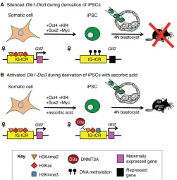
[image:6.612.314.565.55.310.2]An initial report describing the reprogramming of mouse embryonic fibroblasts to a pluripotent state showed that several imprinted genes (H19, Peg1, Peg3 and Snrpn) maintained proper allele-specific DNA methylation after reprogramming (Wernig et al., 2007). Two subsequent studies characterizing imprinting during the induction of human iPSCs showed that LOI is an exceedingly rare but observable event that is evident at early stages in the reprogramming process, is highly cell-line specific, and is maintained through multiple passages (Hiura et al., 2013; Pick et al., 2009). Interestingly, maintenance of the state of imprinting is also evident in iPSCs generated from AS and PWS patient fibroblasts, with pathological errors in imprinting and expression being retained through reprogramming and subsequent culture (Chamberlain et al., 2010). Thus, it appears that imprints present in the somatic cell of origin are, for the most part, faithfully retained in iPSCs after reprogramming. A significant and functionally crucial exception to these trends involves errors during iPSC reprogramming at the Dlk1-Dio3 imprinted cluster (Fig. 4A). In a genome-wide comparison of expression between genetically identical mouse ESCs and iPSCs, the only two significantly downregulated transcripts in iPSCs were the maternally expressed ncRNAGtl2and the long ncRNARian, both of which are found within theDlk1-Dio3imprinting region (Stadtfeld Fig. 4.Dlk1-Dio3expression during iPSC induction.(A)Gtl2, a
non-coding RNA in theDlk1-Dio3imprinted cluster, is expressed from the maternal allele in most somatic tissues. Within these tissues, the ICR (IG-ICR) within the cluster exhibits activating histone marks (H3K4me2 and H3Kac). Induction of pluripotency in somatic cells via exogenous expression ofOct4(Pou5f1),Klf4,Sox2andMycfrequently results in the aberrant silencing of maternal transcripts within theDlk1-Dio3cluster and DNA methylation of the ICR by DNMT3A (not pictured). The subsequent misexpression ofGtl2and otherDlk1-Dio3transcripts results in poor incorporation of these iPSCs into chimeric mice made using the tetraploid (4N) complementation method. No‘all iPSC’mice have been made from iPSCs with silencedDlk1-Dio3. (B) The addition of ascorbic acid (vitamin C) during the iPSC reprogramming process results in activating histone marks at the IG-ICR, including H3K4me3, and the expression ofGtl2. The addition of ascorbic acid prevents the recruitment of DNMT3A (D3a) by an unknown mechanism. These iPSCs can give rise to‘all iPSC’mice.
DEVEL
O
et al., 2010). Analysis of 62 additional iPSC lines showed that only ∼6% of these lines exhibited normal expression ofGtl2, and that lines that misexpress genes in this cluster demonstrated a greatly reduced ability to contribute to chimeras. Further work corroborated that repression of genes in theDlk1-Dio3cluster correlated with reduction in pluripotency hallmarks, specifically the generation of‘all iPSC’ mice (Carey et al., 2011; Liu et al., 2010). Mechanistically, it was found that hypermethylation across Dlk1-Dio3 causes aberrant repression at the cluster and is dependent on the inappropriate recruitment of the de novo DNA-methyltransferase DNMT3A. Surprisingly, recent follow-up work has shown that treatment of iPSCs with ascorbic acid (vitamin C) during passage and reprogramming ensures the maintenance of euchromatic marks across Dlk1-Dio3; ascorbic acid treatment inhibits the recruitment of DNMT3A by a highly specific but unknown mechanism and increases iPSC pluripotency and reprogramming efficiency (Stadtfeld et al., 2012).
Imprinting in adult stem cells
Imprinted genes have been recently implicated in the maintenance and function of adult stem cell populations. A transgenic reporter mouse line for the paternally expressedPeg3 gene has shown that Peg3 expression in adults is restricted to stem cell/progenitor populations in a variety of tissues, including the brain, gut, bone, muscle and skin (Besson et al., 2011). The generation of neurospheres, via anin vitrotechnique used to isolate and amplify neuronal progenitors, resulted in∼100%Peg3-positive cells after a single passage. Additionally, engrafting experiments in the epidermis revealed that transferredPeg3-positive cells have the ability to self-renew within the follicular stem cell niche and differentiate effectively. These experiments suggest thatPeg3plays a functional role in adult stem cells, although it is currently unclear whether this role is different from that observed in early development.
The maternally expressed H19 gene is also involved in the maintenance of adult hematopoietic stem cell (HSC) populations in the mouse (Venkatraman et al., 2013). Conditional maternal deletion of theH19/Igf2ICR in HSCs caused reduced expression of H19 and increased expression of Igf2, accompanied by a reduction in the number of long-term HSCs, an increase in short-term HSCs, and overall compromised hematopoietic potential and function. In addition, maternal deletion of theH19/Igf2ICR caused inappropriate activation of the Igf2-Igf1r pathway via increased expression of Igf2and decreased repression of Igf1r, which is a target ofH19-derived miR-675. This led to inhibited quiescence-associated cell cycle arrest mediated by FOXO3, ultimately resulting in the activation and exhaustion of long-term HSCs.
Additionally, selective loss of Dlk1 imprinting within mouse neural stem cells (NSCs) and their niche has been shown to be crucial for postnatal neurogenesis (Ferrón et al., 2011).Dlk1is a membrane-bound receptor for Notch signaling that is downregulated postnatally in most tissues.Dlk1-deficient mice show decreased pools of slow-dividing NSCs, resulting in depletion of neurons in the adult olfactory bulb. Interestingly, the NSCs and the surrounding astrocytes that make up their niche expressDlk1from both alleles, whereasDlk1is otherwise expressed exclusively from the paternal allele. This coordinated biallelic expression of Dlk1highlights the importance of specific contexts in imprinted gene regulation and underscores the significance of gene dosage for imprinted genes.
Conclusions
Although initial work suggested that imprinted gene expression is crucial for early embryonic growth and differentiation, it is now
clear that genomic imprinting exhibits a much more varied role in mammalian development. It is important to note that this Review is intended to highlight just some of the functions of genomic imprinting and imprinted genes, but is not comprehensive; various roles for many imprinted genes have been documented. For example, LOI at numerous imprinted genes has been associated with cancer and oncogenic phenotypes (Baylin and Jones, 2011); a functional importance not detailed at length in this Review. Additionally, imprinted expression plays a variety of other tissue-specific roles in many other organs and cell types not elaborated here (Prickett and Oakey, 2012). It is almost certain that a full understanding of tissue-specific imprinting is incomplete. Further exploration of the prevalence of imprinting in complex tissues such as the brain and in novel contexts such as adult stem cell populations will undoubtedly lead to exciting discoveries regarding the expression and functions of imprinted genes.
Acknowledgements
We apologize to those investigators whose work was not mentioned in this Review. We thank the members of the Bartolomei lab for helpful comments on the manuscript.
Competing interests
The authors declare no competing financial interests.
Funding
M.S.B. and R.N.P. are supported by the National Institutes of Health. Deposited in PMC for release after 12 months.
References
Abu-Amero, S., Monk, D., Apostolidou, S., Stanier, P. and Moore, G.(2006). Imprinted genes and their role in human fetal growth.Cytogenet. Genome Res.
113, 262-270.
Albrecht, U., Sutcliffe, J. S., Cattanach, B. M., Beechey, C. V., Armstrong, D., Eichele, G. and Beaudet, A. L.(1997). Imprinted expression of the murine Angelman syndrome gene, Ube3a, in hippocampal and Purkinje neurons.Nat. Genet.17, 75-78.
Andrews, S. C., Wood, M. D., Tunster, S. J., Barton, S. C., Surani, A. M. and John, R. M.(2007). Cdkn1c ( p57Kip2) is the major regulator of embryonic growth within its imprinted domain on mouse distal chromosome 7.BMC Dev. Biol.7, 53.
Arima, T., Kamikihara, T., Hayashida, T., Kato, K., Inoue, T., Shirayoshi, Y., Oshimura, M., Soejima, H., Mukai, T. and Wake, N.(2005). ZAC, LIT1 (KCNQ1OT1) and p57KIP2 (CDKN1C) are in an imprinted gene network that may play a role in Beckwith–Wiedemann syndrome. Nucleic Acids Res. 33, 2650-2660.
Arima, T., Hata, K., Tanaka, S., Kusumi, M., Li, E., Kato, K., Shiota, K., Sasaki, H. and Wake, N.(2006). Loss of the maternal imprint in Dnmt3Lmat−/−mice leads to a differentiation defect in the extraembryonic tissue.Dev. Biol. 297, 361-373.
Barlow, D. P., Stöger, R., Herrmann, B. G., Saito, K. and Schweifer, N.(1991). The mouse insulin-like growth factor type-2 receptor is imprinted and closely linked to the Tme locus.Nature349, 84-87.
Bartolomei, M. S. and Ferguson-Smith, A. C.(2011). Mammalian genomic imprinting.Cold Spring Harb. Perspect. Biol.3, a002592.
Bartolomei, M. S., Zemel, S. and Tilghman, S. M.(1991). Parental imprinting of the mouse H19 gene.Nature351, 153-155.
Barton, S. C., Surani, M. and Norris, M. L.(1984). Role of paternal and maternal genomes in mouse development.Nature311, 374-376.
Baylin, S. B. and Jones, P. A.(2011). A decade of exploring the cancer epigenome–
biological and translational implications.Nat. Rev. Cancer11, 726-734. Besson, V., Smeriglio, P., Wegener, A., Relaix, F., Oumesmar, B. N., Sassoon, D. A.
and Marazzi, G.(2011). PW1 gene/paternally expressed gene 3 (PW1/Peg3) identifies multiple adult stem and progenitor cell populations.Proc. Natl. Acad. Sci. U. S.A.108, 11470-11475.
Bourc’his, D., Xu, G.-L., Lin, C.-S., Bollman, B. and Bestor, T. H.(2001). Dnmt3L and the establishment of maternal genomic imprints.Science294, 2536-2539. Broad, K. D., Curley, J. P. and Keverne, E. B.(2009). Increased apoptosis during
neonatal brain development underlies the adult behavioral deficits seen in mice lacking a functional paternally expressed gene 3(Peg3).Dev. Neurobiol.69, 314-325.
Butler, M. G.(2009). Genomic imprinting disorders in humans: a mini-review.
J. Assist. Reprod. Genet.26, 477-486.
DEVEL
O
Carey, B. W., Markoulaki, S., Hanna, J. H., Faddah, D. A., Buganim, Y., Kim, J., Ganz, K., Steine, E. J., Cassady, J. P., Creyghton, M. P. et al.(2011). Reprogramming factor stoichiometry influences the epigenetic state and biological properties of induced pluripotent stem cells.Cell Stem Cell9, 588-598. Caspary, T., Cleary, M. A., Perlman, E. J., Zhang, P., Elledge, S. J. and Tilghman, S. M.(1999). Oppositely imprinted genes p57Kip2 and Igf2 interact in a mouse model for Beckwith–Wiedemann syndrome. Genes Dev. 13, 3115-3124.
Chamberlain, S. J., Chen, P.-F., Ng, K. Y., Bourgois-Rocha, F., Lemtiri-Chlieh, F., Levine, E. S. and Lalande, M.(2010). Induced pluripotent stem cell models of the genomic imprinting disorders Angelman and Prader–Willi syndromes.Proc. Natl. Acad. Sci. U.S.A.107, 17668-17673.
Champagne, F. A., Curley, J. P., Swaney, W. T., Hasen, N. S. and Keverne, E. B. (2009). Paternal influence on female behavior: the role of Peg3 in exploration, olfaction, and neuroendocrine regulation of maternal behavior of female mice.
Behav. Neurosci.123, 469-480.
Charalambous, M., Smith, F. M., Bennett, W. R., Crew, T. E., Mackenzie, F. and Ward, A.(2003). Disruption of the imprinted Grb10 gene leads to disproportionate overgrowth by an Igf2-independent mechanism.Proc. Natl. Acad. Sci. U.S.A.100, 8292-8297.
Chotalia, M., Smallwood, S. A., Ruf, N., Dawson, C., Lucifero, D., Frontera, M., James, K., Dean, W. and Kelsey, G. (2009). Transcription is required for establishment of germline methylation marks at imprinted genes.Genes Dev.23, 105-117.
Choufani, S., Shuman, C. and Weksberg, R. (2013). Molecular findings in Beckwith–Wiedemann syndrome.Am. J. Med. Genet. C Semin. Med. Genet.
163C, 131-140.
Constância, M., Hemberger, M., Hughes, J., Dean, W., Ferguson-Smith, A., Fundele, R., Stewart, F., Kelsey, G., Fowden, A., Sibley, C. et al.(2002). Placental-specific IGF-II is a major modulator of placental and fetal growth.Nature
417, 945-948.
Cooper, E. M., Hudson, A. W., Amos, J., Wagstaff, J. and Howley, P. M.(2004). Biochemical analysis of Angelman syndrome-associated mutations in the E3 ubiquitin ligase E6-associated protein.J. Biol. Chem.279, 41208-41217. Curley, J. P., Barton, S., Surani, A. and Keverne, E. B.(2004). Coadaptation in
mother and infant regulated by a paternally expressed imprinted gene.
Proc. R. Soc. B Biol. Sci.271, 1303-1309.
Davis, T. L., Yang, G. J., McCarrey, J. R. and Bartolomei, M. S.(2000). The H19 methylation imprint is erased and re-established differentially on the parental alleles during male germ cell development. Hum. Mol. Genet. 9, 2885-2894.
Dawlaty, M. M., Breiling, A., Le, T., Raddatz, G., Barrasa, M. I., Cheng, A. W., Gao, Q., Powell, B. E., Li, Z., Xu, M. et al.(2013). Combined deficiency of Tet1 and Tet2 causes epigenetic abnormalities but is compatible with postnatal development.Dev. Cell24, 310-323.
DeChiara, T. M., Robertson, E. J. and Efstratiadis, A.(1991). Parental imprinting of the mouse insulin-like growth factor II gene.Cell64, 849-859.
Dent, C. L. and Isles, A. R.(2014). Brain-expressed imprinted genes and adult behaviour: the example of Nesp and Grb10.Mamm. Genome.25, 87-93. Edwards, C. A. and Ferguson-Smith, A. C. (2007). Mechanisms regulating
imprinted genes in clusters.Curr. Opin. Cell Biol.19, 281-289.
Eggenschwiler, J., Ludwig, T., Fisher, P., Leighton, P. A., Tilghman, S. M. and Efstratiadis, A.(1997). Mouse mutant embryos overexpressing IGF-II exhibit phenotypic features of the Beckwith–Wiedemann and Simpson-Golabi-Behmel syndromes.Genes Dev.11, 3128-3142.
Engel, N., West, A. G., Felsenfeld, G. and Bartolomei, M. S.(2004). Antagonism between DNA hypermethylation and enhancer-blocking activity at the H19 DMD is uncovered by CpG mutations.Nat. Genet.36, 883-888.
Feinberg, A. P.(1999). Imprinting of a genomic domain of 11p15 and loss of imprinting in cancer: an introduction.Cancer Res.59, 1743s-1746s.
Ferguson-Smith, A. C., Cattanach, B. M., Barton, S. C., Beechey, C. V. and Surani, M. A.(1991). Embryological and molecular investigations of parental imprinting on mouse chromosome 7.Nature351, 667-670.
Ferrón, S. R., Charalambous, M., Radford, E., McEwen, K., Wildner, H., Hind, E., Morante-Redolat, J. M., Laborda, J., Guillemot, F., Bauer, S. R. et al.(2011). Postnatal loss of Dlk1 imprinting in stem cells and niche astrocytes regulates neurogenesis.Nature475, 381-385.
Foulstone, E., Prince, S., Zaccheo, O., Burns, J. L., Harper, J., Jacobs, C., Church, D. and Hassan, A. B. (2005). Insulin-like growth factor ligands, receptors, and binding proteins in cancer.J. Pathol.205, 145-153.
Frank, D., Fortino, W., Clark, L., Musalo, R., Wang, W., Saxena, A., Li, C.-M., Reik, W., Ludwig, T. and Tycko, B.(2002). Placental overgrowth in mice lacking the imprinted gene Ipl.Proc. Natl. Acad. Sci. U.S.A.99, 7490-7495.
Gabory, A., Ripoche, M.-A., Le Digarcher, A., Watrin, F., Ziyyat, A., Forné, T., Jammes, H., Ainscough, J. F. X., Surani, M. A., Journot, L. et al.(2009). H19 acts as a trans regulator of the imprinted gene network controlling growth in mice.
Development136, 3413-3421.
Garfield, A. S., Cowley, M., Smith, F. M., Moorwood, K., Stewart-Cox, J. E., Gilroy, K., Baker, S., Xia, J., Dalley, J. W., Hurst, L. D. et al.(2011). Distinct
physiological and behavioural functions for parental alleles of imprinted Grb10.
Nature469, 534-538.
Gicquel, C., Rossignol, S., Cabrol, S., Houang, M., Steunou, V., Barbu, V., Danton, F., Thibaud, N., Merrer, M. L., Burglen, L. et al.(2005). Epimutation of the telomeric imprinting center region on chromosome 11p15 in Silver-Russell syndrome.Nat. Genet.37, 1003-1007.
Greer, P. L., Hanayama, R., Bloodgood, B. L., Mardinly, A. R., Lipton, D. M., Flavell, S. W., Kim, T.-K., Griffith, E. C., Waldon, Z., Maehr, R. et al.(2010). The Angelman syndrome protein Ube3A regulates synapse development by ubiquitinating arc.Cell140, 704-716.
Guillemot, F., Nagy, A., Auerbach, A., Rossant, J. and Joyner, A. L.(1994). Essential role of Mash-2 in extraembryonic development.Nature371, 333-336. Hackett, J. A., Sengupta, R., Zylicz, J. J., Murakami, K., Lee, C., Down, T. A. and
Surani, M. A.(2013). Germline DNA demethylation dynamics and imprint erasure through 5-hydroxymethylcytosine.Science339, 448-452.
Hajkova, P., Ancelin, K., Waldmann, T., Lacoste, N., Lange, U. C., Cesari, F., Lee, C., Almouzni, G., Schneider, R. and Surani, M. A.(2008). Chromatin dynamics during epigenetic reprogramming in the mouse germ line.Nature452, 877-881.
Hao, Y., Crenshaw, T., Moulton, T., Newcomb, E. and Tycko, B.(1993). Tumour-suppressor activity of H19 RNA.Nature365, 764-767.
Hata, K., Okano, M., Lei, H. and Li, E.(2002). Dnmt3L cooperates with the Dnmt3 family of de novo DNA methyltransferases to establish maternal imprints in mice.
Development129, 1983-1993.
Heck, D. H., Zhao, Y., Roy, S., LeDoux, M. S. and Reiter, L. T.(2008). Analysis of cerebellar function in Ube3a-deficient mice reveals novel genotype-specific behaviors.Hum. Mol. Genet.17, 2181-2189.
Henckel, A., Chebli, K., Kota, S. K., Arnaud, P. and Feil, R.(2012). Transcription and histone methylation changes correlate with imprint acquisition in male germ cells.EMBO J.31, 606-615.
Higashimoto, K., Soejima, H., Saito, T., Okumura, K. and Mukai, T.(2006). Imprinting disruption of the CDKN1C/KCNQ1OT1 domain: the molecular mechanisms causing Beckwith–Wiedemann syndrome and cancer.Cytogenet. Genome Res.113, 306-312.
Hiura, H., Toyoda, M., Okae, H., Sakurai, M., Miyauchi, N., Sato, A., Kiyokawa, N., Okita, H., Miyagawa, Y., Akutsu, H. et al.(2013). Stability of genomic imprinting in human induced pluripotent stem cells.BMC Genet.14, 32.
Holm, T. M., Jackson-Grusby, L., Brambrink, T., Yamada, Y., Rideout, W. M., III and Jaenisch, R. (2005). Global loss of imprinting leads to widespread tumorigenesis in adult mice.Cancer Cell8, 275-285.
Holt, L. and Siddle, K.(2005). Grb10 and Grb14: enigmatic regulators of insulin action–and more?Biochem. J.388, 393-406.
Humpherys, D.(2001). Epigenetic instability in ES cells and cloned mice.Science
293, 95-97.
Johnson, M. D., Wu, X., Aithmitti, N. and Morrison, R. S.(2002). Peg3/Pw1 is a mediator between p53 and Bax in DNA damage-induced neuronal death.J. Biol. Chem.277, 23000-23007.
Kagiwada, S., Kurimoto, K., Hirota, T., Yamaji, M. and Saitou, M. (2012). Replication-coupled passive DNA demethylation for the erasure of genome imprints in mice.EMBO J.32, 340-353.
Kaneda, M., Okano, M., Hata, K., Sado, T., Tsujimoto, N., Li, E. and Sasaki, H. (2004). Essential role for de novo DNA methyltransferase Dnmt3a in paternal and maternal imprinting.Nature429, 900-903.
Keniry, A., Oxley, D., Monnier, P., Kyba, M., Dandolo, L., Smits, G. and Reik, W. (2012). The H19 lincRNA is a developmental reservoir of miR-675 that suppresses growth and Igf1r.Nat. Cell Biol.14, 659-665.
Keverne, E. B., Fundele, R., Narasimha, M., Barton, S. C. and Surani, M. A. (1996). Genomic imprinting and the differential roles of parental genomes in brain development.Brain Res. Dev. Brain Res.92, 91-100.
Kishino, T., Lalande, M. and Wagstaff, J.(1997). UBE3A/E6-AP mutations cause Angelman syndrome.Nat. Genet.15, 70-73.
Kumar, S., Talis, A. L. and Howley, P. M.(1999). Identification of HHR23A as a substrate for E6-associated protein-mediated ubiquitination.J. Biol. Chem.274, 18785-18792.
Kühnle, S., Mothes, B., Matentzoglu, K. and Scheffner, M.(2013). Role of the ubiquitin ligase E6AP/UBE3A in controlling levels of the synaptic protein Arc.
Proc. Natl. Acad. Sci. U.S.A.110, 8888-8893.
Kuwajima, T., Nishimura, I. and Yoshikawa, K. (2006). Necdin promotes GABAergic neuron differentiation in cooperation with Dlx homeodomain proteins.J. Neurosci.26, 5383-5392.
Latos, P. A., Pauler, F. M., Koerner, M. V., Senergin, H. B., Hudson, Q. J., Stocsits, R. R., Allhoff, W., Stricker, S. H., Klement, R. M., Warczok, K. E. et al. (2012). Airn transcriptional overlap, but not its lncRNA products, induces imprinted Igf2r silencing.Science338, 1469-1472.
Lee, J. T. and Bartolomei, M. S.(2013). X-inactivation, imprinting, and long noncoding RNAs in health and disease.Cell152, 1308-1323.
Lefebvre, L., Viville, S., Barton, S. C., Ishino, F., Keverne, E. B. and Surani, M. A. (1998). Abnormal maternal behaviour and growth retardation associated with loss of the imprinted gene Mest.Nat. Genet.20, 163-169.
DEVEL
O
Leighton, P. A., Saam, J. R., Ingram, R. S., Stewart, C. L. and Tilghman, S. M. (1995). An enhancer deletion affects both H19 and Igf2 expression.Genes Dev.9, 2079-2089.
Li, L.(1999). Regulation of maternal behavior and offspring growth by paternally expressed Peg3.Science284, 330-334.
Li, X., Ito, M., Zhou, F., Youngson, N., Zuo, X., Leder, P. and Ferguson-Smith, A. C. (2008). A maternal-zygotic effect gene, Zfp57, maintains both maternal and paternal imprints.Dev. Cell15, 547-557.
Liu, L., Luo, G.-Z., Yang, W., Zhao, X., Zheng, Q., Lv, Z., Li, W., Wu, H.-J., Wang, L., Wang, X.-J. et al.(2010). Activation of the imprinted Dlk1-Dio3 region correlates with pluripotency levels of mouse stem cells.J. Biol. Chem.285, 19483-19490. Lucifero, D., Mann, M. R. W., Bartolomei, M. S. and Trasler, J. M.(2004).
Gene-specific timing and epigenetic memory in oocyte imprinting.Hum. Mol. Genet.13, 839-849.
Ludwig, T., Eggenschwiler, J., Fisher, P., D’Ercole, A. J., Davenport, M. L. and Efstratiadis, A.(1996). Mouse mutants lacking the type 2 IGF receptor (IGF2R) are rescued from perinatal lethality in Igf2 and Igf1r null backgrounds.Dev. Biol.
177, 517-535.
Mackay, D. J. G., Callaway, J. L. A., Marks, S. M., White, H. E., Acerini, C. L., Boonen, S. E., Dayanikli, P., Firth, H. V., Goodship, J. A., Haemers, A. P. et al. (2008). Hypomethylation of multiple imprinted loci in individuals with transient neonatal diabetes is associated with mutations in ZFP57. Nat. Genet. 40, 949-951.
Matsuoka, S., Thompson, J. S., Edwards, M. C., Bartletta, J. M., Grundy, P., Kalikin, L. M., Harper, J. W., Elledge, S. J. and Feinberg, A. P. (1996). Imprinting of the gene encoding a human cyclin-dependent kinase inhibitor, p57KIP2, on chromosome 11p15.Proc. Natl. Acad. Sci. U.S.A.93, 3026-3030. McGrath, J. and Solter, D.(1984). Completion of mouse embryogenesis requires
both the maternal and paternal genomes.Cell37, 179-183.
Monnier, P., Martinet, C., Pontis, J., Stancheva, I., Ait-Si-Ali, S. and Dandolo, L. (2013). H19 lncRNA controls gene expression of the Imprinted Gene Network by recruiting MBD1.Proc. Natl. Acad. Sci. U.S.A.110, 20693-20698.
Muscatelli, F., Abrous, D. N., Massacrier, A., Boccaccio, I., Le Moal, M., Cau, P. and Cremer, H. (2000). Disruption of the mouse Necdin gene results in hypothalamic and behavioral alterations reminiscent of the human Prader–Willi syndrome.Hum. Mol. Genet.9, 3101-3110.
Nagano, T. and Fraser, P. (2009). Emerging similarities in epigenetic gene silencing by long noncoding RNAs.Mamm. Genome20, 557-562.
Nakamura, T., Liu, Y.-J., Nakashima, H., Umehara, H., Inoue, K., Matoba, S., Tachibana, M., Ogura, A., Shinkai, Y. and Nakano, T.(2012). PGC7 binds histone H3K9me2 to protect againstconversion of 5mC to 5hmC in early embryos.
Nature486, 415-419.
Okae, H., Hiura, H., Nishida, Y., Funayama, R., Tanaka, S., Chiba, H., Yaegashi, N., Nakayama, K., Sasaki, H. and Arima, T.(2012). Re-investigation and RNA sequencing-based identification of genes with placenta-specific imprinted expression.Hum. Mol. Genet.21, 548-558.
Okano, M., Bell, D. W., Haber, D. A. and Li, E.(1999). DNA methyltransferases Dnmt3a and Dnmt3b are essential for de novo methylation and mammalian development.Cell99, 247-257.
Ono, R., Nakamura, K., Inoue, K., Naruse, M., Usami, T., Wakisaka-Saito, N., Hino, T., Suzuki-Migishima, R., Ogonuki, N., Miki, H. et al.(2006). Deletion of Peg10, an imprinted gene acquired from a retrotransposon, causes early embryonic lethality.Nat. Genet.38, 101-106.
Pagliardini, S., Ren, J., Wevrick, R. and Greer, J. J.(2005). Developmental abnormalities of neuronal structure and function in prenatal mice lacking the Prader–Willi syndrome gene necdin.Am. J. Pathol.167, 175-191.
Papp, B. and Plath, K. (2013). Epigenetics of reprogramming to induced pluripotency.Cell152, 1324-1343.
Pastor, W. A., Aravind, L. and Rao, A.(2013). TETonic shift: biological roles of TET proteins in DNA demethylation and transcription.Nat. Rev. Mol. Cell Biol.14, 341-356.
Pelc, K., Boyd, S. G., Cheron, G. and Dan, B.(2008). Epilepsy in Angelman syndrome.Seizure17, 211-217.
Pick, M., Stelzer, Y., Bar-Nur, O., Mayshar, Y., Eden, A. and Benvenisty, N. (2009). Clone- and gene-specific aberrations of parental imprinting in human induced pluripotent stem cells.Stem Cells27, 2686-2690.
Plagge, A., Isles, A. R., Gordon, E., Humby, T., Dean, W., Gritsch, S., Fischer-Colbrie, R., Wilkinson, L. S. and Kelsey, G. (2005). Imprinted Nesp55 influences behavioral reactivity to novel environments. Mol. Cell. Biol. 25, 3019-3026.
Prickett, A. R. and Oakey, R. J. (2012). A survey of tissue-specific genomic imprinting in mammals.Mol. Genet. Genomics287, 621-630.
Ramamoorthy, S. and Nawaz, Z.(2008). E6-associated protein (E6-AP) is a dual function coactivator of steroid hormone receptors.Nucl. Recept. Signal.6, e006.
Rieusset, A., Schaller, F., Unmehopa, U., Matarazzo, V., Watrin, F., Linke, M., Georges, B., Bischof, J., Dijkstra, F., Bloemsma, M. et al.(2013). Stochastic loss of silencing of the imprinted Ndn/NDN allele, in a mouse model and humans with Prader–Willi syndrome, has functional consequences. PLoS Genet.9, e1003752.
Salehi, A. H., Xanthoudakis, S. and Barker, P. A.(2002). NRAGE, a p75 neurotrophin receptor-interacting protein, induces caspase activation and cell death through a JNK-dependent mitochondrial pathway.J. Biol. Chem. 277, 48043-48050.
Scheffner, M., Huibregtse, J. M., Vierstra, R. D. and Howley, P. M.(1993). The HPV-16 E6 and E6-AP complex functions as a ubiquitin-protein ligase in the ubiquitination of p53.Cell75, 495-505.
Sekita, Y., Wagatsuma, H., Nakamura, K., Ono, R., Kagami, M., Wakisaka, N., Hino, T., Suzuki-Migishima, R., Kohda, T., Ogura, A. et al.(2008). Role of retrotransposon-derived imprinted gene, Rtl1, in the feto-maternal interface of mouse placenta.Nat. Genet.40, 243-248.
Shiura, H., Nakamura, K., Hikichi, T., Hino, T., Oda, K., Suzuki-Migishima, R., Kohda, T., Kaneko-Ishino, T. and Ishino, F.(2009). Paternal deletion of Meg1/ Grb10 DMR causes maternalization of the Meg1/Grb10 cluster in mouse proximal Chromosome 11 leading to severe pre- and postnatal growth retardation.Hum. Mol. Genet.18, 1424-1438.
Srivenugopal, K. S., Yuan, X.-H., Friedman, H. S. and Ali-Osman, F.(1996). Ubiquitination-dependent proteolysis of O6-methylguanine-DNA methyltransferase in human and murine tumor cells following inactivation with O6-benzylguanine or 1,3-bis(2-chloroethyl)-1-nitrosourea.Biochemistry35, 1328-1334.
Stadtfeld, M. and Hochedlinger, K. (2010). Induced pluripotency: history, mechanisms, and applications.Genes Dev.24, 2239-2263.
Stadtfeld, M., Apostolou, E., Akutsu, H., Fukuda, A., Follett, P., Natesan, S., Kono, T., Shioda, T. and Hochedlinger, K.(2010). Aberrant silencing of imprinted genes on chromosome 12qF1 in mouse induced pluripotent stem cells.
Nature465, 175-181.
Stadtfeld, M., Apostolou, E., Ferrari, F., Choi, J., Walsh, R. M., Chen, T., Ooi, S. S. K., Kim, S. Y., Bestor, T. H., Shioda, T. et al. (2012). Ascorbic acid prevents loss of Dlk1-Dio3 imprinting and facilitates generation of all–iPS cell mice from terminally differentiated B cells.Nat. Genet.44, 398-405.
Surani, M. A., Hayashi, K. and Hajkova, P.(2007). Genetic and epigenetic regulators of pluripotency.Cell128, 747-762.
Swaab, D. F.(1997). Prader–Willi syndrome and the hypothalamus.Acta Paediatr.
86, 50-54.
Taniura, H., Matsumoto, K. and Yoshikawa, K.(1999). Physical and functional interactions of neuronal growth suppressor necdin with p53.J. Biol. Chem.274, 16242-16248.
Varrault, A., Gueydan, C., Delalbre, A., Bellmann, A., Houssami, S., Aknin, C., Severac, D., Chotard, L., Kahli, M. and Le Digarcher, A.(2006). Zac1 regulates an imprinted gene network critically involved in the control of embryonic growth.
Dev. Cell11, 711-722.
Venkatraman, A., He, X. C., Thorvaldsen, J. L., Sugimura, R., Perry, J. M., Tao, F., Zhao, M., Christenson, M. K., Sanchez, R., Yu, J. Y. et al.(2013). Maternal imprinting at the H19–Igf2 locus maintains adult haematopoietic stem cell quiescence.Nature500, 345-349.
Wernig, M., Meissner, A., Foreman, R., Brambrink, T., Ku, M., Hochedlinger, K., Bernstein, B. E. and Jaenisch, R.(2007). In vitro reprogramming of fibroblasts into a pluripotent ES-cell-like state.Nature448, 318-324.
Yamaguchi, S., Shen, L., Liu, Y., Sendler, D. and Zhang, Y.(2013). Role of Tet1 in erasure of genomic imprinting.Nature504, 460-464.
Yu, Y., Yoon, S.-O., Poulogiannis, G., Yang, Q., Ma, X. M., Villen, J., Kubica, N., Hoffman, G. R., Cantley, L. C., Gygi, S. P. et al.(2011). Phosphoproteomic analysis identifies Grb10 as an mTORC1 substrate that negatively regulates insulin signaling.Science332, 1322-1326.
Zacharek, S. J., Fillmore, C. M., Lau, A. N., Gludish, D. W., Chou, A., Ho, J. W. K., Zamponi, R., Gazit, R., Bock, C., Jäger, N. et al.(2011). Lung stem cell self-renewal relies on BMI1-dependent control of expression at imprinted loci.Cell Stem Cell9, 272-281.
Zhang, P., Wong, C., DePinho, R. A., Harper, J. W. and Elledge, S. J.(1998). Cooperation between the Cdk inhibitors p27KIP1 and p57KIP2 in the control of tissue growth and development.Genes Dev.12, 3162-3167.
Zwart, R., Verhaagh, S., Buitelaar, M., Popp-Snijders, C. and Barlow, D. P. (2001). Impaired activity of the extraneuronal monoamine transporter system known as uptake-2 in Orct3/Slc22a3-deficient mice. Mol. Cell. Biol. 21, 4188-4196.