Summary
Epigenetic modifications are crucial for the identity and stability of cells, and, when aberrant, can lead to disease. During mouse development, the genome-wide epigenetic states of pre-implantation embryos and primordial germ cells (PGCs) undergo extensive reprogramming. An improved understanding of the epigenetic reprogramming mechanisms that occur in these cells should provide important new information about the regulation of the epigenetic state of a cell and the mechanisms of induced pluripotency. Here, we discuss recent findings about the potential mechanisms of epigenetic reprogramming, particularly genome-wide DNA demethylation, in pre-implantation mouse embryos and PGCs.
Key words: DNA demethylation, Epigenetic reprogramming, Pre-implantation embryos, Primordial germ cells, TET proteins
Introduction
The variety of cellular states in multicellular organisms reflects the diversity of the transcriptional programme of cells, despite the fact that nearly all cells in any given organism bear an identical genome sequence. The transcriptional state of a cell is governed by a specific set of transcriptional regulators and also by chemical modifications of the genome, including cytosine methylation (5-methylcytosine; 5mC) and post-translational modifications of histone tails, which regulate the accessibility of transcriptional regulators to, and the on or off states or expression levels of, all genes in the genome (Bonasio et al., 2010). The stability of the phenotype of a cell upon mitosis/meiosis is considered to be underpinned by the stability of these modifications. Modifications that regulate the identity of a cell without changing the DNA sequence are referred to as epigenetic modifications (Bird, 2007; Bonasio et al., 2010), and the genome-wide state of the epigenetic status of a cell is known as the epigenome (Bernstein et al., 2007). Once specified during development or in adult physiology, the epigenome of a cell is stable upon mitosis/meiosis, and cell identities are maintained essentially for a lifetime (Bonasio et al., 2010).
During mammalian development, however, there are two crucial developmental stages and/or cell types in which the epigenome undergoes profound reprogramming: pre-implantation embryos and primordial germ cells (PGCs), the precursors both for oocytes and spermatozoa (Fig. 1) (Surani et al., 2007). Epigenetic
reprogramming in these cells involves genome-wide demethylation of 5mC; 5mC plays a crucial role in genome imprinting, X inactivation, transposon silencing, the stability of centromeric/telomeric structures and gene expression in general. Over the past decade, the technologies that can be used to determine genome-wide 5mC distribution have dramatically evolved, leading to the identification of global 5mC distribution in a number of cultured cell lines and primary tissues (Lister et al., 2009; Suzuki and Bird, 2008).
In this article, we summarize and discuss recent discoveries about the nature and mechanism of epigenetic reprogramming, particularly those relating to genome-wide DNA demethylation in mouse pre-implantation embryos and PGCs. These findings are important not only for understanding the genetic and epigenetic basis of genome inheritance, but also for elucidating the mechanisms of artificially induced epigenetic reprogramming that may be of medical relevance (Hanna et al., 2010; Hayashi and Surani, 2009; Yamanaka and Blau, 2010). [For reviews on other associated epigenetic events such as histone modification changes and X-chromosome inactivation/reactivation, please see other recent reviews (Brockdorf, 2011; Hemberger et al., 2009; Payer and Lee, 2008; Probst and Almouzni, 2011).]
5-methylcytosine: an overview
Cytosine methylation and 5mC distribution
Genome-wide cytosine methylation states, especially those associated with genes, differ among cell types and function as a form of memory of the identity and developmental state of a cell (Lister et al., 2009). The enzymes that methylate cytosine to form 5-methylcytosine (5mC) have been well characterized (see Table 1). DNA methyltransferase (DNMT) 1 preferentially methylates hemi-methylated cytosines in CpG sequences and thus acts as a maintenance methyltransferase to maintain genome-wide methylation patterns during replication (Bestor et al., 1988; Bestor, 1992; Li et al., 1992). DNMT3A and DNMT3B can methylate unmethylated CpG sequences and hence function as de novo methyltransferases (Okano et al., 1998a). DNMT3L has no catalytic activity but recruits DNMT3A and DNMT3B to their targets by recognizing nucleosomes that carry unmethylated histone H3 lysine 4 (H3K4) (Aapola et al., 2000; Bourc’his and Bestor, 2004; Bourc’his et al., 2001; Hata et al., 2002; Ooi et al., 2007).
5mC occurs mostly in CpG sequences and, to a lesser extent, in CpHpG or CpHpH sequences (where H is A, C or T), especially in pluripotent cells (Lister et al., 2009; Ramsahoye et al., 2000; Tomizawa et al., 2011). Mammalian genomes are globally methylated: genes, transposons, repeat sequences and intergenic DNA are all subjected to methylation (Suzuki and Bird, 2008). 5mC can spontaneously deaminate to form thymine (T), creating T:G mismatches, and thus can be a source of point mutations across the genome (see Box 1 and references therein). Unmethylated Development 139, 0000-0000 (2012) doi:10.1242/dev.050849
© 2012. Published by The Company of Biologists Ltd
Epigenetic reprogramming in mouse pre-implantation
development and primordial germ cells
Mitinori Saitou1,2,3,*, Saya Kagiwada1and Kazuki Kurimoto1,2
1Department of Anatomy and Cell Biology, Graduate School of Medicine, Kyoto University, Yoshida-Konoe-cho, Sakyo-ku, Kyoto 606-8501, Japan. 2JST,
CREST/ERATO, Yoshida-Konoe-cho, Sakyo-ku, Kyoto 606-8501, Japan. 3Institute for Integrated Cell-Material Sciences, Kyoto University, Yoshida-Ushinomiya-cho, Sakyo-ku, Kyoto 606-8501, Japan.
*Author for correspondence ([email protected])
D
E
V
E
LO
P
M
E
N
sequences are most often found in CpG islands (CGIs) (see Glossary, Box 2), which are typically associated with gene promoters (Suzuki and Bird, 2008) and with ~70% of genes. Based on their CpG ratio, GC content and on the length of the CpG-rich region, promoters are classified as being high-, intermediate- or low-CpG promoters (HCPs, ICPs and LCPs, respectively) (Weber et al., 2007).
5mC and histone modifications
5mC and histone modifications act in concert to form an appropriate epigenome during development and in adult cells (Cedar and Bergman, 2009). Generally, 5mCs are associated with transcriptionally repressive histone modifications, such as histone H3 lysine 9 di- (H3K9me2) or tri-methylation (H3K9me3). This is partly because 5mCs are recognized by methyl-CpG binding proteins, which recruit the histone deacetylase complex (see Glossary, Box 2) (Jones et al., 1998; Nan et al., 1998). The interaction of DNMT1 and G9a, a H3K9 methyltransferase, with the replication complex (see Glossary, Box 2) might also connect 5mC to H3K9me2 (Esteve et al., 2006; Hashimshony et al., 2003). Conversely, methylated H3K9 is bound by heterochromatin protein 1 (HP1), which recruits DNMT1 to confer DNA methylation (Fuks et al., 2003; Smallwood et al., 2007). The interaction of the H3K9 methyltransferases SUV39H1 (suppressor of variegation 3-9 homolog 1) and ESET (also known as SETDB1; SET domain, bifurcated 1) with DNMT3A and DNMT3B can also direct DNA methylation at H3K9me3 (Fuks et al., 2003; Lehnertz et al., 2003;
Li et al., 2006). NP95 (also known as UHRF1, ubiquitin-like, containing PHD and RING finger domains 1) is a multi-domain protein that is essential for recruiting DNMT1 to replication foci by interacting with DNMT1 and binding to hemi-methylated DNA (Bostick et al., 2007; Fujimori et al., 1998; Sharif et al., 2007). NP95 also interacts with DNMT3A and DNMT3B (Meilinger et al., 2009), G9a (Kim et al., 2009) and H3K9me3 (Rottach et al., 2010), integrating the DNA methylation and H3K9 methylation pathways.
Unmethylated CpG sequences, most typically CGIs, by contrast, are generally associated with transcriptionally permissive/active acetylated H3K4, H3K4me2 and H3K4me3 (Guenther et al., 2007). CXXC finger protein 1 (CFP1) binds unmethylated CpG sequences via its CXXC zinc-finger domain and recruits the H3K4 methyltransferase SETD1, thereby inducing a H3K4me2/3-positive transcriptionally permissive/active chromatin state (Thomson et al., 2010). Polycomb repressive complex (PRC) 2 (see Glossary, Box 2), which catalyses H3K27me3 formation and induces a repressive chromatin state, also binds unmethylated CGIs and represses their promoter activity during development and in embryonic stem cells (ESCs) (Barski et al., 2007; Bernstein et al., 2006; Mikkelsen et al., 2007; Mohn et al., 2008; Pan et al., 2007; Zhao et al., 2007). In ESCs, PRC target genes are also often marked by H3K4me3, which creates a bivalent modification state that causes the chromatin of a gene to be configured in an active or inactive state depending on the subsequent signal the cell receives. In some cases, PRC-targeted sequences become DNA methylated (Mohn et Epi
E7.25 E8.5 E10.5
E6.25
FertilizationZygote 2 cells4 cells 8 cells MorulaBlastocyst E9.5 E11.5 E12.5 E13.5~ Birth P3 P5 P8 P10 P12 P14 P20 P25
Female
Male
Fertilization
Female Genome-wide DNA demethylation
Imprint erasure X-chromosome reactivation
Active proliferation
G2 arrest
PGC specification
De novo m
ethylation
Female Male
Relative 5mC content
Sex determination
Repression of somatic program
Re-acquisition of potential pluripotency
Dynamic changes to histone modifications
Initiation of meiosis
Folliculogenesis Completion of
first meiosis
Completion of second meiosis Acquisition
of meiotic competence Meiotic arrest
Meiotic homologous recombination
Establishment of maternal imprints Male
Meiotic arrest proliferationActive Meiosis Meiotic homologous recombination
Establishment of paternal
imprints
Spermiogenesis Replacement
of histones by protamines Replacement of
histones by histone variants ZP
PB ICM TE
ExE
VE PGC precursors
Al Migrating PGCs
PGCs (gonad) Sm
A B C
PGCs (genital ridge)
Primary oocyte Secondary oocyte
Fig. 1. A schematic of mouse pre-implantation and germ cell development. (Top) A schematic of pre-implantation and germ-cell
development in mice. (A)Pre-implantation development stages; (B) post-implantation embryonic development, following blastocyst implantation at around E4.5; and (C) postnatal germ cell development and maturation. Primordial germ cell (PGC) precursors (E6.25) and PGCs are shown as green circles in embryos from E6.25 to E12.5. (Bottom) Key genetic and epigenetic events are shown that are associated with pre-implantation and germ cell development, together with relative levels of 5-methylcytosine (5mC) at different developmental stages. Al, allantois; Epi, epiblast; ExE, extra-embryonic ectoderm; ICM, inner cell mass; PB, polar body; PGCs, primordial germ cells; Sm, somite; TE, trophectoderm; VE, visceral endoderm; ZP, zona pelucida.
D
E
V
E
LO
P
M
E
N
Table 1. Proteins involved in cytosine modification
Protein Functions Tissue distribution
Phenotype of knockout/knockdown
mice/ESCs Refs
DNMT1 Methylation of hemimethylated
CpGs during DNA replication Highly expressed inproliferating cells (ubiquitous expression)
Embryonic lethality shortly after
gastrulation; extensive demethylation of the genome.
(Bestor et al., 1988; Lei et al., 1996; Li et al., 1992)
DNMT2 Methylation of small RNA High expression in heart, kidney and testis (ubiquitous expression)
Dnmt2 knockout mice are viable, fertile and show normal genomic methylation pattern; abolishment of RNA
methyltransferase activity.
(Goll et al., 2006; Okano et al., 1998b)
DNMT3A DNA methylation establishment in early development and germ cells (essential for imprint establishment during gametogenesis, with DNMT3L)
Highly expressed in embryonic tissue and undifferentiated ESCs (ubiquitous expression)
Dnmt3a knockout embryos develop to term, but die shortly after birth; loss of imprinting in both male and female germ cells.
(Kaneda et al., 2004; Okano et al., 1999; Okano et al., 1998a)
DNMT3B DNA methylation establishment in early development and germ cells (crucial for methylation of pericentromeric major and minor satellite repeats)
Highly expressed in embryonic tissue and undifferentiated ESCs (ubiquitous expression)
Dnmt3b knockout embryos show embryonic lethality between E13.5 and E16.5, multiple developmental defects and demethylation of major and minor satellite repeats.
(Kaneda et al., 2004; Okano et al., 1999; Okano et al., 1998a; Ueda et al., 2006)
DNMT3L Non-catalytic activity; essential for the establishment of imprints in oocytes and for silencing of dispersed repeated sequences in male germ cells
Specific expression in germ cells during gametogenesis and embryonic stages
Dnmt3l knockout mice are viable; knockout males are sterile, because their germ cells show reactivation of LINE1 and IAP and severe meiosis defects; knockout females fail to deliver viable pups (developing embryos die due to neural tube defects, partly due to bi-allelic expression of maternally imprinted genes owing to loss of imprints).
(Bourc’his and Bestor, 2004; Bourc’his et al., 2001; Hata et al., 2002; Webster et al., 2005)
NP95
(UHRF1) Recruitment of DNMT1 into thereplication foci Highly expressed inproliferating cells (ubiquitous expression)
Embryonic lethality shortly after
gastrulation; extensive demethylation of the genome (similar phenotype to Dnmt1 knockout embryos).
(Bostick et al., 2007; Fujimori et al., 1998; Sharif et al., 2007)
TET1 Conversion of 5mC to 5hmC; limits accessibility of DNA to DNMTs by binding strongly to unmethylated CpG-rich regions via its CXXC domain
Highly expressed in ESCs Tet1 knockdown ESCs show morphological abnormality, decreased AP activity, reduced 5hmC levels and increased DNA methylation in Tet1 binding regions. Tet1 knockdown embryos fail to form
normal blastocysts.
Tet1 knockout ESCs are pluripotent and support full-term mouse development in tetraploid complementation assay. Tet1 knockout mice are viable and fertile
with a reduced litter size; some are small in size.
(Dawlaty et al., 2011; Ito et al., 2010; Koh et al., 2011; Szwagierczak et al., 2010; Tahiliani et al., 2009; Xu et al., 2011)
TET2 Conversion of 5mC to 5hmC Expressed in ESCs and hematopoietic cells (expression in almost all tissues)
Tet2 knockdown ESCs show normal morphology, normal AP activity but reduced 5hmC levels; mutations in Tet2 results in hematopoietic malignancies.
(Figueroa et al., 2010; Ito et al., 2010; Ko et al., 2011; Koh et al., 2011; Langemeijer et al., 2009; Moran-Crusio et al., 2011; Quivoron et al., 2011; Szwagierczak et al., 2010)
TET3 Conversion of 5mC to 5hmC Highly expressed in oocyte and zygote (expression in NSCs, lung, spleen and pancreas, etc.)
Zygotes injected with Tet3 siRNA show reduced 5hmC and elevated 5mC signals in the paternal pronuclei.
A Tet3 maternal knockout leads to a failure in the elevation of 5hmC and the concomitant reduction of 5mC from the paternal genome, and frequent death of the resulting embryos.
(Gu et al., 2011; Iqbal et al., 2011; Ito et al., 2010; Szwagierczak et al., 2010; Wossidlo et al., 2011)
AID Deamination of cytosine to uracil in ssDNA, of 5mC to thymine and of 5hmC to 5hmU
Expressed in activated B cells and testis; expressed in oocytes, ESCs and PGCs (low level)
AID knockout mice fail in class switch recombination and somatic
hypermutation, and result in hyper-IgM phenotype.
AID knockout mice show reduced genome-wide DNA demethylation in PGCs (E13.5). AID knockdown leads to reduced
epigenetic reprogramming.
(Muramatsu et al., 1999; Morgan et al., 2004; Muramatsu et al., 2000; Bhutani et al., 2010; Guo et al., 2011; Popp et al., 2010)
APOBEC1 Deamination of cytosine to uracil in RNA and DNA, of 5mC to thymine and of 5hmC to 5hmU
Expressed in small
intestine APOBEC1 knockout mice show no apparentphenotype. (Guo et al., 2011; Hiranoet al., 1996; Morgan et al., 2004; Morrison et al., 1996; Navaratnam et al., 1993; Teng et al., 1993)
5hmC, 5-hydroxymethylcytosine; 5hmU, 5-hydroxymethyluracil; 5mC, 5-methylcytosine; AID, activation-induced deaminase; AP, apurinic/apyrimidinic; APOBEC1, apolipoprotein B mRNA editing enzyme, catalytic polypeptides; DNMT, DNA methyltransferase; ESC, embryonic stem cells; IAP, intracisternal A particle; IgM, immunoglobulin M; LINE1, long interspersed nuclear element 1; NSC, neural stem cell; PGC, primordial germ cell; siRNA, short interfering RNA; ssDNA, single stranded
DNA; TET, ten-eleven translocation; UHRF1, ubiquitin-like, containing PHD and RING finger domains 1.
D
E
V
E
LO
P
M
E
N
al., 2008; Weber et al., 2007), possibly through the interaction of EZH2 (enhancer of zeste homologue 2), a component of PRC2, with DNMT3A and DNMT3B (Vire et al., 2006).
DNA demethylation in mouse pre-implantation development
Mouse development commences with fertilization – the fusion of an ovulated haploid oocyte with a haploid spermatozoon. Up to blastocyst formation, parental genomes undergo extensive epigenetic reprogramming, most notably genome-wide DNA demethylation. However, some genomic regions escape demethylation at this stage, including centromeric repeats, intracisternal A particle (IAP) retrotransposons (~1000 elements/mouse genome) and the differentially methylated regions (DMRs) (see Glossary, Box 2) that are present in parentally methylated imprinted genes, as well as in some non-imprinted genes (Borgel et al., 2010; Lane et al., 2003; Reik et al., 2001; Rougier et al., 1998).
DNA demethylation in mouse zygotes
A recent genome-wide bisulfite sequence analysis (see Box 3 for more about bisulfite sequence analysis and other techniques for assaying DNA methylation), which covered ~1% of the mouse genome, reported that ~80% of CpG sequences are methylated in sperm (Popp et al., 2010). The maternal genome in mouse oocytes has lower levels of genome-wide DNA methylation than does the paternal genome in sperm, although the precise extent of genome-wide DNA methylation in the maternal genome has yet to be determined (Howlett and Reik, 1991; Monk et al., 1987; Smallwood et al., 2011). Within 1 hour of fertilization, the paternal genome releases protamine and is re-packaged by maternal nucleosomal histones, forming the paternal pronucleus. Either subsequently or concomitantly, the paternal pronucleus enlarges substantially by incorporating further maternal proteins, such as stella (also known as PGC7 and DPPA3, developmental pluripotency associated 3) and nucleoplasmin 2 (NPM2) (Li et al., 2010).
The development of the zygote is defined by the pronuclear stages P0/1 to P5 (Adenot et al., 1997; Santos et al., 2002). P0, P1 and P2 embryos are in the G1 phase, P3 and P4 embryos are largely in the S phase, replicating both the paternal and maternal genomes, and P5 embryos are mostly in the post-replicative G2 phase (Adenot et al., 1997). Several reports have shown that the paternal genome undergoes genome-wide DNA demethylation before replicating its DNA (before or in early P3) via an active
mechanism (Mayer et al., 2000; Oswald et al., 2000; Santos et al., 2002; Wossidlo et al., 2010). By around P3 (7-8 hours post-fertilization), the paternal genome appears to lose a substantial amount of 5mC immunofluorescence (see Box 3 for more about immunofluorescence analysis), whereas the maternal genome retains it at a seemingly constant level. Treatment of zygotes with aphidicolin, which blocks DNA replication, has no effect on the loss of 5mC immunofluorescence from the paternal genome, indicating that a replication-independent, active DNA demethylation mechanism occurs in early zygotes (Mayer et al., 2000; Oswald et al., 2000; Santos et al., 2002; Wossidlo et al., 2010).
In mouse zygotes that lack stella, a maternal-effect protein that is essential for pre-implantation development (Payer et al., 2003), a substantial decrease of 5mC immunofluorescence is also observed in the maternal genome, indicating that the maternal genome is normally protected from demethylation by stella (Nakamura et al., 2007). Bisulfite sequence analysis has also shown that in stella-deficient zygotes, some of the paternally [H19, Rasgrf1(RAS protein-specific guanine nucleotide-releasing factor 1)] and maternally [Peg (paternally expressed gene) 1, Peg3, Peg10] methylated imprinted genes, as well as IAPs, are demethylated at the P5 stage: these genes remain methylated at this stage in wild-type zygotes. Therefore, stella also protects some imprinted genes from demethylation. The reason(s) that stella can protect only paternally imprinted genes and the maternal genome remains unknown, particularly because stella localizes to both the paternal and maternal pronucleus (Nakamura et al., 2007; Payer et al., 2003). Underlying differences in chromatin modifications between the paternal and maternal genome may account, at least in part, for this asymmetric action of stella (Nakamura et al., 2007).
Although the immunofluorescence-based observations described above appear to be significant, bisulfite sequencing analyses offer less substantial evidence for DNA demethylation in mouse zygotes before they undergo DNA replication (Lane et al., 2003; Wossidlo et al., 2010). For example, the levels of 5mC in LINE1 (see Glossary, Box 2) elements (~6⫻105elements/mouse genome, see Glossary, Box 2) in zygotes at P1 is about ~68% and drops to ~53% at early P3 and then to ~27% after DNA replication (Wossidlo et al., 2010). The CpG methylation level of early retrotransposons (ETn) (~300-400 elements/mouse genome) at P1 is about ~77% and drops to ~61% at early P3; after DNA replication, methylation levels somehow then rise to ~73% (Wossidlo et al., 2010). Another study reported on the demethylation of LINE1 and ETn elements by the P4 stage; LINE 1 methylation drops from 87% to 55% and ETn from 89% to 66% (Okada et al., 2010). The demethylation of imprinted genes and IAPs in stella-deficient zygotes is also observed after DNA replication (Nakamura et al., 2007). Thus, although the extent of DNA demethylation in zygotes after DNA replication is substantial, that occurring before DNA replication, by active DNA demethylation, seems less prominent. The discrepancy between the immunofluorescence and bisulfite sequence data thus needs to be resolved. As we discuss below, the recent discovery that 5mC can be oxidized to form 5-hydroxymethylcytosine (5hmC) might offer such a resolution.
5hmC: an intermediate for DNA demethylation?
5hmC, a stable hydroxylated metabolite of 5mC, was first identified in the genome of T-even bacteriophages (Wyatt and Cohen, 1953) and is produced as an oxidation damage product from 5mC (Burdzy et al., 2002; Zuo et al., 1995). However, the
Box 1. 5mC: a major source of point mutations 5mC can be spontaneously deaminated to form thymine (T), creating a T:G mismatch (Duncan and Miller, 1980; Poole et al., 2001). Although T:G mismatches can be repaired by the base-excision repair (BER) system, which uses enzymes that have thymine DNA glycosylase activity, such as MBD4 (methyl CpG binding domain protein 4) and TDG (thymine DNA glycosylase) (Poole et al., 2001), they are often unrecognized and lead to point mutations after DNA replication. Thus, 5mC is a source of point mutations across the genome (Kondrashov, 2003). Indeed, owing to the high mutability of the 5mCpG sequence, the frequency of the CpG sequence is much lower (~0.2-0.25⫻) than would be expected given the GC content of the genome both in mice and humans (Lander et al., 2001; Rollins et al., 2006; Saxonov et al., 2006; Waterston et al., 2002).
D
E
V
E
LO
P
M
E
N
discovery that it is a physiologically relevant DNA modification in mammals, such as in mouse neurons and ESCs (Kriaucionis and Heintz, 2009; Tahiliani et al., 2009), is a more recent novel finding. The hydroxylation of 5mC to 5hmC is catalyzed by a family of dioxygenases, the TET (ten-eleven translocation) 1/2/3 proteins (see Glossary, Box 2), which have different tissue distributions (Cimmino et al., 2011; Szwagierczak et al., 2010; Tahiliani et al., 2009) (Table 1). 5hmC is abundant in the brain (~40% and ~13% as abundant as 5mC in Purkinje and granule cells, respectively), but is present at lower levels in other mouse tissues (Kriaucionis and Heintz, 2009). 5hmC is detected in mouse ESCs (~7-10% as abundant as 5mC) but is undetectable in human T cells and in mouse dendritic cells (Tahiliani et al., 2009). These findings raise the possibility that demethylation of 5mC to cytosine occurs via the
generation of a 5hmC intermediate, which is in turn converted into unmethylated cytosine. Furthermore, recent studies show that TET proteins further convert 5hmC into 5-formylcytosine (5fC) and then into 5-carboxylcytosine (5caC) (He et al., 2011; Ito et al., 2011; Pfaffeneder et al., 2011). The genomic contents of these cytosine derivatives in mouse ESCs are, however, very low, e.g. 20 5fC and three 5caC in every 106Cs (5hmC is about 1.3 ⫻103in every 106Cs), and the significance of these modifications needs to be clarified (Ito et al., 2011).
5hmC may also be a biological end-product of demethylation, as methyl-CpG binding proteins have a significantly lower affinity for 5hmC (Valinluck et al., 2004). DNMT1 also recognizes 5hmC very poorly in vitro (Valinluck and Sowers, 2007); thus, 5hmC might passively convert into cytosine during replication. A more recent report, however, shows that Np95, which recruits DNMT1 to replication foci, recognizes 5hmC as efficiently as it does 5mC (Frauer et al., 2011), raising the possibility that 5hmC may have the same capacity as 5mC for 5mC propagation during replication. The biological significance of 5hmC thus requires further clarification. As we discuss in more detail below, recent studies of the TET proteins have revealed more about their genome-wide binding sites, their functions in epigenetic reprogramming and about the genome-wide distribution of 5hmC.
5hmC and TET proteins in mouse ESCs
Mouse ESCs highly express Tet1, express Tet2to a lesser extent and do not express Tet3(Ito et al., 2010; Koh et al., 2011). Upon ESC differentiation, both Tet1and Tet2are downregulated (Ito et al., 2010; Koh et al., 2011). When knocked down by RNAi, Tet1 and Tet2 were found to be involved in regulating the expression of pluripotency transcription factors, such as Nanog, Esrrb(estrogen-related receptor ) and Prdm14 (PR domain containing 14) (Ito et al., 2010; Koh et al., 2011; Ficz et al., 2011; Williams et al., 2011). The depletion of Tet1 and Tet2 skews ESC differentiation towards the extra-embryonic lineages (Ito et al., 2010; Koh et al., 2011; Ficz et al., 2011; Williams et al., 2011). Surprisingly, however, Tet1knockout ESCs, which show a ~35% reduction in 5hmC levels, exhibit only subtle changes in gene expression, are pluripotent and support full-term mouse development in the tetraploid complementation assay (Dawlaty et al., 2011). To examine the possibility that Tet1and Tet2 are functionally redundant and to investigate 5hmC functions further in ESCs, Tet1and Tet2double knockout ESCs will need to be generated in the near future.
Chromatin immunoprecipitation followed by DNA sequencing (ChIP-seq) for TET1 has shown that most TET1-binding sites are in the transcribed regions of genes, with the highest density around transcription start sites (TSSs) of HCPs and ICPs (Williams et al., 2011; Wu et al., 2011; Xu et al., 2011); Williams et al. (Williams et al., 2011), for example, reported ~6500 TSSs with TET1-binding sites. The CXXC zinc-finger domain of TET1 is required to recruit TET1 onto CpG-rich sequences (Xu et al., 2011). TET1 binding is positively correlated with H3K4me3 and also with bivalent chromatin modifications. 5hmC immunoprecipitation followed by DNA sequencing (hMeDIP-seq, see Box 3) shows that 5hmC is also enriched within gene bodies and at TSSs of HCPs and ICPs (Ficz et al., 2011; Pastor et al., 2011; Williams et al., 2011; Xu et al., 2011). Williams et al. (Williams et al., 2011), for example, identified ~2400 TSSs enriched for 5hmC. In DNMT1/DNMT3A/DNMT3B triple-knockout (TKO) cells that lack all 5mC (Tsumura et al., 2006), nearly all 5hmC signals were found to be absent (Ficz et al., 2011; Szwagierczak et al., 2010;
Box 2. Glossary
Base excision repair (BER).A DNA repair pathway that removes mismatched DNA bases, followed by incision of the 5⬘ phosphodiester bond of the abasic site and gap filling by a DNA polymerase.
CpG island. A genomic region not depleted of CpGs that is typically 200-500 bp in length, has a minimum observed:expected CpG ratio of >0.6 and a minimum GC content of 50-55%. Differentially methylated region (DMR).A genomic region that is differentially DNA methylated between the two parental chromosomes.
Elongator complex.A protein complex that associates with the RNA polymerase II holoenzyme in transcription elongation and exhibits histone acetyltransferase activity.
Endosperm. A nutritive tissue of flowering plant seeds that is formed by the fertilization of the maternal central cell
HIRA.A histone chaperone for H3/H4 for nucleosome assembly independent of DNA replication.
Histone deacetylase complex. A transcription repressor complex that involves Sin3 and histone deacelylases.
LINE1 (long interspersed nuclear element 1).A retrotranspon-derived genetic element that encodes reverse transcriptase and integrase. Around 6⫻105LINE1 elements are present in the mouse genome, constituting ~19% of the genome.
NAP1 (nucleosome assembly protein 1).A histone chaperone for H2A/H2B and H1/B4 that removes histones and is implicated in transcription factor binding to DNA.
Nucleotide excision repair (NER).A DNA repair pathway that recognizes DNA lesions that result in conformational distortions, such as a thymine dimer.
Polycomb repressive complex (PRC).Transcriptional repressor complexes. PRC1 contains ubiquitin ligases RING1A and RING1B, which catalyze mono-ubiquitylation of Lys 119 of histone H2A. PRC2 contains EED, SUZ12, RbAp46/48 and EZH1/2, a histone methyltransferase responsible for di-/tri-methylation of H3K27. Replication complex. A macromolecular structure in which eukaryotic DNA replication occurs.
SIN3A repressor complex.A transcription repressor complex that contains SIN3A and histone deacetylases, together with other proteins.
TET (ten-eleven translocation).Proteins that catalyze oxidization of methylcytosine and produce hydroxymethylcytosine, 5-formylcytosine and 5-carboxylcytosine in a 2-oxyoglutarate- and Fe(II)-dependent manner, through conserved catalytic domains (Cys-rich and dioxygenase domains).
Thymine DNA glycosylase. DNA glycosylases that catalyze the base excision of thymine mismatched with guanine (e.g. TDG and MBD4).
5mC glycosylase/lyase.An enzyme that removes 5-methylated cytosine from the backbone sugar of DNA.
D
E
V
E
LO
P
M
E
N
Williams et al., 2011). These findings indicate that in ESCs, 5hmC is generated from pre-existing 5mC by the action of TET1, and that a significant fraction of 5mC is converted to 5hmC at the TSSs of HCPs and ICPs, where 5mC becomes depleted. One of the functions of TET1 would therefore be to remove aberrant stochastic DNA methylation from HCPs and ICPs, thereby regulating DNA methylation fidelity in ESCs.
TET1 contributes to the transcriptional repression of a fraction [~7-8%, as reported previously (Williams et al., 2011)] of its target genes and to a lesser extent their transcriptional activation [~3% as reported previously (Williams et al., 2011)]. Most of the transcriptional effects of TET1 are independent of the conversion of 5mC to 5hmC, as TET1 has similar transcriptional activity in DNMT TKO ESCs (Williams et al., 2011). Instead, TET1 contributes to transcriptional repression by forming a complex with
the SIN3A repressor complex (see Glossary, Box 2) or indirectly by recruiting PRC2 (Williams et al., 2011; Wu et al., 2011). Whether the catalytic activity of TET1 is required for the functions of TET1 in ESCs thus remains to be explored.
5hmC and DNA demethylation in mouse zygotes
Immunofluorescence analysis has shown that 5hmC levels elevate on the paternal genome at around postnatal day (P) 3, concomitant with the reduction of 5mC (Gu et al., 2011; Iqbal et al., 2011; Wossidlo et al., 2011). This elevation occurs independently of DNA replication, and 5hmC persists at least until the two-cell stage (Iqbal et al., 2011; Wossidlo et al., 2011). In stella-deficient zygotes, 5hmC increases and 5mC decreases on both the paternal and maternal genomes (Wossidlo et al., 2011), and when Tet3is knocked down (Tet3is highly expressed in oocytes and zygotes), 5mC levels increase whereas 5hmC levels reduce, compared with wild type, on the paternal genome (Wossidlo et al., 2011). Importantly, a maternal knockout of Tet3leads to a failure in the elevation of 5hmC and in the reduction of 5mC from the paternal genome, impaired promoter demethylation of Oct4(Pou5f1, POU domain, class 5, transcription factor 1) and Nanog, delay in the activation of a paternally derived Oct4 transgene, and frequent death of the resulting embryos (Gu et al., 2011). These findings suggest that, in normal development, the paternal genome is targeted by TET3, which converts 5mC to 5hmC from around the P3 stage onwards and that the TET3-mediated hydroxylation of 5mC accounts, at least in part, for the active DNA demethylation of the paternal genome.
Crucially, both 5mC and 5hmC are resistant to deamination by bisulfite treatment and are indistinguishable in bisulfite sequence analysis (Hayatsu and Shiragami, 1979). This may explain why the paternal genome seems to retain persistent levels of methylation by bisulfite sequence analysis, despite the fact that it shows highly reduced 5mC immunofluorescence. The development of a technology that can discriminate between cytosine, 5mC and 5hmC by quantitative sequencing analysis is crucial for obtaining more detailed information on DNA demethylation of the paternal genome in zygotes.
As discussed earlier, the mechanism by which 5hmC is converted into cytosine in zygotes remains unclear. In the plant Arabidopsis thaliana, it is already well established that DNA demethylation involves 5mC glycosylases/lyases (see Glossary, Box 2; Box 4) and the base excision repair (BER) pathway (see Glossary, Box 2) (Zhu, 2009). This pathway contributes to the genome-wide DNA demethylation that occurs in the endosperm (see Glossary, Box 2) (Gehring et al., 2009; Hsieh et al., 2009). Although there are as yet no known mammalian homologues of plant 5mC glycosylases/lyases, there is evidence that the BER, but not the nucleotide excision repair (NER) (see Glossary, Box 2), pathway is involved in the DNA demethylation of the mammalian paternal genome (Hajkova et al., 2010; Wossidlo et al., 2010; Ziegler-Birling et al., 2009). Accordingly, H2A.X, the Serine139 phosphorylated form of the histone H2 protein H2AX, which marks DNA strand breaks, and PARP1 [poly(ADP-ribose) polymerase family, member 1], a sensor of single-stranded DNA (ssDNA) breaks and a component of the BER pathway, are recognized specifically on the paternal genome at early P3 (Hajkova et al., 2010; Wossidlo et al., 2010; Ziegler-Birling et al., 2009). At this stage, XRCC1 (x-ray repair complementing defective repair in Chinese hamster cells 1), a core BER component, is tightly bound only to the paternal genome. In stella-deficient zygotes, XRCC1 binds to both the paternal and maternal
Box 3. Methods for genome-wide quantitation of DNA methylation
Methylation-sensitive/dependent enzyme digestion Genomic DNA samples digested with methylationsensitive and -insensitive enzymes (e.g. HpaII and MspI, respectively) are compared by microarrays (Tompa et al., 2002) or by next-generation sequencing (Oda et al., 2009). Genomic DNA can also be digested by McrBC, which digests nearly all methylated CpG islands (Sutherland et al., 1992), and then compared with non-digested DNA (Irizarry et al., 2008; Lippman et al., 2004). These methods depend on enzyme restriction sites.
Methylated/hydroxymethylated DNA immunoprecipitation (MeDIP/hMeDIP)
5-methylcytosine (5mC) or 5-hydroxymethylcytosine (5hmC) in fragmented genomic DNA is enriched by immunoprecipitation using specific antibodies and analyzed using microarray (MeDIP/hMeDP-chip) (Weber et al., 2005) or next-generation sequencing (MeDIP/hMeDIP-seq) (Down et al., 2008). These methods do not quantify absolute 5mC/5hmC levels. The densities of 5mC and 5hmC influence the efficiencies of immunoprecipitation (Pastor et al., 2011; Weber et al., 2007). The GLIB (glucosylation, periodate oxidation, biotinylation) method enables the efficient pulldown of 5hmC, even when it is at a low density (Pastor et al., 2011). MeDIP-chip has been combined with DNA amplification and applied to ~104cells from early mouse embryos (Borgel et al., 2010).
Bisulfite sequencing
Genome DNA is treated with sodium bisulfite, which converts cytosine, but not 5mC/5hmC, to uracil, and analyzed using next-generation sequencing (Cokus et al., 2008; Lister et al., 2008). This method determines methylation sites at single-base resolution, and, with sufficient read depths, absolutely quantifies methylation levels. However, 5mC and 5hmC are indistinguishable by this method. By reducing genome representation with MspI digestion (Meissner et al., 2005), this method has been applied to ~103cells from oocytes and pre-implantation embryos (Smallwood et al., 2011).
Immunofluorescence analysis
5mC/5hmC are marked by specific antibodies and by fluorophore-conjugated secondary antibodies in situ, followed by fluorescent microscopic analysis. This method cannot distinguish the methylation state of specific sequences, but detects genome-wide methylation levels in single cells. 5mC/5hmC antibodies detect densely methylated sequences efficiently but single CpG methylation less efficiently (Pastor et al., 2011; Suzuki and Bird, 2008). As transposon-related elements occupy ~40% of the genome and genes only ~2-3% (Lander et al., 2001; Waterston et al., 2002), most 5mC/5hmC signals should be derived from the methylation of transposon-related elements.
D
E
V
E
LO
P
M
E
N
genome and inhibition of PARP and APE1 (apurinic/apyrimidinic endonuclease 1) activity results in the reduced demethylation of the paternal genome (Hajkova et al., 2010). It is possible that the TET3-mediated hydroxylation of 5mC on the paternal genome directly or indirectly triggers the BER pathway. This possibility needs to be verified experimentally.
Other mechanism of DNA demethylation in mouse zygotes
It has been reported that components of the elongator complex (see Glossary, Box 2), including ELP1, ELP3 and ELP4, are involved in the pre-replicative DNA demethylation of the paternal genome (Okada et al., 2010). The radical SAM (S-adenosylmethionine) domain but not the HAT (histone acetyltransferase) domain of ELP3 appears to be required for this activity. The mechanism by which the elongator complex is involved in demethylating the paternal genome remains to be explored.
DNA demethylation in pre-implantation embryos
There is evidence that DNA methylation is passively removed both from the paternal and maternal genomes from the first S-phase (one-cell stage) up to the morula/early blastocyst stage (Howlett and Reik, 1991; Kafri et al., 1992; Lane et al., 2003; Monk et al., 1987; Oda et al., 2006; Okano et al., 1999; Rougier et al., 1998). Embryos at the morula/early blastocyst stage are therefore considered to bear substantially lower levels of genome-wide DNA methylation than do zygotes. A study that used reduced representation bisulfite sequencing (RRBS) (see Box 3) has recently shown that CGIs that are methylated in mature oocytes are indeed demethylated in blastocysts, but not to the extent that would be expected if passive demethylation occurs at every cleavage division, indicating that mechanisms of DNA demethylation in pre-implantation embryos need to be further investigated (Smallwood et al., 2011). Given that ~10 primitive ectoderm (PEct) cells constitute the inner cell mass (ICM) of ~E4.0-4.5 blastocysts and give rise to all somatic and germ cells, it remains an important challenge to elucidate the epigenome of the primitive ectoderm.
DNA demethylation in pre-implantation embryos could partly be due to a reduction in DNMT1, as DNMT1, but not DNMT3A or DNMT3B, immunofluorescence is excluded from the nucleus during pre-implantation development (Branco et al., 2008; Hirasawa et al., 2008). The DNA methylation of imprinted genes, IAPs and centromeric repeats is, however, maintained during this period (Borgel et al., 2010; Lane et al., 2003; Reik et al., 2001; Rougier et al., 1998). Interestingly, the conditional knockout of both maternal and zygotic Dnmt1leads to a complete erasure of
DNA methylation at imprinted genes in the blastocyst, demonstrating that DNMT1, which is present in the nuclei of pre-implantation embryos at a low level that is undetectable by immunofluorescence analysis, is sufficient to maintain the DNA methylation of imprinted genes (Hirasawa et al., 2008). The conditional knockout of both maternal and zygotic Dnmt3aand Dnmt3bleads to a partial demethylation of a paternally imprinted gene, Rasgrf1, at E9.5 (Hirasawa et al., 2008), indicating that imprinting maintenance requires the presence of all three DNMTs. The maintenance of DNA methylation at IAPs in pre-implantation embryos also depends on DNMT1 function (Gaudet et al., 2004).
Recent studies have re-examined the long-held view that the DMRs of imprinted genes are resistant to genome-wide DNA demethylation during pre-implantation development (Kobayashi et al., 2006; Tomizawa et al., 2011). Accordingly, the DMRs of imprinted genes, particularly of paternally imprinted genes, are partly demethylated during pre-implantation development, especially at their peripheral regions, and are subsequently remethylated, exhibiting an unexpectedly dynamic regulation (Tomizawa et al., 2011). The mechanism that targets DNMTs to demethylation-resistant sequences remains to be clarified.
Functional significance of DNA demethylation
What is the functional significance of DNA demethylation in pre-implantation embryos? It is most likely to be in the creation of the pluripotent epigenome of the primitive ectoderm (PEct) (Surani et al., 2007).
Genome-wide promoter methylation in sperm seems to be generally similar to that in ESCs and embryonic germ cells (EGCs), except at certain loci that encode pluripotency factors, such as Nanogand Brd1(bromodomain containing 1) (Farthing et al., 2008). Therefore, the genome-wide DNA demethylation of the paternal genome in the zygote may occur preferentially at transposons, such as at LINE1 elements, and perhaps in intergenic and intragenic regions (Farthing et al., 2008). Given that most imprints conferred during germ cell development are maternally derived, demethylation of the paternal genome may be a consequence of a need for the maternal cytoplasm to erase paternal imprints (Reik and Walter, 2001).
One report has shown that when round spermatids, the DNA of which is still associated with histones, are injected into oocytes by round spermatid injection (ROSI), paternal genome demethylation is not observed, whereas when mature sperm, the DNA of which is associated mainly with protamines, are injected into oocytes by intracytoplasmic sperm injection (ICSI), the paternal genome is demethylated, indicating that the protamine-histone exchange that occurs in the paternal genome once it is in the oocyte may cause paternal DNA demethylation (Polanski et al., 2008). Notably, both ROSI- and ICSI-derived embryos develop to term at the same ratio, indicating that paternal genome demethylation may have no functional significance in development (Polanski et al., 2008). As this study used only immunofluorescence analysis to detect 5mC, it is possible that functionally important DNA demethylation of the ROSI-derived paternal genome escaped detection.
Epigenetic reprogramming in primordial germ cells
The blastocyst at implantation (~E4.0-E4.5) consists of three cell types, the trophectoderm (TE), PEct and the primitive endoderm (PE) (Rossant and Tam, 2009) (Figs 1, 2). After implantation, the PEct gives rise to the epiblast, the source of all somatic cells, including the PGCs (Figs 1, 2). Genome-wide DNA methylation levels increase in PEct-derived tissues in response to the
Box 4. DNA demethylation in plants
Compelling genetic and biochemical evidence exists in Arabidopsis
thaliana that active DNA demethylation is carried out by
5-methylcytosine (5mC)-specific glycosylases/lyases of the DEMETER family, which consists of four members: DME; repressor of silencing 1 (ROS1, also known as DML1); DML2; and DML3 (Zhu, 2009). These proteins remove 5mC by a glycosylation reaction and cleave one of the phosphodiester bonds. Subsequently, the remaining sugar and phosphate group is removed by an apurinic/apyrimidinic endonuclease and a phosphodiesterase, a proper nucleotide is then inserted by a DNA repair polymerase, and the nick is sealed by a DNA ligase. DME functions to demethylate transposable elements and imprinted genes globally in the endosperm of plants, thereby allowing their parent-of-origin specific (maternal) expression (Gehring et al., 2009; Hsieh et al., 2009).
D
E
V
E
LO
P
M
E
N
activities of DNMT3A and DNMT3B (Borgel et al., 2010; Kafri et al., 1992; Oda et al., 2006; Okano et al., 1999). For example, many gene-specific CpG sequences that are demethylated by the blastocyst stage, are remethylated by E6.5 (Kafri et al., 1992). A more recent study has shown that during implantation, de novo DNA methylation conferred mainly by DNMT3B is primarily targeted to the CGIs of many germline genes, as well as to lineage-specific genes, to repress their expression (Borgel et al., 2010). Moreover, the methylation levels of the major satellite sequences, of LINE1 elements and of IAP elements increase from the blastocyst to the E8.5 stage from 15 to 80% (major satellites), 30 to 80% (LINE1 elements) and from 60 to 95% (IAP elements) (Oda et al., 2006). Compared with the PEct-derived embryonic tissues, the TE-PEct-derived placenta remains hypomethylated, with 30% of major satellites, 40% of LINE1 and 65% of IAPs being methylated at E9.5 (Oda et al., 2006). At E13.5, the genome-wide methylation level of placenta is 43.2% (Popp et al., 2010).
PGC specification takes place in the most proximal epiblast in response to bone morphogenetic protein (BMP) signalling from the extra-embryonic ectoderm at ~E6.0 (Lawson et al., 1999) (Fig. 2A,B). At this stage, epiblast cells are still pluripotent but are being propelled towards somatic fates and are in the process of losing their pluripotency (Kurimoto et al., 2008). BMP signalling induces the expression of the transcriptional regulators BLIMP1 (also known as PRDM1, PR domain containing 1, with ZNF domain) and PRDM14, in the most proximal epiblasts at ~E6.25 and E6.5, respectively; the BLIMP1- and PRDM14-positive cells go on to form a cluster of ~40 alkaline phosphatase (AP)-positive PGCs at the base of the incipient allantois at ~E7.25 (Ginsburg et al., 1990; Ohinata et al., 2009; Ohinata et al., 2005; Vincent et al., 2005; Yamaji et al., 2008) (see Fig. 2). These established PGCs shut down the somatic transcriptional programme (for example, by turning off Hox gene expression), re-acquire the expression of pluripotency factors (such as Sox2) and prepare for the epigenetic reprogramming that manifests after E7.75. From ~E7.5, PGCs start
E6.25
E5.5 E6.75 E7. 25 E8.0 E8.5
E9.0-E10.5
E5.5-E5.75 E6.25
E9.5
PGC specification PGC migration
E12.5
Competence by NODAL and WNT3
Signals from DVE inhibit posteriorization
Inhibitory signals from AVE against posteriorization
BMP4 signal from ExE
PGC precursors
DE DVE
AVE ExE
Epi VE
ExM
EM ExE
VE
DVE AVE
DE ExM
EM PGC precursors
PGCs
PGCs
Node
PGCs
Migrating PGCs
Sm PGCs
settled in gonad
Hindgut
Genital ridge
Sm
PGCs BLIMP1
PRDM14
Stella
AP activity
Embryonic turning Al
Mesentery
Vitelline artery Midgut
B C
Epi
Anterior Posterior
[image:8.612.53.554.280.653.2]A
Fig. 2. Specification and migration of mouse primordial germ cells. (A)A schematic of germ cell specification and migration in developing mouse embryos (prospective anterior is towards the left). Primordial germ cell (PGC) precursors (E6.25) and PGCs are shown as green circles in embryos from E6.25 to E12.5, and the direction of PGC migration is denoted by a green arrow. The timing of expression of key genes (Blimp1,
Prdm14and stella) and alkaline-phosphastase activity is shown below. (B)Signalling activities for PGC specification at E5.5-E5.75 and at E6.25. (C)A
detailed view of PGC migration from the hindgut through the mesentery to genital ridges at E9.0-E10.5. The direction of PGC migration is denoted by a green arrow and anterior is towards the top. Al, allantois; AVE, anterior visceral endoderm; DE, distal endoderm; DVE, distal visceral endoderm; EM, embryonic mesoderm; Epi, epiblast; ExE, extra-embryonic ectoderm; ExM, extra-embryonic mesoderm; PGCs, primordial germ cells; Sm,
somite; VE, visceral endoderm.
D
E
V
E
LO
P
M
E
N
to migrate to the hindgut, from where they migrate to the mesentery and finally to the genital ridges, which they colonize by E10.5 to initiate sexually dimorphic development (Kurimoto et al., 2008; Saitou et al., 2002; Seki et al., 2005; Seki et al., 2007) (Fig. 2A,C). In PGCs, BLIMP1 is required for the repression of the somatic programme, and both BLIMP1 and PRDM14 are involved in the re-expression of pluripotency factors and in epigenetic reprogramming (Kurimoto et al., 2008; Yamaji et al., 2008).
Although the precise nature of the epigenome of the pre-gastrulating epiblast and of established PGCs at E7.25 is unknown and requires further investigation, we do know that, in early PGCs, methylation at imprinted loci is maintained (Hajkova et al., 2002; Lee et al., 2002), that one X chromosome in females is inactivated (Sugimoto and Abe, 2007; Tam et al., 1994) and that transposable elements, such as LINE1 and IAP, are relatively highly methylated (both are ~70% methylated at E11.5) (Hajkova et al., 2002). It is therefore likely that PGCs at their outset bear a genome-wide DNA methylation pattern that is comparable with that of somatic cells at the same stage.
DNA demethylation in PGCs
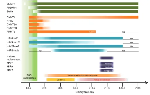
The most striking epigenetic event in PGCs is the genome-wide DNA demethylation that encompasses genic, intergenic and transposon sequences, which is completed in both sexes by E13.5 (see Fig. 3). As a consequence of this demethyation, one inactivated X-chromosome in females is reactivated, imprinted loci are fully demethylated and methylation at most transposable elements is erased (Hayashi and Surani, 2009). Notably, long terminal repeat (LTR) retrotransposon sequences, including IAPs, are more resistant to demethylation (Hajkova et al., 2002; Lane et al., 2003; Popp et al., 2010) and can cause transgenerational epigenetic inheritance (Whitelaw and Whitelaw, 2008).
A genome-wide bisulfite sequence analysis (covering ~1% of the genome) has quantified levels of 5mC in PGCs at E13.5, as well as in various cell types, and has shown that both male and female PGCs are extremely hypomethylated relative to other tissues (Popp et al., 2010). For example, whereas median methylation levels at CpGs in sperm are 85%, in ESCs they are 75%, in E13.5 embryos they are 73.2%, and in placenta they are 43.2%, those in E13.5
BLIMP1 PRDM14
Stella
DNMT1
DNMT3A DNMT3B NP95
H3K9me1/2
H3K27me3 H3K4me2
H4R3me2s
PGC specification
E6.5 E7.5 E8.5 E9.5 E10.5 E11.5 E12.5 E13.5
Nucleus
G2 arrest Active proliferation
Genome-wide DNA demethylation PRMT5
NAP1 Histone replacement
Nucleus
Embryonic day
HIRA CAF1
Nucleus
Cytoplasm
ND
ND
ND
ND ND
[image:9.612.56.541.293.601.2]ND
Fig. 3. A summary of epigenetic reprogramming in mouse primordial germ cells. A summary of the temporal expression patterns of key proteins involved in epigenetic reprogramming of mouse primordial germ cells (PGCs), and of the presence of key histone modifications, as revealed by immunohistochemistry and other methods (Hajkova et al., 2008; Popp et al., 2010; Seki et al., 2005; Seki et al., 2007). Migrating PGCs reduce H3K9me2 and upregulate H3K27me3 when they are arrested at the G2 phase of the cell cycle and become transcriptionally quiescent. Extensive remodelling of histone modifications occurs in the genital ridges at around E11.5, which perhaps reflects extensive histone replacement triggered by DNA repair (Hajkova et al., 2008; Hajkova et al., 2010). The level of genome-wide DNA methylation in PGCs becomes as low as or even lower than that of methylation-defective Np95–/–ES cells (Popp et al., 2010). The light-green box represents the period for PGC specification. The dark-green bars represent the expression of indicated genes associated with PGC specification. The orange bars represent the expression of the chromatin modifiers indicated. The light-blue bars represent the relative levels of the histone modifications indicated. The purple bars represent the occurrence of histone replacement and the expression of histone chaperones indicated (Hajkova et al., 2008; Hajkova et al., 2010). N.D., not determined. BLIMP1 (PRDM1), PR domain containing 1, with ZNF domain; CAF1, chromatin assembly factor 1; DNMT, DNA methyltransferase; HIRA, histone cell cycle regulation defective homolog A; NP95 (UHRF1), ubiquitin-like, containing PHD and RING finger domains 1; PRDM, PR
domain containing; PRMT, protein arginine methyltransferase.
D
E
V
E
LO
P
M
E
N
male and female PGCs are only 16.3% and 7.8%, respectively. Methylation levels in PGCs are substantially lower than the level of 22% that has been recorded in methylation-deficient Np95–/– ESCs. The fact that female PGCs have considerably lower methylation levels than do male PGCs may be because, owing to X-reactivation, female PGCs bear two active X-chromosomes, which may encode a modifier locus to lower genome-wide methylation levels (Zvetkova et al., 2005).
Dynamics of DNA demethylation in PGCs
The genome-wide DNA methylation state of PGCs before E13.5 has not yet been reported. Nonetheless, several studies have examined the timing of the demethylation of imprinted genes, some single-copy genes and transposable elements in PGCs from E10.5 to E12.5/E13.5 (Hajkova et al., 2002; Lane et al., 2003; Lee et al., 2002). In general, the timing of demethylation depends on the genes being analyzed and is thus heterogeneous. This finding might also reflect heterogeneity in the timing of demethylation in each PGC. One study has identified the rapid demethylation of imprinted genes between E11.5 and E12.5, proposing the involvement of active demethylation (Hajkova et al., 2002) when considering the doubling time of PGCs of ~16 hours (Tam and Snow, 1981); the DMRs of maternally methylated genes Snrpn (small nuclear ribonucleoprotein N), Peg3and Lit1[also known as Kcnq1ot1 (potassium voltage-gated channel, subfamily Q, member 1, overlapping transcript 1)] are all nearly fully methylated at E11.5, but become almost fully demethylated at E12.5.
Conversely, other studies have demonstrated a gradual erasure of methylation at several imprinted genes and retrotransposons, such as LINE1 and IAP (Lane et al., 2003; Lee et al., 2002). Notably, imprinted genes such as Nnat (neuronatin), H19 and Peg10are already partly (~50%) demethylated at E10.5 (Lee et al., 2002). These observations are compatible with the occurrence of replication-dependent passive demethylation. More comprehensive measurements of DNA methylation states during PGC development should provide further insights into the dynamics, and hence the mechanism, of DNA demethylation.
Active DNA demethylation in PGCs?
There is evidence that the cytosine deaminases AID (activation-induced deaminase) and APOBEC1 (apolipoprotein B mRNA editing enzyme, catalytic polypeptide 1) (Box 5, Fig. 4, Table 1) can convert 5mCs to thymines by deamination, creating T:G mismatches that might then become targets of thymine glycosylases (see Glossary, Box 2), such as MBD4 (methyl CpG binding domain protein 4) or TDG (thymine DNA glycosylase; see Glossary, Box 2), which then trigger the BER pathway (Morgan et al., 2004). AID-deficient male and female PGCs at E13.5 show median methylation levels of ~22% and 20%, respectively, which are higher than the methylation levels of wild-type male and female PGCs (16.3% and 7.8%, respectively), indicating that AID functions in genome-wide DNA demethylation in PGCs (Popp et al., 2010). Importantly, AID deficiency does not impact genome-wide methylation levels in cells/tissues other than PGCs (Popp et al., 2010). As the methylation levels of AID-deficient PGCs are still lower compared with those of earlier wild-type PGCs, the demethylation events occur even without AID, possibly owing to compensation by other deaminases, including APOBEC1/2/3. However, it is important to note that both AID-deficient male and female mice are relatively healthy (except for their B-cell-derived phenotype) and fertile, although some abnormalities in
litter size and progeny birth weights have been reported (Popp et al., 2010). Considering that the deregulated dose of even a single imprinted gene profoundly affects development and adult physiology, the genome-wide DNA demethylation that occurs in PGCs, which erases imprints and contributes to the creation of proper imprinted gene dose, should be a crucial event. Therefore, the finding that DNA demethylation deficiencies in AID-mutant PGCs does not lead to profound reproductive defects, such as infertility, subfertility or marked adult phenotypes, appears to be counter-intuitive. It has also been shown that AID is unable to act on double-stranded DNA and that 5mC is a much more inefficient target for AID-mediated deamination than is unmethylated cytosine in vitro (Bransteitter et al., 2003; Di Noia and Neuberger, 2007; Larijani et al., 2005), raising the issue of whether AID can directly deaminate 5mC in vivo.
In support of the active removal of 5mC during DNA demethylation in PGCs, the BER, but not the NER, pathway has been reported to operate in PGCs during their genome-wide DNA demethylation (Hajkova et al., 2010). As in the paternal pronucleus of the zygote, in ~E11.5 PGCs, components of this pathway, including XRCC1, APE1 and PARP1 are found to be enriched in their nuclei, and XRCC1 is found bound to PGC chromatin, suggesting that ssDNA breaks are present in PGCs. Hajkova et al. have shown that, as a potential consequence of the DNA demethylation mediated by the DNA repair mechanisms, PGCs at ~E11.5 show dramatic changes in their chromatin states, including rapid loss of linker histone H1, loss of detectable chromophores, significant enlargement of nuclei, and a concomitant loss of H3K9me3, H3K27me3, H4/H2AR3me2s and H3K9ac (Fig. 3). The loss of H3K9me3 and H3K27me3 seems transient, with these modifications being recovered after E12.5, whereas the loss of H4/H2AR3me2s and H3K9ac seems persistent. These dynamic
Box 5. AID and APOBECs: cytidine deaminases
AID (activation-induced deaminase) and APOBECs (apolipoprotein B mRNA editing enzyme, catalytic polypeptides) are a group of cytidine deaminases in vertebrates that can introduce mutations in DNA and RNA by deaminating cytidine to uridine. AID is involved in class switch recombination (CSR) and in somatic hypermutation (SHM) of the immunoglobulin (Ig) genes (Muramatsu et al., 2000; Revy et al., 2000). In one model, in activated B-cells, AID deaminates cytosines into uracils on the Ig loci, which creates U:G mismatches, triggering the error-prone DNA repair system (Di Noia and Neuberger, 2007). Consequently, U:G mismatches occurring in the V, D and J genes lead to affinity maturation or gene conversion, whereas U:G mismatches occurring in the switch regions lead to CSR (Di Noia and Neuberger, 2007). Another model posits that AID deaminates unidentified mRNA, leading to the production of a potential endonuclease that cleaves DNA during the immune response (Honjo et al., 2005). In the absence of AID, none of these events occurs (Muramatsu et al., 2000; Revy et al., 2000). APOBEC1 is an RNA deaminase that converts cytidine to uridine. Most typically, it edits apolipoprotein B RNA, generating a truncated apolipoprotein B in a tissue-specific manner (Conticello, 2008). APOBEC3 functions to restrict the activity of viruses and retrotransposons in primates by editing their DNAs (Conticello, 2008). Interestingly, both Aidand Apobec1are located in close proximity to Nanog and stella/Pgc7 (Dppa3, developmental pluripotency-associated 3) on mouse chromosome 6. This may account for the expression of Aidand Apobec1in pluripotent cell lineages, such as oocytes, embryonic stem cells and primordial germ cells (Morgan et al., 2004).
D
E
V
E
LO
P
M
E
N
changes are possibly mediated through histone replacement, perhaps by the histone chaperone HIRA (histone cell cycle regulation defective homolog A) or NAP1 (nucleosome assembly protein 1) (see Glossary, Box 2) (Hajkova et al., 2008).
Although the expression of Aid, Apobec1 (apolipoprotein B mRNA editing enzyme, catalytic polypeptide 1),Mbd4and Tdg (thymine DNA glycosylase) is low in PGCs from E10.5 to E12.5, significant expression of Tet1 has been found in these cells (Hajkova et al., 2010), indicating that, in PGCs, TET1 may convert 5mC into 5hmC, which could be cleaved by an as yet unidentified 5hmC glycosylase, leading to the activation of BER. However, recently generated Tet1knockout mice are viable and fertile, and mating between homozygous mutant males and females produces viable progeny, although with a reduced average litter size (three
to six pups compared with wild-type litters of five to nine pups) (Dawlaty et al., 2011). The role of TET proteins in gametogenesis and fertility thus requires further investigation.
Passive DNA demethylation in PGCs?
The extent of genome-wide DNA demethylation in PGCs is extraordinary and more global compared even with that in pre-implantation embryos, because, in PGCs, genome imprints are erased and the demethylation of transposable elements is more extensive (Hajkova et al., 2002; Lane et al., 2003; Lee et al., 2002; Popp et al., 2010). This suggests the presence of mechanisms unique to PGCs, allowing nearly complete DNA demethylation. Although AID and TET1 are suggested to be part of this process, these molecules are expressed elsewhere (e.g. B-cells and ESCs), AP lyase 5mC glycosylase OH O O P O O H O H OH P O O H O H AP endonuclease Polymerize and ligation A Deamination AID, APOBEC1 T/G mismatch, DNA glycosylase B TDG, MBD4 AP lyase, AP endonuclease Oxidation TET 5hmC glycosyla se Deami nation
?
mCN N O C H3 AID, APOB ECs G mCN N O O P+ O H OH O O O NH2 O N N NH N O NH2 O P O H O O H C H3 P OH O O H O O P+ O H OH O O O N N NH N O NH2 P O H O O H O H G O P OH O O H C NH N O O P+ O H OH O O O O O N N NH N O P O H O O H C H3 T G O P OH O O H N N O O P+ O H OH O O O O N N NH N O P O H O O H O H G hmC O P OH O O H SMUG1, TDG Methylated CpG hmUNH N H O O O HNH2 OH
P O H O O O N N NH N O G O P OH O O H NH2 C H2 OH O OH P O H O O O N N NH N O G O P OH O O H NH2 C G N N O O P+ O H OH O O O O N N NH N O P O H O O H O P OH O O H CpG NH2 NH2 NH2 NH2 NH2 G mCN N O O P+ O H OH O O O NH2 O N N NH N O NH2 O P O H O O H C H3 P OH O O H Methylated CpG G mCN N O O P+ O H OH O O O NH2 O N N NH N O NH2 O P O H O O H C H3 P OH O O H Methylated CpG O O P+ O H OH O O O N N NH N O NH2 P O H O O H OH G O P OH O O H OH P O H O O O N N NH N O G O P OH O O H NH2 C H2 OH O OH P O O H O
H CN
[image:11.612.52.565.60.448.2]N O O P+ O H OH O O O O N N NH N O P O H O O H O P OH O O H CpG NH2 NH2 (1) (2) (4) 5mC 5mC 5mC (in plants) T 5hmC 5hmU N N H NH2 O O H fC N N H NH2 O O O H caC 5fC 5caC Oxidation TET Oxidat ion TET CO2 Iso-orotate decar boxy lase (3) G TD G Polymerize and ligation
Fig. 4. Potential chemical pathways for active DNA demethylation. (A)Direct excision of 5mC (orange) by a 5mC glycosylase followed by repair via the base excision repair (BER) pathway (green and pink), as occurs in plants. (B)Cytosine deamination by AID/APOBEC1 (red), followed by base excision mismatch repair, involving the TDG/MBD4 (pale blue) and BER pathways. (C)Hydroxylation by TET (blue) initiates four potential pathways leading to demethylated cytosine: (1) removal of 5hmC by an unidentified 5hmC glycosylase, followed by BER; (2) deamination of 5hmC by AID or APOBECs creates 5hmU, which is removed by SMUG1 (single-strand selective monofunctional uracil DNA glycosylase) or TDG, followed by BER; (3) further oxidization of 5hmC to 5fC and then to 5caC, which then may be converted to C by a decarboxylase or by TDG followed by BER; and (4) direct conversion of 5hmC to 5mC by an unidentified enzyme (?). 5caC, carboxylcytosine; 5fC, formylcytosine; 5hmC, 5-hydroxymethylcytosine; 5hmU, 5-hydroxymethyluracil;5mC, 5-methylcytosine; AP, apurinic/apyrimidinic; AID, activation-induced deaminase; APOBEC1, apolipoprotein B mRNA editing enzyme, catalytic polypeptide 1; C, cytosine; G, guanine; MBD4, methyl CpG binding domain protein 4; SMUG1, single-strand selective monofunctional uracil DNA glycosylase; T, thymidine; TDG, thymine DNA glycosylase; TET, ten-eleven translocation.