http://dx.doi.org/10.4174/astr.2016.91.3.97 Annals of Surgical Treatment and Research
Is focused parathyroidectomy appropriate for patients
with primary hyperparathyroidism?
Won Woong Kim, Yumie Rhee1, Eun Jeong Ban, Cho Rok Lee, Sang-Wook Kang, Jong Ju Jeong,
Kee-Hyun Nam, Woong Youn Chung, Cheong Soo Park
Department of Surgery, Yonsei University College of Medicine, Seoul, 1Department of Internal Medicine, Endocrine Research Institute, Yonsei University College of Medicine, Seoul, Korea
INTRODUCTION
Primary hyperparathyroidism (PHPT) is the third most common endocrine disorder in the United States, with an annual incidence of 100,000 new cases [1]. In the advanced industrialized world, PHPT is detected through a screening test. Accordingly, asymptomatic hypercalcemia is more prominent than symptomatic PHPT [2]. Previously, bilateral neck explo ration was considered the gold standard for the treatment of
PHPT. However, with the advent of the sestamibi (MIBI) scan and radioguided parathyroidectomy in the late 1990s, many groups advocated for a singlegland operation [36]. Fortunately, most PHPT cases are due to single adenomas. Although focused parathyroidectomy (FP) has been the preferred treatment of PHPT, advocates of conventional parathyroidectomy (CP) have continued to debate the efficacy of CP relative to that of FP. Although in most cases a single adenoma is found, double ade nomas and multiglandular disease (MGD) have been reported
Purpose: The aim of this study was to determine whether focused or complete parathyroidectomy was more appropriate and to compare follow-up data in primary hyperparathyroidism (PHPT).
Methods: We retrospectively analyzed 225 operations for PHPT at Yonsei University Health System between 2000 and 2012. After excluding 93 patients, the remaining 132 were divided into 2 groups: those who underwent focused parathyroidectomy (FP) and those who underwent conventional parathyroidectomy (CP). We compared clinicopathological features; preoperative calcium, parathyroid hormone (PTH), phosphorus, vitamin D, 24-hour urine calcium, and alkaline phosphatase levels; postoperative calcium and PTH levels; pathologic diagnosis; multiplicity; and results of a localization study between the 2 groups.
Results: There was no significant difference in the rates of development of postoperative persistent hyperparathyroidism (1/122 FP patients and 1/10 CP patients) between the 2 groups due to a technical reason (FP 0.8% vs. CP 10.0%, P = 0.146). Multiglandular disease (MGD) was uncommon in all cases (6 of 132, 4.5%). All MGD cases were diagnosed using a preoperative localization study. Sestamibi scan and ultrasonography sensitivity were 94.2% and 90.2%, respectively.
Conclusion: We suggest that FP is appropriate in PHPT, except in cases of MGD if detected before the operation using preoperative imaging. Knowledge of hereditary PHPT and improved preoperative localization studies, such as high-resolution ultrasonography, contributed to the decision to perform FP rather than CP in all cases of unilateral results of the localizing study.
[Ann Surg Treat Res 2016;91(3):97-103]
Key Words: Primary hyperparathyroidism, Parathyroidectomy, Ultrasonography, Technetium Tc 99m Sestamibi
Reviewed January February March April May June July August September October November December
Received February 16, 2016, Revised May 21, 2016, Accepted June 2, 2016
Corresponding Author: Jong Ju Jeong
Department of Surgery, Yonsei University College of Medicine, 50-1 Yonsei-ro, Seodaemun-gu, Seoul 03722, Korea
Tel: +82-2-2228-2100, Fax: +82-2-313-8289 E-mail: Jungjongj@yuhs.ac
Copyright ⓒ 2016, the Korean Surgical Society
cc Annals of Surgical Treatment and Research is an Open Access Journal. All articles are distributed under the terms of the Creative Commons Attribution Non-Commercial License (http://creativecommons.org/licenses/by-nc/4.0/) which permits unrestricted non-commercial use, distribution, and reproduction in any medium, provided the original work is properly cited.
at rates of 4% and 14%, respectively [7]. Recently, the leading group advocating unilateral parathyroidectomy has abandoned unilateral exploration and reversed its position on CP due to the high recurrence rate (FP 5% vs. CP 0.6% at 10 years) [8].
However, the incidence of PHPT has differed according to race, with the highest rates among blacks, followed by whites, and the lowest rates among Asians (prevalence of 321 vs. 201 vs. 103 per 100,000, respectively) [9]. It has also been reported that multiple parathyroid lesions were rare in Asians when PHPT was diagnosed [911].
In addition, we have also recognized two discrete forms, such as asynchronous and synchronous MGD [12]. Preoperative localization studies have been more successful due to the advent of sensitive imaging techniques, thus allowing pre determination of single or multiple gland involvement [1319].
Consequently, we hypothesized that FP may be an operation method that offersh a high success rate in PHPT. The aim of this study was to determine whether CP or FP is appropriate and to compare followup data in PHPT.
METHODS
This study was approved by the Yonsei University Institutional Review Board (approval number: 420140015). The need for informed consent was waived because of the
retrospective nature of the study. We retrospectively analyzed 225 operations for PHPT at Yonsei University Health System between 2000 and 2012. Ninetythree cases met the exclusion criteria, and 30 cases were excluded owing to multiple endocrine neoplasm, familial isolated hyperparathyroidism, pending chronic re nal failure, parathyroid cyst, or parathyroid carcinoma. Fur thermore, 51 cases were excluded owing to concomitant thyroid disease requiring thyroidectomy, and 12 cases were excluded owing to an incomplete preoperative study (Fig. 1). All patients underwent preoperative ultrasonography (USG) and a MIBI scan, and all pathologybased diagnoses were confirmed histopathologically after the operation. We did not use intraoperative parathyroid hormone (PTH) monitoring. Instead, we assessed postoperative PTH levels within 2 hours after the operation. A total of 132 patients were divided into 2 groups according to the operation method: those who underwent FP (n = 122) and those who underwent CP (n = 10). The FP group included minimally invasive radioguided parathyroidectomy and endoscopic parathyroidectomy. We compared the clinicopathological features, including symptoms and signs; preoperative calcium, PTH, phosphorus, vitamin D, 24hour urine calcium, and ALP levels; postoperative calcium and PTH levels; pathologic diagnosis; multiplicity; and results of the localization study. Recurrence was defined as elevated calcium and PTH occurring after 6 months postoperatively.
Fig. 1. Characteristics of the cases ex cluded. PHPT, primary hyperparathyroidism; CRF, chro nic renal failure; MEN, mul tiple endocrine neoplasia; HPT, hyper parathyroidism; MIBI, sestamibi; USG, ultrasonography.
225 Patients for PHPT
51 Concomitant thyroid disease 30 Thyroid cancer
20 Thyroid nodule
12 Parathyroid cyst 2 Parathyroid carcinoma 4 Pending CRF
12 Inherited parathy oid disease 10 MEN 1
1 MEN 2a
1 Familial isolated HPT r
12 Incomplete imaging study (no preceding MIBI scan)
132 Enrolled patients
How many parathyroid lesion are detected using preoperative localization studies (MIBI scan and USG)?
122 Focused parathy oidectomyr Single
10 Conventional parathyroidectomy Multiple
Persistence was defined as elevated calcium and PTH within 6 months postoperatively [20].
Data are expressed as mean ± standard deviation. The Fisher exact test or Pearson chisquare test was used to compare clinicopathological findings in FP and CP groups. Statistical analysis was performed using IBM SPSS Statistics ver. 20.0 (IBM Co., Armonk, NY, USA), and differences were considered significant when Pvalues were less than 0.05.
RESULTS
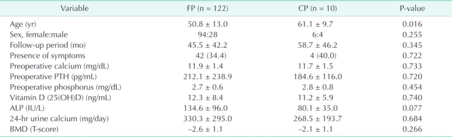
There were no differences in symptomatic characteristics between FP and CP groups (Table 1). About 60%–65% of cases were asymptomatic. Age was significantly higher in the CP group compared to that in the FP group, but there were no differences in demographic and laboratory characteristics bet
ween FP and CP groups for sex, followup period, presence of symptoms, preoperative serum calcium, preoperative PTH, pre operative serum phosphate and ALP levels, and preoperative bone mineral density. Operation time was shorter in the FP than that in the CP group. Our study contained a variable followup period, with some patients followed for almost 12 years. The mean followup time was about 46 months in the FP group and 59 months in the CP group (Table 2).
There was one failed preoperative localization using both USG and MIBI in the FP group. However, this failed localization was followed by a successful additional magnetic resonance study. Our study showed that sensitivity in detecting parathyroid lesions was 94.3% using MIBI scans and 90.2% using USG. Of the 122 patients in the FP group, only 103 patients were positive using both USG and the MIBI scan; 7 patients were positive only in the USG study, and 12 patients were positive only in the MIBI study. Therefore, almost all parathyroid lesions were localized using a combination of USG and the MIBI scan (Table 3).
There was no significant difference between the 2 groups for postoperative calcium, PTH level, or size and weight of the parathyroid specimen. A large percentage (95.5%) of all pa tients with PHPT had a single parathyroid lesion. MGD was un common in all cases (6 of 132, 4.5%). In all cases of multiple para thyroid lesions identified in the preoperative localization study, we preferred CP due to the possibility of MGD or double adenoma. However, of the 10 CP patients, four underwent CP owing to another USGdetected ambiguous parathyroid lesion that was confirmed using a frozen biopsy. In these cases, disease in only a single parathyroid gland was reported at the final pathology.
There was no significant difference in the rate of failed sur Table 1. Characteristic symptoms and signs of primary hy
per parathyroidism
Symptom and sign FP (n = 122) CP (n = 10)
Asymptomatic (hypercalcemia based
on a screening test) 80 (65.6) 6 (60.0)
Symptomatic 42 (34.4) 4 (40.0)
Bone pain/osteoporosis 18 (14.8) 1 (10.0)
Renal or ureter stone 12 (9.8)
Fatigue/general weakness 9 (7.4) 2 (20.0)
Gastroduodenal ulcer 5 (4.1)
Palpable neck mass 3 (2.5) 1 (10.0)
Itching and aching pain, hot flush 5 (4.1)
Values are presented as number (%).
FP, focused parathyroidectomy; CP, conventional parathyroidec tomy.
Table 2. Preoperative demographics and clinical data bet ween focused parathyroidectomy and conventional para thy roi dec tomy groups
Variable FP (n = 122) CP (n = 10) Pvalue
Age (yr) 50.8 ± 13.0 61.1 ± 9.7 0.016
Sex, female:male 94:28 6:4 0.255
Followup period (mo) 45.5 ± 42.2 58.7 ± 46.2 0.345
Presence of symptoms 42 (34.4) 4 (40.0) 0.722 Preoperative calcium (mg/dL) 11.9 ± 1.4 11.7 ± 1.5 0.733 Preoperative PTH (pg/mL) 212.1 ± 238.9 184.6 ± 116.0 0.720 Preoperative phosphorus (mg/dL) 2.7 ± 0.6 2.8 ± 0.8 0.454 Vitamin D (25(OH)D) (ng/mL) 12.3 ± 8.4 11.2 ± 5.9 0.740 ALP (IU/L) 134.6 ± 96.0 80.1 ± 35.0 0.077
24hr urine calcium (mg/day) 330.3 ± 295.0 268.5 ± 193.7 0.684
BMD (Tscore) –2.6 ± 1.1 –2.1 ± 1.1 0.266
Values are presented as mean ± standard deviation or number (%).
Reference values for our institution: calcium, 8.6–10.6 mg/dL; PTH, 15–65 pg/mL; phosphorus, 2.3–4.5 mg/dL; vitamin D (25(OH)D), >30 ng/mL; ALP, 120–360 IU/L; 24hr urine calcium, 50–200 mg/day.
FP, focused parathyroidectomy; CP, conventional parathyroidectomy; PTH, parathyroid hormone; BMD, bone mineral density (Tscore from Lspine).
gery between the 2 groups (FP 0.8% vs. CP 10.0%, P = 0.146). Post operative persistent hyperparathyroidism developed in one FP patient and one CP patient due to technical failure. All MGD cases were diagnosed using the preoperative localization study. There was no significant difference in the sizes or weights of specimens between the 2 groups. However, the rate of hyperplasia was higher in the CP group than in the FP group (50% vs. 12.3%, P = 0.02) (Table 4).
In the 2 cases of persistent hyperparathyroidism, the postop
erative PTH level exceeded the upper limit of the normal range.
Case 1
A 65yearold male (sCa 12.2 mg/dL, PTH 88.2 pg/mL) underwent right superior and left superior parathyroidectomy. Postoperatively, sCa (10.6 mg/dL) and PTH (75.2 pg/mL) were still elevated. One right superior parathyroid hyperplasia and one left superior lymph node in one CP patient were reported at the final pathology. The patient denied the proposed additional reoperation.
Case 2
A 63yearold female (sCa 11.1 mg/dL, PTH 300 pg/mL) underwent right inferior parathyroidectomy. Postoperatively, sCa (9.4 mg/dL) and PTH (151 pg/mL) were checked. One para thyroid tissue in one FP patient was reported at the final patho logy. Ten months after initial operation, she was cured after resection of right inferior parathyroid adenoma.
In the 2 failed surgery cases, an additional localization study such as methionine PETCT, selective venous sampling, or USG guided indigo carmine tattooing was used for reoperation. After the preceding additional localization study, 2 patients had reexploration after a previous procedure failed to locate and remove an abnormally functioning parathyroid gland. After Table 3. Comparison of preoperative localization studies
bet ween focused parathyroidectomy and conventional para thyroidectomy groups
Preoperative study FP (n = 122) CP (n = 10) Pvalue
USG Positive 110 (90.2) 9 (90.0) 0.987 Negative 12 (9.8) 1 (10.0) MIBI Positive 115 (94.3) 10 (100) 0.436 Negative 7 (5.7) 0 (0)
Values are presented as number (%).
FP, focused parathyroidectomy; CP, conventional para thy roi dec tomy; USG, ultrasonography; MIBI, sestamibi.
Table 4. Comparison of postoperative characteristic findings
Variable FP (n = 122) CP (n = 10) Pvalue
Operation time (min) 52.0 ± 19.1 94.5 ± 30.8 0.001
Postoperative calcium (mg/dL)
Within 2 hours after surgery 9.2 ± 0.9 9.1 ± 1.0 0.862
6 Months after surgery 9.27 ± 0.5 9.3 ± 0.7 0.846
Postoperative PTH (pg/mL)
Within 2 hours after surgery 19.0 ± 21.8 25.6 ± 28.1 0.373
6 Months after surgery 35.4 ± 20.8 40.8 ± 25.8 0.469
Parathyroid lesion 0.001
Single lesion 122 (100) 4 (40.0)
Right superior:left superior 17:20 0:1
Right inferior:left inferior 47:38 2:1
Multiple lesion 0 (0) 6 (60.0) Unilateral 0 (0) 1 DA Bilateral 0 (0) 5 (MGD 2, DA 3) Pathology 0.002 Adenoma 107 (87.7) 5 (50.0) Hyperplasia 15 (12.3) 5 (50.0) Size (mm) 19.3 ± 8.9 21.4 ± 13.9 0.497 Weight (mg) 2,354 ± 2,969 3,200 ± 812 0.574 Failed surgery 0.146 Persistent 1 (0.8) 1 (10.0) Recurrent 0 (0) 0 (0)
Values are presented as mean ± standard deviation or number (%).
Reference values for our institution: calcium, 8.6–10.6 mg/dL; PTH, 15–65 pg/mL.
FP, focused parathyroidectomy; CP, conventional parathyroidectomy; PTH, parathyroid hormone; DA, double adenoma; MGD, multi glandular disease.
removal of the abnormal gland, all patients became normo calcemic, and the PTH level decreased below the upper limit of the normal range.
DISCUSSION
Before advances in preoperative localization studies and intraoperative PTH analysis, bilateral exploration was consi dered the gold standard treatment of PHPT because of the possi bility of MGD. However, with improved understanding of the disease pattern in PHPT, unilateral approaches have emerged with high cure rates. Nonetheless, Duh et al. [7] reported that unilateral exploration was associated with a risk of missing a contralateral double adenoma with or without a localization study. Despite the possibility of MGD, many groups have advocated for using a singlegland operation such as radioguided or FP [36,21]. Before the first application of minimally invasive FP in 2001, we performed 32 radio guided parathyroidectomies beginning in 1999. Our previous initial study showed that minimally invasive radioguided parathyroidectomy and minimally invasive FP achieved a high success rate compared with bilateral or unilateral exploration [22]. Among the 122 patients with FP who were enrolled in this study, only 9 underwent radioguided parathyroidectomy. Accordingly, Radioguided parathyroidectomy could not have influenced our results. This study had somewhat descriptive nature, making it difficult to identify significant differences between FP and CP. As more data have become available, there has been a gradual evolution of the operation method used to
avoid recurrence. About 10 years ago, we confirmed that FP is sufficient to treat PHPT due to the advent of sensitive imaging techniques such as highresolution USG that are used for preoperative localization studies [22].
Furthermore, the group of Norman et al. [8]—the leading group advocating unilateral parathyroidectomy—have recently abandoned this position and reversed their position toward CP. They showed that the recurrence rate after unilateral para thyroidectomy was significantly higher than that after bila teral operation. Nevertheless, it is possible that the authors might have missed false negative parathyroid lesions in the parathyroid scan, because they only used MIBI scan. Consequently, surgical controversies are more complicated in PHPT, and there was a need to confirm treatment of PHPT using longterm followup data. It is notable that MGD was un common in our study and that cure rates were as high as 98.5%, with an especially high rate of 99.2% in the FP group (Table 5).
In addition, some groups have reported that microscopically hyperplastic glands of normal size are of no clinical significance and that the removal of such glands is unnecessary [23]. After experienced surgeons inspected ambiguous parathyroid lesions in patients with MGD and double adenoma, they decided whether a frozen biopsy should be performed. Even though we used frozen biopsies to confirm pathology, some of the pathologic reports did not provide enough information to obtain a precise determination of whether the parathyroid lesion included pathological change. Although histologic differences between adenomas and hyperplastic glands have been proposed, the diagnosis of singlegland disease versus Table 5. Comparison of cure rates between procedures
Study Operative procedure No. of patients Preoperative studies IoPTH Cure rate (%)
Chen et al. [3] (1999) MIP 33 SestamibiSPECT (+) 100
BE 184 (+) 97.3
Udelsman [21] (2002) MIP 255 Sestamibi (+) 99.0
BE 401 (+) 98.0
Yoon et al. [22] (2004) BE+UE 60 Sestamibi (–) 96.7
MIRP 31 Sestamibi (–) 96.8
MIFP 27 USG + Sestamibi (–) 96.3
Lee et al. [10] (2008) BE 70 USG + Sestamibi (–) 100
UE 34 (–) 100
MIP 75 (–) 98.7
Elaraj et al. [25] (2010) BE 153 USG + Sestamibi (+) 91.5
FP +UE 339 (+) 98.2
Norman et al. [6] (2010) BE 2,048 Sestamibi (+) 99.9
UE 952 (+) 99.0
Gill et al. [18] (2011) BE 62 USG + Sestamibi (+) 98.0
UE 14 (+) 100
Kim et al. 2016 (present study) FP 122 USG + Sestamibi (–) 99.2
CP 10 (–) 90.0
BE, bilateral exploration; UE, unilateral exploration; SPECT, single photon emission computerized tomography; MIRP, minimal invasive radioguided parathyroidectomy; MIFP, minimal invasive focused parathyroidectomy; IoPTH, intraoperative parathyroid hormone.
fourgland hyperplasia has traditionally been made using a combination of gross surgical and histologic findings. On the other hand, a Wisconsin group suggested that parathyroid gland weight helps to predict the likelihood of an additional hyperfunctioning parathyroid gland [24]. In this regard, high resolution USG is very useful and can supplement MIBI scans, which do not precisely measure parathyroid size. Elaraj et al. [25] suggested additional imaging using neck USG is helpful for selecting minimally invasive parathyroidectomy in most patients with PHPT who have negative MIBI scan results. We decided to perform FP in both of the following situations: when USG and MIBI scan findings were discordant in unilateral location, and when there was single positive lesion on both the USG and MIBI scan. However, when USG and MIBI scan findings were discordant in bilateral location, we decided to perform CP. Our results showed that USG is useful when MIBI scans are negative and that it may also be useful when tattooing a parathyroid lesion in failed surgery.
Nonetheless, reoperation for recurrent PHPT was associated with higher failure rates and complications, especially in CP cases. The occurrence of asynchronous MGD has been re ported after an initially successful operation. Worsey et al. [12] reported an average recurrentfree period of 3.8 years after a successful operation with 15 years of followup. Because of the possibility of asynchronous MGD, using FP to avoid unnecessary dissection is more beneficial than CP in recurrent cases. Patients who are not cured after such an approach are able to undergo a simple, noncomplex, second operation that is performed in naïve tissues [8].
Our study has limitations. First, the followup period for
this series was different for each patient (6–160 months). In addition, the incidence of PHPT in Korea is lower than in America and Europe. Moreover, we did not use intraoperative PTH monitoring; instead, we checked postoperative PTH within 2 hours postoperatively. In spite of these limitations, we have previously reported that there was a gradual evolution of the operation method used to avoid recurrence at our institution over about 24 years [22].
Ning et al. reported that elevated PTH after successful para thyroidectomy for PHPT is not associated with recurrent hyper parathyroidism [26,27]. Many studies have reported that intraoperative PTH monitoring is useful in successful para thyroidectomy for PHPT [28,29]. In some cases, intraoperative PTH monitoring is thought to confuse the decisionmaking process [27,30]. Furthermore, undetected hyperplastic glands of normal size in the preoperative imaging study are of no clinical significance, and the removal of such glands is unnecessary [23].
In conclusion, FP is appropriate in cases of a single lesion confirmed preoperatively. Consequently, we suggest FP is more appropriate than CP for PHPT, except for cases in which MGD is detected in the preoperative imaging study. Improved preoperative localization studies such as highresolution USG and additional knowledge regarding hereditary PHPT may help to determine whether FP or CP should be performed for all cases in which the localization study has unilateral results.
CONFLICTS OF INTEREST
No potential conflict of interest relevant to this article was reported.
1. Adler JT, Sippel RS, Chen H. New trends in parathyroid surgery. Curr Probl Surg 2010;47:9581017.
2. Macfarlane DP, Yu N, Donnan PT, Leese GP. Should ‘mild primary hyper para thy roidism’ be reclassified as ‘insidious’: is it time to reconsider? Clin Endocrinol (Oxf) 2011;75:7307.
3. Chen H, Sokoll LJ, Udelsman R. Outpatient minimally invasive parathyroidectomy: a combination of sestamibiSPECT locali za tion, cervical block anesthesia, and intra opera tive parathyroid hormone assay. Surgery 1999;126:101621.
4. Flynn MB, Bumpous JM, Schill K, Mc Masters KM. Minimally invasive radio guided parathyroidectomy. J Am Coll Surg 2000;191:2431.
5. Nagar S, Reid D, Czako P, Long G, Shanley C. Outcomes analysis of intraoperative ad juncts during minimally invasive para thy roidectomy for primary hyper para thyroidism. Am J Surg 2012;203:17781. 6. Norman J, Politz D. Prospective study in
3,000 consecutive parathyroid operations demonstrates 18 objective factors that in fluence the decision for unilateral versus bilateral surgical approach. J Am Coll Surg
2010;211:2449.
7. Duh QY, Uden P, Clark OH. Unilateral neck exploration for primary hyperpara thy roidism: analysis of a controversy using a mathematical model. World J Surg 1992;16:65461.
8. Norman J, Lopez J, Politz D. Abandoning unilateral parathyroidectomy: why we reversed our position after 15,000 para thyroid operations. J Am Coll Surg 2012; 214:2609.
9. Yeh MW, Ituarte PH, Zhou HC, Nishimoto S, Liu IL, Harari A, et al. Inci dence and prevalence of primary hyper para thy roi
REFERENCES
dism in a racially mixed population. J Clin Endocrinol Metab 2013;98:11229. 10. Lee SH, Hong SJ. Minimally invasive
parathyroidectomy without an Intra opera tive iPTH test for patients with primary hyperparathyroidism. J Korean Surg Soc 2008;75:914.
11. Lo CY, Lang BH, Chan WF, Kung AW, Lam KS. A prospective evaluation of pre operative localization by techne tium99m sestamibi scintigraphy and ultrasonography in primary hyper para thyroidism. Am J Surg 2007;193:1559. 12. Worsey MJ, Carty SE, Watson CG. Success
of unilateral neck exploration for sporadic primary hyperparathyroidism. Surgery 1993;114:10249.
13. Mihai R, Simon D, Hellman P. Imaging for primary hyperparathyroidisman evi dencebased analysis. Langenbecks Arch Surg 2009;394:76584.
14. Agcaoglu O, Aliyev S, Heiden K, Neumann D, Milas M, Mitchell J, et al. A new classi fication of positive sestamibi and ultra sound scans in parathyroid locali za tion. World J Surg 2012;36:251621.
15. Akram K, Parker JA, Donohoe K, Kolodny G. Role of single photon emission com puted tomography/computed tomography in localization of ectopic parathyroid ade noma: a pictorial case series and review of the current literature. Clin Nucl Med 2009;34:5002.
16. Rodgers SE, Hunter GJ, Hamberg LM, Schellingerhout D, Doherty DB, Ayers GD, et al. Improved preoperative planning for directed parathyroidectomy with 4dimen sional computed tomography. Surgery 2006;140:93240.
17. Alexandrides TK, Kouloubi K, Vagenakis
AG, Yarmenitis S, Spyridonidis T, Vassila kos P, et al. The value of scinti gra phy and ultrasonography in the pre op era tive localization of parathyroid glands in patients with primary hyper para thy roi dism and concomitant thyroid disease. Hormones (Athens) 2006;5:4251. 18. Gill MT, Dean M, Karr J, Aultman DF,
Nathan CO. Intraoperative parathyroid hor mone assay: a necessary tool for multi glandular disease. Otolaryngol Head Neck Surg 2011;144:6917.
19. Yip L, Pryma DA, Yim JH, Virji MA, Carty SE, Ogilvie JB. Can a lightbulb sestamibi SPECT accurately predict singlegland dis ease in sporadic primary hyper para thyroidism? World J Surg 2008;32:78492. 20. Wirowski D, Goretzki PE, Schwarz K,
Lammers BJ, Dotzenrath C, Roher HD. Failed surgery in primary hyper para thy roi dism: what has changed with time. Exp Clin Endocrinol Diabetes 2013;121:3238. 21. Udelsman R. Six hundred fiftysix con
secutive explorations for primary hyper parathyroidism. Ann Surg 2002;235:665 70.
22. Yoon JH, Chang HS, Park CS. Surgical strategy in the management of primary hyperparathyroidism. J Korean Surg Soc 2004;66:3728.
23. Harrison TS, Duarte B, Reitz RE, Prin centhal R, Seaton JF, Badder EM, et al. Pri mary hyperparathyroidism: four to eightyear postoperative followup demon strating persistent functional in signi ficance of microscopic parathyroid hyper plasia and decreased autonomy of para thyroid hormone release. Ann Surg 1981;194:42937.
24. Mazeh H, Chen H, Leverson G, Sippel
RS. Creation of a “Wisconsin index” nomo gram to predict the likelihood of addi tional hyperfunctioning parathyroid glands during parathyroidectomy. Ann Surg 2013;257:13841.
25. Elaraj DM, Sippel R S, Lindsay S, Sansano I, Duh QY, Clark OH, et al. Are additional localization studies and re ferral indicated for patients with pri mary hyper parathyroidism who have nega tive sestamibi scan results? Arch Surg 2010; 145:57881.
26. Ning L, Sippel R, Schaefer S, Chen H. What is the clinical significance of an elevated parathyroid hormone level after curative surgery for primary hyperparathyroidism? Ann Surg 2009;249:46972.
27. Carneiro DM, Solorzano CC, Nader MC, Ramirez M, Irvin GL 3rd. Comparison of intraoperative iPTH assay (QPTH) criteria in guiding parathyroidectomy: which cri terion is the most accurate? Surgery 2003; 134:9739.
28. Day KM, Elsayed M, Monchik JM. No need to abandon focused unilateral exploration for primary hyperparathyroidism with intra operative monitoring of intact para thyroid hormone. J Am Coll Surg 2015; 221:51823.
29. Kim HG, Kim WY, Woo SU, Lee JB, Lee YM. Minimally invasive para thy roi dec tomy with or without intraoperative para thyroid hormone for primary hyper para thyroidism. Ann Surg Treat Res 2015;89: 1116.
30. Miura D, Wada N, Arici C, Morita E, Duh QY, Clark OH. Does intraoperative quick parathyroid hormone assay improve the results of parathyroidectomy? World J Surg 2002;26:92630.