CASE REPORT
Coccidioidomycosis with cutaneous manifestation of erythema
nodosum in Taiwan
Chien-Ming Chen
1, Hua-En Lee
1*, Shu-Ying Li
21Department of Dermatology, Chang Gung Memorial hospital and Chang Gung University College of Medicine, Tao-Yuan, Taiwan
2Mycotic and Sexually Transmitted Disease Laboratory Research and Diagnostic Center, Centers for Disease Control, Taipei, Taiwan
ABSTRACT
Coccidioidomycosis is a fungal disease endemic to the southwestern USA, northern Mexico, and focal areas in Central and South America, but it is rarely reported in Taiwan. We report a case of coccidioidomycosis of probable pulmonary origin in a young woman, who came back to Taiwan after a 10-month stay in the Central Valley of California. Coccidioidomycosis was suspected by typical constellation of symptoms and signs of Valley fever and subsequent desert rheumatism. The diagnosis was con-firmed by positive results of latex agglutination, immunodiffusion tube precipitation and immunodiffusion complement fixation tests. We prescribed symptomatic treatment without any anti-fungal agent according to the Infectious Disease Society of America guidelines, and the clinical symptoms and signs gradually subsided 3 weeks later.
Although coccidioidomycosis is rare in Taiwan, it is believed that the real incidence has been underestimated. Clinicians must be familiar with the typical manifestations and consider the diagnosis of coccidioidomycosis when an individual presents with fever, cough, arthralgia and skin eruptions such as erythema nodosum and/or erythema multiforme with a traveling history to the endemic area.
Copyright © 2010, Taiwanese Dermatological Association. Published by Elsevier Taiwan LLC. All rights reserved. KEYWORDS
Coccidioidomycosis Desert rheumatism Immunodiffusion complement-fixation test
Immunodiffusion tube precipitation Valley fever
Introduction
Coccidioidomycosis (San Joaquin fever, Valley fever) is a fungal disease found mostly in the Western hemisphere.1 It is
caused by two nearly identical fungal species, Coccidioides immitis, and C. posadasii. Endemic regions include south-western USA with California (mainly San Joaquin Valley), western Texas, New Mexico and the desert areas of Arizona, and Central and South America. Many tourists and visitors acquire the infection. The classic symptoms of Valley fever include fever, cough, shortness of breath, pleuritic chest pain, fatigue, weight loss and headaches, and it may be followed by a triad of desert rheumatism (fever, erythema nodosum, and arthralgia). We report a case of coccidioidomycosis and it was confirmed by latex agglutination (LA), immunodiffusion
tube precipitation (IDTP) and immunodiffusion complement fixation (IDCF) tests in Taiwan.
Case report
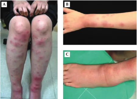
A 29-year-old female Taiwanese copilot had been in the Central Valley of California for flight training since July 2008. Generalized malaise, dry cough, night sweating, and mild fever, appeared half a month before she came back to Taiwan. Intermittent headaches, generalized nonitchy mac-ulopapules, hemorrhagic vesicles within the lip and buccal mucosa and tender swelling over the left ankle were also found. The lesions subsided without treatment. She returned to Taiwan on 2 May, 2009; tender erythematous nodules over bilateral lower extremities, which progressed to her upper extremities, and aggravation of the left ankle swelling (Figure 1) were found.
The patient came to our emergent department because of fever and progressive tender skin lesions on 19 May, 2009.
*Corresponding author. Department of Dermatology, Chang Gung Memorial Hospital and Chang Gung University College of Medicine, No. 123, Dinghu Road, Guishan Township, Taoyuan County 333, Taiwan.
E-mail: sharonrose@adm.cgmh.org.tw
A physical examination showed mild fever (37.6°C) and tachy-cardia. Fine bilateral basilar crackles and painless, movable, lymphadenopathy at the right of the base of the neck were also discovered. No photophobia, conjunctivitis, maculo-papules, oral lesions, or abdominal discomfort were found. A chest radiograph at our emergent department revealed mild exaggerated lung markings and a suspicious cavitating lesion at the right upper lobe (Figure 2). A lab examination showed leukocytosis with a white blood cell count of 11.2 × 10⁹/L (normal range: 3.9 × 10⁹–10.6 × 10⁹/L), atypical
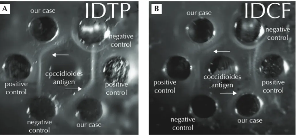
lymphocytes were 3.0% and the erythrocyte sedimentation rate was elevated (66 mm/hr) (normal range: 1–20 mm/hr). A basic metabolic panel showed elevation of C-reactive pro-tein levels (1169 nmol/L) (< 47.6 nmol/L), but there were no remarkable findings for a liver function test, serum creatinine, antinuclear antibody, C3 and RPR-VPRL (Rapid Plasma Reagin-Venereal Disease Research Laboratory). Urinary analysis showed no pyuria. Blood cultures for bacteria and fungi were collected and showed negative results. Sputum cul-ture was not available because of scanty sputum and the patient refused bronchoalveolar lavage. A skin biopsy was performed. Histopathology of the skin specimen showed septal fibrosis and thickened vessels with mononuclear cells in fat lobules. Focal granulomatous inflammation with occasional multinucleated giant cells was also observed but no remarkable pathogens were found (Figure 3). These findings were consistent with erythema nodosum. We also contacted the Taiwan Centers for Disease Control to submit the patient’s serum for LA, IDTP and IDCF because of a high suspicion of coccidioidomycosis. The results of LA (not shown), IDTP and IDCF were all positive (Figure 4) and the diagnosis of coccidioidomycosis was confirmed.
We did not administer antifungal agents because there was no evidence of systemic spread and clinically evanes-cent disease course. However, we gave symptomatic medi-cation and scheduled regular outpatient visits every 3 months
for 1 − 2 years according to the Infectious Disease Society of America guidelines for uncomplicated primary pulmonary infection.
Three weeks later, follow-up chest radiography (Figure 5) and computed tomography (Figure 6) showed that the previ-ous cavitating lesion observed on a chest radiograph over the right upper lobe had eventuated into a pulmonary nodule. Computed tomography revealed multiple reactive lymphad-enopathy within bilateral lung fields and the mediastinum. Erythema nodosum had improved with remnant dull-brownish patches. Follow-up laboratory tests showed no leukocytosis and normal C-reactive protein levels. No other noteworthy symptoms and signs were found.
Discussion
Coccidioidomycosis is a fungal infection acquired by inhala-tion of arthroconidia in the arid desert soil in southwestern
B A
C
Figure 1 Erythema nodosum-like lesions over (A) bi-lateral lower extremities and (B) the left upper arm. (C) Erythematous tender swelling over the left ankle.
R
Figure 2 Chest radiograph at the emergency department. The arrow-head indicates a cavitating lesion over the right upper lobe.
USA, northern Mexico and several areas of South America.1
Only two cases have been reported in Taiwan. One case devel-oped disseminated macules on the trunk and four limbs with pulmonary and lymph node involement,2 and the other died
from fulminant disseminated coccidioidomycosis without
cutaneous manifestation.3 Our case presented with cutaneous
erythema nodosum, arthralgia, and mild pulmonary symp-toms, which suggested the diagnosis of coccidioidomycosis. The fungus grows in a hardy mycelial phase in the soil and can remain viable for months to years. As the soil dries
A B
Figure 3 (A) Prominent septal fibrosis and thickened vessels with mononuclear cells in fat lobules (H&E, 40×). (B) Focal granulomatous inflammation with occasional multinucleated giant cells is seen but there are no spherules (H&E, 200×).
our case
IDTP
our caseIDCF
our case our case positive
control
positive control coccidioides
antigen positive coccidioidesantigen control positive control negative control negative control negative control negative control A B
Figure 4 Results of (A) immunodiffusion tube precipitation (IDTP) and (B) immunodiffusion complement fixation (IDCF). White arrows indicate positive results (immune complex deposition). The two wells of each sample represent duplicate tests.
R
Figure 5 Chest radiograph 3 weeks later. The arrow indicates the pre-vious cavitating lesion that eventuated into a nodule.
Figure 6 Computed tomography 3 weeks later reveals a nodule in the superior segment of the right lower lobe and other tiny nodules in both lungs.
or nutrients become limiting, the fungus rapidly reproduces asexually by disarticulating the hyphae into small, environ-mentally resistant arthroconidia (reproductive spores) after rain. Wind disperses these arthroconidia into the air and they are inhaled by animals or humans.4 Within the lung,
one arthroconidia is able to transform into new, multinucle-ated spherules, which vary in size from 10 μm to 80 μm, with the average size being approximately 40 μm containing 800– 1000 endospores (2–5μm), and each endospore grows into a new spherule that causes the disease.5
Coccidioidomycosis is seldom contagious in the spherule phase. Human-to-human spread is extremely rare. Primary exposure to contaminated soil or dust is the only risk factor for the acquisition of this disease. Of infected individuals, 60% are asymptomatic and the remaining 40% manifest as self-limited “influenza-like” symptoms after 7–21 days of inhalation of arthroconidia,4 and 0.5–1.5% develop
dissemi-nation to the skin, bones, joints, or central nervous system.6
Disseminated coccidioidomycosis most frequently occurs in African Americans, Filipinos, Hispanics, and Native Americans, the elderly, pregnant women, and those with cellular immunodeficiencies.7,8
The cutaneous manifestations of C. immitis are different between acute pulmonary coccidioidal exanthem, the primary cutaneous form and the secondary cutaneous form. During the acute pulmonary coccidioidal exanthem, toxic erythema, erythema nodosum, and/or erythema multiforme are mostly encountered. These are classified as reactive or nonspecific manifestations in which no detectable organisms are found on biopsy4 as in our case. A generalized toxic erythema occurs
in 12% of patients with symptomatic pulmonary coccidioi-domycosis.8 Both erythema nodosum and erythema
multi-forme are more commonly found in females and they are associated with a favorable prognosis (disseminated disease is unlikely).6
In the present case, symptoms of Valley fever such as fever, cough, equivocal pleuritic chest pain, fatigue and severe headaches with toxic erythema were initially noted, as well as influenza-like symptoms and prominent left ankle arthral-gia. Erythema nodosum was then found over the patient’s four limbs. Skin biopsy of the erythema nodosum-like lesion re-vealed no pathogens within the tissue and this represented a reactive process. Furthermore, this was then followed by the triad of fever, erythema nodosum, and arthralgia, also known as desert rheumatism. The arthralgia seemed to be a reactive process. However, the presence of pathogens in the tissue remains to be elucidated.
Although it is extremely rare, C. immitis may directly in-volve the skin by primary puncture of the organism into the skin after an inoculation period of 2–3 weeks (primary cu-taneous coccidioidomycosis) resulting in a protean mani-festation such as papules, nonhealing chancriform ulcers, verrucous nodules or granulomatous plaques with local lymphadenopathy.9 More commonly, the skin is involved
as a secondary locus after systemic dissemination (second-ary cutaneous coccidioidomycosis) a few months after a primary pulmonary infection.10
Disseminated coccidioidomycosis may be initially difficult to identify, as it is also a great imitator of other diseases11
and secondary cutaneous involvement may occur without historical, physical, or radiographic evidence of prior or co-existing infection.5 Dissemination of coccidioidomycosis to
the skin may also mimic other diseases including contact der-matitis, tuberculosis, leprosy, sarcoid, acne, basal or squa-mous cell carcinoma, scars, warts and lupus vulgaris.6,7,10–13
Infections of coccidioidomycosis should be considered in persons who reside or have traveled from endemic areas and present with upper respiratory tract symptoms, arthral-gia and skin eruptions. Diagnosis of coccidioidomycosis depends on direct histological identification of the fungus in tissue or respiratory secretions, culture, or serology.14
Tissue specimens stained with hematoxylin and eosin, periodic acid-Schiff, or Grocott-methenamine silver stain will all show the organism, although silver stain is consid-ered the most sensitive method.4 The histopathology may
show granulomas, abscess formation with necrosis, and/or vascular proliferation and inflammatory cell reactions, which may resemble vasculitis.10 In addition, fungal cultures are
helpful in confirming the diagnosis. Laboratory personnel, however, should be informed when cultures are submitted, as C. immitis is a laboratory biohazard.6
Serologic testing is most frequently used to identify coc-cidioidal infections. Most of the available tests are highly specific for an active infectious process. However, a nega-tive result does not exclude a coccidioidal infection.4
The LA test is a sensitive and rapid screening test (sen-sitivity: 66%, specificity: 93%), but it has at least a 6% false-positive rate.15 Although tube precipitation and
com-plement fixation assays are traditional methods, few labo-ratories currently perform these assays.14 IDTP and IDCF
tests are simplified qualitative analyses at least as sensitive as their original counterparts.15 Since the LA test is not as
specific as the IDTP test and a false-positive rate remains, confirmation of results of LA by immunodiffusion is recom-mended.15 No single test is adequate for detecting all
posi-tive coccidioidal specimens. The LA, IDTP and IDCF tests performed simultaneously can detect 93% of coccidioido-mycosis cases.15
Treatment of coccidioidomycosis (Table 116) initially
in-volves recognizing a coccidioidal infection, defining the extent of infection, and identifying host factors that predis-pose to disease severity, i.e. AIDS, high dose glucocorticoid administration, diabetes mellitus, pregnancy, and antitu-mor necrosis factor therapy. Patients with localized acute pulmonary infections and no risk factors for complications often require only periodic reassessment to demonstrate resolution of their self-limited process. Nevertheless, pa-tients with extensive involvement or who are at high risk of
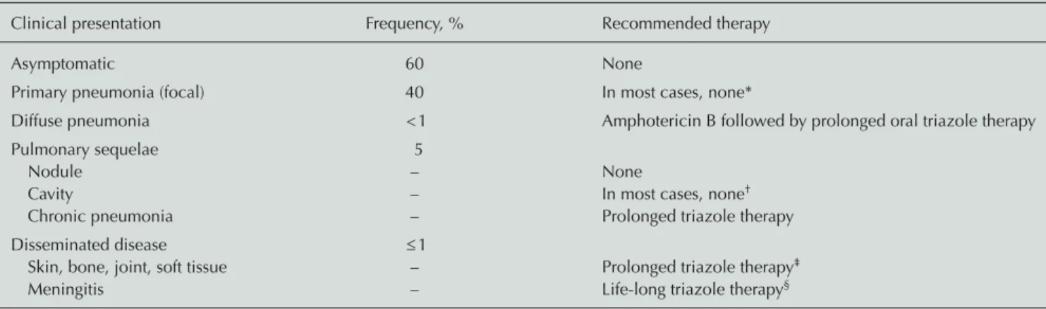
Table 1 Clinical presentation of coccidioidomycosis, their frequency, and recommended initial therapy for the immunocompetent host.
Clinical presentation Frequency, % Recommended therapy
Asymptomatic 60 None
Primary pneumonia (focal) 40 In most cases, none*
Diffuse pneumonia < 1 Amphotericin B followed by prolonged oral triazole therapy
Pulmonary sequelae 5
Nodule – None
Cavity – In most cases, none†
Chronic pneumonia – Prolonged triazole therapy
Disseminated disease ≤ 1
Skin, bone, joint, soft tissue – Prolonged triazole therapy‡
Meningitis – Life-long triazole therapy§
*Treatment is indicated for hosts with depressed cellular immunity as well as for those with prolonged symptoms and signs of increased severity, includ-ing night sweats for > 3 weeks, weight loss of > 10%, a complement-fixation titer of > 16, and extensive pulmonary involvement on chest radiography;
†treatment (usually the oral triazoles fluconazole and itraconazole) is recommended for persistent symptoms; ‡in severe cases, some clinicians use
ampho-tericin B as the initial therapy; §intraventricular or intrathecal amphotericin B is recommended in cases of triazole failure. Hydrocephalus may occur,
requiring a cerebrospinal fluid shunt.
complications because of immunosuppression or other preexisting factors require a variety of therapeutic strategies that may include antifungal agents, surgical debridement, or a combination of both. Azole antifungals, primarily flu-conazole and itraflu-conazole, have replaced amphotericin B as the initial therapy for most chronic pulmonary or dissem-inated infections. Amphotericin B is now reserved for pa-tients with respiratory failure due to coccidioidal infection, those with rapidly progressive coccidioidal infections, or women during pregnancy. Therapy often ranges from months to years in duration, and in some patients, lifelong suppressive therapy is needed to prevent relapses.17
Although coccidioidomycosis is an endemic disease chiefly of the western hemisphere, and one which rarely occurs in Taiwan, it should be considered whenever there is a differential diagnosis of pneumonia resistant to standard medication with small cavitating and/or nodular lesions on a chest radiograph associated with variable skin manifestations such as erythema nodosum and arthralgia, as well as ar-thralgia and/or arthritis. Inquiring into the traveling history to endemic areas plays a pivotal role in the correct diagno-sis of the disease. If there are any problems detecting the infection in an individual, the patient’s serum and probable sputum or other tissue fluid can be submitted to the Taiwan Centers for Disease Control. Treatment is not routinely neces-sary because most infections are self-limited and the thera-peutic protocol must be individualized according to the Infectious Disease Society of America guidelines.
References
1. Galgiani JN. Coccidioidomycosis: a regional disease of national importance. Ann Intern Med 1999;130:293–300.
2. Chen CH, Shih JF, Hsu YT, et al. Disseminated coccidioidomycosis with lung, skin and lymph node involvement: report of a case.
J Formos Med Assoc 1991;90:788–92.
3. Wang CY, Jerng JS, Ko JC. Disseminated Coccidioidomycosis.
Emerg Infect Dis 2005;11:177–8.
4. Saubolle MA, McKellar PP, Sussland D. Epidemiologic, clinical, and diagnostic aspects of coccidioidomycosis. J Clin Micro 2007; 45:26–30.
5. Hinshaw M, Longley BJ. Fungal disease. In: Elder DE, Elenitsas R, Johnson BL, Murphy GF, eds. Lever’sHistopathology of the Skin, 10th ed. Philadelphia: Lippincott Williams & Wilkins, 2009:608–9.
6. Crum NF. Disseminated coccidioidomycosis with cutaneous
lesions clinically mimicking mycosis fungoides. Int J Dermatol
2005;44:958–60.
7. Stevens DA. Coccidioidomycosis. N Engl J Med 1995;332:1077–82. 8. DiCaudo DJ, Yiannias JA, Laman SD, Warschaw KE. The exan-them of acute pulmonary coccidioidomycosis: clinical and his-topathologic features of 3 cases and review of the literature.
Arch Dermatol 2006;142:744–6.
9. Chang A, Tung RC, McGillis TS, et al. Primary cutaneous coc-cidioidomycosis. J Am Acad Dermatol 2003;49:944–9. 10. Quimby SR, Connolly SM, Winkelmann RK, et al. Clinicopathologic
spectrum of specific cutaneous lesions of disseminated coccidi-oidomycosis. J Am Acad Dermatol 1992;26:79–85.
11. Oldfield EC, Olson PE, Bone WD, et al. Coccidioidomycosis presenting as neoplasia: another great imitator disease. Infect Dis Clin Pract 1995;4:87–92.
12. Kim A, Parker SS. Coccidioidomycosis, case report and update on diagnosis and management. J Am Acad Dermatol 2002;46: 743–7.
13. Rance BR, Elston DM. Disseminated coccidioidomycosis discov-ered during routine skin cancer screening. Cutis 2002;70:70–2. 14. Ampel NM. Coccidioidomycosis: a review of recent advance.
Clin Chest Med 2009;30:241–51.
15. Pappagianis D, Zimmer BL. Serology of coccidioidomycosis.
Clin Microbiol Rev 1990;3:247–68.
16. Neil MA. Coccidioidomycosis. In: Fauci AS, Braunwald E, Kasper DL, et al, eds. Harrison’s Principles of Internal Medicine, 17th ed. New York: McGraw-Hill, 2008:1247–9.
17. Galgiani JN, Ampel NM, Blair JE, et al. Coccidioidomycosis. Clin Infect Dis 2005;41:1217–23.