Research Article
Sars-CoV-2 Envelope and Membrane Proteins: Structural
Differences Linked to Virus Characteristics?
Martina Bianchi
,
1Domenico Benvenuto
,
2Marta Giovanetti
,
3Silvia Angeletti
,
4Massimo Ciccozzi
,
2and Stefano Pascarella
11Department of Biochemical sciences“A Rossi Fanelli”, Sapienza University of Rome, 00185 Rome, Italy 2Unit of Medical Statistics and Molecular Epidemiology, University Campus Bio-Medico of Rome, Rome, Italy 3Flavivirus Laboratory, Oswaldo Cruz Institute, Oswaldo Cruz Foundation, Rio de Janeiro, Brazil
4Unit of Clinical Laboratory Science, University Campus Bio-Medico of Rome, Rome, Italy
Correspondence should be addressed to Stefano Pascarella; [email protected]
Received 1 April 2020; Revised 23 April 2020; Accepted 7 May 2020; Published 30 May 2020
Academic Editor: Miguel A. Andrade
Copyright © 2020 Martina Bianchi et al. This is an open access article distributed under the Creative Commons Attribution License, which permits unrestricted use, distribution, and reproduction in any medium, provided the original work is properly cited. The Coronavirus Disease 2019 (COVID-19) is a new viral infection caused by the severe acute respiratory coronavirus 2 (SARS-CoV-2). Genomic analyses have revealed that SARS-CoV-2 is related to Pangolin and Bat coronaviruses. In this report, a structural comparison between the Sars-CoV-2 Envelope and Membrane proteins from different human isolates with homologous proteins from closely related viruses is described. The analyses here reported show the high structural similarity of Envelope and Membrane proteins to the counterparts from Pangolin and Bat coronavirus isolates. However, the comparisons have also highlighted structural differences specific of Sars-CoV-2 proteins which may be correlated to the cross-species transmission and/or to the properties of the virus. Structural modelling has been applied to map the variant sites onto the predicted three-dimensional structure of the Envelope and Membrane proteins.
1. Introduction
COVID-19 has become a planetary emergency which is seriously threatening human health [1, 2]. Development of effective therapeutic and prevention strategies is significantly hampered also by the lack of detailed structural information on virus proteins, although several crystallographic struc-tures of Sars-CoV-2 proteins are now available [3–5]. In this report, a structural comparison between the Sars-CoV-2 surface proteins from different isolates with homologous proteins from closely related viruses such as those from Bat and Pangolin is described. This work has been focussed onto the Envelope (E) and Membrane (M) proteins that, along with the Spike, form the virus protein interface to the exter-nal environment. The Spike glycoprotein has been already extensively studied, and a few crystallographic structures are available in the Protein Data Bank [3–6]; consequently, this protein has not been specifically addressed within this note. Identification of local structural differences, even
minimal, to the closest virus proteins may indicate the muta-tions that enabled Sars-CoV-2 to cross species and/or to acquire its peculiar pathogenic properties [7, 8]. Indeed, a number of examples have been reported in the scientific liter-ature suggesting how even single point mutations in virus proteins can significantly alter their biology and pathogenesis [9, 10]. Therefore, comparative studies may shed light on the molecular mechanisms through which an epidemic of epizootic origin can emerge and may also suggest molecular targets for therapeutics or reverse vaccinology experiments.
2. Material and Methods
Nucleotide and protein sequences have been taken from GenBank [11] data repository. Blast suite [12] has been used for databank searches; Jalview [13] and MAFFT [14] have been used for multiple sequence display and alignment, respectively. Transmembrane helix prediction has been obtained by TMHMM [15], MEMSAT [16], and Protter
[17]. Cd-hit program [18] has been used for sequence clus-tering. Homology modelling relied on Swiss-Model [19], Modeller [20], or HHpred [21] and structure display and analysis on Open-Source PyMOL [22]. When necessary, I-Tasser [23] has been used as an alternative source of ab initio homology models.
3. Results
3.1. Databank Searches and Structure Modelling. From the GenBank repository, 797 complete genomes of Sars-CoV-2 have been collected (the full list is reported in Supplementary Data). The TblastN program has been used to extract the sequences of E and M proteins from each genome. To remove redundancy within each E and M protein set, cd-hit clustering has been applied at 100% sequence identity level: identical sequences have been assigned to the same group for which only one representative has been considered for further analysis. The Sars-CoV-2 E and M protein sets have been grouped into three and seven clusters, respectively. This finding suggests that within the 797 genomes three and seven variants of the E and M proteins can be observed, respec-tively. E and M homologous proteins from closely related virus have been retrieved from the GenBank using the TblastN tool.
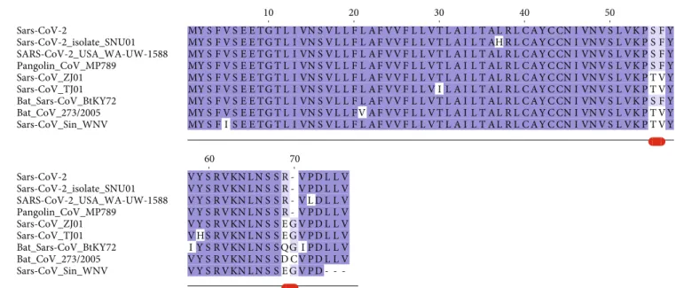
3.2. Envelope Protein. The E protein is conserved across β-coronaviruses. Only three variants have been found in the Sars-CoV-2 E set collected. Sequence comparisons show that the Sars-CoV-2 E protein from the reference genome (RefSeq code YP_009724392) is identical to the sequences from Pangolin CoV MP798 and Bat CoV CoVZXC21, CoVZC45, and RaTG13 isolates. The multiple sequence alignment reported in Figure 1 demonstrates that a distin-guishing feature of Sars-2-CoV E variants is the presence of
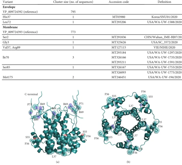
Arg at position 69 that substitutes Glu, Gln, Asp in other homologous Sars-CoV E proteins. This site is followed by a deletion in position 70 corresponding to Gly or Cys in the other proteins. Sars-CoV-2 E sequences differ from the homologous proteins also at positions 55-56, where the dyad Ser-Phe replaces Thr-Val (except in Bat coronavirus isolate BtKY72, accession code KY352407). Variants of the Sars-CoV-2 E protein differ at positions 37 and 72 where His substitutes a Leu and Leu replaces a conserved Pro, respectively. The size of each Envelope variant cluster is reported in Table 1 along with accession codes and definitions of the isolates. A homology model of the E protein has been built with Modeller using as a template the pentameric ion channel structure of Sars-CoV protein identified by the PDB code 5X29. This sequence shares 91% identity to Sars-CoV-2 E protein and covers the segment encompassed by positions 8-65. Figure 2 displays the structure of the homology model of the Sars-CoV-2 E protein assembled as a pentameric viroporin-like protein. Figure 2 displays also the position of the variant sites onto the three-dimensional model. Prediction of the transmembrane helices is difficult in a short protein. Therefore, transmembrane topology cannot be assigned reli-ably. Likewise, experiments have not clarified definitively which portions of the E protein are exposed to the external or internal side of the virus membrane [24].
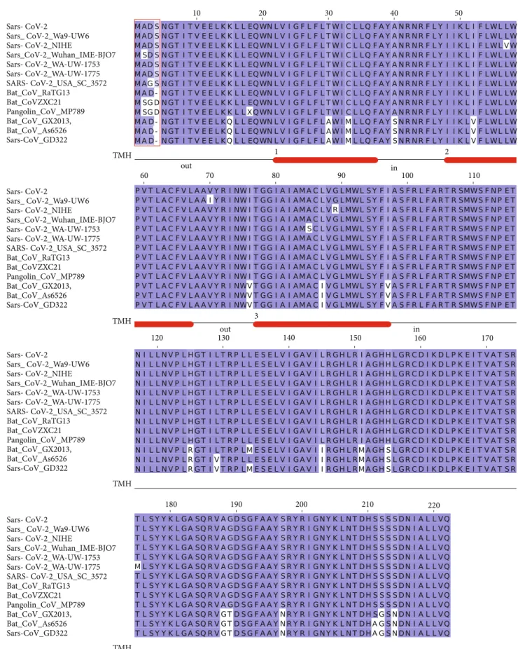
3.3. Membrane Glycoprotein. The M glycoprotein is con-served across theβ-coronaviruses. However, seven variants of Sars-CoV-2 M protein were identified in the collected set, while only three variants were observed for the E protein (Figure 3). The multiple sequence alignment shows a remarkable similarity (98% identity) among the Sars-CoV-2 M variants and the sequences from Bat and Pangolin isolates. However, a difference at the N-terminal position (Figure 3) can be observed: the insertion of a Ser residue at position 4 Sars-CoV-2 Sars-CoV-2_isolate_SNU01 SARS-CoV-2_USA_WA-UW-1588 Pangolin_CoV_MP789 Sars-CoV_ZJ01 Sars-CoV_TJ01 Bat_Sars-CoV_BtKY72 Bat_CoV_273/2005 Sars-CoV_Sin_WNV Sars-CoV-2 Sars-CoV-2_isolate_SNU01 SARS-CoV-2_USA_WA-UW-1588 Pangolin_CoV_MP789 Sars-CoV_ZJ01 Sars-CoV_TJ01 Bat_Sars-CoV_BtKY72 Bat_CoV_273/2005 Sars-CoV_Sin_WNV 10 20 30 40 50 60 70
Figure1: Multiple sequence alignment among Sars-CoV-2 Envelope protein variants and a set of the most similar homologous proteins. The sequence labelled Sars-CoV-2 corresponds to the reference sequence identified by the RefSeq code YP_009724392. Red lines below the alignment indicate the changed sites discussed in the text. Blu background denotes conserved alignment positions.
of human Sars-CoV-2 seems to be a unique feature of this protein. In the corresponding position, the RaTG13 Bat M protein displays a deletion, while Bat CoVZXC21, CoVZC45, and Pangolin MP789 proteins have an Asp residue. The seven M protein variants differ at positions 2, 3, 57, 70, 85, 89, and 175. The size of each Membrane variant cluster is reported in Table 1 along with accession codes and defi ni-tions of the isolates. Noteworthy, the protein from the Sars-CoV-2 NIHE isolate (accession code MT127115) possesses an Arg instead of a conserved Gly at position 89 (Figure 3). The mutation occurs within a predicted transmembrane
helix and, if confirmed, may have a significant impact on the protein properties (Figure 3).
The three-dimensional model of the M protein has been taken from the I-Tasser server (code QHD43419) as other methods failed to find any suitable template. However, it should be mentioned that HHpred found a weak local affi n-ity, albeit below the statistical significance level, to 4N31, a peptidase-like protein fromStreptococcus pyogenes essential for pilus polymerisation. Figure 4 displays the positions of the variant sites onto the model structure. This model has been predicted by ab initio techniques. Therefore, it should C-terminal R69 P71 R69 P71 R69 S55 F56 S55 F56 L37 R69 R69 (a) F56 F56 F56 F56 F56 L37 L37 L37 L37 L37 S55 S55 S55 S55 S55 (b)
Figure2: Three-dimensional model of the viroporin-like tetrameric assembly of the E protein from Sars-CoV-2 represented as a cartoon model. Residues corresponding to the mutated sites indicated in Figure 1 are displayed as transparent space-filling spheres and labelled with the amino acid one-letter code. The C-terminal segments of the model are reported for completeness. However, they convey no structural information due to lack of a corresponding segment in the structural template used in homology modelling. Structure in panel (b) is rotated by approximately 180°along thexaxis with respect to the orientation shown in panel (a).
Table1: Size of the variant clusters of the Sars-CoV-2 Envelope and Membrane proteins.
Variant Cluster size (no. of sequences) Accession code Definition
Envelope YP_009724392 (reference) 795 His37 1 MT03980 Korea/SNU01/2020 Leu72 1 MT293206 USA/WA-UW-1588/2020 Membrane YP_009724393 (reference) 773 Ser2 1 MT291836 CHN/Wuhan_IME-BJ07/2020 Gly3 1 MT325626 USA/SC_3572/2020
Val57, Arg89 1 MT127115 VIE/NIHE/2020
MT293184 USA/WA-UW-1297/2020 lle70 3 MT326166 USA/WA-UW-1735/2020 MT293211 USA/WA-UW-1591/2020 Ser85 1 MT326167 USA/WA-UW-1753/2020 MT326093 USA/WA-UW-1775/2020 Met175 2 MT246451 USA/WA-UW-194/2020
Sars- CoV-2 Sars_ CoV-2_Wa9-UW6 Sars- CoV-2_NIHE Sars_CoV-2_Wuhan_IME-BJO7 Sars- CoV-2_WA-UW-1753 Sars- CoV-2_WA-UW-1775 SARS- CoV-2_USA_SC_3572 Bat_CoV_RaTG13 Bat_CoVZXC21 Pangolin_CoV_MP789 Bat_CoV_GX2013, Bat_CoV_As6526 Sars-CoV_GD322 Sars- CoV-2 Sars_ CoV-2_Wa9-UW6 Sars- CoV-2_NIHE Sars_CoV-2_Wuhan_IME-BJO7 Sars- CoV-2_WA-UW-1753 Sars- CoV-2_WA-UW-1775 SARS- CoV-2_USA_SC_3572 Bat_CoV_RaTG13 Bat_CoVZXC21 Pangolin_CoV_MP789 Bat_CoV_GX2013, Bat_CoV_As6526 Sars-CoV_GD322 Sars- CoV-2 Sars_ CoV-2_Wa9-UW6 Sars- CoV-2_NIHE Sars_CoV-2_Wuhan_IME-BJO7 Sars- CoV-2_WA-UW-1753 Sars- CoV-2_WA-UW-1775 SARS- CoV-2_USA_SC_3572 Bat_CoV_RaTG13 Bat_CoVZXC21 Pangolin_CoV_MP789 Bat_CoV_GX2013, Bat_CoV_As6526 Sars-CoV_GD322 Sars- CoV-2 Sars_ CoV-2_Wa9-UW6 Sars- CoV-2_NIHE Sars_CoV-2_Wuhan_IME-BJO7 Sars- CoV-2_WA-UW-1753 Sars- CoV-2_WA-UW-1775 SARS- CoV-2_USA_SC_3572 Bat_CoV_RaTG13 Bat_CoVZXC21 Pangolin_CoV_MP789 Bat_CoV_GX2013, Bat_CoV_As6526 Sars-CoV_GD322 10 20 30 40 50 60 out 70 in 2 1 80 90 100 110 120 TMH TMH TMH TMH 130 3 140 150 in160 out 170 180 190 200 210 220
Figure3: Multiple sequence alignment among Sars-CoV-2 M protein variants and a set of most similar homologous proteins. The sequence label Sars-CoV-2 indicates the reference sequence identified by the RefSeq code YP_009724393. Red box indicates the variant sites at the N-terminal discussed in the text. Numbered red bars under the multiple alignment mark the prediction of transmembrane helices. The location of the connect loop with respect to the virion surface is indicated as“in” or“out”. Blu background denotes conserved alignment positions.
be considered with great caution and should be treated as a low-resolution approximation of the real structure. Accord-ing to the prediction of the transmembrane helix topology, the N- and C-terminal portions of the M protein are exposed outside and inside the virus particle, respectively (Figure 4).
4. Discussion
Previous studies pointed out that E and M proteins could be important for viral entry, replication, and particle assembly within the human cells [24, 25]. According to the accepted theories, the current COVID-19 pandemic has been caused by the cross-species transmission of a β-coronavirus nor-mally hosted by Bats and, perhaps, Pangolin to humans [3, 26]. In this paper, E and M proteins from 797 Sars-CoV-2 genomes have been compared to the counterparts taken from the most closely related virus also to evaluate the potential role of amino acid mutations in the epizootic origin of COVID-19. E protein is a minor component of the virus membrane though it is deemed to be important for many stages of virus infection and replication [24, 25]. Sequence comparison has shown that this protein is identical to the counterparts of specific Bat and Pangolin coronavirus isolates, even though the Sars-CoV-2 sequence seems to pos-sess specific modifications and characteristics with respect to other Sars CoVs. In particular, Arg69, a positively charged amino acid, replaces Glu or Gln residues, negatively charged and neutral, respectively, in the homologous CoV proteins. Moreover, a deletion specific to Sars-CoV-2 proteinsflanks this position. Unfortunately, it is not possible to predict reli-ably whether the sites of these modifications are exposed to the internal or external side of the membrane. In any case, the substitution and the deletion appear a rather drastic change and may have a significant impact on conformational properties and possibly on protein-protein interactions. Further structural studies are needed. However, it may be hypothesized that these changes can also affect the
olig-omerization process necessary to form a transmembrane ion channel.
It has been demonstrated that M protein is more prev-alent within the virus membrane, and it is deemed to be important for the budding process of coronaviruses. Indeed, during the process of virus particle assembly, this protein interacts with the Nucleocapsid, Envelope, Spike, and Membrane glycoprotein itself [25]. Moreover, in Alphacoronaviruses, it has been demonstrated that this protein cooperates with the Spike during the cell attach-ment and entry [27]. Therefore, mutations occurring at the N-terminus region, which is exposed to the virus sur-face, could play a key role in the host cell interaction.
In conclusion, the analyses here reported show the struc-tural similarity of E and M proteins to the counterparts from Pangolin and Bat coronavirus isolates. At the same time, comparisons have highlighted structural differences specific of Sars-CoV-2 proteins which may be correlated to the cross-species transmission and/or to the properties of the virus. Although further studies are needed, it is clear that these amino acid variations have been important for the virus evolutionary history, and the results may hint at how similar mutations within the coronavirus family can lead in the next years to other epizootic epidemic events similar to the one that we are experiencing these days.
Data Availability
All sequence data are available in the GenBank repository. The complete list is available in the Supplementary Materials.
Conflicts of Interest
The authors declare that there is no conflict of interest regarding the publication of this paper.
A2 D3 S4 T175 V70 L57 A85 G89
Figure4: I-Tasser model of the Membrane protein represented as cartoon model. Variant positions are displayed as transparent space-filling spheres and labelled with the amino acid one-letter code.
Acknowledgments
This work has been in part funded by a grant to SP from Sapienza University of Rome (RP11916B74B27C4D).
Supplementary Materials
The supplementary data consist of an Excelfile containing the list of the 797 Sars-CoV-2 genomes retrieved from GenBank and analysed in the article. For each genome, the sequence header is displayed as found in the GenBank. The header reports the accession code in thefirstfield of each line after the“>”character.(Supplementary Materials)
References
[1] D. Benvenuto, M. Giovanetti, A. Ciccozzi, S. Spoto, S. Angeletti, and M. Ciccozzi, “The 2019-new coronavirus epidemic: evidence for virus evolution,” Journal of Medical Virology, vol. 92, no. 4, pp. 455–459, 2020.
[2] C.-C. Lai, T.-P. Shih, W.-C. Ko, H.-J. Tang, and P.-R. Hsueh,
“Severe acute respiratory syndrome coronavirus 2 (SARS-CoV-2) and coronavirus Disease-2019 (COVID-19): the epidemic and the challenges,”International Journal of Antimi-crobial Agents, vol. 55, no. 3, article 105924, 2020.
[3] D. Benvenuto, M. Giovanetti, M. Salemi et al., “The Global Spread of 2019-NCoV: A Molecular Evolutionary Analysis,”
Pathogens and Global Health, vol. 114, no. 2, pp. 64–67, 2020. [4] A. C. Walls, Y.-J. Park, M. A. Tortorici, A. Wall, A. T. McGuire, and D. Veesler,“Structure, Function, and Antigenic-ity of the SARS-CoV-2 Spike Glycoprotein,” Cell, vol. 181, no. 2, pp. 281–292.e6, 2020.
[5] D. Wrapp, N. Wang, K. S. Corbett et al.,“Cryo-EM Structure of the 2019-NCoV Spike in the Prefusion Conformation,”
Science, vol. 367, no. 6483, pp. 1260–1263, 2020.
[6] R. Yan, Y. Zhang, Y. Li, L. Xia, Y. Guo, and Q. Zhou,
“Structural Basis for the Recognition of SARS-CoV-2 by Full-Length Human ACE2,” Science, vol. 367, no. 6485, pp. 1444–1448, 2020.
[7] S. Angeletti, D. Benvenuto, M. Bianchi, M. Giovanetti, S. Pascarella, and M. Ciccozzi,“COVID‐2019: the role of the Nsp2 and Nsp3 in its pathogenesis,”Journal of Medical Virol-ogy, vol. 92, no. 6, pp. 584–588, 2020.
[8] W. Ji, W. Wang, X. Zhao, J. Zai, and X. Li, “Cross‐species transmission of the newly identified coronavirus 2019-NCoV,”
Journal of Medical Virology, vol. 92, no. 4, pp. 433–440, 2020. [9] N. M. André, B. Cossic, E. Davies, A. D. Miller, and G. R. Whittaker,“Distinct mutation in the feline coronavirus spike protein cleavage activation site in a cat with feline infectious peritonitis-associated Meningoencephalomyelitis,” Journal of Feline Medicine and Surgery Open Reports, vol. 5, no. 1, 2019.
[10] Y. Sakai, K. Kawachi, Y. Terada, H. Omori, Y. Matsuura, and W. Kamitani,“Two-amino acids change in the Nsp4 of SARS coronavirus abolishes viral replication,” Virology, vol. 510, pp. 165–174, 2017.
[11] D. A. Benson, M. Cavanaugh, K. Clark et al., “GenBank,”
Nucleic Acids Research, vol. 46, no. D1, pp. D41–D47, 2018. [12] S. Altschul, T. L. Madden, A. A. Schäffer et al., “Gapped
BLAST and PSI-BLAST: a new generation of protein database
search programs,” Nucleic Acids Research, vol. 25, no. 17, pp. 3389–3402, 1997.
[13] A. M. Waterhouse, J. B. Procter, D. M. A. Martin, M. Clamp, and G. J. Barton,“Jalview version 2–a multiple sequence align-ment editor and analysis workbench,”Bioinformatics, vol. 25, no. 9, pp. 1189–1191, 2009.
[14] K. Katoh and D. M. Standley, “MAFFT multiple sequence alignment software version 7: improvements in performance and usability,” Molecular Biology and Evolution, vol. 30, no. 4, pp. 772–780, 2013.
[15] Y. Chen, P. Yu, J. Luo, and Y. Jiang,“Secreted protein predic-tion system combining CJ-SPHMM, TMHMM, and PSORT,”
Mammalian Genome, vol. 14, no. 12, pp. 859–865, 2003. [16] L. J. McGuffin, K. Bryson, and D. T. Jones, “The PSIPRED
protein structure prediction server,” Bioinformatics, vol. 16, no. 4, pp. 404-405, 2000.
[17] U. Omasits, C. H. Ahrens, S. Müller, and B. Wollscheid,“ Prot-ter: interactive protein feature visualization and integration with experimental proteomic data,” Bioinformatics, vol. 30, no. 6, pp. 884–886, 2014.
[18] L. Fu, B. Niu, Z. Zhu, S. Wu, and W. Li,“CD-HIT: accelerated for clustering the next-generation sequencing data,” Bioinfor-matics, vol. 28, no. 23, pp. 3150–3152, 2012.
[19] A. Waterhouse, M. Bertoni, S. Bienert et al.,“SWISS-MODEL: homology Modelling of protein structures and complexes,”
Nucleic Acids Research, vol. 46, no. W1, pp. W296–W303, 2018.
[20] B. Webb and A. Sali,“Protein structure modeling with MOD-ELLER,”Methods in Molecular Biology, vol. 1654, pp. 39–54, 2017.
[21] L. Zimmermann, A. Stephens, S.-Z. Nam et al.,“A completely Reimplemented MPI bioinformatics toolkit with a new HHpred server at its Core,” Journal of Molecular Biology, vol. 430, no. 15, pp. 2237–2243, 2018.
[22] L. L. C. Schrodinger, The AxPyMOL Molecular Graphics System, Version 2.0, Schrödinger, LLC., 2015.
[23] J. Yang, R. Yan, A. Roy, D. Xu, J. Poisson, and Y. Zhang,“The I-TASSER suite: protein structure and function prediction,”
Nature Methods, vol. 12, no. 1, pp. 7-8, 2015.
[24] D. Schoeman and B. C. Fielding, “Coronavirus envelope protein: current knowledge,”Virology Journal, vol. 16, no. 1, p. 69, 2019.
[25] E. A. J. Alsaadi and I. M. Jones,“Membrane binding proteins of coronaviruses,” Future Virology, vol. 14, no. 4, pp. 275– 286, 2019.
[26] R. Lu, X. Zhao, J. Li et al., “Genomic characterisation and epidemiology of 2019 novel coronavirus: implications for virus origins and receptor binding,”The Lancet, vol. 395, no. 10224, pp. 565–574, 2020.
[27] A. Naskalska, A. Dabrowska, A. Szczepanski, A. Milewska, K. P. Jasik, and K. Pyrc,“Membrane protein of human corona-virus NL63 is responsible for interaction with the adhesion receptor,” Journal of Virology, vol. 93, no. 19, pp. 455–459, 2019.