REVIEW ARTICLE
Glioblastoma stem cells (GSCs) epigenetic
plasticity and interconversion between
differentiated non-GSCs and GSCs
Ahmad R. Safa
a,b,*
, Mohammad Reza Saadatzadeh
a,c,
Aaron A. Cohen-Gadol
c, Karen E. Pollok
a,b,d,
Khadijeh Bijangi-Vishehsaraei
b,ea
Indiana University Simon Cancer Center, Indiana University School of Medicine, Indianapolis, IN 46202, USA
b
Department of Pharmacology and Toxicology, Indiana University School of Medicine, Indianapolis, IN 46202, USA
cDepartment of Neurosurgery, IU School of Medicine and Goodman Campbell Brain and Spine,
Indiana University School of Medicine, Indianapolis, IN 46202, USA
dHerman B. Wells Center for Pediatric Research, Indiana University School of Medicine, Indianapolis,
IN 46202, USA
eOtolaryngology-Head and Neck Surgery, Indiana University School of Medicine, Indianapolis, IN 46202,
USA
Received 22 January 2015; accepted 1 February 2015 Available online 14 February 2015
KEYWORDS
Cancer stem cells; Epigenetic; GBM plasticity; Glioblastoma; Stemness; Dedifferentiation; GBM stem cells
Abstract Cancer stem cells (CSCs) or cancer initiating cells (CICs) maintain self-renewal and multilineage differentiation properties of various tumors, as well as the cellular heterogeneity consisting of several subpopulations within tumors. CSCs display the malignant phenotype, self-renewal ability, altered genomic stability, specific epigenetic signature, and most of the time can be phenotyped by cell surface markers (e.g., CD133, CD24, and CD44). Numerous studies support the concept that non-stem cancer cells (non-CSCs) are sensitive to cancer ther-apy while CSCs are relatively resistant to treatment. In glioblastoma stem cells (GSCs), there is clonal heterogeneity at the genetic level with distinct tumorigenic potential, and defined GSC marker expression resulting from clonal evolution which is likely to influence disease progres-sion and response to treatment. Another level of complexity in glioblastoma multiforme (GBM) tumors is the dynamic equilibrium between GSCs and differentiated non-GSCs, and the poten-tial for non-GSCs to revert (dedifferentiate) to GSCs due to epigenetic alteration which confers phenotypic plasticity to the tumor cell population. Moreover, exposure of the differentiated
* Corresponding author. Indiana University Simon Cancer Center, Indiana University School of Medicine, 980 W. Walnut Street, R3-C524, Indianapolis, IN 46202, USA. Tel.:þ1 317 278 4952 (office); fax:þ1 317 274 8046.
E-mail address:asafa@iupui.edu(A.R. Safa).
Peer review under responsibility of Chongqing Medical University. http://dx.doi.org/10.1016/j.gendis.2015.02.001
2352-3042/Copyrightª2015, Chongqing Medical University. Production and hosting by Elsevier B.V. This is an open access article under the CC BY-NC-ND license (http://creativecommons.org/licenses/by-nc-nd/4.0/).
H O S T E D BY Available online atwww.sciencedirect.com
ScienceDirect
GBM cells to therapeutic doses of temozolomide (TMZ) or ionizing radiation (IR) increases the GSC pool bothin vitroandin vivo. This review describes various subtypes of GBM, discusses the evolution of CSC models and epigenetic plasticity, as well as interconversion between GSCs and differentiated non-GSCs, and offers strategies to potentially eliminate GSCs.
Copyrightª2015, Chongqing Medical University. Production and hosting by Elsevier B.V. This is an open access article under the CC BY-NC-ND license (http://creativecommons.org/licenses/
by-nc-nd/4.0/).
Introduction
Glioblastoma multiforme (GBM) comprises the largest group of brain tumors which respond very poorly to current therapies.1 In the United States, approximately 13,000 people die annually from GBM, and it is disappointing that only about 10% of patients survive 5 years.2e4The combi-nation of radiotherapy and adjunct temozolomide (TMZ) has increased the survival of patients with GBM, but the median survival of GBM patients is only about 14.6 months.5 The highly aggressive nature of GBM is due to multiple ge-netic alterations which result in augmented cytoprotective and survival pathways as well as numerous defects in the apoptotic signaling machinery and epigenetic alterations (Fig. 1).
A growing body of evidence indicates that rare pop-ulations of cancer cells, termed cancer stem cells (CSCs) or cancer initiating cells (CICs), play a significant role in several cancers, including GBM.6e8GBM tumors display high degree of phenotypic, cellular, genetic, and epigenetic heterogeneity, and it is believed that a major problem in the unresponsiveness of GBM tumors to therapy is the ex-istence of GBM stem cells (GSCs) within the tumor which are most crucial for driving invasive tumor growth and relapse.6,9Emerging results have revealed that in GBM and other malignancies, CSC enrichment may occur either from an increased symmetric self-renewal division rate of CSCs or a reprogramming of non-CSC to CSCs and conferring phenotypic plasticity to the tumor population.10 The concept of interconversion of CSCs and non-CSCs has pro-vided major complexity in understanding the role of CSCs in tumor heterogeneity, a potential mechanism for thera-peutic relapse, resistance to anticancer therapies, and developing therapeutic strategies. In this review we describe various subtypes of GBM, discuss the evolution of CSC models and epigenetic plasticity as well as intercon-version between GSCs and differentiated non-GSCs, and offer strategies to potentially eliminate GSCs. Under-standing GBM tumor cell plasticity and its underlying mo-lecular mechanisms will help in the design of more effective therapies against GBM and preventing tumor recurrence.
Glioblastoma multiforme (GBM)
GBM comprises the most common and very aggressive form of primary brain tumors which respond very poorly to the current therapies.1,2 This most malignant brain tumor is designated as World Health Organization (WHO) grade IV astrocytoma which expresses the astrocyte marker, glial
fibrillary acidic protein (GFAP).11e14 Initiation and recur-rence of primary GBM may be caused by a subpopulation of GSCs which may derive from mutated neural stem and precursor cells.8e14 GBM tumors developed from lower-grade astrocytomas or oligodendrogliomas are termed sec-ondary GBMs (Fig. 1). While primary and secsec-ondary GBM’s are histologically similar, they are genetically different.15,16 Primary GBM frequently displays molecular alterations in EGFR, PDGFRA, PTEN, p53 tumor suppressor protein, NF1, CDKN2A/B, and telomerase reverse transcriptase (TERT) promoter mutations (seeFig. 1).16,17Furthermore, as re-ported by Cadieux et al, global hypomethylation is frequently observed in primary human GBM.18
Primary GBM is heterogeneous in nature, and based on its patterns of gene expression and genetic changes, four different subtypes including proneural, neural, classical and mesenchymal have been identified.19,20 While the biological significance and origin of these GBM subtypes are unclear, patients with specific GBM subtypes exhibit distinct survival times and different responses to ther-apy.12,19,20A high frequency of isocitrate dehydrogenase 1 (IDH1) mutations and O6-methylguanine-DNA methyl-transferase (MGMT) promoter methylation among young adult patients with primary GBM compared to other sub-types correlates with increased survival.21 The classical subtype is associated with a high frequency of EGFR aber-rations and low expression of p53 tumor suppressor protein mutations.22The mesenchymal subtype displays loss of the tumor suppressor gene NF1 with high CD44 and MERTK expression, and the neural subtype does not express any particular alterations of specific genes or pathways.12,22
The most complete information has been provided by The Cancer Genome Atlas (TCGA) Research Network which published a report by analysis of copy number, methylation patterns, expression profiling, and whole-genome sequencing of GBM samples.20Many genes including EGFR, PDGFRA, CDK4, MDM2, MDM4, MET, CDK6, N-Myc, Cyclin D2, PIK3CA, and AKT3 have been found amplified in GBM, further contributing to the complexity in developing ther-apies to treat GBM.20Moreover, significant abnormalities in several signaling pathways including the receptor tyrosine kinase pathway, the p53 pathway, and the RB pathway were found.12,16,20
Cancer stem cell model
GBM tumors display a great degree of phenotypic and functional heterogeneity.7,8,12,13 Heterogeneity among tumor cells arises within a single tumor as a result of genetic and epigenetic changes (Fig. 2) as well as different
microenvironments within different regions of tumor.23,24 The genetic alterations and epigenetic changes of the cells within the same tumor is not well characterized, and for future personalized medicine strategies, it is necessary to explore intratumoral heterogeneity with respect to the phenotype and genotype of the tumor as well as evaluating its epigenetic alterations to achieve effective treatment for
GBM.25,26To better understand intratumoral heterogeneity in a given GBM tumor, Sottoriva et al demonstrated that investigating genome-wide GBM intratumoral genomic het-erogeneity can be used to reveal tumor evolution.25 Furthermore, the authors showed that based on gene expression levels, tumor fragments from different anatom-ical regions of the same patient tumor may be classified into
Figure 1 Genetic alterations and aberrant signaling pathways in primary and secondary GBM.A. The continued growth and recurrence of primary GBM is due to the presence of GSCs which express various protein markers and display self-renewal and tumorigenic potential. Modified from Masui et al.15B. Epigenetic changes in GBM. Numerous molecular alterations shown in this figure and described in the text occur in primary GBM. Mutations in p53 tumor suppressor protein (p53) and ATRX typically occur in low-grade gliomas and secondary GBM. Mutation of the IDH1 gene is commonly found in low-grade gliomas and secondary GBM, but is rare in primary GBM. Mutation of IDH1 leads to aberrant DNA methylation and mutations in the important chromatin modifier ATRX, affecting chromatin structure. Figure was modified from Kondo et al.16
different GBM subtypes.25 Significantly, by using single-molecule techniques, the authors described the clonal composition of single tumor fragments and showed that a hierarchy of mitotic clones coexists within the same frag-ment of tumor. These impressive results unraveled the complexity of GBM tumors with respect to their heteroge-neity which represents the signature of GBM clonal evolution at the single patient level.25These results demonstrate the urgent need for personalized medicine and the difficulty in developing effective therapies for each GBM patient.
The origin of tumor cell heterogeneity may occur from clonal evolution and from differentiation of CSCs.7,27e32The CSC model well explains the versatility and plasticity of heterogeneous tumor populations. This model discusses how very small subpopulations of CSCs drive cancer progression and how small subpopulations of cancer cell types with specific features are produced within a given tumor.26CSCs are characterized by their ability to generate xenografts representing the initial tumor in immunodeficient animals and to divide asymmetrically to allow self-renewal as well as differentiation into a non-CSC population (Fig. 3). However, recent experimental evidence showing CSC plasticity sug-gests that the tumor cell populations are dynamic, and both CSCs and non-CSCs are capable of interconversion (Figs. 3 and 4) due to environmental factors.7,8,33e39 The dediffer-entiation of non-CSCs to CSCs further complicates the gen-eration of tumor heterogeneity and CSC-targeted therapy.39,40 As stated by Vries et al, any tumor cell can revert to a CSC after gaining a clonal advantage over the original CSC during its development.28While much evidence supports the CSC model in several cancers, reliability on cell surface markers for identifying authentic CSCs is limited. However, clonal analysis and lineage tracing demonstrating
the hierarchical organization of tumors in vivo provide strong evidence in support of the CSC concept.41,42In sup-port of this CSC concept, Cheng et al byin vivocell lineage tracing also showed that GSCs contribute to vascular peri-cytes that may remodel perivascular niches.43
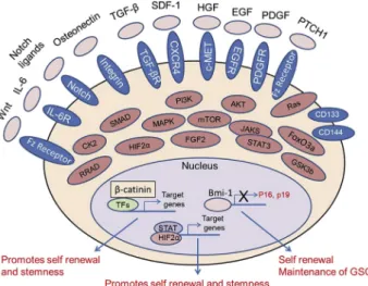
The relationship between neuronal stem cells (NSCs) and GSCs as well as differentiation of these stem cells are shown inFig. 2. GSCs like other CSCs are a rare population of slow growing cells in tumors which display various “stemness” properties including (1) the ability to self-renew and differentiate into distinct lineages through different intermediate progenitors, (2) co-existence or heterogeneity of cells with different differentiation ca-pacities providing the cellular hierarchy within the tumor, and (3) GSCs have the ability to initiate tumors in intra-cranial xenograft models in immunodeficient animals that recapitulate phenotypic characteristics of the initial tumor including tumor cell heterogeneity, invasiveness, migration and metastasis, tumor hypoxic response; resistance to drugs and radiation; resistance of tumors to apoptosis stimuli, and vascular characteristics.2,6e8,44,45 Mounting evidence shows that the stem cell niche, i.e., the envi-ronment in which GSCs reside, is responsible for the maintenance of these cells with respect to “stemness” and therapeutic response.36,46e48 The intimate network of various cell types and niche paracrine factors are respon-sible for controlling the necessary signaling pathways that regulate the properties of GSCs. As shown in Fig. 3, numerous signaling pathways maintain stemness and regu-late the tumor propagating capacity of CSCs including GSCs.
GSC specific markers
The role of the cell surface protein CD133 (pronin) as a cancer stem cell marker in GBM has been extensively investigated. While the CD133 identifies GSCs that form
Figure 3 Multiple signaling networks in GSCs. A complex and integrated signaling network governs self-renewal, stem-ness, and maintaince of CSCs including GSCs. As shown in this figure, this network of proteins belong to many pivotal cellular pathways and include several plasma membrane receptors, cytoplasmic signaling proteins, specific transcription factors, growth factors, and ligands.
Figure 2 Relationship between neuronal stem cells, dif-ferentiation, GSCs, cancer initiation, and dedifferentiation. NSCs are able to differentiate into neural progenitors. Neural progenitors differentiate into neurons and glial progenitors differentiate to oligodendrocytes, ependymal cells, and as-trocytes. GBM is initiated from the transformation of NSCs into GSCs. Similarly, glial progenitors are able to trigger tumor development following malignant transformation of normal progenitor cells. Astrocytes, neurons, oligodendrocytes, and ependymal cells also have the potential to initiate tumorigenesis.33
neurospheres and generate heterogeneous tumors when transplanted in immune-compromised mice, CD133-negative cells displaying similar properties have also been reported.49e54 Interestingly, Brescia et al through clonal analysis reported that actually there is not a hierarchical relation between CD133-positive and CD133-negative cells, and in fact CD133 is capable of changing its subcellular localization between the cytoplasm and the plasma mem-brane of GSC neurospheres.49 Significantly, these authors demonstrated that silencing CD133 in human GBM neuro-spheres using lentivirus-mediated short hairpin RNA impaired the self-renewal and tumorigenic capacity of neurosphere cells. Interestingly, hypoxia significantly increased the percentage of CD133-positive cells from 69% to 92%.55 These data collectively suggest that CD133 is indispensible for GSC function and essential for maintaining the self-renewal and tumorigenic potential of GBM stem cells.55 Moreover, Denysenko et al demonstrated that CD133-positive cell lines showed increased proliferation rates in neurospheres and increased differentiation poten-tial towards neuronal lineages, while cell lines with low CD133 expression showed mesenchymal properties
in vitro.56 Moreover, other factors may collaborate with CD133 and increase the stemness of GSCs. For instance, EGFRvIII contributes to stemness through coexpression with CD133.57 Moreover, while other biomarkers have been investigated in GBM including L1CAM, SOX2, CXCR4, Integrin a-6, and CD36, their roles in GSCs are not well defined.57
While tumor heterogeneity is evident in all four clinically relevant subtypes of GBM as described above, molecular signaling in GSCs in individual subtypes is poorly character-ized.58 In light of this, Mao et al recently identified and
characterized two mutually exclusive GSC subtypes, pro-neural (PN) and mesenchymal (Mes) GSCs.58 Mes GSCs showed more aggressive phenotypes both in vitro and in intracranial xenografts of GBM in mice, and were very resistant to radiation compared with PN GSCs. Interestingly, both the glycolytic pathway and ALDH1A3 activities were robustly elevated in Mes but not PN GSCs, and inhibition of ALDH1A3 attenuated the growth of Mes but not PN GSCs.
Recent results clearly show the heterogeneity of GSCs that display intrinsically distinct tumorigenic ability. By combining ploidy-based flow sorting with array-comparative genomic hybridization, Stieber et al found that primary GBMs are either mono- or polygenomic tumors (64% versus 36%, respectively) within primary GBMs.26 The authors showed that monogenomic tumors are composed of a pseudodiploid tumor clone and normal stromal cells, whereas polygenomic tumors consisted of multiple tumor clones and always contain a pseudodiploid subpopulation. While multiple tumor GSC clones could generate spheroids as well as spheroid-based xenografts, genetically distinct clones had different tumori-genic potential. Interestingly, genetically distinct tumor cell populations displayed putative GSC markers including CD133, CD15 (SSEA-1), A2B5, and CD44. Therefore, the clonal het-erogeneity at the genetic level, tumorigenic potential, and GSC marker expression may influence GBM progression and govern its response to treatment.26
GBM heterogeneity and GSC plasticity
Recent research efforts have been directed toward selec-tively targeting CSCs for therapy.29 However, therapeutic
Figure 4 MicroRNAs identified in GSCs. A summary of deregulated microRNAs regulating various cellular processes is listed. This is summary of the previously reported publications cited in the reference list.16,64,96,97,130
response is influenced by the stemness of a tumor which is defined by cancer genetics, epigenetics, microenviron-ment, and dedifferentiation or conversion of non-CSCs to CSCs (Fig. 2).7,8,59e63These processes determine stemness and resistance to drugs and ionizing radiation in GBM tu-mors. Moreover, growing evidence reveals a high degree of plasticity of cancer cells with the ability to effectively and reversibly transit between differentiated and CSC pheno-types in response to microenvironmental factors like hypoxia.62e67 Therefore, the capacity of tumor cells to mutually interconvert is directed by genetic, epigenetic, and microenvironmental regulation by which tumor cells alter their phenotypic and functional role which contrib-utes to tumor growth.62e67 A new model explaining the differential ability of tumor cells to interconvert explains the concept of “CSC plasticity” in which many cells within the tumor can serve as stem cells with various degrees of “stemness” regulated by microenvironmental factors.68,69 Indeed, Chaffer et al demonstrated that CSC cells can arisede novofrom more differentiated cell types and that hierarchical models of stem cell biology achieve bidirec-tional interconversion between stem and non-stem com-partments (Fig. 2).68
It has been demonstrated that GSCs can be more resis-tant to conventional anticancer agents like TMZ than their differentiated GBM cells.70,71 Conversely, other reports have shown that primary GSCs are sensitive to TMZ therapy, and significant expansion of different GSC subpopulations after treatment of GBM patients with TMZ has been detected.37,72,73 It has been reported that the chemo-resistance of GSCs correlated with elevated levels of the detoxifying protein MGMT, which confers strong intrinsic resistance to these cells, and that extrinsic factors and conversion of non-CSCs to new CSCs contributes to the resistance of CSC to TMZ.74e76 To understand GBM post-therapy, Auffinger et al recently investigated the proper-ties of GSCs after primary chemotherapy with TMZ.37These authors first showed that exposure of patient-derived as well as established GBM cell lines to therapeutic doses of TMZ increases the GSC pool over time both in vitro and
in vivo. Secondly, by performing lineage-tracing analysis of the expanded GSC pool, they showed that such increase by TMZ was the result of a phenotypic shift in the non-GSC population to a GSC-like state which expressed pluripo-tency and stemness markers such as CD133, SOX2, Oct4, and Nestin. Moreover, these new GSCs served as a reservoir for initiating relapse of the tumors.37 The phenomenon of spontaneous conversion of a non-CSC population into a CSC-like population has also been reported in breast cancer.61 Therefore, collectively, these results plus published data on other tumors indicate that the tight cellular hierarchy within a tumor (i.e., the initial CSC hypothesis) does not control CSCs, and the cellular heterogeneity of the tumor plus cellular plasticity control the stemness of CSCs including GSCs.37,61,77,78
The identification of GSCs has advanced our knowledge of the molecular mechanisms involved in regulating GBM development. However, the specific intrinsic factors that govern GSCs self-renewal, stemness, differentiation, and dedifferentiation of GBM tumor cells to GSCs are not un-derstood.7,8,37,79 Moreover, emerging evidence has revealed that specific GBM microenvironments (niches) also
play a crucial role in maintaining the stemness of GSCs, and that changes in the niches may lead to these processes in GSCs.47,80,81 Delineating the molecular mechanisms by which cellular plasticity is influenced by niche factors can govern the interconversion of non-CSCs to CSCs and enhance the “stemness” of the tumor. This information should provide an important direction for developing potentially effective therapies and therapeutic strategies for targeting the heterogeneous GSC subpopulations as well as the bulk of the tumor population with the aim of erad-icating GBM.
Transcription factors and GCSs
The cellular epigenetic state of an organism (or “epi-genome”) incorporates a landscape of complex and flexible molecular events that create dynamic plasticity in response to environmental cues, and enables cells to function under different conditions with phenotypic and functional versa-tility within cell populations having identical genetic backgrounds.82e84 This morphological and functional flexi-bility or plasticity is particularly important for CSCs which generate tumor cells that transiently expand and then un-dergo differentiation to form the bulk of the tumor.60,85 However, the underlying molecular mechanisms operating this tumor cell plasticity is not clear. Interestingly, using combinatorial mapping of various epigenetic markers and gene expression results from GSCs, Suva` et al recently identified a core set of four neurodevelopmental tran-scription factors (TFs) including POU3F2, SOX2, SALL2, and OLIG2 essential for GBM propagation.86 Significantly, more than 50% of the cells with all four TF (4 TF) also expressed the CSC marker CD133 compared to 4 TF-negative cells, which lack CD133. These TFs coordinately bind and activate stem-like tumor propagating cell (TPC)-specific regulatory elements. Interestingly, they are sufficient and essential to totally reprogram differentiated GBM cells and interconvert these cells to TPCs.86 These exciting results revealed that these 4TFs are able to reproduce the epigenetic charac-teristic and phenotype of native or initial TPCs. Moreover, by reconstructing the transcriptional network controlled by these factors, Suva` et al highlighted critical interactions and a regulatory role for a chromatin-modifying complex involving RCOR2 and LSD1.86 These significant findings identified the RCOR2/LSD1 histone demethylase complex as a candidate therapeutic target in human GBM stem-like TPCs.86 These data establish the epigenetic basis of plas-ticity and evolutionary and developmental hierarchies within GBM.86
Another critical transcription factor playing an impor-tant role in the GSC phenotype is FOXM1, a master regulator of mitotic progression of cancer cells. FOXM1 forms a pro-tein complex with the mitotic kinase maternal embryonic leucine zipper kinase (MELK) in GSCs, leading to phos-phorylation and activation of FOXM1.87 Activated FOXM1 results in increased mitotic regulatory genes in GSCs. TMZ treatment enriches both FOXM1- and MELK- positive GSCs, and adding Siomycin A, a CSC-targeted agent, to TMZ treatment in mice harboring GSC-derived intracranial tu-mors enhanced the effects of TMZ.87 Identifying and developing therapeutic agents to inhibit TFs has been very
complex. Since the protein complex of FOXM1 with the mitotic kinase MELK in GSCs plays a critical role in GSC maintenance, a specific MELK inhibitor, OTSSP167, has been shown to havein vitroandin vivoeffects on various human cancer xenograft models and is a promising agent for GBM therapy.88 Moreover, Minata et al used the multi-kinase inhibitor C1 and showed that it induces mitotic catastro-phe in GBMs, primarily through MELK kinase inhibition.89
To further understand the regulation of GSC sub-populations, Chudnovsky et al recently identified a 397-kDa transcription factor, ZFHX4, which regulates differentia-tion, and its suppression increased GBM-free survival in intracranial xenografts.90 The authors showed that ZFHX4 interacts with CHD4, a core member of the NuRD (nucleo-some remodeling and deacetylase) complex. Furthermore, using expression data derived from GBM patients, they found that ZFHX4 is a regulatory factor that links the chromatin remodeling NuRD complex and the GBM tumor initiating cells (TIC) or GSC state.
Epigenetic regulation of GSCs
Known mechanisms of epigenetic gene regulation include (1) chromatin remodeling and histone modification, (2) DNA methylation, (3) regulation by polycomb group proteins (PcGs), and (4) control and regulation by microRNAs (miR-NAs). Chromatin remodeling and histone modification re-sults in histone acetylation and phosphorylation, ubiquitination, sumoylation, and ADP-ribosylation. DNA methylation results in covalent modification of cytosine nucleotides at the C5 position of particular areas of unmethylated CpG dinucleotides.91 PcGs play crucial roles in regulating many cellular processes including develop-ment, pluripotency, senescence, and cancer.92 PcGs are essential epigenetic factors and some members have his-tone methyltransferase activity.91,93
MicroRNAs and other epigenetic factors in
GBCs
miRNAs are non-coding regulatory RNAs that are dysregu-lated in GSCs, suggesting they play an important role in posttranscriptional gene regulation and function in a vari-ety of cellular processes.94 Recent results have revealed that miRNAs play important regulatory roles in the GSC apoptotic pathway, differentiation, proliferation, migra-tion and invasion, drug resistance, and radiamigra-tion resis-tance.94,95Like CSCs from other types of cancer, GSCs are controlled by specific receptor signaling and the regulation of stem cell genes by transcription factors and miRNAs. Recently, a number of new targets for these regulators for GBM treatment have been identified (Fig. 4) and demon-strated that miRNA expression patterns are correlated with the developmental lineage and differentiation state of tumor cells, as well as innovative biomarkers.94e100Several published articles have summarized a wide range of miRNAs in GSCs and the molecular mechanisms of miRNAs involved in the signaling pathways regulating these processes, as well as potential usefulness of miRNAs for eliminating GSCs (Fig. 4).96,101e103From the viewpoint of the CSC hypothesis,
several deregulated miRNAs have been strongly implicated in regulating the GSCs self-renewal capacity, maintenance of stemness and plasticity, and resistance to drugs and ra-diation therapy, as well as unresponsiveness to apoptotic stimuli (Fig. 4).8,103e107 Therefore, miRNAs can serve as potential targets for anti-GSC therapeutics.103,108e110
Godlewski et al demonstrated a link between miR-128, which is significantly downregulated in GBM, and the loss of GSC self-renewal, which occurs by direct regulation of the neural stem cell (NSC) self-renewal factor B lymphoma Mo-MLV insertion region 1 homolog (BMI1).110 The polycomb repressor complex (PRC) is an epigenetic regulator of transcription and its action is mediated by two protein complexes, PRC1 and PRC2. PRC functions as an oncogene in GBM where it is involved in GSC maintenance and radi-oresistance.111 miR-128 directly targets the mRNA of SUZ12, an important component of PRC2, in addition to BMI1, a component of PRC1.111 This reduction of SUZ12 expression blocks the partially redundant functions of PRC1/PRC2, thereby significantly reducing PRC activity and its associated histone modifications.
Epigenetic modifications regulate intratumoral hetero-geneity, which is usually regulated by specific GSC niches, particularly, perivascular and hypoxic region microenvi-ronments.112 Moreover, GSC survival, proliferation, and maintenance is regulated by oncogenic cytoprotective signaling pathways and epigenetic modifications (Fig. 3).113 Recently, Nabilsi et al investigated the extent to which epigenetic differences contribute to intratumoral cellular heterogeneity by developing a high-throughput method, termed MAPit-patch.113 The authors found several differ-entially expressed and methylated promoters that are associated with altered gene expression between NSC and GBM cell populations. In addition, considering each pro-moter individually, substantial epigenetic heterogeneity was observed across the sequenced molecules, indicating the presence of epigenetically distinct cellular sub-populations within a GBM tumor.113 Their results showed the biological relevance of epigenetically distinct sub-populations to the phenotypic heterogeneity of tumor cell populations. Moreover, Schonberg et al demonstrated that changes in chromatin accessibility without alterations in DNA methylation may comprise a novel class of epigenetic biomarkers of GBM.112A summary of the significance and targets of GSC miRNAs is shown inFig. 4.
While the underlying mechanisms of GSC plasticity are not well established, as discussed above, it is regulated by interconversion of GBM tumor cells to GSCs. Mechanisti-cally, Natsume et al have shown that this conversion is accompanied by the gain or loss of polycomb repressive complex 2 (PRC2), which modifies chromatin structure.114 PRC2 mediates lysine-27 trimethylation on histone H3 and affects pluripotency or development-associated genes (e.g., Nanog, Wnt1, and BMP5) in GSCs as well as alterations in the subcellular localization of EZH2, a catalytic compo-nent of PRC2. Mechanistic studies revealed that epigenetic regulation by PRC2 is a key mediator of tumor cell plas-ticity, which is required for the adaptation of GBM cells to their microenvironment.114
Transcriptional mechanisms that control the phenotypic conversion of differentiated tumor cells into tumor-propagating stem-like cells remain to be found.
Lopez-Bertoni recently showed that the reprogramming tran-scription factors Oct4 and Sox2 trigger GBM cells to change into stem-like and tumor-propagating cells via a mechanism involving direct DNA methyltransferase (DNMT) promoter transactivation, leading to global DNA methylation and DNMT-dependent downregulation of multiple miRNAs.115 They showed that one of the miRNAs, miRNA-148a, inhibi-ted GBM cell stem-like properties and tumor-propagating potential. These findings identify methylation- and microRNA-based strategies for inhibiting the GSCs, their functions, and contributions to tumor growth and recurrence.115
Epigenetic therapy
The identification and development of drugs to correct aberrant epigenetic processes in CSCs requires an in depth understanding of the extent and roles of epigenetic reprogramming in these cells. Among many alterations, amplification and rearrangements of the epidermal growth factor receptor (EGFR) gene are frequently found in GBM. The most common variant is EGFR variant III (EGFRvIII) and this variant could be a marker for GSCs showing that epigenetic mechanisms have a role in maintaining hetero-geneous EGFRvIII expression.116 Demethylation induced a 20%e60% increase in the percentage of EGFRvIII-positive cells, indicating that some cells could re-express EGFRvIII. Interestingly, inhibition of histone deacetylation resulted in a 50%e80% reduction in EGFRvIII expression.116
Two main features of cancer are aberrant gene function and altered patterns of gene expression, and evidence shows that epigenetic changes in collaboration with genetic alterations cause dysregulation in cancer.117,118 However, the epigenetic changes in cancer are potentially reversible, and treating CSCs with demethylating agents or HDAC in-hibitors may potentially reactivate silenced tumor sup-pressor and TF genes.118 The DNA methyltransferase (DNMT) inhibitor 5-azacytidine is an effective anticancer agent and inhibitor of GSCs.119e121Another class of epige-netic inhibitors are HDAC inhibitors. HDACs are a family of 18 deacetylating enzymes that remove acetyl groups from lysine residues of histone proteins and other proteins including TFs.122 HDACs regulate the conformation and activity of chromatin and mostly function as transcriptional co-repressors as part of large multi-protein complexes.122 HDAC inhibitors and DNA damaging agents synergistically inhibit the growth and induce apoptosis in GSC cells possibly because they promote an open chromatin confor-mation and allow more effective access of DNA damaging agents to the chromatin, resulting in the increased effec-tiveness of these agents.12
Clinical significance of GSC plasticity
For the future of personalized medicine for cancer pa-tients, delineating the molecular mechanisms to predict the therapeutic response in GBM is critically important. A major challenge is to identify molecular predictors of response to new drugs. However, in the absence of such detailed molecular mechanisms, it is still possible to some degree to predict the response of GBM tumors to therapy.
For example, in GBM cells TMZ is cytotoxic to cells by triggering DNA damage, but it can be rapidly repaired by the protein MGMT. In a subset of GBM, the MGMT promoter methylation, impairs the repair mechanism and confers chemosensitivity.123 While numerous GSC targeted thera-pies have been identified, the usefulness of these com-pounds from the viewpoint of pharmacokinetics and toxicity profiles and whether they cross the bloodebrain barrier (BBB) remain to be found. Repurposing FDA-approved drugs which are clinically used for other dis-eases may identify effective agents for GBM therapy. For example, several drugs that target epigenetic alterations, including HDAC inhibitors and DNA methyltransferase (DNMT), approved for hematological malignancies, are available for solid tumor therapy.124Recently, Jiang et al used GBM cells and GSCs to identify several FDA-approved compounds that potentially could be useful in GBM treat-ment.125Their findings provided the basis for the rational combination of statins and topoisomerase inhibitors for GBM therapy. Moreover, using high-throughput chemical screens, Hothi et al identified an FDA-approved agent for the treatment of alcoholism, disulfiram (DSF), as an inhib-itor of human GSCs.126Interestingly, DSF is a relatively non-toxic drug that can cross the BBB, and it is a direct and potent inhibitor of human MGMT in brain tumor cells.126,127 These results support the repurposing of DSF for GBM therapy.127Another group of agents potentially useful for GBM therapy are epigenetic inhibitors. For example, treating GSCs with the histone deacetylase inhibitors trichostatin A (TSA) and valproic acid (VPA) significantly reduced proliferation rates, decreased the expression of stem cell markers, and induced differentiation of these cells.128 Using these agents may increase the efficacy of conventional cancer treatments for eliminating GSCs. Moreover, it has been shown that GBM patients have dis-played stable disease and partial responses to the redox agent perylene-quinone hypericin (HYP), a compound tar-geting multiple epigenetic mechanisms.129
Future directions
While considerable progress has been made toward isolating GSCs, it is still not clear what the molecular characteristics of authentic GSCs are. Therefore, identi-fying the specific and reliable biomarkers of GSCs is critical. Current studies have shown the presence of distinct sub-populations of GSCs within a single GBM tumor. Therefore, it would be critically important to develop therapeutic strategies that contain agents targeting different signaling pathways and/or employing effective multi-targeting agents to eradicate these GSCs which display several phenotypic, genotypic and epigenetic characteristics. Mounting evidence supports a model of tumorigenicity with considerable plasticity between the non-GSC and GSC subpopulations within a GBM tumor, and particularly interconversion of the differentiated non-GSCs to GSCs upon chemotherapy treatment. Investigating specific niche factors which influence the interconversion between GSCs and non-GSCs will provide significant information on the role of microenvironment on GSC plasticity. Moreover, un-derstanding the molecular mechanisms of how cellular
plasticity can govern the interconversion of non-CSCs to CSCs and enhance the “stemness” of the tumor is required for developing effective therapeutic strategies to treat GBM. Targeting the mechanisms associated with drug-and ionizing radiations (IR)-induced dedifferentiation and plas-ticity may potentially lead to the development of rational therapeutic strategies for treatment of GBM.
Conflicts of interest
No author has a conflict of interest
Acknowledgments
We would like to thank Dr. Mary D. Kraeszig for her excel-lent editorial assistance. This publication was supported in part by the National Cancer Institute of the National In-stitutes of Health under award number RO1CA138798 (KP), the Riley Children’s Foundation, the Jeff Gordon Children’s Foundation (KP), and the support of the IUPUI Signature Center Initiative for the Cure of Glioblastoma.
References
1.Jemal A, Murray T, Ward E, et al. Cancer statistics.CA Cancer J Clin. 2005;55:10e30.
2.Stupp R, Hegi ME, Mason WP, et al. Effects of radiotherapy with concomitant and adjuvant temozolomide versus radio-therapy alone on survival in glioblastoma in a randomised phase III study: 5-year analysis of the EORTC-NCIC trial. Lan-cet Oncol. 2009;10:459e466.
3.Hegi ME, Diserens AC, Godard S, et al. Clinical trial sub-stantiates the predictive value of O-6-methylguanine-DNA methyltransferase promoter methylation in glioblastoma pa-tients treated with temozolomide.Clin Cancer Res. 2004;10: 1871e1874.
4.Hegi ME, Diserens AC, Gorlia T, et al. MGMT gene silencing and benefit from temozolomide in glioblastoma. N Engl J Med. 2005;352:997e1003.
5.Stupp R, Mason WP, van den Bent MJ, et al. Radiotherapy plus concomitant and adjuvant temozolomide for glioblastoma.N Engl J Med. 2005;352:987e996.
6.Singh SK, Hawkins C, Clarke ID. Identification of human brain tumour initiating cells.Nature. 2004;432:396e401.
7.Singh AK, Arya RK, Maheshwari S, et al. Tumor heterogeneity and cancer stem cell paradigm: updates in concept, contro-versies and clinical relevance. Int J Cancer. 2014;136: 1991e2000.
8.Yan K, Yang K, Rich JN. The evolving landscape of glioblas-toma stem cells.Curr Opin Neurol. 2013;26:701e707. 9.Jackson M, Hassiotou F, Nowak A. Glioblastoma stem-like
cells: at the root of tumor recurrence and a therapeutic target.Carcinogenesis. 2015;36:177e185.
10.Gao X, McDonald JT, Naidu M, et al. A proposed quantitative index for assessing the potential contribution of reprogram-ming to cancer stem cell kinetics. Stem Cells Int. 2014, 249309.
11.Louis DN, Ohgaki H, Wiestler OD, et al. The 2007 WHO clas-sification of tumours of the central nervous system. Acta Neuropathol. 2007;114:97e109.
12.Care´n H, Pollard SM, Beck S. The good, the bad and the ugly: epigenetic mechanisms in glioblastoma.Mol Asp Med. 2013; 34:849e862.
13. Hamaya K, Doi K, Tanaka T, et al. The determination of glial fibrillary acidic protein for the diagnosis and histogenetic study of central nervous system tumors: a study of 152 cases.
Acta Med Okayama. 1985;39:453e462.
14. Jacque CM, Vinner C, Kujas M, et al. Determination of glial fibrillary acidic protein (GFAP) in human brain tumors.Neurol Sci. 1978;35:147e155.
15. Masui K, Cloughesy TF, Mischel PS. Review: molecular pa-thology in adult high-grade gliomas: from molecular di-agnostics to target therapies. Neuropathol Appl Neurobiol. 2012;38(3):271e291.
16. Kondo Y, Katsushima K, Ohka F, et al. Epigenetic dysregula-tion in glioma.Cancer Sci. 2014;105:363e369.
17. Appin CL, Brat DJ. Molecular genetics of gliomas.Cancer J. 2014;20:66e72.
18. Cadieux B, Ching TT, VandenBerg SR, et al. Genome-wide hypomethylation in human glioblastomas associated with specific copy number alteration, methylenetetrahydrofolate reductase allele status, and increased proliferation.Cancer Res. 2006;66:8469e8476.
19. Phillips HS, Kharbanda S, Chen R, et al. Molecular subclasses of high-grade glioma predict prognosis, delineate a pattern of disease progression, and resemble stages in neurogenesis.
Cancer Cell. 2006;9:157e173.
20. Verhaak RG, Hoadley KA, Purdom E, et al. Integrated genomic analysis identifies clinically relevant subtypes of glioblastoma characterized by abnormalities in PDGFRA, IDH1, EGFR, and NF1.Cancer Cell. 2010;17:98e110.
21. Leibetseder A, Ackerl M, Flechl B, et al. Outcome and mo-lecular characteristics of adolescent and young adult patients with newly diagnosed primary glioblastoma: a study of the Society of Austrian Neurooncology (SANO). Neuro Oncol. 2013;15:112e121.
22. Popova SN, Bergqvist M, Dimberg A, et al. Subtyping of gli-omas of various WHO grades by the application of immuno-histochemistry.Histopathology. 2014;64:365e379.
23. Vartanian A, Singh SK, Agnihotri S, et al. GBM’s multifaceted landscape: highlighting regional and microenvironmental heterogeneity.Neuro Oncol. 2014;16:1167e1175.
24. Gill BJ, Pisapia DJ, Malone HR, et al. MRI-localized biopsies reveal subtype-specific differences in molecular and cellular composition at the margins of glioblastoma.Proc Natl Acad Sci U. S. A. 2014;111:12550e12555.
25. Sottoriva A, Spiteri I, Piccirillo SG, et al. Intratumor hetero-geneity in human glioblastoma reflects cancer evolutionary dynamics.Proc Natl Acad Sci U. S. A. 2013;110:4009e4014. 26. Stieber D, Golebiewska A, Evers L, et al. Glioblastomas
are composed of genetically divergent clones with
distinct tumourigenic potential and variable stem
cell-associated phenotypes. Acta Neuropathol. 2014;127:
203e219.
27. Meacham CE, Morrison SJ. Tumour heterogeneity and cancer cell plasticity.Nature. 2013;501:328e337.
28. Vries RG, Huch M, Clevers H. Stem cells and cancer of the stomach and intestine.Mol Oncol. 2010;4:373e384. 29. Humphries A, Cereser B, Gay LJ, et al. Lineage tracing reveals
multipotent stem cells maintain human adenomas and the pattern of clonal expansion in tumor evolution. Proc Natl Acad Sci U. S. A. 2013;110:E2490eE2499.
30. Andor N, Harness JV, Mu¨ller S, et al. EXPANDS: expanding ploidy and allele frequency on nested subpopulations. Bioin-formatics. 2014;30:50e60.
31. Nakano I. Stem cell signature in glioblastoma: therapeutic development for a moving target.J Neurosurg. 2014;14:1e7. 32. Shackleton M, Quintana E, Fearon ER, et al. Heterogeneity in cancer: cancer stem cells versus clonal evolution.Cell. 2009; 138:822e829.
33. Goffart N, Kroonen J, Rogister B. Glioblastoma-initiating cells: relationship with neural stem cells and the micro-environment.Cancers (Basel). 2013;5:1049e1071.
34. Tang DG. Understanding cancer stem cell heterogeneity and plasticity.Cell Res. 2012;22:457e472.
35. Binda E, Reynolds BA, Vescovi AL. Glioma stem cells: turpis omen in nomen? (The evil in the name?).J Intern Med. 2014; 276:25e40.
36. Lathia JD, Heddleston JM, Venere M, et al. Deadly teamwork: neural cancer stem cells and the tumor microenvironment.
Cell Stem Cell. 2011;8:482e485.
37. Auffinger B, Tobias AL, Han Y, et al. Conversion of differen-tiated cancer cells into cancer stem-like cells in a glioblas-toma model after primary chemotherapy.Cell Death Differ. 2014;21:1119e1131.
38. Luo J, Zhou X, Yakisich JS. Stemness and plasticity of lung cancer cells: paving the road for better therapy.Onco Targets Ther. 2014;7:1129e1134.
39. Dahan P, Martinez Gala J, Delmas C, et al. Ionizing radiations sustain glioblastoma cell dedifferentiation to a stem-like phenotype through survivin: possible involvement in radio-resistance.Cell Death Dis. 2014;5:e1543.
40. Friedmann-Morvinski D. Glioblastoma heterogeneity and cancer cell plasticity.Crit Rev Oncog. 2014;19:327e336. 41. Schepers AG, Snippert HJ, Stange DE, et al. Lineage tracing
reveals Lgr5þ stem cell activity in mouse intestinal ade-nomas.Science. 2012;337:730e735.
42. Driessens G. Deciphering tumor growth by clonal analysis.Crit Rev Oncog. 2014;19:317e325.
43. Cheng L, Huang Z, Zhou W, et al. Glioblastoma stem cells generate vascular pericytes to support vessel function and tumor growth.Cell. 2013;153:139e152.
44. Wakimoto H, Mohapatra G, Kanai R, et al. Maintenance of primary tumor phenotype and genotype in glioblastoma stem cells.Neuro Oncol. 2012;14:132e144.
45. Bayin NS, Modrek AS, Placantonakis DG. Glioblastoma stem cells: molecular characteristics and therapeutic implications.
World J Stem Cells. 2014;6:230e238.
46. Mao XG, Yan M, Xue XY, et al. Overexpression of ZNF217 in glioblastoma contributes to the maintenance of glioma stem cells regulated by hypoxia-inducible factors.Lab Invest. 2011; 91:1068e1078.
47. Motegi H, Kamoshima Y, Terasaka S, et al. Type 1 collagen as a potential niche component for CD133-positive glioblastoma cells.Neuropathology. 2014;34:378e385.
48. Denysenko T, Gennero L, Roos MA, et al. Glioblastoma cancer stem cells: heterogeneity, microenvironment and related therapeutic strategies.Cell Biochem Funct. 2010;28:343e351. 49. Brescia P, Ortensi B, Fornasari L, et al. CD133 is essential for glioblastoma stem cell maintenance. Stem Cells. 2013;31: 857e869.
50. Gambelli F, Sasdelli F, Manini I, et al. Identification of cancer stem cells from human glioblastomas: growth and
differenti-ation capabilities and CD133/prominin-1 expression.
Neoplasia. 2012;14:150e158.
51. Jamal M, Rath BH, Tsang PS, et al. The brain
microenviron-ment preferentially enhances the radioresistance of
CD133(þ) glioblastoma stem-like cells. Stem Cells. 2010;28: 5e16.
52. Fan X, Khaki L, Zhu TS, et al. NOTCH pathway blockade de-pletes CD133-positive glioblastoma cells and inhibits growth of tumor neurospheres and xenografts.Front Biosci (Elite Ed). 2011;1:701e710.
53. Beier CP, Beier D. CD133 negative cancer stem cells in glio-blastoma.PLoS One. 2009;4:e6869.
54. Nishide K, Nakatani Y, Kiyonari H, et al. Glioblastoma for-mation from cell population depleted of Prominin1-expressing cells.PLoS One. 2009;4:e6869.
55.Lehnus KS, Donovan LK, Huang X, et al. CD133 glycosylation is enhanced by hypoxia in cultured glioma stem cells. Int J Oncol. 2013;42:1011e1017.
56.Denysenko T, Gennero L, Juenemann C, et al. Heterogeneous phenotype of human glioblastoma: in vitro study. Cell Bio-chem Funct. 2014;32:164e176.
57.Liu XJ, Wu WT, Wu WH, et al. A minority subpopulation of CD133(þ)/EGFRvIII(þ)/EGFR() cells acquires stemness and contributes to gefitinib resistance.CNS Neurosci Ther. 2013; 19:494e502.
58.Mao P, Joshi K, Li J, et al. Mesenchymal glioma stem cells are maintained by activated glycolytic metabolism involving aldehyde dehydrogenase 1A3. Proc Natl Acad Sci U. S. A. 2013;110:8644e8649.
59.Scatena R, Bottoni P, Pontoglio A, et al. Cancer stem cells: the development of new cancer therapeutics.Expert Opin Biol Ther. 2011;11:875e892.
60.Kreso A, Dick JE. Evolution of the cancer stem cell model.Cell Stem Cell. 2014;14:275e291.
61.Klevebring D, Rosin G, Ma R, et al. Sequencing of breast cancer stem cell populations indicates a dynamic conversion between differentiation states in vivo. Breast Cancer Res. 2014;16:R72.
62.Berezovsky AD, Poisson LM, Cherba D, et al. Sox2 promotes malignancy in glioblastoma by regulating plasticity and astrocytic differentiation.Neoplasia. 2014;16:193e206. 63.Jun HJ, Bronson RT, Charest A. Inhibition of EGFR induces a
c-MET-driven stem cell population in glioblastoma.Stem Cells. 2014;32:338e348.
64.Gao X, Jin W. The emerging role of tumor-suppressive microRNA-218 in targeting glioblastoma stemness. Cancer Lett. 2014;353:25e31.
65.Li Y, Laterra J. Cancer stem cells: distinct entities or dynam-ically regulated phenotypes?Cancer Res. 2012;72:576e580. 66.Soeda A, Park M, Lee D, et al. Hypoxia promotes expansion of
the CD133-positive glioma stem cells through activation of HIF-1alpha.Oncogene. 2009;28:3949e3959.
67.Pistollato F, Abbadi S, Rampazzo E, et al. Intratumoral hyp-oxic gradient drives stem cells distribution and MGMT expression in glioblastoma.Stem Cells. 2010;28:851e862. 68.Chaffer CL, Brueckmann I, Scheel C, et al. Normal and
neoplastic nonstem cells can spontaneously convert to a stem-like state.Proc Natl Acad Sci U. S. A. 2011;108:7950e7955. 69.Marjanovic ND, Weinberg RA, Chaffer CL. Cell plasticity and
heterogeneity in cancer.Clin Chem. 2013;59:168e179. 70.Bao S, Wu Q, McLendon RE, et al. Glioma stem cells promote
radioresistance by preferential activation of the DNA damage response.Nature. 2006;444:756e760.
71.Chen J, Fu X, Wan Y, et al. miR-125b inhibitor enhance the chemosensitivity of glioblastoma stem cells to temozolomide by targeting Bak1.Tumour Biol. 2014;35:6293e6302. 72.Riganti C, Salaroglio IC, Caldera V, et al. Temozolomide
downregulates P-glycoprotein expression in glioblastoma stem cells by interfering with the Wnt3a/glycogen synthase-3 kinase/b-catenin pathway.Neuro Oncol. 2013;15:1502e1517. 73.Beier D, Ro¨hrl S, Pillai DR, et al. Temozolomide preferentially depletes cancer stem cells in glioblastoma.Cancer Res. 2008; 68:5706e5715.
74.Fouse SD, Nakamura JL, James CD, et al. Response of primary glioblastoma cells to therapy is patient specific and inde-pendent of cancer stem cell phenotype.Neuro Oncol. 2014; 16:361e371.
75.Pointer KB, Clark PA, Zorniak M, et al. Glioblastoma cancer stem cells: biomarker and therapeutic advances.Neurochem Int. 2014;71:1e7.
76.Beier D, Schulz JB, Beier CP. Chemoresistance of glioblastoma cancer stem cellsemuch more complex than expected.Mol Cancer. 2011;10:128.
77.Yang G, Quan Y, Wang W, et al. Dynamic equilibrium between cancer stem cells and non-stem cancer cells in human SW620 and MCF-7 cancer cell populations. Br J Cancer. 2012;106: 1512e1519.
78.Zhu H, Wang D, Liu Y, et al. Role of the Hypoxia-inducible factor-1 alpha induced autophagy in the conversion of non-stem pancreatic cancer cells into CD133þpancreatic cancer stem-like cells.Cancer Cell Int. 2013;13:119.
79.Bayin NS, Modrek AS, Placantonakis DG, et al. Glioblastoma stem cells: molecular characteristics and therapeutic impli-cations.World J Stem Cells. 2014;6:230e238.
80.Hide T, Kuratsu J. Progress in the study of brain tumor stem cells as treatment targets.Brain Nerve. 2009;61:781e789. 81.Schiffer D, Mellai M, Annovazzi L, et al. Stem cell niches in
glioblastoma: a neuropathological view. Biomed Res Int. 2014;2014:725921.
82.Finer S, Holland ML, Nanty L, et al. The hunt for the epiallele.
Environ Mol Mutagen. 2011;52:1e11.
83.Easwaran H, Tsai HC, Baylin SB. Cancer epigenetics: tumor heterogeneity, plasticity of stem-like states, and drug resis-tance.Mol Cell. 2014;54:716e727.
84.Ting AH, McGarvey KM, Baylin SB. The cancer
epi-genomeecomponents and functional correlates. Genes Dev. 2006;20:3215e3231.
85.Gronych J, Pfister SM, Jones DT. Connect four with glioblas-toma stem cell factors.Cell. 2014;157:525e527.
86.Suva` ML, Rheinbay E, Gillespie SM, et al. Reconstructing and reprogramming the tumor-propagating potential of glioblas-toma stem-like cells.Cell. 2014;157:580e594.
87.Joshi K, Banasavadi-Siddegowda Y, Mo X, et al. MELK-depen-dent FOXM1 phosphorylation is essential for proliferation of glioma stem cells.Stem Cells. 2013;31:1051e1063.
88.Cho YS, Kang Y, Kim K, et al. The crystal structure of MPK38 in complex with OTSSP167, an orally administrative MELK se-lective inhibitor. Biochem Biophys Res Commun. 2014;447: 7e11.
89.Minata M, Gu C, Joshi K, et al. Multi-kinase inhibitor C1 trig-gers mitotic catastrophe of glioma stem cells mainly through MELK kinase inhibition.PLoS One. 2014;9:e92546.
90.Chudnovsky Y, Kim D, Zheng S, et al. ZFHX4 interacts with the NuRD core memberCHD4 and regulates the glioblastoma tumor-initiating cell state.Cell Rep. 30 Jan 2014;6:313e324. 91.Mathews LA, Crea F, Farrar WL. Epigenetic gene regulation in stem cells and correlation to cancer.Differentiation. 2009; 78:1e17.
92.Gil J, O’Loghlen A. PRC1 complex diversity: where is it taking us?Trends Cell Biol. 2014;24:632e641.
93.Kim E, Kim M, Woo DH, et al. Phosphorylation of EZH2 acti-vates STAT3 signaling via STAT3 methylation and promotes tumorigenicity of glioblastoma stem-like cells.Cancer Cell. 2013;23:839e852.
94.Asuthkar S, Velpula KK, Chetty C, et al. Epigenetic regulation of miRNA-211 by MMP-9 governs glioma cell apoptosis, che-mosensitivity and radiosensitivity. Oncotarget. 2012;3: 1439e1454.
95.Gao X, Jin W. The emerging role of tumor-suppressive microRNA-218 in targeting glioblastoma stemness. Cancer Lett. 2014;353:25e31.
96.Chu PM, Ma HI, Chen LH, et al. Deregulated microRNAs identified in isolated glioblastoma stem cells: an overview.
Cell Transpl. 2013;22:741e753.
97.Bier A, Giladi N, Kronfeld N, et al. MicroRNA-137 is down-regulated in glioblastoma and inhibits the stemness of glioma stem cells by targeting RTVP-1.Oncotarget. 2013;4:665e676. 98.Lee HK, Bier A, Cazacu S, et al. MicroRNA-145 is down-regulated in glial tumors and regulates glioma cell migration by targeting connective tissue growth factor.PLoS One. 2013; 8:e54652.
99. Yao Y, Ma J, Xue Y, et al. MiR-449a exerts tumor-suppressive functions in human glioblastoma by targeting Myc-associated zinc-finger protein.Mol Oncol. 2015;9(3):640e656.
100. Fazi F, Blandino G. MicroRNAs: non coding pleiotropic factors in development, cancer prevention and treatment.Microrna. 2013;2:81.
101. Ma J, Yao Y, Wang P, et al. MiR-152 functions as a tumor suppressor in glioblastoma stem cells by targeting Kru ¨ppel-like factor 4.Cancer Lett. 2014;355:85e95.
102. Shang C, Guo Y, Hong Y, et al. MiR-21 up-regulation mediates glioblastoma cancer stem cells apoptosis and proliferation by targeting FASLG.Mol Biol Rep. 2015:721e727.
103. Rathod SS, Rani SB, Khan M. Tumor suppressive miRNA-34a suppresses cell proliferation and tumor growth of glioma stem cells by targeting Akt and Wnt signaling pathways.FEBS Open Bio. 2014;4:485e495.
104. Gonza´lez-Go´mez P, Sa´nchez P, Mira H, et al. MicroRNAs as regulators of neural stem cell-related pathways in glioblas-toma multiforme.Mol Neurobiol. 2011;44:235e249. 105. Liu J, Albrecht AM, Ni X, et al. Glioblastoma tumor initiating
cells: therapeutic strategies targeting apoptosis and micro-RNA pathways.Curr Mol Med. 2013;13:352e357.
106. Liu Q, Nguyen DH, Dong Q, et al. Molecular properties of CD133þ glioblastoma stem cells derived from treatment-refractory recurrent brain tumors.J Neurooncol. 2009;94:1e19. 107. Ruan J, Lou S, Dai Q, et al. Tumor suppressor miR-181c
at-tenuates proliferation, invasion, and self-renewal abilities in glioblastoma.Neuroreport. 2015;26(2):66e73.
108. Tezcan G, Tunca B, Bekar A, et al. microRNA expression pattern modulates temozolomide response in GBM tumors with cancer stem cells. Cell Mol Neurobiol. 2014;34(5): 679e692.
109. Tezcan G, Tunca B, Bekar A, et al. Olea europaea leaf extract improves the treatment response of GBM stem cells by modu-lating miRNA expression.Am J Cancer Res. 2014;4:572e590. 110. Godlewski J, Nowicki MO, Bronisz A, et al. Targeting of the
Bmi-1 oncogene/stem cell renewal factor by microRNA-128 inhibits glioma proliferation and self-renewal. Cancer Res. 2008;68:9125e9130.
111. Peruzzi P, Bronisz A, Nowicki MO, et al. MicroRNA-128 coor-dinately targets Polycomb Repressor Complexes in glioma stem cells.Neuro Oncol. 2013;15:1212e1224.
112. Schonberg DL, Lubelski D, Miller TE, et al. Brain tumor stem cells: molecular characteristics and their impact on therapy.
Mol Asp Med. 2014;39:82e101.
113. Nabilsi NH, Deleyrolle LP, Darst RP, et al. Multiplex mapping of chromatin accessibility and DNA methylation within targeted single molecules identifies epigenetic heterogeneity in neural stem cells and glioblastoma.Genome Res. 2014;24:329e339. 114. Natsume A, Ito M, Katsushima K, et al. Chromatin regulator
PRC2 is a key regulator of epigenetic plasticity in glioblas-toma.Cancer Res. 2013;73:4559e4570.
115. Lopez-Bertoni H, Lal B, Li A, et al. DNMT-dependent sup-pression of microRNA regulates the induction of GBM tumor-propagating phenotype by Oct4 and Sox2. Oncogene. 2014 [Epub ahead of print].
116. Del Vecchio CA, Giacomini CP, Vogel H, et al. EGFRvIII gene rearrangement is an early event in glioblastoma tumorigen-esis and expression defines a hierarchy modulated by epige-netic mechanisms.Oncogene. 2013;32:2670e2681.
117. Jones PA, Baylin SB. The epigenomics of cancer.Cell. 2007; 128:683e692.
118. Baylin SB, Esteller M, Rountree MR, et al. Aberrant patterns of DNA methylation, chromatin formation and gene expression in cancer.Hum Mol Genet. 2001;10:687e692.
119. Christman JK. 5-Azacytidine and 5-aza-2-deoxycytidine as in-hibitors of DNA methylation: mechanistic studies and their im-plications for cancer therapy.Oncogene. 2002;21:5483e5495.
120. So AY, Jung JW, Lee S, et al. DNA methyltransferase controls stem cell aging by regulating BMI1 and EZH2 through micro-RNAs.PLoS One. 2011;6:e19503.
121. Chang HW, Wang HC, Chen CY. 5-azacytidine induces anoikis,
inhibits mammosphere formation and reduces
metal-loproteinase 9 activity in MCF-7 human breast cancer cells.
Molecules. 2014;19:3149e3159.
122. Witt O, Deubzer HE, Milde T, et al. HDAC family: what are the cancer relevant targets?Cancer Lett. 2009;277:8e21. 123. Tabouret E, Chinot O, Sanson M, et al. Predictive biomarkers
investigated in glioblastoma.Expert Rev Mol Diagn. 2014;14: 883e893.
124. Connolly R, Stearns V. Epigenetics as a therapeutic target in breast cancer. J Mammary Gland Biol Neoplasia. 2012;17: 191e204.
125. Jiang P, Mukthavaram R, Chao Y. Novel anti-glioblastoma agents and therapeutic combinations identified from a collection of FDA approved drugs.J Transl Med. 2014;12:13.
126. Hothi P, Martins TJ, Chen L, et al. High-throughput chemical screens identify disulfiram as an inhibitor of human glioblas-toma stem cells.Oncotarget. 2012;3:1124e1136.
127. Paranjpe A, Zhang R, Ali-Osman F, et al. Disulfiram is a direct and potent inhibitor of human O6-methylguanine-DNA meth-yltransferase (MGMT) in brain tumor cells and mouse brain and markedly increases the alkylating DNA damage. Carci-nogenesis. 2014;35:692e702.
128. Alvarez AA, Field M, Bushnev S, et al. The effects of histone deacetylase inhibitors on glioblastoma-derived stem cells.J Mol Neurosci. 2015;55:7e20.
129. Dror N, Mandel M, Lavie G. Unique anti-glioblastoma activities of hypericin are at the crossroad of biochemical and epige-netic events and culminate in tumor cell differentiation.PLoS One. 2013;8:e73625.
130. Katsushima K, Kondo Y. Non-coding RNAs as epigenetic regu-lator of glioma stem-like cell differentiation.Front Genet. 3 Feb 2014;5:14.