0022-538X/84/080479-05$02.00/0
Copyright C 1984. American Society forMicrobiology
Electron
Microscopy
of
Bacteriophage ¢6 Nucleocapsid:
Two-Dimensional Image Analysis
H. THOMAS STEELY, JR., AND DIMITRIJ LANG*Molecslari-Biology
Progi-gram,
The UniversityoJ
Texvas atDallas, Ric1hardsoni, Texas 75080 Received 22November 1983/Accepted 23 March 1984Electron micrographs of negatively stained nucleocapsids isolated from intact, wild-type 4)6bacteriophage revealed three distinctmorphologicalforms. Two-dimensional analysis of electron micrographs oftwoof these forms and image averaging of all forms are consistent with a dodecahedral structure embodied in the 4)6
nucleocapsid.
The Pseludomnonas plhaseolicola bacteriophage 4)6 (18) is
theonly known bacteriophage having double-strandedRNA asthegenomicagent(16). Itsisometric nucleocapsid (NC) is enclosed inaphospholipid-protein envelope(6, 17). The NC can be separated from the envelope by nonionic detergent
and remains structurally intact in the presence of divalent cations (17). Chemically, the NC consists of the four early proteins P1,P2, P4, and P7 plus oneofthe lateproteins, P8, as well as three polycistronic double-stranded RNA seg-ments,each of distinct length (1, 4).
The morphology of the NC and NC derivatives, as
re-vealed by electron microscopy (EM), is clearly different
from that of other known isometric virus particles. NCs isolated frommature 4)6virions are 45to50nm indiameter
and display a complex shape (2). From cells infected by either wild-type phage or nonsense mutants deficient in P8 synthesis, Mindich and Davidoff-Abelson (14) have isolated
a particle which they propose to be a precursor of mature
NCs. These particles (120S) contain the phage proteinsP1, P2, P4, andP7but notP8 and RNA.The authors (14)found thatthe120S fractioncontainsthreetypesof particles: stars,
circles with 10 dots, and hexagonal structures. They have suggested that the star form results froma small number of
substructuraltransitionsin the 120S particle. Emorietal. (5) have foundevidence oftwonucleocapsid precursors (previr-ions I and II)which display morphologies similarto that of the 120S particle describedabove.
Inthisstudy,weisolatedtheNC from wild-type4)6phage
by usingTriton X-100. NC preparations, negatively stained by phosphotungstic acid (PTA) for EM, confirmed the existence of threetypeofparticles: (i) ringswith 10dots,(ii) irregular hexagons,and(iii) sixfoldstars.Rotational
averag-ing ofimages of these three forms showed five-, two-, and threefold axes ofsymmetry, respectively. Measurement of the geometric proportions of particle types (i) and (ii) showed thatthey were, withinmeasurementerror, identical tocorrespondingproportionsofaregular dodecahedron, but differentfromthose ofaregularicosahedron. On thisbasis, we propose that the three morphological forms of the NC seen in this study by EM and the three types of 120S particles (14) are different views of a single structure,
common to matureand immature NCs, and that this struc-ture is dodecahedral, at least when stained byPTA.
(This work was carried out in partial fulfillment of the requirements for the Ph.D. degree of H. T. Steely. Jr., Universityof Texas at Dallas, Richardson.)
* Correspondingauthor.
MATERIALS AND METHODS
Propagation of4)6bacteriophage. Pseiudomtionatis plitiseoli-cola HB1OY (ATCC 21781) and bacteriophage 4)6 (ATCC 21781-B1; Fig. la) were provided by J. L. Van Etten, University of Nebraska. The bacteria were grown at 24 +
2°C in SSM medium and inoculated with phage 4)6 as
previously described (18). Precipitation ofbacteriophage in thelysatewasaccomplished by additionofflaked polyethyl-eneglycol 6000 and 0.5 M NaCl (19). Furtherisolation and purification of the phage proceeded asgiven by Vidaveret
al. (18).
Isolation of 4)6 NC. In a modification of the technique established by Bamford et al. (2), 2 units, as measured by
optical densityat260nm(OD.260),of4)6 phagewere suspend-ed in 5 ml of KM buffer (12.5 mM potassium phosphate-I mM magnesium sulfate [pH 7.0])and brought to0.1% (vol/ vol) in Triton X-100 (polyethylene glycol p-isooctylphenyl
ether).Thesolutionwasgently mixed,allowedtostand for 5 minat 4°C, and thencentrifuged (8 min,60,000rpm, Spinco SW-65Ti rotor). The supernatant phospholipid-protein and detergentwerediscarded,thepelletwaswashed in 0.5 ml of KMbuffer, and thewashings werealso discarded. The final
pelletwasallowed toresuspendin KM buffertogiveanNC
concentrationof0.6 to 1.0OD260 U/ml.
EM.Intact 4)6phageor 4)6 NCwasappliedtoPtspecimen grids covered with carbon films(prepared byvacuum
subli-mationonto mica). After 5 min, excessliquid was removed with a wedge of Whatman no. 1 filter paper, and a drop of
aqueous2%PTA, neutralized withKOH, wasappliedtothe still-wet grids. Alternatively, samples were mixed 1:1 with 2%PTA(pH 7)and thendepositedontogrids. Excessliquid
wasremoved almost immediately withfilterpaper. Electron
micrographsweremadewithaSiemensElmiskopIAat60or 80 kV on Kodak electron image plates at electron optical magnificationsof 2 x 10⁴to8 x 104.
Rotational averaging of NC images. The signal-to-noise
ratioof selectedimages,and thus therecognitionof rotation-al symmetry of the three morphological types ofNCs, was
improved by rotational photographic averaging (12). Measurement of particle proportions. Geometric propor-tionswithintwoof themorphological forms, (i) ringswith 10 dots and (ii) irregular hexagons, were measured on
photo-graphic printsof theoriginalelectronimage plates by usinga
calibratedruler with 0.5-mm divisions. The NC diameteron
printswasabout 15 mm. Therewas nosignificantdifference between proportions in freeand clustered NCs.
Recovery of double-stranded RNA from 4)6 NC. Purified NCs were disrupted with sodium dodecyl sulfate and sub-479
on November 10, 2019 by guest
http://jvi.asm.org/
480 STEELY AND LANG
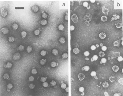
FIG. 1. Electronmicrograph of intact (a) and partially disrupted (b)46bacteriophages negatively stained with2%PTA. The bar represents 100 nm.
jected to double-phase phenol extraction with 100 mM sodiumphosphatebuffer(pH 7.0). Afterextraction,theNa+ waslowered to 1 mM by dialysis in the presenceof0.1 mM disodium EDTA. Thermal denaturation of the recovered nucleic acid was monitored at 258 nm with a Gilford 2000 recording spectrometer. The samplechamberwasheated at a constant rate of 0.2°C
min-'
by a circulating ethylene glycol-water solution.Zone sedimentation of
4)6
NC. A suspension ofpurified NCs (2OD26(6
units) was layeredon 11 ml, 10to35%
linear sucrose densitygradients containing KM buffer and centri-fuged (23,500 rpm, 90 min) in a Spinco SW41 rotor. After centrifugation, vertical illumination of the centrifuge tubes revealed only one light-scattering band near the center of each tube. Thegradient
was thenpartitioned
into 0.25-mlfractions. Absorbance measurementsofeachfractionat 280 and 258 nm confirmed the presence of a single band. Fractionsincludingtheleadingandtrailing edgesof the band werethen examined by EM.
RESULTS
EM of 46 and
+6
NC. Bacteriophage 46, negatively stainedbyPTA(pH 7), showed little internal detail(Fig.la), but partial disruption of theenvelope, duringstorageat 4°C before being mounted on grids, revealed both ring and hexagonal forms (Fig. lb). After removal of the envelope,threedistinctformswerepersistently seen(Fig.2a), descrip-tively identified as (i) irregular hexagons, (ii) rings (often with 10dots), and (iii) sixfold stars. The four long edgesof
the negatively stained, irregularhexagons were consistently seeninhighercontrastthan the tworemainingedges. Forms (i)and(ii)werefoundwithhigh frequency, and form (iii) was found withlowfrequency. Oursixfold stars differ fromthe stars, identified earlier(14), which have no symmetry axis.
Rotational imageaveraging.Figure 2btod shows mutiple exposures of NC images on photographic paper at n rota-tions of
Thin,
in which n is an integer generating the best structural definition (12). Not shown are other n-fold rota-tionsresultingin blurredimages. Weconcluded that eachof the threedistinct forms hasanaxis of symmetry perpendicu-lar to the plane of the image corresponding to these NC symmetry axes: fivefold for the ring, twofold forthe irregu-larhexagon, and threefold for the sixfold star.ZonesedimentationofNC. Thequestionarosewhether the three observed forms represented three different NC spe-cies.Wethereforeattemptedtoseparate andisolate themby zonal sedimentation butwere unsuccessful. Hence, the NC sampleswereeitherhomogeneousorcontained amixtureof particleswhosedifferences indensityandshapecombined in suchawayas to result in identical sedimentation velocities. EMexamination of NCs from theleadingandtrailing edges of the single band, and from its centerfraction, showed a uniformfrequencyratio ofhexagonaltoringforms ofca.2:1. Geometryof
46
NC.The fact thatorthogonalaxesof two-, three-, and fivefold symmetry define icosahedral symmetry (3) ledus to compare the NC images with the twosimplest bodies of icosahedral symmetry:theregularicosahedron and the regular dodecahedron. Parallel projections of the edgesJ. VIROL.
a
I.-.on November 10, 2019 by guest
http://jvi.asm.org/
[image:2.612.107.509.78.393.2]of these bodies, viewed alongsymmetry axes, areshown in Fig. 3. A projection along the twofold symmetry axis was strikingly similar to the irregular hexagon seen in Fig. 2b. This comparison isjustified by the fact that electron micro-graphs are also parallel projections of the specimen since the numerical aperture of the objective lens is about
10-2
and << 1. Similarly, inspection of Fig. 3 and of the ringlike forms (e.g., Fig. 2aand d) also suggested adodecahedron, but nota
b
forms:irregular hexagons star(inset),andringswith10dots Three different forms from (a) were enlarged by the same factor and
photographically averaged (12): (b) irregular hexagon rotatedtwice
about twofold symmetry axis; (c) star, rotated 6 times about
threefold axis, and (d) ring, rotated 10 times about fivefold axis.
[image:3.612.344.525.77.234.2]Magnification reference:outerdiameter of theringformsis46nm.
FIG. 3. Parallel projections of the edges along the two-, five-, and threefoldaxes ofsymmetry for thedodecahedron(a toc) and
the icosahedron (dto f). If these bodies were virus particles on a
specimen gridintheelectron microscope. theywould be restingona
single edge(aandd).onapentagonal (b)ortriangularface(f).oron avertex (cand e).
anicosahedron. Measurements of the dimensions of thetwo
mostfrequent morphological forms, given asratios (Fig. 4), supported thisobservation quantitatively.
Recovery of double-stranded RNA from
46
NC. After phenol extraction of purified NCs. we observed peak UVabsorptionat 258nm, an
OD,6(10D28()
ratio of2.18, and, inthe presence of 1 mM sodiumphosphate buffer and 0.1 mM disodium EDTA, a thermally induced hyperchromicity of
35%,
clearly indicative ofdouble-stranded nucleic acid. The presence of double-stranded RNA in purified NCs shows that our NC samples and the 120S particles (14) differchemically, although the 120S particles and mature NCs appeartohaveidentical morphology when stained with PTA
atpH 7.
DISCUSSION
We infer that the 46 NC contains a structure which has icosahedralsymmetryandcanbest bedescribedasaregular
dodecahedron and that the three remarkably different forms determined by EM after PTA negative staining are merely different views ofthat structure. If this is true, the (b6 NC would be the first known example ofa virus-related particle
having such an architecture. Evidence in favor of this
inference is asfollows.
(i)The failure ofourattempts to separatethe threedistinct morphological forms by zonal sedimentation suggests that there is onlyonespeciesof NC. Support forthis inferenceis
presented in the accompanying paper(20).
(ii) Each of the three forms observed hasitsown
symme-try axis (Fig. 2) corresponding to NC axes ofsymmetry. If there is indeed onlyoneNCspecies,the NCmustthenhave
two-, three-, and fivefold axes of symmetry and hence. icosahedral symmetry. This would satisfy the constraintson
the geometry ofan isometric virus as discussed by Caspar and Klug (3, 11)and Klug(10).
The surface/volume ratio of a regular dodecahedron is only 3.2% higher than that ofa regular icosahedron. Thus.
containment of the genome in a dodecahedral framework should not be asignificant disadvantage in thisregard.
(iii) Electron micrographs are parallel projections of the
NC particlesandthereforedirectly comparable withparallel projections of model structures. The simplest bodies of
a
b
e
c
f
d
on November 10, 2019 by guest
http://jvi.asm.org/
[image:3.612.67.280.180.663.2]482 STEELY AND LANG
FIG. 4. Geometric proportions of the hexagonal and the ring form seen in nucleocapsid preparations in comparison with the corresponding proportions for a regular dodecahedron and icosahe-dron.Measured were 19 hexagonal and 22 circular particles. The u/v andx/yratios,respectively, were as follows: observed,1.16 + 0.04
and 1.86 + 0.13 (the errors are sample standarddeviations); for a dodecahedron,1.176 and1.902; for anicosahedron,1.070and 1.236.
icosahedral symmetry are the regular icosahedron and the regular dodecahedron, having parallel projections as illus-tratedin Fig. 3. We have shown (Fig. 4) that the geometric proportions in the NC images of rings and irregular hexagons are, within measurement error, those of projected regular dodecahedraand not of regular icosahedra. Among the more complex bodies of icosahedral symmetry is the deltahedral family of polyhedra (3) whose faces are equilateral triangles
("icosadeltahedra").
Anexample is the pentakisdodecahe-dron(9), illustrated in reference 13. We did not find evidence that would favor an icosadeltahedron over a simple, regular dodecahedron.
(iv) Parallel projection along the threefold axis of symme-try implies that the NC rests on a vertex if it is a dodecahe-dron andon afaceif it isanicosahedron(Fig. 3). The rarity of starforms in our samples, together with the assumption that anNC is unlikely to rest on a vertex(as yet unproven), would suggest that the NC has a dodecahedral rather than icosahedral surface.
The question must be raised as to whether or not the observed structures are preparative artifacts. We are un-aware of proof that negative staining with PTA generates virus structures that are absent without PTA. However, it hasbeen shown that PTA revealsstructuresinvacciniavirus
selectively, dependingonthepH(15), and that this selectiv-ityis reversible(8). Wethereforeassumethat the structures in ourmicrographs ofNCs negatively stained with neutral-ized PTA are real but that the NC may contain (besides RNA) other structural elements.
Deformation of NCs in PTA during drying may pose anotherproblem. NCs dried from suspensions withoutPTA and shadowed withplatinum (not shown)were significantly flattened,presumably by surface forcesatthetime when the solution-air interface passed through the NCs. However, one may expectthat flattening is reduced in the presence of PTA, when the glassy PTA precipitate forms a supporting matrix around the NCs, before the interface has passed them. Indeed, tilting the specimen grids has demonstrated (20) that the spatial structure is sufficiently preserved to permit athree-dimensional analysis.
Elimination of structures by PTA is another
possi-bility. Using uranyl acetate as a negative stain, the
mor-phologies of
06
NCs differ from those obtained by PTA(reference 14andourunpublished data). Mindich and Davi-doff-Abelson(14) point outthat +6NCs stained with
uranyl
acetateappeartohavean outerlayernotobserved withPTA and suggestthat PTAmayremovetheouterlayer
containing
the protein P8. Unfortunately, the geometric structures
discussed above are in general not clearly delineated in mature NCs stained withuranyl acetate sothatit isdifficult
to view the isometric structure and the outer layer simulta-neously. However, Fig. 4d of reference 14 does show a uranyl-stained 120S particle in star form, indicating that this form is either real or that both stains induce the same artifact. The only proof of structural reality when employing EMis indirect: bydemonstrating that many different prepa-ration methods produce consistent results. It remains to be seen in thiscasewhether ornot ourproposedNC structure can be confirmed by other techniques such as cryofixation withcarbon replication and double-angle shadowing.
We have notobserved irradiationdamage of the morpho-logical features as described; these appear to remain stable in the electron beam after the first viewing.
The structures in our NC micrographs were not induced byTriton X-100 since(k6 preparations,partially disruptedby storage at 4°C but not treated with the detergent (Fig. lb), showed ring, hexagonal and star forms indistinguishable from those of isolated NCs. Also, the same three forms are seen among sus297 NC particles isolated in the absence of Triton X-100 (14).
Itis presently not clear howan icosahedralNC structure orthe proposed dodecahedralstructurecould be reconciled with thecopy numberofproteincomponents per NC (4) and theCaspar-Klug theory(3) ofcapsidstructure. Anintriguing possibilityconsistent withourdata and supported by Fig.4 in reference 20 is that the structure is a rather open dode-cahedral cage whose edges are assembled from rodlike proteins.Anexampleofsuch adodecahedralproteincageis seen in themultienzymic complexesof pyruvate dehydroge-naseisolated fromBacilliusstearothermnophilus(7). Depend-ingontheirorientationonthespecimengrid, these complex-esclearly displayedthree forms in the EM similartothose in Fig. 3a, b, and c.
ACKNOWLEDGMENTS
We thank James VanEtten. of the University ofNebraska, who generously donated phage 46 plus its host and helped us with excellent advice and Young Yang and Carla W. Gray of this department fora critical discussion ofthe manuscript.
Wearealsogratefulfor support ofthisworkbytheUniversity of TexasatDallasOrganized Research Fund.
LITERATURE CITED
1. Bamford, D. H., and E. T. Palva. 1980. Structure ofthe lipid-containingbacteriophage f6: disruption by TritonX-100
treat-ment.Biochim. Biophys.Acta 601:245-259.
2. Bamford, D. H., E. T. Palva, and K. Lounatmaa. 1976.
Ultra-structureandlifecycleof thelipid-containing bacteriophage46. J. Gen. Virol. 32:249-259.
3. Caspar, D.L. D.,and A. Klug. 1962. Physical principles in the construction of regular viruses. Cold Spring Harbor Symp. Quant. Biol. 27:1-24.
4. Day, L. A., and L. Mindich. 1980. The molecular weight of bacteriophage k6and itsnucleocapsid. Virology 103:376-385.
5. Emori, Y.,H.Iba,and Y.Okada.1982.Morphogenetic pathway of bacteriophage j6: a flow analysis of subviral and viral particlesininfected cells. J. Mol. Biol. 154:287-310.
6. Gonzales,C.F.,W.G.Langenberg, J.L. VanEtten,and A. K.
Vidaver. 1977.Ultrastructure ofbacteriophage46:arrangement of the double-stranded RNA and envelope. J. Gen. Virol. 35:353-359.
7. Henderson, C. E., R. N. Perham, and J. T. Finch. 1979. Structure and symmetry of B. stearothermophilus pyruvate dehydrogenase multi-enzyme complex andimplicationsfor
eu-caryoticevolution. Cell 17:85-93.
8. Herzberg, K., D. Lang, K. Reuss, R. Dahn, andC. Plescher.
1964. Uber das Verhalten der Filamentedes Kanarienpocken-virus und der Capsomeren des Varizellen-Zostervirus gegen-uber Losemitteln und Fermenten. Zentralbl. Bakteriol. Parasi-J. VIROL.
on November 10, 2019 by guest
http://jvi.asm.org/
tenkd. Infektionskr. Hyg. Abt. 1 Orig.Reihe A 195:133-148. 9. Jamison, R. M. 1969. Morphology ofechovirus 22. J. Virol.
4:904-906.
10. Klug,A.1983. Architecturaldesignofsphericalviruses. Nature
(London) 303:378-379.
11. Klug, A., and D. L. D. Caspar. 1960. The structure ofsmall
viruses. Adv. Virus Res. 7:225-325.
12. Markham, R., S. Frey, andG. J. Hills. 1963. Methods forthe
enhancement of image detail and accentuation ofstructure in electronmicroscopy. Virology 20:88-102.
13. Mayor, H. D. 1964. Picornavirus symmetry. Virology
22:157-159.
14. Mindich, L., and R. Davidoff-Abelson. 1980. The
characteriza-tion ofa 120S particle formed during 4)6 infection. Virology 103:386-391.
15. Muller, G., and D. Peters. 1963. Substrukturen des
Vaccine-virus, dargestellt durch Negativkontrastierung. Arch.
Virus-forsch. 13:435-451.
16. Semancik, J. S., A. K. Vidaver, and J. L. Van Etten. 1973.
Characterization of segmented double-helicalRNA from bacte-riophage4)6. J. Mol. Biol. 78:617-625.
17. Van Etten, J., L. Lane, C. Gonzales, J. Partridge, and A. Vidaver. 1976.Comparative properties ofbacteriophage 4)6 and
4)6nucleocapsid. J. Virol. 18:652-658.
18. Vidaver, A. K., R. K. Koski, and J. L. Van Etten. 1973.
Bacteriophage 4)6: a lipid-containing virus of Pseuidolmoi(s phaseolicola.J. Virol. 11:799-805.
19. Yamamoto, K., and B. Alberts. 1970. Rapid bacteriophage
sedimentation in the presence of polyethylene glycol and its
application to large-scale virus production. Virology
40:734-744.
20. Yang, Y.,and D.Lang. 1984. Electronmicroscopyof
bacterio-phage 4)6 nucleocapsid: three-dimensional image analysis. J.
Virol. 51:484-488.