This is a repository copy of
Development of a Generic Wound Care Assessment Minimum
Data Set
.
White Rose Research Online URL for this paper:
http://eprints.whiterose.ac.uk/121237/
Version: Accepted Version
Article:
Coleman, S orcid.org/0000-0002-1437-421X, Nelson, EA, Vowden, P et al. (17 more
authors) (2017) Development of a Generic Wound Care Assessment Minimum Data Set.
Journal of Tissue Viability, 26 (4). pp. 226-240. ISSN 0965-206X
https://doi.org/10.1016/j.jtv.2017.09.007
© 2017 Published by Elsevier Ltd on behalf of Tissue Viability Society. This manuscript
version is made available under the CC-BY-NC-ND 4.0 license
http://creativecommons.org/licenses/by-nc-nd/4.0/
[email protected] https://eprints.whiterose.ac.uk/ Reuse
Items deposited in White Rose Research Online are protected by copyright, with all rights reserved unless indicated otherwise. They may be downloaded and/or printed for private study, or other acts as permitted by national copyright laws. The publisher or other rights holders may allow further reproduction and re-use of the full text version. This is indicated by the licence information on the White Rose Research Online record for the item.
Takedown
If you consider content in White Rose Research Online to be in breach of UK law, please notify us by
1
Development of a Generic Wound Care Assessment Minimum Data Set
Abstract
Background
At present there is no established national minimum data set (MDS) for generic wound
assessment in England, which has led to a lack of standardisation and variable assessment
criteria being used across the country. This hampers the quality and monitoring of wound
healing progress and treatment.
Aim
To establish a generic wound assessment MDS to underpin clinical practice.
Method
The project comprised 1) a literature review to provide an overview of wound assessment
best practice and identify potential assessment criteria for inclusion in the MDS and 2) a
structured consensus study using an adapted Research and Development/ University of
California at Los Angeles Appropriateness method. This incorporated experts in the wound
care field considering the evidence of a literature review and their experience to agree the
assessment criteria to be included in the MDS.
Results
The literature review identified 24 papers that contained criteria which might be considered
as part of generic wound assessment. From these papers 68 potential assessment items
were identified and the expert group agreed that 37 (relating to general health information,
baseline wound information, wound assessment parameters, wound symptoms and
specialists) should be included in the MDS.
Discussion
Using a structured approach we have developed a generic wound assessment MDS to
underpin wound assessment documentation and practice. It is anticipated that the MDS will
facilitate a more consistent approach to generic wound assessment practice and support
providers and commissioners of care to develop and re-focus services that promote
improvements in wound care.
Keywords
Wounds
Assessment
Management
Audit
Nursing
Abbreviations
Minimum Data Set: MDS
Commissioning for Quality and Innovation: CQUIN
National Health Service: NHS
2
1. Background
Chronic wounds, sometimes referred to as
‘
difficult to hea
l’
wounds are usually defined in
relation to wound duration with parameters of 4 -12 weeks being used [1-4]. Chronic
wounds, which commonly incorporate pressure ulcers, venous ulcers, arterial ulcers and
diabetic ulcers represent a significant burden to patients and health care providers
worldwide. It is estimated that almost 1% of the worlds population experiences difficult to
heal wounds which are associated with negative quality of life [5]. In the United States,
chronic wounds affect approximately 6.5 million patients with an estimated $25 billion
treatment cost per annum [6]. This is also reflected in the United Kingdom where recent
information from the Health Improvement Network (THIN) Database which collects data from
primary care, indicated 4.5% (2.2 million) of the adult population were estimated to have a
wound (excluding surgical wounds that healed within 4 weeks of the procedure) in 2012/13,
accounting for 40.6 million healthcare professional/patient visits, 97.1 million drug
prescriptions, 344.6 million dressings/bandages and costing £4.5-5.1 billion [7]. The study
also found that 12% of wounds had no recorded diagnosis and 56% of the wounds recoded
as leg ulcers lacked a differential diagnosis, suggesting a lack of evidence-based wound
care/assessment [7]. This is a substantial problem to the NHS and an important part of
nursing practice.
At present there is no established national minimum data set (MDS) for generic wound
assessment, which has led to a lack of standardisation and variable criteria being used
across England. This is particularly important for difficult to heal or chronic wounds as the
lack of standardisation hampers decision making about diagnosis and treatment as well as
the quality and monitoring of wound healing progress. Work to establish an MDS for generic
wound assessment was taken
forward as part of NHS England’s
Leading Change Adding
Value Framework - Improving Wound Care Project. This aims to underpin wound
assessment practice and to support commissioners and providers in developing and
re-focussing services that promote improvements in wound care. The work is supported by a
new quality indicator for improving the assessment of wounds as part of the 2017-19
Commissioning for Quality and Innovation (CQUIN) framework [8]. The Improving Wound
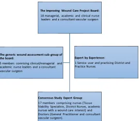
Care Project is led by a Board (Figure 1) which provides oversight for the development of the
generic wound assessment MDS. The project incorporates:
1) A literature review to identify potential assessment criteria for the MDS and;
2) A structured consensus study to agree the assessment criteria to be included in the
MDS to facilitate a standardised approach to wound assessment practice.
3
Figure 1 Groups involved in the development of the Generic Wound Assessment MDS
2. Literature Review
2.1 Method
A literature review was undertaken to identify potential assessment criteria to be included in
the MDS. The review considered any literature relating to wound assessment criteria and
was not limited by any particular study design and incorporated guidance papers [9]. A
simple key word search (chronic wound, assessment, management, validity, reliability,
guideline, documentation) of the MEDLINE database (Jan 1996 - Aug 2016) was undertaken
using Boolean operators
‘and’
‘or’
. Citations of relevant studies were also considered.
The abstracts of these papers were screened to identify those which potentially provided
comprehensive information about criteria considered when conducting wound assessment.
Papers considered potentially relevant were reviewed in full by the researcher (SC). The
wound assessment criteria contained in relevant papers were extracted and mapped against
wound assessment domains (key assessment areas) and sub-domains (detailed
assessment concepts). The initial framework for the domains and sub-domains were
informed by the generic wound assessment MDS sub-group (Figure 1)). These were
amended as new concepts emerged from the literature review and the final domains and
sub-domains were reviewed and agreed by the Improving Wound Care project Board.
2.2 Results
The search identified over 300 papers, of which 24 identified wound assessment domains
and sub-domains incorporating the following papers types:
9 wound healing/monitoring instruments [10-18]
10 wound assessment guidance [19-28]
2 primary wound care studies [29, 30]
2 literature/systematic review [31, 32]. The systematic review provided citations for
other wound assessment instruments included in this review.
1 wound care quality improvement initiative [33]
The Improving Wound Care Project Board:
18 managerial, academic and clinical nurse leaders and a consultant vascular surgeon
Consensus Study Expert Group: 17 members comprising nurses (Tissue Viability Specialists, District Nurses, academic nurses with a wound care interest) and Doctors (General Practitioner and consultant vascular surgeon).
The generic wound assessment sub-group of the board:
5 members comrising clinical/managerial and academic nurse leaders and a consultant vascular surgeon
Expert by Experience:
4
Table 1 provides a summary of findings indicating 6 key domains comprising general health
information wound history/baseline information, wound assessment parameters, wound
symptoms, infection and specialist information and an associated 69 sub-domains. Most of
these sub-domains were considered potential assessment criteria in the subsequent
consensus study. Further information for each paper can be found in Appendix 1.
Table 1 Wound assessment domains and sub-domains (potential assessment criteria)
included in papers of the literature review
Domain Sub-domain
Of 24 papers General health
information
Allergies 3
Mobility Status 2
Risk Factors for Delayed Healing
Factors affecting systemic blood supply to wound (e.g. vascular or arterial disease, smoking, anaemia)
10 Factors affecting local blood supply to wound (e.g.
pressure/shear, pressure ulcer)
8 Factors affecting skin (e.g. malnutrition, obesity, peripheral
neuropathy) 10
Susceptibility to infection (e.g. immune-supressed) 10 Medication affecting wound healing (e.g. steroids,
chemotherapy) 9
Generic delayed healing (non-specific) 2
Quality of life
Patient Information 2
Physical - Fatigue/lack of sleep 1
Physical - Reduced mobility 2
Physical- Health and wellbeing 2
Physical - Changes to eating habits 1
Physical- Daily activities 2
Emotional – Depression 1
Emotional – Emotions 2
Social- Friendships 2
Social – Hobbies 1
Social - Frequency of dressing changes 1
Social- Pain 1
Social – Odour 1
Social- Social isolation 2
Non-specific 1
Wound history/ baseline information
Number of wounds 6
Wound Location 14
Wound type/classification (e.g., venous leg ulcer, pressure
ulcer) 16
5
Domain Sub-domain
Of 24 papers Wound
Assessment Parameters
Wound size width 19
Wound size length 19
Wound size (non-specific) 1
Wounds size depth 21
Undermining/tunnelling 16
Shape 3
Wound bed tissue type (e.g. necrotic, sloughy) 20 Wound bed tissue amount (e.g. percentage of wound) 11
Wound bed (non- specific) 1
Wound margins/edges description (e.g. epithelialisation
undermining) 17
Wound margins/edges (non-specific)
Surrounding skin colour (e.g. redness) 17 Surrounding skin condition (e.g. maceration, oedema,
induration) 16
Surrounding skin (non-specific) 2
Current wound status (e.g. progress/deterioration) 12 Surrounding skin condition (e.g. maceration, oedema,
induration) 16
Surrounding skin (non-specific) 2
Current wound status (e.g. progress/deterioration) 12
Wound symptoms
Pain frequency (e.g. at dressing change) 11
Pain severity 11
Pain (non-specific) 4
type (i.e. inflammatory neuropathic) 2
Full pain assessment 5
Current pain status (progress/change) 4 Exudate amount (e.g. high, moderate) 18 Exudate consistency/type/colour (e.g. serous, blood,
sero-sanguineous, thick, thin) 16
Exudate (non-specific)
Current exudate status (progress/deterioration) 5
Exudate problem to patient 1
The performance of the current dressing in absorbing
wound exudate 10
Odour occurrence (e.g. on dressing removal, when dressing
intact) 9
Odour intensity (e.g. acceptable
minimal, problem) 4
Odour (non-specific) 2
Current odour status (progress/ changes) 5
Odour problem to patient 1
6
Domain Sub-domain
Of 24 papers
Signs of local wound infection (e.g. cellulitis, abscess/pus, increasing pain, exudate, odour; deterioration (wound breakdown and dehiscence), healing slower than anticipated, friable granulation tissue, bleeds easily,
pocketing at wound base) 16
Signs of systemic infection relating to the wound (e.g. high
temp) 4
Management of infection 6
Specialist Information
Tissue viability team referrals 0 Hospital consultant referrals (e.g. vascular, plastics) 0
Pressure ulcer risk assessment 8
Doppler ABPI 5
3. Consensus study
3.1 Methods
A modified RAND/UCLA (Research and Development/University of California at Los
Angeles) appropriateness method [34] based on a previous consensus study was used [35].
This incorporated face-to-face interaction of an expert group and pre- and post- meeting
questionnaire completion (incorporating 9 point Likert scales) considering what should be
included in a generic wound assessment MDS.
3.2 Sample
Participants forming the expert group were purposefully sampled to incorporate
multi-speciality clinical/academic leaders in the wound care field, identified by their previous work
and/or related publications [36]. Seventeen members were recruited to allow for attrition and
to maximize reliability while preventing facilitation problems [37].
3.3 Ethical considerations
The study was reviewed and approved by a University of Leeds Research Ethics Committee.
Prior to recruitment expert group members were provided with a study information sheet and
encouraged to ask questions. Following this informed consent was sought by the researcher
(SC)
3.3 Data Collection
The initial literature review provided the framework for the pre and post meeting
questionnaires which were developed to seek expert group members
’
individual views
regarding the important elements to be included in the MDS. The questionnaires
incorporated statements relating to potential assessment criteria and participants were
asked to reflect on the results of the literature review and their clinical experience and/or
expertise in the field when rating their level of support for including these in the MDS on a 9
point Likert scale (where 1 indicates poor support and 9 indicates strong support).
The questionnaires comprised fewer items than the sub-domains identified in literature
review because the primary papers did not always specify the underlying concept being
considered e.g. ‘none specific exudate’
sub-domain (did not specify whether this related to
amount, consistency or something else). In contrast some sub-domains were excluded
because they were considered too specific for the MDS and were only encountered in a few
7
ratings in light of discussions and/or where necessary for questionnaire items to be clarified
and amended.
The face-to-face meeting was undertaken in a pleasant hotel setting and refreshments were
provided throughout. The meeting was facilitated by the researcher (SC) to ensure all
members had the opportunity to discuss their opinions [37]. The focus of the meeting was
discussing the scope of the MDS and reviewing the results of the pre-meeting questionnaire.
This allowed areas of disagreement and uncertainty to be discussed and for members to
consider this before privately re-rating their level of support in the post-meeting
questionnaire.
3.4 Analysis
Questionnaire statements were summarised using the median group response and
categorised into tertiles, 1-3 disagree, 4-6 uncertain, 7-9 agree. Within-group agreement was
measured using the RAND Disagreement index [34], which considers the dispersion of
individual scores and identifies areas of disagreement (where panellists rate at both ends of
the Likert Scale). Using the group median response and the disagreement index for each
statement the following principles were applied following post meeting questionnaire
completion to identify MDS items:
Group medians of 1-3 without disagreement will be excluded
Group medians of 7-9 without disagreement will be included
Disagreement index is >1 or median 4-6 will be excluded but are potential areas for
further research
A directed content analysis approach [38] was used to code data from transcripts of the
expert group meetings and a summary report was written and checked for accuracy the
expert group.
3.5 Results
Expert group members comprised nurses and Doctors with a wound care interest (Figure 1)
and incorporated 2 males and 15 females. All seventeen members fully completed the
pre-meeting questionnaire. Sixteen members attended the face-to-face pre-meeting and fully
completed the post-meeting questionnaire.
The expert group indicated their support for inclusion of 42 of the 46 MDS items (Table 2)
included in the pre-meeting questionnaire (with one area of disagreement relating to odour
status) and 4 areas of uncertainty (quality of life (physical, social and emotional) and
pressure ulcer risk assessment).
The discussions at the face-to-face meeting (Table 3) led to some changes in opinion and
amendments to the post-meeting questionnaire which incorporated 47 items. The
amendments related to requested additional items of skin sensitivities, wound location,
treatment aim, re-assessment date, pressure ulcer category, healing and some items being
combined (all noted by italics in Table 2). The results of the post-meeting questionnaire
indicated there was support for 33 items with a group median of 7-9 (without disagreement),
lack of support for 7 items, uncertainty about 4 items,1 item with a median of 8 but with
disagreement, 2 items where the median fell in-between 2 tertiles.
8
Table 2 Questionnaire Results
Pre-Meeting Potential Assessment Criteria GM DI Post-Meeting Potential Assessment Criteria GM DI
Allergies should be recorded in the generic wound
assessment MDS. 9.00 0.75
Allergies should be recorded in the generic wound
assessment MDS. 3.50* 2.26
Sk in sensitivities should be recorded in the generic wound
assessment MDS. 9.00 0.00
Mobility status should be recorded in the generic wound
assessment MDS. 7.00 0.75
Mobility status should be recorded in the generic wound
assessment MDS. 2.00 0.65
Factors affecting the patient’s systemic blood supply to wound (e.g. vascular or arterial disease, smoking anaemia) should be recorded in the generic wound
assessment MDS. 9.00 0.05
Factors affecting the patient’s systemic blood supply to wound (including vascular or arterial disease, smoking anaemia) should be recorded in the generic wound
assessment MDS. 7.00 0.61
Factors affecting the patient’s local blood supply to the wound (e.g. pressure/shear, pressure ulcer) should be
recorded in the generic wound assessment MDS. 9.00 0.13
Factors affecting the patient’s local blood supply to the wound (including pressure/shear, pressure ulcer) should
be recorded in the generic wound assessment MDS. 7.00 0.70 Factors affecting the patient’s skin integrity (e.g.
malnutrition, obesity, peripheral neuropathy) should be
recorded in the generic wound assessment MDS. 9.00 0.29
Factors affecting the patient’s skin integrity (including malnutrition, obesity, peripheral neuropathy) should be
recorded in the generic wound assessment MDS. 6.50* 0.78 Factor affecting the patient’s susceptibility to infection
(e.g. immune-supressed) should be recorded in the
generic wound assessment MDS. 8.00 0.33
Factor affecting the patient’s susceptibility to infection (including immune-supressed) should be recorded in the
generic wound assessment MDS. 7.50 0.49 Medication affecting wound healing (e.g. steroids,
chemotherapy) should be recorded in the generic wound
assessment MDS. 9.00 0.16
Medication affecting wound healing (including steroids, chemotherapy) should be recorded in the generic wound
assessment MDS. 7.00 0.52
The number of wounds should be recorded in the
generic wound assessment MDS. 9.00 0.00
The number of wounds should be recorded in the generic
wound assessment MDS. 9.00 0.00
The location of the wound should be recorded in the
generic wound assessment MDS. 9.00 0.00 The wound type/classification (e.g., venous leg ulcer,
pressure ulcer) should be recorded in the generic wound
assessment MDS. 9.00 0.00
The wound type/classification (including, venous leg ulcer, pressure ulcer) should be recorded in the generic wound
9
Pre-Meeting Potential Assessment Criteria GM DI Post-Meeting Potential Assessment Criteria GM DI
The duration of the wound (e.g. weeks, months, years) should be recorded in the generic wound assessment
MDS. 9.00 0.00
The duration of the wound (including weeks, months, years) should be recorded in the generic wound
assessment MDS. 9.00 0.00
The treatment aim should be recorded in the generic
wound assessment MDS. 9.00 0.29
A planned re-assessment date should be recorded in the
generic wound assessment MDS. 9.00 0.00 The width of the wound should be recorded in the
generic wound assessment MDS. 9.00 0.00
The maximum width of the wound should be recorded in
the generic wound assessment MDS. 9.00 0.00 The length of the wound should be recorded in the
generic wound assessment MDS. 9.00 0.00
The maximum length of the wound should be recorded in
the generic wound assessment MDS. 9.00 0.00 The depth of the wound should be recorded in the
generic wound assessment MDS. 9.00 0.02
The maximum depth of the wound should be recorded in
the generic wound assessment MDS. 9.00 0.00 The category of a pressure ulcer wound should be
recorded in the generic wound assessment MDS. 9.00 0.00 Undermining/tunnelling of the wound should be
recorded in the generic wound assessment MDS. 9.00 0.00
Undermining/tunnelling of the wound should be recorded
in the generic wound assessment MDS. 9.00 0.00 The shape of the wound should be recorded in the
generic wound assessment MDS. 7.00 0.75
The shape of the wound should be recorded in the generic
wound assessment MDS. 1.00 0.21
The wound bed tissue type (e.g. necrotic, sloughy) should be recorded in the generic wound assessment MDS.
9.00 0.00
The wound bed tissue type after cleansing (including necrotic, sloughy, granulating, epithelialisation, tendon, bone) should be recorded in the generic wound
assessment MDS. 9.00 0.00
The wound bed tissue amount (e.g. percentage of wound) should be recorded in the generic wound
assessment MDS. 9.00 0.54
The wound bed tissue amount after cleansing should be quantified and recorded in the generic wound assessment
MDS. 9.00 0.00
A description of the wound margins/edges (e.g.
epithelialisation, undermined) should be recorded in the
generic wound assessment MDS. 9.00 0.05
A description of the wound margins/edges (including epithelialisation, undermined) should be recorded in the
generic wound assessment MDS. 9.00 0.13
The colour of the skin surrounding the wound (e.g. redness) should be recorded in the generic wound assessment MDS.
9.00 0.02
The colour (including redness) and condition (including oedema, maceration, induration of the skin surrounding the wound should be recorded in the generic wound
10
Pre-Meeting Potential Assessment Criteria GM DI Post-Meeting Potential Assessment Criteria GM DI
The condition of the skin surrounding the wound (e.g. oedema, maceration, induration) should be recorded in
the generic wound assessment MDS. 9.00 0.00 The current overall wound status (e.g. improving,
deteriorating) should be recorded in the generic wound
assessment MDS. 9.00 0.29
The current overall wound status (including improving, deteriorating) should be recorded in the generic wound
assessment MDS. 2.50 0.37
Whether the wound has healed should be recorded in the
generic wound assessment MDS. 9.00 0.00 Pain frequency (e.g. constant, at dressing change)
should be recorded in the generic wound assessment
MDS. 9.00 0.13
Wound pain frequency (including constant, at dressing change) should be recorded in the generic wound
assessment MDS. 9.00 0.13
Pain severity (e.g. on a visual analogue scale) should be recorded in the generic wound assessment MDS.
8.00 0.13
Wound pain severity (including on a visual analogue scale) should be recorded in the generic wound
assessment MDS. 8.00 0.13
The type of pain (i.e. inflammatory or neuropathic) should be recorded in the generic wound assessment
MDS. 7.00 0.75
The type of wound pain (i.e. inflammatory or neuropathic) should be recorded in the generic wound assessment
MDS. 6.00 1.94
A full validated pain assessment instrument should be used and recorded in the generic wound assessment
MDS. 7.00 0.75
A full validated wound pain assessment instrument should be used and recorded in the generic wound assessment
MDS. 5.00 1.61
The current pain status (e.g. improving, deteriorating, changes) should be recorded in the generic wound
assessment MDS. 7.00 0.54
The current pain status (including improving, deteriorating, changes) should be recorded in the generic wound
assessment MDS. 3.00 0.83
The exudate amount (e.g. high, moderate) should be
recorded in the generic wound assessment MDS. 9.00 0.02
The exudate amount (including high, moderate) should be
recorded in the generic wound assessment MDS. 9.00 0.00 Exudate consistency/type/colour (e.g. serous, blood,
sero-sanguineous, thick, thin) should be recorded in the
generic wound assessment MDS. 8.00 0.16
Exudate consistency/type/colour (including serous, blood, sero-sanguineous, thick, thin) should be recorded in the
generic wound assessment MDS. 9.00 0.00 The current exudate status (e.g. static, reducing,
increasing) should be recorded in the generic wound
assessment MDS. 8.00 0.37
The current exudate status (including static, reducing, increasing) should be recorded in the generic wound
assessment MDS. 2.00 0.50
The performance of the current dressing in absorbing wound exudate (e.g. strikethrough, leakage) should be
recorded in the generic wound assessment MDS. 8.00 0.33
The performance of the current dressing in absorbing wound exudate (including strikethrough, leakage) should
11
Pre-Meeting Potential Assessment Criteria GM DI Post-Meeting Potential Assessment Criteria GM DI
Whether exudate is a problem to the patient should be
recorded in the generic wound assessment MDS. 9.00 0.29
Whether exudate is a problem to the patient should be
recorded in the generic wound assessment MDS. 2.50 1.29
The occurrence of odour (e.g. on dressing removal, when dressing intact) should be recorded in the generic wound assessment MDS.
9.00 0.33
The occurrence of odour (including on dressing removal, when dressing intact) should be recorded in the generic wound assessment MDS. (b and c deleted as felt already
covered in a and d) 8.50 0.13
The intensity of odour (e.g. acceptable, minimal, problem) should be recorded in the generic wound
assessment MDS. 8.00 0.67
The current odour status (e.g. changes) should be
recorded in the generic wound assessment MDS. 7.00 1.14 Whether odour is a problem to the patient should be
recorded in the generic wound assessment MDS. 9.00 0.13
Whether odour is a problem to the patient should be
recorded in the generic wound assessment MDS. 5.00 2.55 Signs of local wound infection (e.g. cellulitis,
abscess/pus, increasing pain, exudate, odour; deterioration (wound breakdown and dehiscence), healing slower than anticipated, friable granulation tissue, bleeds easily, pocketing at wound base) should
be recorded in the generic wound assessment MDS. 9.00 0.00
Signs of local wound infection (including cellulitis, abscess/pus, increasing pain, exudate, odour;
deterioration (wound breakdown and dehiscence), healing slower than anticipated, friable granulation tissue, bleeds easily, pocketing at wound base) should be recorded in
the generic wound assessment MDS. 9.00 0.00 Signs of systemic infection (e.g. high temperature)
relating to the wound should be recorded in the generic
wound assessment MDS. 9.00 0.37
Signs of systemic infection (including high temperature) relating to the wound should be recorded in the generic
wound assessment MDS. 8.00 1.30
The management of infection should be recorded in the generic wound assessment MDS.
9.00 0.29
Whether a wound swab has been tak en should be recorded in the generic wound assessment MDS.
(replaces the management of wound infection) 9.00 0.06 Referrals to the Tissue Viability Nurse/Team should be
recorded in the generic wound assessment MDS. 9.00 0.13
Referrals to the Tissue Viability Nurse/Team should be
recorded in the generic wound assessment MDS. 8.50 0.29 Referrals to a hospital consultant (e.g. vascular, plastics)
should be recorded in the generic wound assessment
MDS. 9.00 0.13
Referrals to a hospital consultant (including vascular, plastics) should be recorded in the generic wound
assessment MDS. 8.50 0.29
Information provided to patients and carers should be
recorded in the generic wound assessment MDS. 7.00 0.54
Information provided to patients and carers should be
12
Pre-Meeting Potential Assessment Criteria GM DI Post-Meeting Potential Assessment Criteria GM DI
The impact of the wound on the physical aspects of the patients quality of life (e.g. fatigue/lack of sleep, activities of daily living, mobility, altered eating habits) should be recorded in the generic wound assessment
MDS. 6.00 0.65
The impact of the wound on the physical, emotional and social aspects of the patient’s quality of life should be recorded in the generic wound assessment MDS if it is not included in the patients generic record.
8.50 0.13 The impact of the wound on the emotional aspects of
the patients quality of life (e.g. emotions, depression) should be recorded in the generic wound assessment
MDS. 6.00 0.65
The impact of the wound on the social aspects of the patients quality of life (e.g. hobbies, friendships, social isolation, pain, odour) should be recorded in the generic
wound assessment MDS. 6.00 0.65
A pressure ulcer risk assessment should be recorded in
the generic wound assessment MDS. 5.00 0.72
A pressure ulcer risk assessment should be recorded in
the generic wound assessment MDS. 1.50 0.21 Specialist investigations (e.g. ABPI, Doppler) should be
recorded for chronic wounds of the lower limb and
recorded in the generic wound assessment MDS. 9.00 0.33
Specialist investigations (including ABPI, Doppler) should be recorded for chronic wounds of the lower limb and
recorded in the generic wound assessment MDS. 8.00 0.13
Additional post-meeting questionnaire items Allergies should be recorded in the generic wound
assessment MDS if it is not included in the patients
generic record. 9.00 0.13
Signs of systemic infection (including high temperature) relating to the wound should be recorded in the generic wound assessment MDS if it is not included in the patients
generic record. 9.00 0.13
An initial item about whether the patient experiences wound pain should be recorded in the generic wound
assessment MDS. 9.00 0.13
13
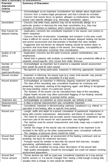
Table 3 Summary of discussions
Potential Assessment Criteria
Summary of Discussion
Allergies - Acknowledged as an important consideration but debate about duplication - Concern from a medico-legal perspective and that it should be included. - Concern that nurses focus on generic allergies to medications rather than wound care specific allergies (e.g. dressings, emollients, tapes ).
Sensitivities - Concern that while many patients may not be technically allergic to a particular product or dressing the may have sensitivities (e.g. they may complain of itchiness or redness) which should be considered.
Risk Factors for delayed healing
- Duplication concerns but considered important in the wound care context to inform treatment.
- Concern that lack of education, knowledge and research in this area could make it difficult for nurses to make the link between delayed healing risk factors and wound assessment and that prompts to facilitate this were needed.
- Suggested that risk factors for delayed healing should be broken down to systemic and local blood supply to the wound, skin integrity, susceptibility to infection and medication to prompt their consideration
Quality of Life (QOL)
- Duplication concerns but the need to ensure patient centeredness was emphasised.
- Noted that in patients with multiple co-morbidities it was sometimes difficult to separate wound-specific QOL issues from wider illnesses.
Number of wounds
- Acknowledged as important but in practice a separate wound assessment form would be used for each wound.
Wound
type/classification
- Recognised as being particularly important in informing appropriate treatment.
Wound Location - Important in informing the wound type (e.g. lower limb wounds may prompt the nurse to consider the possibility of a leg ulcer).
Wound duration - Acknowledged as important in informing ongoing treatment and referrals. - The date of first wounding was considered important for continuity and preventing ‘everybody starting at the beginning again’ and failing to recognised the long-standing nature of a particular wound.
- The duration of the wound can be calculated from date of first wounding. - Digital health records may allow automated flagging of non-healing wounds after specific time periods to prompt further action.
Treatment aim - Acknowledged as important as some wounds are unlikely to heal. Reassessment - A date to prompt reassessment was considered important.
Referrals - Considered important in demonstrating pathway compliance e.g. referral to specialist if non-healing after to specific time period.
Wound size - Length, width and depth of a wound considered of paramount importance in being able monitor improvement, deterioration and failure of wounds to heal. - The need for consistent and accurate wound measurement undertaken at the maximum part of the wound for each parameter was highlighted.
- The method used for wound measurement and body maps was a matter for local policy.
- Overall the shape of the wound was not considered to be important to be included in the MDS as the other measurements were considered sufficient. - The category of pressure ulcers should be noted.
Wound Bed Tissue Type
- Recognised as providing an assessment of the healing continuum and as providing the basis for treatment decisions.
- Prompts would be needed to describe the wound bed (necrotic, sloughy, granulating, epithelialisation, tendon, bone)
- This could be simplified by using colour descriptors of the wound bed.
- High quality photography should be encouraged but could not replace existing assessment parameters.
Wound Bed Tissue amount
14
Potential Assessment Criteria
Summary of Discussion
- Overall the group leaned towards the use of percentage measurement for quantifying tissue amount but noted the need for education and guidance. Description of
wound margins
- Considered important in the ongoing monitoring of the wound particularly relating to the colour and condition of the surrounding skin.
Current overall wound status and healing
- Concern was raised about the subjectivity of the overall wound status item. - Other wound assessment items provide more objective measures of improvement/deterioration.
- Need to record whether a wound had healed as a key outcome.
Pain - Suggested that one leading item was needed about whether they had wound pain or not, which could lead to other items i.e. frequency and severity. - Acknowledged that some areas use specific pain care plans with metrics and there was concern about duplication for some pain items.
- It was also noted that we needed to make it clear that the assessment related to ‘wound pain’ rather than other pain.
Exudate - Overall the ‘exudate amount’ item was considered to provide an objective measure of wound response (which would be informed by dressing performance).
- The item relating to whether exudate was a problem to the patient was considered redundant as exudate was always an issue and is addressed in the other items.
- The current exudate item was not considered an objective measure.
Odour - Recognised odour as a very important symptom, particularly relating to a sign of infection (especially in the presence of increasing exudate and pain), - Also acknowledged as being very subjective measure with a lack of a reliable tool to assist with odour measurement in practice.
- Patient (or carer/family) concerns were the most important consideration. - Suggested that only the presence of odour item was needed in the MDS . Referrals - Considered important in demonstrating pathway compliance e.g. referral to
specialist if non-healing after to specific time period. Pressure Ulcer
Risk Assessment
- Concern about duplication
- Thought to be more relevant to the holistic patient assessment as undertaken on all patient to facilitate prevention.
Specialists - Generic assessment should prompt consideration of a Doppler for wounds on the lower limb (when appropriate), to facilitate a diagnosis and guide
subsequent treatment/referral pathways.
- The appropriateness of undertaking a Doppler should be informed by the holistic assessment of the patient.
- The inclusion of a Doppler could be usefully included to prompt a second tier more specialised assessment of the lower limb. MDS Scope and
Implementation Issues
- Acknowledged that the MDS would be supported by appropriate clinical policies.
- Concerns about duplication between information in the standard holistic patient assessment and MDS.
- Recognised that those with electronic records may be able to pull through information of relevance to the wound assessment from the wider patient assessment.
- Acknowledged that there is great variation in the implementation of electronic records and that the MDS would need to work for both electronic and paper-based systems.
15
4. Further Consultation
The Improving Wound Care Project Board (Figure 1) met to discuss the results and drawing
on the thematic summary (Table 3) it was agreed that an additional post-meeting follow-up
questionnaire would be sent to the expert group to seek clarity on the item with a median of
3.5 (allergies), the item with a median of 8 with disagreement (signs of systemic infection)
and an additional pain item. This led to the inclusion of these items (Table 2). In addition,
the Board agreed that
‘
factors affecting the patient
’
s skin integrity
’
should be recorded in the
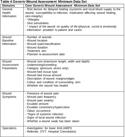
MDS. Table 4 provides a summary of the agreed generic wound assessment MDS
comprising 37 items.
Further consultation about the MDS was also undertaken with the ‘Expert by Experience’
[image:16.596.71.491.312.769.2]Group which was brought together to support the Improving Wound Care Project and
comprised service users and practising District Nurses and Practice Nurses (Figure 1). The
group were supportive of the MDS and particularly about the inclusion of the quality of life
item as they saw this as an opportunity for the patient to express the impact of the wound on
their life and raise any issues that needed to be addressed as part of their treatment plan.
The group also considered the use of photography to be a useful way of monitoring wound
progress.
Table 4 Generic Wound Assessment Minimum Data Set
Domains Core Generic Wound Assessment Minimum Data Set
General Health Information
- Risk factors for delayed healing (systemic and local blood supply to the wound, susceptibility to infection, medication affecting wound healing, skin integrity)
- *Allergies - Skin sensitivities
- * Impact of the wound on quality of life (physical, social & emotional) - Information provided to patient and carers
Wound Baseline Information
- Number of wounds - Wound location
- Wound type/classification - Wound duration
- Treatment aim
- Planned re-assessment date
Wound Assessment Parameters
- Wound size (maximum length, width and depth) - Undermining/tunnelling
- Category (pressure ulcers only) - Wound bed tissue type - Wound bed tissue amount
- Description of wound margins/edges - Colour and condition of surrounding skin - Whether the wound has healed
Wound Symptoms
- Presence of wound pain - Wound pain frequency - Wound pain severity - Exudate amount
- Exudate consistency/type/colour - Odour occurrence
- *Signs of systemic infection - Signs of local wound infection
- Whether a wound swab has been taken
16
* should be recorded in generic wound assessment MDS if not recorded in wider the patient record5.
Discussion
This project comprising a literature review and a consensus study was undertaken to
establish a generic wound assessment MDS. The literature review identified 6 key domains
and 69 sub-domains relating to wound assessment. This underpinned the development of
consensus questionnaires comprising 46 (pre-meeting) and 47 (post-meeting) items that
were considered by an expert group and led to the agreement of 37 items in the generic
wound assessment MDS (Table 4).
In keeping with those who used structured consensus methods in the development of
pressure ulcer risk assessment MDS [35], the method provided a transparent approach to
the development of a generic wound assessment MDS, informed, in this case by a literature
review (rather than a more robust systematic review) and the opinions of experts in the field.
The discussions of the expert group face-to-face meeting allowed challenges and
differences in opinion to be explored and understood. This highlighted the different
approaches to wound assessment and the complexities of standardising this variation across
different systems for patient records including paper-based, electronic and various
combinations of the two. Avoiding duplication between the MDS and the standard holistic
patient assessment was recognised as a challenge given the differing approaches being
used. This led to some flexibility where it was agreed that some items should be recorded in
the MDS if not recorded in wider the patient record e.g. allergies, quality of life. Another
important discussion point of the expert group and expert by experience group related to the
use of photography for wound assessment and monitoring purposes. Due to recognition of
the varying availability of high quality cameras in clinical practice, the use of photography
was not included in the MDS. However, it was recognised as good practice, something
which should be encouraged and could potentially be included in the MDS in the future.
The MDS was developed using a bottom-up manner involving predominantly clinical
practitioners with facilitation by policy makers (NHS England) and academia. This approach
enabled academic scientific theoretical knowledge to be balanced with technical knowledge
and practical wisdom from clinical practice to inform the MDS. The resulting MDS is
therefore more likely to be acceptable and usable in clinical practice. The decision to focus
on a MDS rather than seeking to develop a pre-specified assessment form also allows
flexibility. Many healthcare providers will already use a wound assessment form that
incorporate many or all of the items in the MDS. The MDS will allow review of existing
documentation systems against a set of evidence-based criteria to assess whether
supplementation or simplification is required.
The development of the MDS has sought to address concerns over inadequate wound
assessment practice [7] by providing a framework upon which healthcare provider
organisations can base their assessment documentation. However, the MDS can only
provide a starting point for improving care and measures to encourage implementation are
needed. The MDS will be supported by a guidance manual for practice, sample assessment
forms and a specific CQUIN to monitor progress. It is anticipated that the MDS will facilitate
a more consistent approach to wound assessment potentially leading to improved
subsequent t clinical decision making about wound care treatment, escalation plans,
pathways and patient outcomes. The MDS may lead to the development of large NHS
data-sets that can be used for research and to monitor wound care practice and service
17
The development of the MDS is only one important component of the Improving Wound
Care Project and other work streams including React to Red, Education & Competencies
and Wound Care Commissioning Support (Primary Care & Community Services) all have a
role to play in supporting improvements in wound care practice. Of particular note is the
need for further development of more specialist assessment MDS for specific types of
wounds such as those of the lower limb.
5.1 Limitations
The literature review was undertaken by one reviewer and only incorporated the search of
one database over limited years, with the potential that a more systematic scoping review
may have identified additional relevant studies. However, given that the literature review
aimed to identify potential items for inclusion in the MDS (rather than identifying every paper
that considered wound assessment parameters) and we drew on the collective wisdom of
experts in the wound care field throughout the consensus process, it seems unlikely that any
important aspects of wound assessment would have been missed.
Another area of concern relates to limited service user involvement in this study. While other
consensus studies have incorporated limited numbers of patients/carers in their expert
groups [40, 41], we used a different approach in an effort to avoid under-representation of
service user views [35] and some of the problems associated with reviewing complex
information and facilitating mixed groups of professionals and patients [42]. This involved
consultation with the ‘expert by experience group’ following the consensus process.
Unfortunately despite best efforts, there were difficulties in identifying patients/carers who
were able to join the group and only one service user representative was involved. Increased
numbers of service users may have identified additional important issues to influence the
final MDS. In addition, it could be argued that the involvement of patients and carers earlier
in the consensus process would have facilitated increased integration of their views to shape
the MDS. Involving patients in research of cross
–
speciality problems has been identified as
challenging due to lack of support infrastructure and the complex health needs of potential
participants [42]. The Wound Care Project Board are committed to increasing service user
involvement through a range of methods in the wider programme of work.
6. Conclusion
18
References
1.
Mustoe, T.A., K. O'Shaughnessy, and O. Kloeters, Chronic wound pathogenesis and
current treatment strategies: a unifying hypothesis. Plast Reconstr Surg, 2006. 117(7
Suppl): p. 35s-41s.
2.
Järbrink, K., et al., Prevalence and incidence of chronic wounds and related
complications: a protocol for a systematic review. Systematic Reviews, 2016. 5(1): p.
152.
3.
Werdin, F., et al., Evidence-based Management Strategies for Treatment of Chronic
Wounds. Eplasty, 2009. 9: p. e19.
4.
Mekkes, J.R., et al., Causes, investigation and treatment of leg ulceration. Br J
Dermatol, 2003. 148(3): p. 388-401.
5.
Gottrup, F., et al., A new concept of a multidisciplinary wound healing center and a
national expert function of wound healing. Archives of Surgery, 2001. 136(7): p.
765-772.
6.
Sen, C.K., et al., Human Skin Wounds: A Major and Snowballing Threat to Public
Health and the Economy. Wound repair and regeneration : official publication of the
Wound Healing Society [and] the European Tissue Repair Society, 2009. 17(6): p.
763-771.
7.
Guest, J.F., et al., Health economic burden that wounds impose on the National
Health Service in the UK. BMJ Open, 2015. 5(12).
8.
England, N., CQUIN Indicator 10: Improving the assessment of wounds
N. England, Editor. 2017.
9.
Pham, M.T., et al., A scoping review of scoping reviews: advancing the approach and
enhancing the consistency. Res Synth Methods, 2014. 5(4): p. 371-85.
10.
Barber, S., A clinically relevant wound assessment method to monitor healing
progression. Ostomy Wound Manage, 2008. 54(3): p. 42-9.
11.
Bates-Jensen, B.M., The Pressure Sore Status Tool a few thousand assessments later.
Adv Wound Care, 1997. 10(5): p. 65-73.
12.
Ferrell, B.A., The Sessing Scale for measurement of pressure ulcer healing. Adv
Wound Care, 1997. 10(5): p. 78-80.
13.
Krasner, D., Wound Healing Scale, version 1.0: a proposal. Adv Wound Care, 1997.
10(5): p. 82-5.
14.
NPUAP, PUSH Tool 3.0. National Pressure Ulcer Advisory Panel
(www.npuap.org/resources/educational-and-clinical-resources/push-tool), 1998.
15.
Restrepo Medrano, J.C. and J. Verdú Soriano, Development of a wound healing index
for chronic wounds.
16.
Sussman, C. and G. Swanson, Utility of the Sussman Wound Healing Tool in
predicting wound healing outcomes in physical therapy. Advances in Wound Care.
10(5): p. 74-7.
17.
Woodbury, M.G., et al., Development, validity, reliability, and responsiveness of a
new leg ulcer measurement tool. Adv Skin Wound Care, 2004. 17(4 Pt 1): p. 187-96.
18.
Beitz, J.M. and L. van Rijswijk, A cross-sectional study to validate wound care
algorithms for use by registered nurses. Ostomy Wound Manage, 2010. 56(4): p.
46-59.
19.
Brown, G., Wound documentation: managing risk. Adv Skin Wound Care, 2006.
19(3): p. 155-65, quiz 165-7.
20.
Collier, M., The elements of wound assessment. Nurs Times, 2003. 99(13): p. 48-9.
21.
Doughty, D.B., Four steps to successful chronic wound management. Home Healthc
19
22.
Grey, J.E., S. Enoch, and K.G. Harding, Wound assessment. BMJ, 2006. 332(7536):
p. 285.
23.
Jones, K.R., K. Fennie, and A. Lenihan, Evidence-based management of chronic
wounds. Adv Skin Wound Care, 2007. 20(11): p. 591-600.
24.
Keast, D.H., et al., MEASURE: A proposed assessment framework for developing best
practice recommendations for wound assessment. Wound Repair & Regeneration,
2004. 12(3 Suppl): p. S1-17.
25.
Schultz, G.S., et al., Wound bed preparation: a systematic approach to wound
management. Wound Repair Regen, 2003. 11 Suppl 1: p. S1-28.
26.
Hess, C.T., The art of skin and wound care documentation. Home Healthc Nurse,
2005. 23(8): p. 502-13; quiz, 514-5.
27.
Stotts, N.A. and P.S. Sparacino, Assessing the patient with a wound as the basis for
home care wound management. Home Healthc Nurse, 2005. 23(2): p. 82-92; quiz
93-4.
28.
Morrison, M., A framework for patient assessment and care planning, in Chronic
Wound Care: A problem-based learning approach, M. Morison, L. Ovington, and K.
Wilkie, Editors. 2004,
mosby: China.
29.
Cullum, N., et al., in Wounds research for patient benefit: a 5-year programme of
research. 2016: Southampton (UK).
30.
Pokorna, A. and D. Leaper, Assessment and documentation of non-healing, chronic
wounds in inpatient health care facilities in the Czech Republic: an evaluation study.
Int Wound J, 2015. 12(2): p. 224-31.
31.
Lait, M.E. and L.N. Smith, Wound management: a literature review. J Clin Nurs,
1998. 7(1): p. 11-7.
32.
Pillen, H., et al., Assessment of wound healing: validity, reliability and sensitivity of
available
instruments. Wound Practice and Research, 2008. 17(4): p. 208-217.
33.
Stewart, S., et al., " Measurement Monday" : one facility's approach to standardizing
skin impairment documentation. Ostomy Wound Management, 2009. 55(12): p.
49-54.
34.
Fitch, K., et al.,
The RAND/UCLA Appropriateness Method User’s Manual
. 2001,
RAND Corporation: Santa Monica CA.
35.
Coleman, S., et al., Developing a pressure ulcer risk factor minimum data set and risk
assessment framework. Journal of Advanced Nursing, 2014. 70(10): p. 2339-2352.
36.
Hutchings, A. and R. Raine, A systematic review of factors affecting the judgments
produced by formal consensus development methods in health care. Journal of Health
Services & Research Policy, 2006. 11(3): p. 172-9.
37.
Murphy, M.K., et al., Consensus development methods, and their use in clinical
guideline development. Health Technol Assess, 1998. 2(3): p. i-iv, 1-88.
38.
Hsieh, H.F. and S.E. Shannon, Three approaches to qualitative content analysis.
Qualitative Health Research, 2005. 15(9): p. 1277-88.
39.
Coleman, S., et al., Patient risk factors for pressure ulcer development: Systematic
review. International Journal of Nursing Studies, 2013. 50(7): p. 974-1003.
40.
Rycroft-Malone, J., Formal consensus: the development of a national clinical
guideline. Qual Health Care, 2001. 10(4): p. 238-44.
41.
Jackson, A., et al., Using consensus methods in developing clinical guidelines for
exercise in managing persistent low back pain. Physiotherapy, 2009. 95(4): p. 302-11.
42.
Nixon, J., et al., Pressure UlceR Programme Of reSEarch (PURPOSE): using mixed
21
Appendix 1
–
Detailed Wound assessment sub-domains included in papers of literature review
Dom ain Sub-domain Barber
2008 Bat es -J ens en 1997 Beit z & R ijs w ijk 2010 Brow n 2006 C ollier 2003 C ullum et al
2016 Dought
y 2004 F errell 1997 Grey et al
2006 Hes
s 2005 J ones et al 2006 Keas t et al 2004 Kras ner 1997 Lait & Sm it h
1997 NPU
AP 1998 M orris on et al
2004 Pork
orna & Leaper 2014 R es treppo -M edrano & Soriano 2010 St ew art et al 2009 Sc hult z et al 2003 St ot ts & Sparac ino 2005 Sus s m an & Sw ans on 1997 W oodbury et al 2004 T ot al General health
information Allergies 1 1 1 3
Mobility Status 1 1 2
Risk Factors for Delayed Healing
Factors affecting blood supply to w ound (e.g. vascular or arterial disease, smoking anaemia)
1 1 1 1 1 1 1 1 1 1 10
Factors affecting local blood supply to w ound (e.g. pressure/shear,
pressure ulcer) 1 1 1 1 1 1 1 1 8
Factors affecting skin (e.g. Malnutrition, Obesity, peripheral neuropathy)
1 1 1 1 1 1 1 1 1 1 10
Susceptibility to infection (e.g.
immune-supressed) 1 1 1 1 1 1 1 1 1 1 10
Medication affecting w ound healing (e.g. steroids,
chemotherapy) 1 1 1 1 1 1 1 1 1 9
Generic delayed
healing (non-specific) 1 1 2
Wound history/ baseline
information Number of w ounds 1 1 1 1 1 1 6
Wound Location 1 1 1 1 1 1 1 1 1 1 1 1 1 1 14
Wound
type/classification (e,g, venous leg ulcer,
pressure ulcer) 1 1 1 1 1 1 1 1 1 1 1 1 1 1 1 1 16
Wound Duration (e.g.
22
Dom ain Sub-domain Barber
2008 Bat es -J ens en 1997 Beit z & R ijs w ijk 2010 Brow n 2006 C ollier 2003 C ullum et al
2016 Dought
y 2004 F errell 1997 Grey et al
2006 Hes
s 2005 J ones et al 2006 Keas t et al 2004 Kras ner 1997 Lait & Sm it h
1997 NPU
AP 1998 M orris on et al
2004 Pork
orna & Leaper 2014 R es treppo -M edrano & Soriano 2010 St ew art et al 2009 Sc hult z et al 2003 St ot ts & Sparac ino 2005 Sus s m an & Sw ans on 1997 W oodbury et al 2004 T ot al Wound
Assessment Wound size w idth 1 1 1 1 1 1 1 1 1 1 1 1 1 1 1 1 1 1 1 19
Wound size length 1 1 1 1 1 1 1 1 1 1 1 1 1 1 1 1 1 1 1 19
Wound size
(non-specific) 1 1
Wounds size depth 1 1 1 1 1 1 1 1 1 1 1 1 1 1 1 1 1 1 1 1 1 21
Undermining/tunnelling 1 1 1 1 1 1 1 1 1 1 1 1 1 1 1 1 16
Shape 1 1 1 3
Wound Bed tissue type
(e.g necrotic, sloughy) 1 1 1 1 1 1 1 1 1 1 1 1 1 1 1 1 1 1 1 1 20
Wound Bed tissue amount (e.g.
percentage of w ound) 1 1 1 1 1 1 1 1 1 1 1 11
Wound bed (non-
specific) 1 1
Wound margins/edges description (e.g. epithelialisation
unattached) 1 1 1 1 1 1 1 1 1 1 1 1 1 1 1 1 1 17
Wound margins/edges
(non-specific) 1
Surrounding skin
colour (e.g. redness) 1 1 1 1 1 1 1 1 1 1 1 1 1 1 1 1 1 17
Surrounding skin condition (e.g. maceration, oedema,
induration) 1 1 1 1 1 1 1 1 1 1 1 1 1 1 1 1 16
Surrounding skin
(non-specific) 1 1 2
Current Wound Status (e.g.
progress/deterioration) 1 1 1 1 1 1 1 1 1 1 1 1 12
Wound sym ptoms
Pain Frequency (e.g. at
dressing change) 1 1 1 1 1 1 1 1 1 1 1 11
Pain Severity 1 1 1 1 1 1 1 1 1 1 1 11
Pain (non-specific) 1 1 1 1 4
type (i.e. inflammatory
neuropathic) 1 1 2
23
Dom ain Sub-domain Barber
2008 Bat es -J ens en 1997 Beit z & R ijs w ijk 2010 Brow n 2006 C ollier 2003 C ullum et al
2016 Dought
y 2004 F errell 1997 Grey et al
2006 Hes
s 2005 J ones et al 2006 Keas t et al 2004 Kras ner 1997 Lait & Sm it h
1997 NPU
AP 1998 M orris on et al
2004 Pork
orna & Leaper 2014 R es treppo -M edrano & Soriano 2010 St ew art et al 2009 Sc hult z et al 2003 St ot ts & Sparac ino 2005 Sus s m an & Sw ans on 1997 W oodbury et al 2004 T ot al
Current pain status
(progress/change) 1 1 1 1 4
Exudate amount (e.g.
high, moderate) 1 1 1 1 1 1 1 1 1 1 1 1 1 1 1 1 1 1 18
Exudate
consistency/type/colour (e.g. serous, blood, sero-sanguineous,
thick, thin) 1 1 1 1 1 1 1 1 1 1 1 1 1 1 1 1 16
Exudate (non-specific) 1
Current exudate status
(progress/deterioration) 1 1 1 1 1 5
Exudate problem to
patient 1 1
The performance of the current dressing in absorbing w ound
exudate 1 1 1 1 1 1 1 1 1 1 10
Odour occurrence (e.g. on dressing removal,
w hen dressing intact) 1 1 1 1 1 1 1 1 1 9
Odour intensity (e.g. acceptable
minimal, problem) 1 1 1 1 4
Odour (non-specific) 1 1 2
Current odour status
(progress/ changes) 1 1 1 1 1 5
Odour problem to
patient 1 1
Infection
Signs of local w ound infection (e.g. cellulitis; Abscess/pus; Increasing pain, exudate, odour; deterioration (w ound breakdow n and dehiscence); healing slow er than anticipated; friable granulation tissue; bleeds easily pocketing at w ound
24
Dom ain Sub-domain Barber
2008 Bat es -J ens en 1997 Beit z & R ijs w ijk 2010 Brow n 2006 C ollier 2003 C ullum et al
2016 Dought
y 2004 F errell 1997 Grey et al
2006 Hes
s 2005 J ones et al 2006 Keas t et al 2004 Kras ner 1997 Lait & Sm it h
1997 NPU
AP 1998 M orris on et al
2004 Pork
orna & Leaper 2014 R es treppo -M edrano & Soriano 2010 St ew art et al 2009 Sc hult z et al 2003 St ot ts & Sparac ino 2005 Sus s m an & Sw ans on 1997 W oodbury et al 2004 T ot al
Signs of systemic infection relating to the
w ound (e.g. high temp) 1 1 1 1 4
Management of
infection 1 1 1 1 1 1 6
Referrals Specialist Referrals 0
Quality of Life Patient information 1 1 2
Physical - frequency of dressing changes 1 1
Physical - fatigue/lack of sleep 1 1
Social - pain 1 1
Social- odour 1 1
Physical - reduced mobility 1 1 2
Physical - health and w ellbeing 1 1 2
Physical - altered eating habits 1 1
Social - social isolation 1 1 2
Emotional- depression 1 1
Emotional - emotions 1 1 2
Physical - daily activities 1 1 2
Social - friendships 1 1 2
Social - hobbies 1 1
Non-specific 1 1 Pressure Ulcer specific Elem ents of Assessment Pressure ulcer risk assessment 1 1 1 1 1 1 1 1 8