PROSPECTIVE EVALUATION OF VOCAL CORD FUNCTION
WITH INTRA-OPERATIVE NERVE IDENTIFICATION IN
TOTAL THYROIDECTOMY PATIENTS
Institution
DEPARTMENT OF ENDOCRINE SURGERY
MADRAS MEDICAL COLLEGE
CHENNAI
Dissertation submitted in partial fulfilment of
BRANCH IX – M.CH ENDOCRINE SURGERY
EXAMINATION
August 2013
The Tamilnadu Dr. M.G.R Medical University
Acknowledgement
I am grateful to Prof. V. Kanagasabai, Dean, Madras Medical College, for
permitting me to conduct this study.
I would be failing in my duty if I didn’t acknowledge the keen interest shown
and guidance imparted by Prof. M. Chandrasekaran, Head of the Dept.
(Retd.), Department of Endocrine Surgery, Madras Medical College during the
course of this study.
I thank Prof. S. Deivanayagam, Head, Department of Endocrine Surgery in all
earnest for his guidance.
I wish to express my heartfelt gratitude to Dr. S. Zahir Hussain and Dr. S.
Dhalapathy, Asst. Professors, Department of Endocrine Surgery, Madras
Medical College, for their invaluable advice on intellectual aspects as well as
technical details and their emotional inputs throughout the course of this study.
I would also like to thank Prof. V. Sucharita, Associate Professor (Retd.),
Department of Endocrine Surgery, Madras Medical College, for the insightful
It is with utmost pleasure that I take the names of Dr. Himagirish. K. Rao, Dr.
Uma Devi, Dr. Sai Vishnu Priya, Dr. Poongkodi, Dr. Senthil Kumar, Dr.
Mohana Priya and indeed, all the staff members of this department, without
whose help this study wouldn’t have been possible.
I am eternally grateful to my wife, Dr K. Sasirekha and my family for the
unwavering, invaluable emotional support.
Last, but definitely not the least, I wish to express my sincere, heartfelt thanks
to all the patients for their enthusiastic participation and diligent co-operation
during the course of treatment and follow-up.
MADRAS MEDICAL COLLEGE
CHENNAI
CERTIFICATE
This is to certify that this dissertation on ‘Prospective evaluation of vocal cord
function with intra-operative nerve identification in total thyroidectomy
patients’ is a bonafide dissertation by Dr. M. P. Kumaran conducted in Madras
Medical College under the supervision and guidance of and is submitted to The
TamilNadu Dr. M. G. R. Medical University, Chennai in partial fulfilment of
the requirement for the M.Ch (Endocrine Surgery) degree.
Date: Professor and Head
Department of Endocrine Surgery
Date: Dean
CONTENTS
1.
Introduction and Objectives
1
2.
Review of literature
3
2.1
Anatomy of the larynx5
2.2
Neurophysiology of the larynx and pathophysiology of vocal cordpalsy
13
2.3
Vocal cord palsy20
2.4
Treatment options for vocal cord palsy27
3.
Patients and Methods32
4.
Results36
5.
Discussion46
6.
Conclusion51
1
The recurrent laryngeal nerve and the external branch of the superior
laryngeal nerve are at risk of injury during total thyroidectomy. This may
result in change of voice post-operatively. The recurrent laryngeal nerve is at
particular risk due to its proximity to the inferior thyroid artery and the
ligament of Berry. Injury to the recurrent laryngeal nerve may result in
dreaded complications like stridor, loss of voice sometimes necessitating
tracheostomy as a life saving procedure.
One’s voice is one of the integral aspects of one’s identity. Change of
voice, to any degree, is not acceptable regardless of whether one is
dependent on voice for one’s profession. Post operative voice change is a
major concern for the patients undergoing total thyroidectomy as well as for
surgeons performing the surgery. This could be mild or it may be severe.
While it is often transient, it could be permanent as well.
In order to analyze voice change after surgery, an objective method of
assessment of the patient’s voice is necessary, which depends on reliable and
accurate feedback from the patients. This necessitates a questionnaire, which
can be administered to the patients in question. In this respect, compliance of
2
which is easy to understand. In addition, the questionnaire should be so
constructed as to enable objective assessment of severity of the symptoms.
This can be achieved by permitting the patients to grade each voice-related
symptom on a simple scale on the basis of severity. Validation of the
questionnaire affords credibility to this method of assessment.
Direct laryngoscopy, either with the naked eye, or with the help of a
fibre-optic video-laryngoscope will enable objective assessment and
documentation of vocal cord movement and function. By this method, vocal
cord palsy, which occurs due to RLN injury, during thyroid surgery can be
assessed objectively and documented.
With this background, this study was conducted in the Department of
Endocrine Surgery, Madras Medical College with the following objectives.
1. Objective evaluation of voice and vocal cord status pre-operatively,
with the help of a validated voice assessment score (VAS) and
videolaryngoscopy (VLS), respectively.
2. Documentation of identification of the external branch of superior
laryngeal nerve (EBSLN) and the recurrent laryngeal nerve (RLN)
3
3. Assessment of voice and vocal cord status post-operatively, on the 5th
post-operative day and again on the 180th post-operative day with the
help of VAS and VLS respectively.
4
REVIEW OF LITERATURE
Voice change in the post-operative period is one of the most bothersome
complications in thyroid surgery, both for the patient and the operating
surgeon. Regardless of the degree of surgical expertise, voice change after
thyroid surgery is sometimes unavoidable, especially after difficult surgery.
Although a significant number of patients undergoing thyroidectomy
experience some change in voice after surgery, it is transient in the large
majority.
There are various factors which influence voice change, including
innocuous causes like acute upper respiratory infection including laryngitis,
upper airway edema as a result of trauma during intubation, arytenoids
dislocation and injury to the recurrent laryngeal nerve (RLN). In a small
minority of subjects (0.3%), idiopathic vocal cord palsy is present
pre-operatively as well [1].
Unilateral RLN injury will result in unilateral vocal cord palsy, which
can present with post-operative aspiration and hoarseness of voice. Bilateral
life-5
threatening stridor in the immediate post-operative period, often
immediately after extubation.
In order to minimize the rate of post-operative voice change,
understanding voice production apparatus and process, including anatomy
of the larynx, physiology and neurophysiology of voice production and the
anatomy of the recurrent laryngeal nerves and their relation to the thyroid
gland and the surrounding structures is essential. In addition to these, it is
important to know about the patterns of clinical presentation of
post-operative voice change, the methods of voice assessment and the methods of
objective evaluation of vocal cord function. Knowledge of the natural
history of events following surgery with regard to voice, including the
6
2.1ANATOMY OF THE LARYNX
The larynx is made up of a cartilaginous skeleton with many
ligaments connecting the various cartilages, which provide attachment to
various intrinsic and extrinsic muscles of larynx.
2.1.1 Cartilages of larynx
The larynx has paired as well as unpaired cartilages making up the
skeleton. The thyroid, the cricoid and the epiglottic cartilages are the
unpaired cartilages, aligned in the median plane. The arytenoid, cuneiform
and the corniculate cartilages are the paired cartilages of the laryngeal
framework. In addition, the hyoid bone provides support to the main
framework of the larynx, although it is not a part of the larynx itself. A
schematic representation of the laryngeal framework is included in Figures
7
2.1.2 Muscles of the larynx
The intrinsic muscles of the larynx, which are present entirely within
the laryngeal framework, are primarily involved in movements of the vocal
and vestibular folds. The intrinsic muscles of the larynx include the posterior
cricoarytenoid, the lateral cricoarytenoids, the transverse arytenoids or the
inter-arytenoids, the oblique arytenoids, the thyro-arytenoids, the vocalis and
the crico-thyroid. The vocalis and the thyro-arytenoids are the muscles of the
true vocal folds, while the crico-arytenoids, the inter-arytenoids and the
crico-thyroid muscles attach the cricoids, thyroid and arytenoids cartilages.
The majority of the laryngeal muscles are adductors of the vocal cords,
while the posterior cricoarytenoids are believed to be the main abductors of
the glottis aperture. The various muscles and their actions are listed in Box 1
overleaf. Their pictorial representation is as shown in Figures 4, 5 and 6.
2.1.3 The laryngeal lumen
The vocal and the vestibular folds are the most conspicuous structures
in the laryngeal lumen, which extends from the epiglottic aperture above to
pseudo-8
stratified ciliated columnar (respiratory) epithelium. The vocal folds are the
true cords, while the vestibular folds are the false vocal cords.
Box 1: Intrinsic laryngeal muscles.
Muscle and innervation Action
Posterior cricoarytenoid (RLN) Only abductor of the vocal cords [NS – RLN]
Abduction of vocal cords
Open glottis by rotation of arytenoids on the cricoid
Tenses cords during phonation
Lateral cricoarytenoid [NS – RLN]
Close glottis by rotating arytenoids medially
Transverse arytenoid / Inter-arytenoid [NS – RLN]
Close glottis
Adduction of arytenoids – slide medially towards each other on the cricoid
Oblique arytenoid [NS – RLN]
Close glottis
Synchronous with transverse arytenoid Closure of the glottic aperture especially during swallowing
Thyroarytenoid
[Wide, fan-like muscle; 3 functional parts
Thyroarytenoidus internus / Vocalis Thyroarytenoidus externus
Thyroepiglotticus]
Adductor, tensor of free edge of vocal cord Major adductor of vocal folds
Shortens vocal ligaments
Cricothyroid [NS – EBSLN]
9
2.1.4 Anatomy of the recurrent laryngeal nerve
The recurrent laryngeal nerve (RLN), a branch from the vagus, is
made up of myelinated and unmyelinated axons containing 1200 myelinated
axons and thousands of unmyelinated axons [1]. It provides both sensory as
well as motor innervations to the larynx.
The Vagus nerve contains three nuclei in the medulla oblongata - the
Dorsal nucleus of vagus nerve, the Nucleus Ambiguus and the Nucleus of
Tractus Solitarius. The dorsal nucleus is the parasympathetic nucleus, which
innervates the heart, the involuntary muscles of the bronchi and the
esophagus and the organs of the gastro-intestinal tract including the
stomach, small bowel and parts of the large bowel. The nucleus of Tractus
Solitarius is sensory to the pharynx, the larynx and the esophagus. The
Nucleus Ambiguus, which is the motor nucleus of the Vagus, supplies the
muscles of pharynx, larynx, hypo-pharynx and soft palate. So the RLN
contains fibres from the Nucleus Ambiguus and the Nucleus of Tractus
Solitarius.
Once the vagus nerve emerges from the medulla the recurrent
10
jugular foramen on each side. There are a number of fascicles in the vagus
nerve within the jugular foramen [2]. The nodose ganglion, which is present
just caudal to the jugular foramen, gives off nerves that contribute to the
pharyngeal plexus. The superior laryngeal nerve is given off from the vagus
at this ganglion.
The vagus traverses the neck within the carotid sheath. The recurrent
laryngeal nerves on either side are given off at different levels.
The right RLN is given off at the thoracic inlet at the point where the
vagus passes anterior to the sub-clavian artery. The RLN loops around the
sub-clavian artery, passing upwards and medially in relation to the pleura,
behind the common carotid artery. In the neck, it is found in the
para-tracheal areolar tissue, related to the postero-lateral aspect of the thyroid
lobe as it passes upwards, ultimately approaching the trachea-esophageal
groove as it exits the para-tracheal space and enters into the larynx at the
crico-thyroid junction.
In about 1% of the subjects, this nerve emerges directly from the
11
recurrent. This may be associated with anomalous right subclavian artery
[3].
On the left side, the vagus follows the carotid artery into the
mediastinum where it lies in relation to the aortic arch [4]. The RLN is given
off at the lower border of the aortic arch, at the level of the ligamentum
arteriosum. It loops around the aortic arch and ascends behind it to approach
the trachea-esophageal groove. It ascends in this groove to enter the neck,
which it traverses in the above-said groove, postero-medial to the thyroid
lobe. It exits the paratracheal space as it enters the larynx at the crico-thyroid
junction.
On the right side, the RLN is approximately 5-6 cm long. It is much
longer on the left side, with an approximate length of 10-12 cm [5].
The relationship of the RLN to the inferior thyroid artery (ITA) and
its branches is significant [3]. Ligation of the inferior thyroid artery is an
essential step during thyroid surgery and the RLN is at risk of injury by
virtue of its relation to the ITA and its branches.
The left RLN passes postero-medial to the main twig of the ITA in
12
The RLN passes in front of the left ITA in 11%-12% of the population,
while it is so on the right side in 26%-33%. The RLN passes in between the
branches of the ITA in 33% on the left side and in 50% of the subjects on
the right side [3].
The RLN enters the larynx deep to the inferior constrictor muscle and
posterior to the crico-thyroid joint. Traditionally it is believed that the RLN
divides into motor and sensory branches after entering the larynx. However,
extra-laryngeal branches of the RLN are seen as well, in about 35% - 80% of
the patients [6]. The motor branch, which is directed anteriorly, is made up
of 500 – 1000 axons. The posterior cricoarytenoid muscle is supplied by a
quarter of these.
The inferior thyroid artery supplies blood to the proximal RLN, while
the distal, intra-laryngeal part of the RLN derives its blood supply from the
inferior laryngeal artery.
A pictorial representation of the anatomy of the RLN, the EBSLN and
13
2.2 NEUROPHYSIOLOGY OF LARYNX AND PATHOPHYSIOLOGY OF VOCAL
CORD PALSY
2.2.1 Neurophysiology of larynx
The primary functions of the larynx include protection of the
tracheo-bronchial airway and vocalization, in addition to aiding the process of
ventilation. When a person breathes silently, the glottic aperture is open,
while it is closed during phonation or deglutition. The epiglottis, which is
oriented in the form of a trapdoor, hinges down to close the airway during
deglutition, while the epiglottic aperture is open during phonation.
The movement of the vocal folds, which is integral to the opening and
closing of glottic aperture, is controlled by two sets of mutually antagonistic
muscles. When the glottic aperture is open, the abductors of the vocal cords,
the posterior cricoarytenoids, are contracted. Simultaneously, the adductors
relax to facilitate smooth, synchronous vocal cord movement. When the
glottis is closed, the adductors contract while the abductors relax. Tensing of
the vocal folds, which is necessary for appropriate vocalization and
14
muscles. The cricothyroid muscles are in contraction during abduction as
well as adduction of the cords [7].
The RLN and the internal branch of the SLN are responsible for
sensory innervation of the laryngeal mucosa. The IBSLN supplies the
supra-glottic larynx, while the RLN supplies the supra-glottic aperture and infra-supra-glottic
larynx. Occasionally, there is cross-innervation of the sensory supply via the
anastomosis of Galen, which is a communicating nerve between the RLN
and the SLN.
Recently, several articles have been published that have revealed that
each laryngeal muscle is not a single entity but rather an assembly of
anatomically distinct compartments adapted for different functions. The
intrinsic muscles, viz. the posterior cricoarytenoids, the thyro-arytenoids and
the cricothyroid muscles, can be grouped into two to three functional
compartments on the basis of fascial barriers and differences in the direction
of orientation and the site of insertion of muscle fibres [7]. Muscle
biochemistry studies support this theory of functional compartmentalization
of the laryngeal muscles. The compartments of muscle receive innervations
15
Han, et al. have demonstrated that the horizontal division of posterior
cricoarytenoid muscle has a greater proportion of slow-twitch (type 1)
fibres, as does the vocalis, which is the supero-medial division of the
thyro-arytenoid muscle. In general, these slow-twitch fibres are adapted for tonic
functions, like quiet respiration or vocalization [8].
The vocalis muscle, with its slow fibre composition, multilobar
construction and muscle density, is suited for the accurate generation of
sound used in communication. Bei-Lian, et al have reported that in some
cases, the vocalis may receive additional innervation via the EBSLN, apart
from the regular innervation through the RLN [9].
The idea of slow and fast motor units is fundamental to the laryngeal
behavior. With many nerve branches and anostomotic channels, which
terminate in plexuses, the innervation of larynx is intricate and complex.
Another aspect of vocal fold physiology which is integral to voice
production is the phenomenon of the mucosal wave. This refers to the
movement of the vocal fold in three dimensions. Movement of the cords
along a two-dimesnional, seemingly axial plane is evident by laryngoscopy
16
vocal cords move in three diemensions. Apart from abduction and
adduction, the vocal folds move upwards and downwards as well, along the
cranio-caudal axis.
In addition, rippling of the mucosa on the vocal cords is appreciable
by strobo-videolaryngoscopy. The mucosal wave refers to this rippling
movement, much akin to the ripples of waves that can be seen on the ocean
from a beach. Like fingerprints and lip prints, the mucosal wave is unique to
a particular individual, conferring identity to one’s voice [1]. Alteration of
the architecture of the vocal cords will result in alteration of the mucosal
wave and consequent voice change.
2.2.2 Mechanism of voice production
Phonatory signals generated in the motor cortex proceed via bilateral
brainstem nuclei (Nucleus Ambiguus) and through vagus nerves and their
branches, the RLN and the SLN, on either side to reach the larynx. They
terminate in the motor end plate of the intrinsic laryngeal muscles, resulting
17
voice production can be accomplished in 90ms, it needs close coordination
of the respiratory musculature [10].
Lalwani, et al. have described voice as a product of the semicyclical
vibrations of the vocal cords [10]. Normal voice emanates from oscillation
of the vocal cord mucosa as it moves relative to the underlying vocal cord
musculature and the laryngeal skeleton. This cordal vibration is controlled
by vocal cord muscular tension, the mucosal wave and other elastic vocal
cord properties and aerodynamic forces of sub-glottic air as it passes through
the relative constriction of the partially closed glottis. Cordal vibration is
generated as the air expelled under pressure from the lung passes between
the vocal cords and sets cords into oscillatory motion.
Vibration of the vocal cord is age and gender dependent. In addition
to intrinsic muscles extrinsic muscles of the larynx are subject to contraction
during phonation, singing, respiration, yawning and during swallowing.
These extrinsic muscles make vertical laryngeal motion during the
contraction. When this vertical movement is affected voice production may
18
2.2.3 Mechanisms of nerve damage and regeneration
The various mechanisms of nerve injury that result in vocal cord
paralysis include neuropraxia, axonotmesis and neurotmesis.
Neuropraxia refers to the loss of signal conduction as a result of
demyelination without disruption of axons. Functional recovery occurs
within days to months as a result of remyelination of the Schwann cell that
lines these axons.
Axonotmesis refers to mechanical disruption, or transection, of the
involved axons. Regeneration occurs in due course, after a period of weeks
to months. It involves regeneration of the Schwann cells, which form the
neural tubes into which the regenerating axons grow. Thus, structural
continuity is established and is followed by recovery of nerve transmission.
Neurotmesis refers to mechanical disruption, transection or avulsion
of the nerve or a part of it. After the injury, reinnervation may be
inappropriate, inadequate or nonexistent. Inapprorpirate regeneration may
result in the regenerating axons entering the endoneural sheaths randomly,
thus resulting in cross-innervation, akin to that seen in Frey’s syndrome after
19
Dysfunctional reinnervation may result in simultaneous contraction of
corresponding, mutually antagonistic groups of muscle fibres, resulting in
dysfunctional cord mobility. This phenomenon is known as synkinesis [7].
Synkinetic contraction is a feature of chronic, vocal cord paralysis as a result
of irreversible RLN injury.
In cases of extensive nerve injury, axonal and neural regeneration may
be completely impeded by formation of neuromas at the cut ends of the
nerve [7].
It is remarkable, though, that reinnervation of laryngeal muscles can
and does occur in face of severe RLN injury. In their study, Crumly and
McCabe [11] reported regeneration of the RLN after resection of a segment
of the nerve 2.5cm in length and ligation of the cut ends. Reinnervation of
PCA muscle was reported [12] even after resection of 10cm of the nerve and
ligation of the stumps. The proximal stump was reported to be the source of
20
2.3VOCAL CORD PALSY
2.3.1 Epidemiology and Etiology
The exact incidence of RLN paralysis is difficult to estimate, since a
significant proportion of patients - as many as 30% to 50% of them - with
vocal cord paralysis may be asymptomatic [13]. Idiopathic RLN paralysis is
one situation where the exact etiology for nerve paralysis is not known and
its incidence can vary. According to Yumoto, et al. [14], the incidence of
idiopathic RLN palsy in the population is between1.5% and 14%. According
to some other reports, it was between 25.9% and 41.3%. The incidence of
RLN paralysis is more common in the elderly, due increased incidence of
malignancy, increased fragility and decreased potential for recovery.
While the RLN has a longer course on the left side, it is more
obliquely oriented within the neck on the right side. So, the right RLN is
more prone to injury in the neck when compared to the left RLN, but
overall, the incidence of RLN injury is more on the left side. Pathological
processes within the mediastinum including carcinoma of the lung,
carcinoma of the esophagus, lymphoma, tuberculosis, sarcoidosis, silicosis
21
Operations on the cervical spine through the anterior approach,
carotid endarterectomy, thyroid surgery and skull base surgery are some of
the common operations responsible for RLN paralysis. Thyroid surgery
remains most common operation associated with RLN paralysis.
Vocal cord palsy associated with endotracheal intubation, which
sometimes results from dislocation of the arytenoid cartilages, can occur due
to compression of the nerve between the thyroid cartilage and arytenoids as
well [15]. It is important to distinguish these two causes of
intubation-associated vocal cord palsy.
Injury to the recurrent laryngeal nerves and the vagus nerves can
occur anywhere along its course. Surgical and traumatic causes apart, certain
other causes like vascular insults, viruses, bacterial infections and neurotoxic
drugs have been implicated.
Nerve injury during surgery can be due to thermal injury, stretch,
cutting, compression and vascular compromise. Surgically induced recurrent
laryngeal nerve injury may not be recognized at the time of injury. The
potential for recovery is generally proportional to the degree of injury.
22
Slow growing tumors that infiltrate the nerve generally allow
compensation for paralysis and even with immobile fold symptoms may not
be evident. The recovery time for RLN paralysis after carotid
endarterectomy, anterior surgical approach to cervical spine and
thyroidectomy with benign pathologic findings is shorter than for that after
skull base surgery and thoracic surgery [15]. The latter, which are mainly
performed in the setting of malignancy, require extensive dissection and so
are associated with a more severe degree of injury when compared to the
former group.
2.3.2 Laryngoscopy
Inspection of the vocal cords with the help of laryngoscopy is
generally performed before, as well as after surgery. Pre-operative
evaluation will enable confirmation of the vocal cord status. Vocal cord
paralysis that may be present pre-operatively, can be confirmed. This will
aid in determining the disease load and in evaluating the therapeutic options
in case of thyroid malignancy. In the post-operative setting, laryngoscopy
23
optic laryngoscopy is a simple and well tolerated office procedure for
surgical patients.
The appearance of the larynx should be symmetrical. During silent
respiration, the glottic aperture assumes a triangular configuration, with the
base directed posteriorly. In health, the triangle should be symmetrical.
During phonation, the glottic aperture is closed and assumes a slit-like
configuration in the median plane.
Unilateral vocal cord paralysis is visualized as thick, short, immobile
vocal fold, associated with anterior prolapse of the ipsilateral arytenoid
cartilage. In addition, the affected cord is displaced inferiorly so that during
phonation, the mucosal edges of the two vocal cords don’t appose each
other. As a result, the voice is breathy and vocal fatigue occurs[1].
When both the cords are affected, as in bilateral RLN injury, both
cords are immobile and oriented in the paramedian plane. Abduction, if
possible at all, is very minimal. These patients often present with serviceable
voice but with signs of upper airway obstruction. Not infrequently, the
presentation is dramatic with stridor. Unless immediately managed, this
24
Unilateral EBSLN injury, in the setting of which the vocal range and
pitch is limited, may be observed as a rotation of the posterior larynx
towards the side of the injury[16]. The affected cord is bowed and somewhat
caudal with respect to that on the unaffected side.
Preoperative vocal cord paralysis was found in 70% of patients with
invasive disease versus 0.3% of patients with benign thyroid lesions[15].
Preoperative vocal cord paralysis may be completely asymptomatic.
Knowledge of preoperative paralysis is helpful in evaluating treatment
options. For instance, in the setting of preoperative unilateral vocal cord
palsy, when contra-lateral surgery is planned, tracheostomy may be
necessary and this possibility has to be considered.
2.3.3 Voice assessment in patients undergoing thyroid surgery
The incidence of voice change in the immediate post-operative period
after thyroid surgery is 30% [17]. Changes in voice character have been
documented in the absence of laryngeal nerve injury as many as 30% of
cases [17]. Lasting functional voice change after thyroid surgery has been
25
factors that influence post-operative voice change, apart from nerve injury
[1].
The patterns of lymphatic drainage, blood supply and venous drainage
of the larynx may be altered after surgery. The strap muscles, including the
sternohyoid and the sternothyroid, may be adherent to the laryngeal
framework, as a result of which the vertical movement of the larynx may be
restricted. The mucosa overlying the cords may be injured or rendered
edematous after endo-tracheal intubation. The cricothyroids, which are
intimately related to the supero-medial border of the thyroid lobe, may be
injured during dissection. In addition, although the thyroid bed may be
structurally intact, movement may be splinted in the immediate
post-operative period due to post-post-operative pain. Psychological aspects like pain
threshold and pain perception may also influence post-operative voice.
Some of the most common voice-related symptoms after thyroid
surgery are vocal fatigue, hoarseness and decreased voice stamina.
The voice symptoms in the immediate post operative period of total
thyroidectomy patients usually are temporary. In the absence of nerve injury,
26
In a study by Watt-Boolsen, et al. [18], voice-related symptoms
improved significantly after thyroid surgery. This was probably due to
alleviation of mass effect of the goitre and improvement of thyroid status
27
2.4TREATMENT OPTIONS FOR VOCAL CORD PALSY
As a result of glottic incompetence, the various functions of the
larynx, including airway protection, breathing and voice production are
impaired. The primary concern for the patient is dysphonia. The presentation
of dysphonia is varied. Patients complain of various symptoms including
insufficient loudness, vocal fatigue, globus sensation, hoarseness, impaired
singing quality, breathlessness on speaking and laryngospasm. A wide
spectrum of treatment options are available, ranging from voice therapy to
injection laryngoplasty, medialization laryngoplasty and laryngeal
reinnervation.
2.4.1 Voice therapy
The surgeon and the speech therapist must work as a team in the
management of vocal cord paralysis. The speech therapist should be
consulted soon after the diagnosis of vocal cord paralysis. The nearer the
vocal fold is to the midline, the less breathy and hoarse the voice. The
further away it is from the midline, the weaker the voice. Diplophonia or
28
effort to attain glottic closure [19]. Odynophonia, or pain on vocalization,
can occur as well.
Production of voice requires glottic competence sufficient for the
mucosa of the vocal fold to vibrate. Changes in pitch occur with alteration in
length, mass and tension of the vocal cord. In patients with vocal fold
paralysis, impairment of vocal power and quality occur because of
inadequate closure of vocal fold and loss of vocal fold bulk. The affected
fold is flaccid, with a bowed edge.
The speech therapist evaluates the quality of voice and breathing
pattern of the patient. It is important to understand the patient’s vocal
demands at work and at home. Dysphagia and other co morbid symptoms, if
present, should be recorded.
Patients with unilateral or bilateral paresis with adequate glottic
competence and patients with superior laryngeal nerve injury should be
subjected to voice therapy. Patients undergoing surgical procedures for
correction of vocal fold paralysis also benefit from voice therapy sessions.
In the past, forced adduction exercises were taught to help the
29
during phonation. These exercises were used successfully as well. Today
most voice therapists avoid this technique for fear of creating supraglottic
hyper functioning. However, pushing exercises are still used by some voice
therapists to enable patients to achieve glottic closure.
Voice therapy aims to improve glottic closure without causing
supraglottic hyper-function while developing abdominal support for
breathing and improving intrinsic muscle strength. A variety of these
therapy approaches are available. To mention a few, hard glottal attacks and
pushing, half swallow boom, abdominal breathing, appropriate tone focus,
accent method, lip and tongue trills, head, neck and shoulder relaxation are
some of these [19].
2.4.2 Injection Laryngoplasty
Injection laryngoplasty was introduced in 1911 by Bruenning [20] for
the purpose of correcting glottic insufficiency due to unilateral vocal fold
immobility. The glottic defect was corrected by means of injection of liquid
paraffin. However, due to operational difficulties, this technique was
30
In 1955,Arnold [21] revisited injection laryngoplasty for unilateral
vocal cord palsy. The principle behind this technique was to reposition the
edge of the affected vocal cord medially, in order to decrease or eliminate
the gap between the edges of the two cords. As a result, the affected cord,
which was in the intermediate or paramedian position, would be medialized.
Instead of the earlier practice of injection into the vocal cord, a foreign
substance would be injected lateral to the cord, into thyro-arytenoid muscle
or the paraglottic space – the space between this muscle and the vocal fold.
This paraglottic space is away from the vibratory surface of the vocal fold.
The viscosity of the injected substance would not affect on the movement of
vocal fold.
2.4.3 Medialization laryngoplasty
Laryngeal framework surgery was conceptualized and described by
Dr Isshiki in 1974 [22]. The concept of Isshiki’s thyroplasty involves
removal of the upper border of the thyroid ala to fashion an implant, which
is inserted through a window in the thyroid cartilage into the paraglottic
31
closure with decreased effort in phonation, resolution of vocal fatigue,
resolution of discomfort with voice use and restoration of normal vocal fold
pitch, quality and range.
Anterior laryngeal support is provided via implants placed adjacent to
the lateral aspect of the thyroarytenoid muscle. The posterior laryngeal
procedure, which is performed to effect closure of a posterior glottal gap,
includes adduction and repositioning of the arytenoid cartilage on the
affected side, along with posterior flange thyroplasty [23].
2.4.4 Laryngeal reinnervation
Laryngeal reinnervation refers to surgical procedures performed to
restore the neural connection to the intrinsic laryngeal muscles and the
laryngeal mucosa. The functions of the RLN, the SLN or both nerves may
be restored. After reinnervation, the motor as well as the sensory function
may be restored. The techniques employed may be any one of the following,
including end to end neural anastomosis, direct implantation of nerve into a
muscle, the nerve muscle pedicle[NMP] technique and muscle-nerve-muscle
32
PATIENTS AND METHODS
This study was conducted over the past 15 months, from December
2011 to February 2013,spanning fifteen months in the Department of
Endocrine Surgery, Madras Medical College. Consecutive patients
undergoing total thyroidectomy were included in the study.
Informed consent was taken from all the patients who were included
in this study. On admission, the thyroid functional status was assessed,
pre-operative cytological diagnosis obtained and imaging studies were
performed to ascertain the anatomical, biochemical and pathological
diagnosis. Voice was assessed preoperatively with the help of the validated
voice assessment score. Vocal cord status was confirmed by
videolaryngoscopy. If required, the patients were adequately prepared
pre-operatively before surgery.
Exclusion criteria
Patients with preoperative evidence of vocal cord palsy, revision
thyroid surgery and additional neck dissection procedures for thyroid
33
Validated Voice Assessment Scoring
The validated voice assessment score (VAS) consists of a
questionnaire with fifteen symptoms, which was administered. The patients
were advised to grade each symptom on a scale of 0 to 5, depending upon
the severity of the individual symptoms. A score of 0 signify no symptom,
while a score of 5 corresponds to maximum severity of the symptom.
The various voice-related symptoms included in the questionnaire
were as follows – sore throat, hoarseness of voice, loudness of voice, loss of
voice, cough, weak voice, mental depression, throat obstruction, presence of
neck nodes, voice fatigue, ability to raise voice or the lack of it, variability
of voice, voice straining, breakage of voice and loneliness as a result of
voice change. The individual scores for each symptom were summed up to
arrive at an overall voice assessment score for each patient.
Operative details
A low collar crease incision was employed, after which sub-platysmal
34
the sternothyroid muscles were divided close to the cranial attachment. On
either side, the Reeve’s space was dissected to look for the external branch
of the superior laryngeal nerve (EBSLN). The peri-thyroidal areolar tissue
was dissected on either side to identify the recurrent laryngeal nerve (RLN)
throughout its course within the neck. Relation of the RLN to the inferior
thyroid artery (ITA) and its branches was noted. These structures were
identified and preserved, along with the parathyroid glands. Total
thyroidectomy was performed.
Post-operative follow-up
The patients’ voice was evaluated with the help of VAS again on the
5th post-operative day. Vocal cord status post-operatively was assessed
immediately after voice assessment on the same day, with the help of
videolaryngoscopy (VLS). If post-operative complications including vocal
cord palsy and post-operative hypocalcemia occurred, they were noted and
appropriately managed. The patients were discharged when deemed fit.
Voice and the vocal cord status were assessed again on the 180th
35
with regard to VAS and vocal cord palsy on the 5th post-operative day as
well as the 180th post-operative day were documented, tabulated and
analyzed.
Vocal cord palsy, if occurred, was deemed transient if the vocal cord
function recovered within a period of 180 days post-operatively, as
evidenced by VLS. If it persisted for longer than 180 days, it was considered
permanent.
Statistical analysis
After tabulating, the data were analyzed with regards to the
anatomical orientation of the RLN with respect to the ITA, the VAS on the
5th post-operative day and the 180th post-operative day and the rate of
transient as well as permanent vocal cord palsy.
Analysis was performed using the Chi-squared test and Pearson’s
correlation. Multivariate analysis was performed with Logistic regression.
36
RESULTS
A total of 105 patients were included in this study. Of these, 86(81.9
%) were women and 19 (18.1 %) were men. The patients were aged between
12 and 75 years, with a mean of 41.4 ± 13.7 years.
Sex of the Patient
Frequency Percent Valid Percent
Cumulative Percent
Valid Male 19 18.1 18.1 18.1
Female 86 81.9 81.9 100.0 Total 105 100.0 100.0
Of the total 105 patients, 74 (70.5%) had benign thyroid disease and
18(17.1%) had thyroid malignancy. 13 (12.5%) patients had thyroiditis.
Pathological Diagnosis
Frequency Percent Valid Percent
Cumulative Percent
Valid Benign 74 70.5 70.5 70.5
Malignant 18 17.1 17.1 87.6
Thyroiditis 11 10.5 10.5 98.1
Benign+ Thyroiditis 1 1.0 1.0 99.0
Malignancy+Thyroiditis 1 1.0 1.0 100.0
37
Among these 13 patients, thyroiditis was found co-existing with benign and
malignant pathology in 1 patient each.
With regards to thyroid function at diagnosis, 75 patients (71.4%)
were euthyroid, 24 (22.9%) were hyperthyroid and 6 patients (5.7%) were
hypothyroid. None of the patients had pre-operative vocal cord palsy, since
such patients were excluded at the outset.
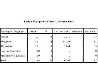
The mean pre-operative voice assessment score for the 74 patients
with benign thyroid pathology was 1.3 ± 3.3 ranging from 0 to 24 (Table 3).
For the 18 patients with malignant pathology, the mean score was 4.6 ±
14.3, ranging from 0 to 61. In the 11 patients with thyroiditis alone, the
mean score was 1.9 ± 2.8, with a range of 0 to 9. In the patients with
thyroiditis co-existing with benign thyroid disease and malignant thyroid
disease (one patient each), the score was 0.0. There was no significant
difference between these groups with respect to the pre-operative voice
38
Table 3: Pre-operative Voice Assessment Score
Pathological Diagnosis Mean N Std. Deviation Minimum Maximum
Benign 1.27 74 3.270 0 24
Malignant 4.61 18 14.255 0 61
Thyroiditis 1.91 11 2.844 0 9
Benign+ Thyroiditis .00 1 . 0 0
Malignancy+Thyroiditis .00 1 . 0 0
Total 1.89 105 6.567 0 61
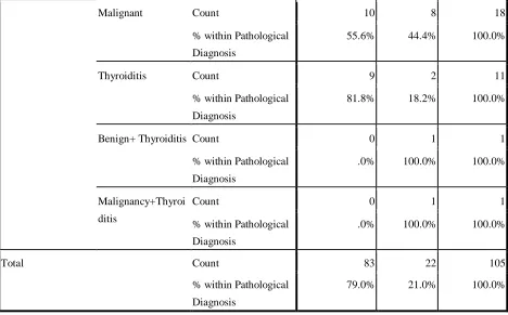
The overall rate of transient post-operative vocal cord palsy was
21.0% (22 of 105 patients). The incidence of transient post-operative vocal
cord palsy with various pathological diagnoses has been detailed in Table 4.
VC palsy at POD 5
Total no palsy palsy
Pathological Diagnosis
Benign Count 64 10 74
% within Pathological Diagnosis
39
Malignant Count 10 8 18
% within Pathological Diagnosis
55.6% 44.4% 100.0%
Thyroiditis Count 9 2 11
% within Pathological Diagnosis
81.8% 18.2% 100.0%
Benign+ Thyroiditis Count 0 1 1
% within Pathological Diagnosis
.0% 100.0% 100.0%
Malignancy+Thyroi
ditis
Count 0 1 1
% within Pathological Diagnosis
.0% 100.0% 100.0%
Total Count 83 22 105
% within Pathological Diagnosis
[image:44.612.91.559.92.383.2]79.0% 21.0% 100.0%
Table 4
Chi-Square Tests
Value df
Asymp. Sig. (2-sided)
Pearson Chi-Square 16.067a 4 .003
Likelihood Ratio 14.024 4 .007
40
The mean voice assessment score on the 5th post-operative day for the
105 patients was 10.3 ± 10.2(0 – 43) (Plate 1). There was a significant
difference in the mean scores among the groups (p<0.05), as shown in Plate
2. In addition, the mean voice assessment score on the 5th post-operative day
was significantly more than that during pre-operative assessment when
considered overall for the 105 patients in this study (p < 0.0001).
The overall rate of permanent vocal cord palsy was 7.6% (8 of 105
patients). Among the 74 patients with benign disease, 2 patients had
permanent palsy (2.7%). Among the 18 patients with malignant thyroid
disease, 4 patients (22.2%) had permanent vocal cord palsy. Of the 11
patients with thyroiditis, one patient had permanent vocal cord palsy (9.1%).
There was no permanent vocal cord palsy in the lone patient with thyroiditis
existent with benign disease. In the patient with thyroiditis and
co-existent malignant disease, permanent vocal cord palsy was seen (100%).
There was a significant difference in the incidence of permanent vocal
cord palsy between the benign and malignant subgroups (p = 0.005). Details
of correlation of permanent vocal cord palsy with thyroid pathology are as
41
When the laterality of vocal cord palsy on the 5th POD was analyzed
(Plate 4), the incidence of left vocal cord palsy was 11.4% (12 out of 105
patients), while that on the right side was 8.6% (9 out of 105 patients). This
difference in incidence was found to be statistically significant (p < 0.005).
One patient had bilateral vocal cord palsy in the early post-operative period.
When transient post-operative vocal cord palsy was correlated with
individual post-operative voice symptoms, significant correlation was
established between transient post-operative vocal cord palsy and all the
voice symptoms except for sore throat and cervical lymphadenopathy.
Patient with temporary vocal cod palsy were correlated with the voice
assessment scores done on days 5 & 180. 83 patients had normal functioning
vocal cords among the 105 patients and the rest 22 had vocal cord palsy
done on day 5.we analysed the various parameters in the VAS on day 5 &
180 for its implication with the cord status. Among the 15 parameters in the
42
an impact with greater scores in patients with temporary palsy than without
palsy having a statistical difference(p<0.05).
In those patients who developed vocal cord palsy, the incidence of
transient palsy was correlated with the voice assessment score on post-op
day 5 and the VAS on post-op day 180 (Plate 7a and 7b). There was
significant correlation between the VAS on op day 5 and that on
post-op day 180 (p<0.005). Those patients with vocal cord palsy who had high
VAS on post-op day 5 had persistent high scores on day 180 as well. In such
patients, there was a higher risk of permanent vocal cord palsy. This fact
underscores the importance of voice assessment score for its prediction of
permanent vocal cord palsy.
There was significant correlation (p<0.005) of the various parameters
of VAS on post-op day 180 with vocal cord palsy, as shown in plate 9. The
comparison of the pre-op VAS with that on post-op day 5 is as shown in
plates 10 & 11. The comparison of the overall VAS on post-op day 5 with
that on post-op day 180 is as shown in plates 12 & 13. The comparison of
the overall pre-op VAS with that on post-op day 180 is as shown in plates 14
43
Of the 105 patients who underwent surgery, EBSLN was identified
on the left side in 94 patients. Of the remaining 11 patients, it was not
identified. On analysis with the VAS on post-op days 5 and 180, it was
found that this aspect correlated with VAS on post-op day 5(p=0.043), but
not with that on post-op day 180 (p=0.78).
With regards to the right EBSLN, it was identified in 96 patients,
while it was not seen in the other 9 patients. On analysis with the VAS on
post-op days 5 and 180, there was no correlation with the post-op VAS,
44
Correlation of temporary palsy with anatomical relationship of RLN
On left side when the relationship of RLN and ITA was analysed,in
the majority of cases {n=92} RLN was deep to the ITA.In 9 cases RLN was
in between branches of ITA where as in one case RLN was superficial to
ITA and in three RLN was not identified.The rate of temporary vocal cord
palsy was correlaeted with ITA and RLN relationship and there was no
statistical significance (p = 0.244.)
On right side when the relationship of RLN and ITA was analysed,in
the majority of cases {n=86} RLN was deep to the ITA.In 14 cases RLN
was in between branches of ITA where as in one case RLN was superficial
to ITA and in 4 cases RLN was not identified.The rate of temporary vocal
cord palsy was correlaeted with ITA and RLN relationship and there was no
statistical significance ( p = 0.76).
Relation of RLN with ITA and correlation with vocal cord palsy
When vocal cord palsy was correlated with regard to the relation of
45
On the left side, the RLN was deep to the main trunk of the ITA in 92
patients, found coursing in between the ITA branches in 9 patients and
found superficial to the main trunk of ITA in 1 patient. The RLN was not
identified I on the left side in 3 patients.
On the right side, the RLN was deep to the ITA in 86 patients, found
coursing in between the branches of the ITA in 8 patients and superficial to
the main trunk of ITA in 4 patients. The RLN was not identified on the right
side in 7 patients.
The rate of transient or permanent RLN palsy was not influenced by
46
DISCUSSION
The incidence of transient RLN paralysis after thyroid surgery ranges
from 2.6% to 7.2% according to various researchers [15]. With regards to
permanent RLN paralysis, various studies have reported incidence ranging
from 0.5% to about 1%[15].
In the present study, the incidence of transient RLN palsy was 21.0%
and that of permanent RLN palsy was 7.6%. Evidently, the incidence of both
types of RLN palsy were higher when compared with other similar studies.
In the present study, the post-operative vocal cord status was
documented for each patient with the help of videolaryngoscopy (VLS).
This is not a routine practice. In addition, the rate of RLN palsy, especially
that of permanent RLN palsy, may be under-reported [1] due to follow-up
issues. During thyroid surgery, the EBSLN and RLN are routinely sought
and identified at this centre, which is a tertiary care, teaching institution. A
significant proportion of the procedures are performed by surgeons with
variable experience and as a result, the learning curve is a factor which has
47
Moreover, the fact that most of the cases of transient palsy recover
over a period of six months. Hence, regular follow up of the patients with
VLS is important. Direct examination of the vocal cords by VLS, both
pre-operatively and post-pre-operatively (on post-op days 5 and 180) is a routine
practice at this centre.
With regards to the exact time for laryngeal inspection after surgery
and time of diagnosis of post-operative RLN palsy, there is no consensus.
Different study groups advocate follow up at different times
operatively. While some advocate examination in the immediate
post-operative period, during extubation after surgery, some others are of the
opinion that the optimum time for laryngoscopy is two months after surgery.
If the vocal cord palsy persists even after two months post-operatively, then
laryngoscopy is advocated at six months and again after one year.[25]
In the present study, patients with malignancy had the maximum risk
of vocal cord palsy in the immediate post operative period. Risk of
permanent injury was also high in this group. The incidence of transient
palsy was 40% (8 out of 10 patients) in this group. The lone patient with
48
In those with thyroiditis (n=11), 2 patients (18.2%) developed
transient palsy. The incidence in those with benign pathology was 13.5% (10
of 74 patients).
Mountain JC, et al. [26] have reported pattern of incidence somewhat
similar to the present study, as regards thyroid pathology. In those with
carcinoma, the incidence was 8%. Chronic lymphocytic thyroiditis was
associated with an incidence of 5% for transient palsy. Risk of injury
increases four-fold when the nerve is not identified. [26].
The increased rate of palsy present in malignancy may be due to
difficult dissection encountered during surgery, apart from the possibility of
malignant infiltration of the nerve. In thyroiditis, the chronic inflammatory
infiltrate and the fibrosis prevalent in the perithyroidal tissues renders
dissection difficult. This may render the RLN more vulnerable.
The incidence of left vocal cord palsy was 11.4% (12 out of 105
patients), while that on the right side was 8.6% (9 out of 105 patients). This
difference in incidence was found to be statistically significant (p < 0.005).
49
and the patient recovered from vocal cord palsy in one side within six month
and other side continue to have palsy even after six months.
In the present study, the relationship of the RLN to the ITA and its
branches did not influence the rates of RLN injury, regardless of the side.
In the present study, a validated voice assessment score (VAS) was
used as a tool for objective evaluation of laryngeal symptoms, both before
surgery and in the post-operative period (POD 5 and POD 180). Individual
symptoms were rated with the help of a score, on the basis of severity.
Overall score for each assessment was calculated.
When compared to the delayed post-operative VAS (POD 180), the
early post-operative score (VAS on POD 5) was significantly higher, when
considered overall for the 105 patients in this study (p < 0.0001).
The mean post operative score was 10.3 ± 10.2. Also, there was a
significant difference of the VAS with regards to various subgroups divided
on the basis of histopathology (p < 0.005).
According to Byung-Joon Chun, et al., 30% of the patients experience
50
appropriate postoperative voice rehabilitation may improve voice in many
patients. Early post operative therapy such as voice therapy may improve
many of the symptoms.
When transient palsy was correlated with the VAS on POD 5 and
POD 180, significant correlation was observed (p < 0.05) between the
incidence of palsy and the VAS on POD 5 with regards to all the symptoms,
except for sore throat and cervical lymphadenopathy. Patients who had
permanent vocal cord palsy had higher VAS on POD 5, when compared to
those who have temporary palsy. The VAS was persistently high after 180
days as well.
Of the 105 patients who underwent surgery, the left EBSLN was
identified in 94 patients, while it was not seen in the other 11 patients. When
analyzed with VAS on POD 5 and POD 180, VAS was higher in those in
whom EBSLN was not seen (p=0.043), but there was no significant
correlation with regards to VAS on POD 180 (p=0.78).
The right EBSLN was identified in 96 patients out of the 105. Whern
analyzed with the VAS scores on POD 5 and 180, there was no significant
51
CONCLUSION
In the present study, the incidence of transient vocal cord palsy after
total thyroidectomy, as evidenced by videolaryngoscopy, was 21%. In about
two-thirds of patients, the vocal cords recovered within 6 months of surgery,
so that the incidence of permanent vocal cord palsy was 7.6%.
With regards to the validated voice assessment score, the score on the
5th post-operative day was invariably higher than that pre-operatively. Vocal
cord palsy was associated with a higher score post-operatively. Further, in
those patients who had vocal cord palsy and high voice assessment scores on
the 5th post-operative day, persistently high scores on the 180th
post-operative day and permanent vocal cord palsy were likely.
Voice assessment scoring is a reliable, noninvasive method of
assessment of vocal cord function, vocal cord palsy and pattern of recovery
1. Randolph et al. Surgery 2006; 139: 357-62.
2. Sen C et al. Neurosurgery 2001; 48: 838-47.
3. Hollinshead WH. Anatomy for surgeons, Vol 1. The head and neck
4. Hartl DM. Ann Otolaryngol 2000; 117: 60-84
5. Weisberg NK, et al. Stretch-induced nerve injury as a cause of paralysis
secondary to he anterior cervical approach. Otolryngol Head Neck Surg
1997; 116: 317-26
6. Moreau S, et al. The recurrent laryngeal nerve: related vascular anatomy.
Laryngoscope 1998; 108: 1351-3.
7. Neuro physiology of vocal fold paralysis. Otolaryngol Clin N Am 2004;
379: 1-23
8. Sanders I. The innervations of the human larynx. Arch Otolaryngol Head
Neck Surg 1993; 119: 934-9
9. Bei-Lian W, et al. The Human communicating nerve. Arch Otolaryngol
Head Neck Surg 1994; 120: 1321-8
10.Lalwani AK. Current diagnosis & treatment in otolaryngology-head &
neck surgery 2nd ed. New York: McGraw-Hill; 2008
11.Crumley RL. Regeneration of recurrent laryngeal nerve. Otolyngol Head
Neck Surg 1982; 90: 442-7
12.Zealear DL, et al. Neurophysiology of vocal fold paralysis. Otolaryngol
13.Collazo-Clavell ML. Relationship between vocal cord paralysis and
benign thyroid disease. Head Neck 1993; 17: 24-30
14.Yumoto, et al. Causes of recurrent laryngeal nerve paralysis. Auris Nasus
Larynx 2002; 29: 41-5
15.Recurrent laryngeal nerve paralysis :anatomy and etiology. Otolaryngol
Clin N Am 2004; 37: 25-44
16.Arnold GE. Physiology and pathology of the cricothyroid muscle.
Laryncoscope 1961; 71: 687-753
17.M.Santhosh, et al. Perceptual and acoustic analysis of voice in individuals
with Total thyroidectomy:Pre-Post surgery comparison . Indian J
Otolaryngol Head Neck Surg 63: 32-39
18.Watt- Boolsen et al. Influence of thyroid surgery on voice function and
laryngeal symptoms. Br J Surg 1979; 66: 535-6.
19.Susan Miller, et al. Voice therapy for vocal fold paralysis. Otolaryngol
Clin N Am 2004; 37: 25-44
20.Bruening W, et al. Uber eine neue Behandlungsmethode der
Rekurrenslahmung. Verh Dtsch Laryg1911; 18: 23
21.Arnold GE, et al. Vocal rehabilitation of paralytic dysphonia, 1955; 62:
1-17
22.Isshiki N, et al. Thyroplasty as anew phonosurgical technique. Acta
23.Isshiki, et al. Arytenoid adduction for unilateral vocal cord paralysis Arch
Otolaryngol 1978:104:555-8
24.Randall C, et al. Laryngeal reinnervation. Otolaryngol clin N Am 2004;
37: 161-181
25.Dionigi, et al. Post-operative laryngoscopy in thyroid surgery: proper
timing to detect recurrent laryngeal nerve injury. Langenbech Arch Surg
2010; 395:327-331
26.Mountain JC, et al. The recurrent laryngeal nerve in thyroid operations.
Surg Gynecol Obstet 1971; 133; 978-80.
27.Aluffi P, Policarpo M, Cherovac C, et al. Post-thyroidectomy sureprior
Plate 10: Comparison of pre-op VAS with that on POD 5
Mean N Std. Deviation Std. Error Mean
Pre Sore Throat .29 105 .743 .073
Pair 1
Post 5 sore throat 1.47 105 1.373 .134
Pre Hoarseness .16 105 .652 .064
Pair 2
Post 5 Hoarseness 1.19 105 1.462 .143
Pre Loudness .18 105 .769 .075
Pair 3
Post 5 Loudness .66 105 1.142 .111
Pre Loss of Voice .09 105 .557 .054
Pair 4
Post5 Loss of voice .41 105 .997 .097
Pre Cough .30 105 .759 .074
Pair 5
Post5 Cough 1.27 105 1.085 .106
Pre Weak Voice .13 105 .651 .064
Pair 6
Post5 weak voice .70 105 1.126 .110
Pre Mental Depression .09 105 .557 .054
Pair 7
Post5 Mental depression .38 105 .934 .091
Pre Throat Obstruction .17 105 .642 .063
Pair 8
Post5 Throat Obstruction .65 105 1.101 .107
Pre Cervical lymph nodes .12 105 .675 .066
Pair 9
Post5 Cervical LN .17 105 .563 .055
Pre Voice tiredness .11 105 .543 .053
Pair 10
Post5 Voice tiredness .52 105 1.001 .098
Pre inability to shout .13 105 .651 .064
Pair 11
Post5 inability to shout .99 105 1.369 .134
Pre Voice Variability .00 105 .000 .000
Pair 12
Post5 voice variability .64 105 1.084 .106
Pre Voice straining .06 105 .435 .042
Pair 13
Post5 Voice straining .48 105 1.066 .104
Pre Voice Breaking .09 105 .557 .054
Pair 14
Post5 Voice Break .66 105 1.117 .109
Pre Lonliness .05 105 .488 .048
Pair 15
Post5 Voice Loneliness .21 105 .703 .069
Scoring Parameter P value
Sore throat 0.03
Hoarsenes of voice <.001
Loudness <.001
Loss of voice 0.774
Cough <.001
Weak voice <.001
Mental depression <.001
Throat obstruction <.001
Cervical lymphadenopathy 1.00
Voice tiredness <.001
Inability to shout <.001
Voice variability <.001
Voice straining <.001
Voice break <.001
Loneliness <.001
Plate
9:
Significance
of
various
components
of
VAS
on
POD
180
and
Scoring Parameter P value
Sore throat 0.69
Hoarsenes of voice <.001
Loudness <.001
Loss of voice 0.008
Cough <.001
Weak voice <.001
Mental depression 0.004
Throat obstruction 0.010
Cervical lymphadenopathy 0.37
Voice tredness 0.02
Inability to shout <.001
Voice variability 0.04
Voice straining <.001
Voice break 0.003
Loneliness 0.002
Plate 7: Correlation of VAS on POD 5 with vocal cord palsy on POD 5
VC palsy at
POD 180 N Mean Rank
no palsy 97 53.31
Post 5 sore throat
VC palsy 8 49.19
no palsy 97 50.28
Post 5 Hoarseness
VC palsy 8 85.94
no palsy 97 50.77
Post 5 Loudness
VC palsy 8 80.00
no palsy 97 51.56
Post5 Loss of voice
VC palsy 8 70.50
no palsy 97 50.51
Post5 Cough
VC palsy 8 83.25
no palsy 97 50.20
Post5 weak voice
VC palsy 8 86.94
no palsy 97 51.44
Post5 Mental depression
VC palsy 8 71.88
no palsy 97 51.25
Post5 Throat Obstruction
VC palsy 8 74.19
no palsy 97 53.37
Post5 Cervical LN
VC palsy 8 48.50
no palsy 97 51.61
Post5 Voice tiredness
VC palsy 8 69.81
no palsy 97 50.31
Post5 inability to shout
VC palsy 8 85.56
no palsy 97 51.69
Post5 voice variability
VC palsy 8 68.94
no palsy 97 50.46
Post5 Voice straining
VC palsy