0022-538X/97/$04.0010
Copyright © 1997, American Society for Microbiology
Partial Resistance to gD-Mediated Interference Conferred by
Mutations Affecting Herpes Simplex Virus Type 1 gC and gK
PETER E. PERTEL1,2
ANDPATRICIA G. SPEAR2*
Division of Infectious Disease, Department of Medicine,1and Department of Microbiology-Immunology,2 Northwestern University Medical School, Chicago, Illinois 60611
Received 28 May 1997/Accepted 30 June 1997
Cells expressing herpes simplex virus (HSV) gD can be resistant to HSV entry as a result of gD-mediated interference. HSV strains differ in sensitivity to this interference, which blocks viral penetration but not binding. Previous studies have shown that mutations or variations in virion-associated gD can confer resis-tance to gD-mediated interference. Here we show that HSV-1 mutants selected for enhanced ability to bind and penetrate in the presence of inhibitory concentrations of heparin were partially resistant to gD-mediated interference. The resistance was largely due to the presence of two mutations: one in gC (the major heparin-binding glycoprotein) resulting in the absence of gC expression and the other in gK resulting in a syncytial phenotype. The results imply that heparin selected for mutants with altered postbinding requirements for entry. Resistance to gD-mediated interference conferred by mutations affecting gC and gK has not been previously described.
Initial absorption of herpes simplex virus type 1 (HSV-1) to cells is mediated by interaction of a viral glycoprotein, gC (UL44), with cell surface heparan sulfate (18, 20, 41, 44, 50). Glycoprotein B (UL27) mediates initial binding for virus that does not express gC and is also required for subsequent pen-etration (5, 19, 29, 39). Other viral glycoproteins essential for penetration include gD (US6), gH (UL22), and gL (UL1), and possibly the tegument protein UL25 (1, 15, 28, 38). Penetration of virus may also involve interaction with cell surface heparan sulfate (40). Soluble heparin, which is chemically similar to heparan sulfate (24), can inhibit viral binding and penetration (36, 41, 50). Penetration of HSV into cells proceeds by a series of steps requiring several viral glycoproteins and cell surface molecules, culminating in fusion of the viral envelope with the cell plasma membrane and release of the nucleocapsid into the cell cytoplasm (16, 17, 26, 32, 35, 49).
Recently, a gene was cloned that encodes a cell surface protein capable of mediating HSV entry (31). The protein, designated herpesvirus entry mediator (HVEM), is a member of the tumor necrosis factor-nerve growth factor receptor fam-ily and appears to be one of several cellular proteins that can mediate viral entry (31, 46). Expression of HVEM in nonhu-man cells resistant to HSV entry confers susceptibility to viral infectivity. In addition, anti-HVEM antibodies inhibit HSV-1 infection of HVEM-expressing Chinese hamster ovary (CHO) cells and of activated human T cells, indicating that HVEM mediates HSV-1 entry into certain types of human cells (31). HVEM interacts directly with gD and can be considered a coreceptor for HSV entry (48). Other cell surface molecules have been shown to interact with gD but either fail to mediate HSV entry or have not yet been identified (3, 4, 22).
Cells that express HSV gD are resistant to infection with wild-type virus as a result of gD-mediated interference (6, 7, 25). Interference has been noted with several cell lines engi-neered to express gD, including HEp-2 cells, BHK cells, L
cells, and HVEM-expressing CHO cells (6, 7, 25, 47). Inter-ference has also been noted with other alphaherpesviruses, including HSV-2, pseudorabies virus, and bovine herpesvirus type 1 (10, 11, 14, 37, 43). Cross interference between alpha-herpesviruses can occur but is not always reciprocal (10, 11, 27, 37). Although the mechanism is not fully defined, gD-mediated interference appears to occur after binding at the level of penetration (6, 13, 25). One proposal is that interference in-volves competition between cell-associated gD and virion-as-sociated gD for a cellular coreceptor for entry (6, 7, 13, 25).
HSV-1 mutants that are resistant to gD-mediated interfer-ence have been isolated. Some have been selected for their ability to propagate in cells expressing gD (7, 13). These mu-tants contain altered forms of gD that can partially or fully account for any noted resistance. Other interference-resistant mutants, again containing altered forms of gD, were selected for resistance to neutralization by a specific anti-gD monoclo-nal antibody (30). In addition, wild-type strains of HSV can vary in sensitivity to gD-mediated interference, due to poly-morphisms in gD and other undefined loci (14). Mutants with alterations in gD that confer resistance to interference have greatly reduced ability to utilize HVEM for entry but can infect human cells via other pathways (13, 31, 47). Moreover, wild-type gD interacts directly with HVEM, while the binding of the mutant forms of gD to HVEM cannot be detected (48). It may be that differences in HSV sensitivity to gD-mediated interfer-ence correlate with relative ability to use HVEM and other mediators of entry.
Recently we described the properties of mutants obtained by selection for variants of HSV-1(KOS) (designated KOS) that could bind and penetrate in the presence of heparin (36). The heparin-selected uncloned virus pools contained predomi-nantly syncytial and gC-negative mutants. Syncytial mutants were plaque purified from the uncloned pools and designated HS, for heparin-selected: two were gC positive (HS2 and HS4), and two were gC negative (HS1 and HS3). The gC-negative mutants were significantly more resistant to heparin inhibition than were the gC-positive mutants. For all four plaque-purified mutants, the syncytial phenotype mapped to a missense muta-tion (A40V) in gK (UL53). The gK mutamuta-tion did not contrib-ute to heparin resistance of HS1 and HS3; absence of gC * Corresponding author. Mailing address: Department of
Microbi-ology-Immunology, Northwestern University Medical School, Ward Bldg., Room 6-241, MC W213, 303 E. Chicago Ave., Chicago, IL 60611. Phone: (312) 503-8230. Fax: (312) 503-1339. E-mail: p-spear @nwu.edu.
8024
on November 9, 2019 by guest
http://jvi.asm.org/
expression alone was found to be sufficient to confer resistance to heparin inhibition. Because the infectivity of gC-negative virus is reduced (19, 20), the syncytial mutation in gK may have conferred a selective advantage by enhancing the spread of the virus through the cell monolayer by cell fusion.
Interestingly, HSV-1(ANG), a syncytial strain of HSV that was plaque purified under agar medium (33, 34), is resistant to gD-mediated interference because of amino acid substitutions at the same positions in gD as for other interference-resistant mutants (13). Because agar, like heparin, contains sulfated polysaccharides that can inhibit HSV binding (42), this study was undertaken to determine whether the heparin-selected mutants were altered in their ability to infect cells expressing gD.
Resistance to gD-mediated interference.To test the relative
susceptibility of the heparin-selected mutants and the un-cloned pools to gD-mediated interference, virus was inocu-lated onto HEp-2 cells that either express HSV-1 gD (H-gD-1 cells) or do not (H-control cells) (25). Shown in Fig. 1 are the number of plaques on H-gD-1 cells as a percentage of the number on H-control cells. All four uncloned virus pools ex-hibited partial resistance to gD-mediated interference (left panel), as did the four heparin-selected cloned mutants (right panel). The number of plaques formed on H-gD-1 compared to that on control cells was approximately 1 to 10% for the uncloned pools and the four heparin-selected mutants. In con-trast, the number of plaques on gD-expressing cells compared to that on control cells obtained with interference-resistant mutants altered in gD was 10 to 100% (7, 13).
To define mutations in the heparin-selected mutants that contribute to the partial resistance to gD-mediated interfer-ence, several virus strains were used. HS3 was selected because its syncytial phenotype and heparin resistance were previously
shown to be determined entirely by a single amino acid sub-stitution (A40V) in gK and a mutation resulting in absence of gC expression (36). A gC-positive syncytial recombinant virus, designated SyngK, was constructed by transfer of the HS3 gK gene to KOS (36). The gC-negative recombinant virus HSV-1(KOS)DgC2-3 (designatedDgC2-3), in which the gC gene is replaced by theEscherichia coli lacZgene, has been previously described (19). In order to construct a virus strain carrying both mutations known to be present in HS3, but lacking other mutations that might also be present, a gC-negative syncytial recombinant (designatedDgC2-3syngK) was isolated from the progeny of cells coinfected with SyngK andDgC2-3 (36). The genotypes and phenotypes of each recombinant were con-firmed by detection of characteristic restriction endonuclease sites in amplified copies of the gK genes and by monitoring of gC andb-galactosidase expression (data not shown). The gD-negative mutant HSV-1(KOS)gDb(designated KOSgDb) (13) was used as the recipient in marker transfer experiments. Fi-nally, the interference-resistant mutant HSV-1(KOS)Rid1 (designated Rid1) was used as the donor for a mutant form of gD that confers a high level of resistance to gD-mediated interference (13).
Absence of interference resistance mutations in HS3 gD.
Comparison of the ability of gD genes from HS3, KOS,
DgC2-3, and Rid1 to transfer to recombinant viruses resistance to gD-mediated interference tested the possibility that muta-tions in HS3 gD were responsible for the noted resistance. The gD gene from Rid1 served as a positive control, while the gD gene from KOS, which is extremely sensitive to gD-mediated interference (14), served as a negative control. Vero cells were transfected with genomic DNA from KOSgDbalong with gD genes obtained by PCR amplification from each virus of inter-est (14). Because gD is essential for HSV propagation, progeny capable of forming plaques on noncomplementing cells will be recombinant virus carrying the gD gene of the donor strain. The titer of recombinant virus produced by the transfected cells on H-gD-1 cells and H-control cells was determined. Figure 2 shows that only the Rid1 gD gene was able to confer resistance to gD-mediated interference, whereas the HS3 and
DgC2-3 gD genes gave results indistinguishable from those obtained with the KOS gD gene. Results similar to those ob-tained with the HS3 gD gene were also obob-tained with the HS1 gD gene (data not shown).
To exclude the possibility of mutations in the gD gene that can confer partial resistance to interference only when ex-pressed with other mutations, such as those altering gC or gK, the gD gene from HS3 was sequenced. The gD gene was amplified by PCR in two separate reactions, and both products were cloned into pGEM4 (Promega). The nucleotide sequence was determined with an ABI Prism 377 DNA sequencer (Uni-versity of Chicago Cancer Research Center DNA Sequencing Facility). The nucleotide sequence of HS3 gD did not differ from the published KOS gD sequence (data not shown).
Because the amount of gD incorporated into the viral enve-lope may influence susceptibility to gD-mediated interference, the relative amounts of gD present in purified KOS and HS3 virions were assessed. Shown in Table 1 are the relative amounts of gD and the major capsid protein VP5 present in preparations of KOS and HS3 virions as assessed by a bioim-ager as follows. Basically, purified KOS and HS3 (9) were solubilized in sodium dodecyl sulfate sample buffer, boiled for 5 min, and then loaded onto a sodium dodecyl sulfate–7% polyacrylamide gel (N,N9-methylene-bisacrylamide) (Bio-Rad laboratories). After electrophoresis, the proteins were trans-ferred to nitrocellulose (Schleicher & Schuell) with a Trans-Blot electrophoretic transfer cell apparatus (Bio-Rad
labora-FIG. 1. Resistance of the uncloned virus pools and plaque-purified heparin-selected mutants to gD-mediated interference. Virus diluted in complete phos-phate-buffered saline was inoculated onto six-well cultures of H-gD-1 cells or H-control cells for 2 h at 37°C (25). After the inoculum was removed, the cells were overlaid with medium 199 supplemented with 1% heat-inactivated newborn calf serum and 0.1% pooled human gamma globulin (Armour; 199O) and then
incubated for 3 days at 37°C in 5% CO2. Plaques were visualized by an anti-gB
immunoassay and counted (20, 21). Results are presented on a logarithmic scale as the number of plaques on H-gD-1 cells as a percentage of the number on H-control cells. The geometric means and standard deviations for three different experiments each done in duplicate are shown for each pool and virus. Differ-ences between KOS and each pool (A, B, C, and D) or mutant (HS1, HS2, HS3,
and HS4) were statistically significant (left panel,P50.002,P,0.001,P5
0.002, andP50.012, respectively; right panel,P,0.001; two-tailedttest).
VOL. 71, 1997 NOTES 8025
on November 9, 2019 by guest
http://jvi.asm.org/
[image:2.612.80.272.70.251.2]tories). The nitrocellulose blot was incubated in blocking buffer (150 mM NaCl, 10 mM Tris-HCl [pH 7.4], 0.2% Tween 20, 0.02% thimerosal, 5% milk) and then was incubated with a pool of polyclonal rabbit antiserum at 4°C followed by 35
S-labeled anti-rabbit immunoglobulin (Amersham) at room tem-perature. Rabbit anti-gD antibody R-7 (diluted 1:1,000) and anti-VP5 antibody NC-1 (diluted 1:2,000) were used (12, 23). Reactive bands were visualized, and the photostimulated lu-minescence value was quantified with a FUJIX BAS 2000 bioimager (Fuji). No significant difference in the gD/VP5 ra-tios for KOS and HS3 was noted.
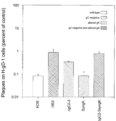
[image:3.612.334.535.67.276.2]Mutations responsible for resistance to gD-mediated inter-ference.To evaluate whether lack of gC expression or a syn-cytial mutation could confer partial resistance to gD-mediated interference, recombinants expressing these phenotypes either alone or together were studied. The titers of the engineered recombinants along with KOS and HS3 on gD-1 and H-control cells were determined. As shown in Fig. 3, both HS3 andDgC2-3syngK were approximately 10-fold less sensitive to gD-mediated interference than KOS, with no difference noted between HS3 andDgC2-3syngK. Interestingly, the engineered gC-negative virus, DgC2-3, was approximately threefold less sensitive to gD-mediated interference than was KOS. No dif-ference was noted between KOS and SyngK. The revertants for SyngK (36) andDgC2-3 (19) also did not differ from KOS (data not shown). Results similar to those shown in Fig. 3 were obtained with a second engineered gC-negative virus, HSV-1(KOS)DgC6, and its revertant (45) and a double recombinant constructed by transfer of the gK allele from HS3 to DgC6 (data not shown). Thus, the lack of gC expression conferred slight resistance to gD-mediated interference, while the syncy-tial mutation in gK did not. Combination of the two mutations resulted in a recombinant virus that was as resistant to inter-ference as HS3. The resistance to gD-mediated interinter-ference conferred by these two mutations, however, is less than that conferred by single mutations in HSV-1 gD.
FIG. 2. Absence of gD mutations that could confer resistance to gD-medi-ated interference. Subconfluent Vero cells were transfected with Lipofectamine reagent (Gibco BRL) with 1mg of genomic KOSgDbDNA and 1mg of the gD gene obtained by PCR amplification from each virus (14). Genomic viral DNA was purified from infected cell lysates as previously described (36). PCR was performed with Vent polymerase (New England Biolabs) and purified viral DNA. The gD gene along with flanking sequences was amplified with the sense primer gD1-5 (59-GGAGTTGTTCGGTCATAAGCTTCAGC-39) and the anti-sense primer gD1-7 (59-GACTTATCGACTGTCCGCCTTTC-39) (13), obtained from the Northwestern University Biotechnology Facility. After the transfection mixture was removed, the cells were overlaid with medium 199 supplemented with 1% heat inactivated newborn calf serum (199V) and then were incubated for 48 h at 37°C in 5% CO2. The cells were then harvested and lysed to release
recombinant progeny. Three separate PCR amplifications and transfections were done, generating three recombinant stocks that each contain the gD gene of interest. The titer of each recombinant stock on H-gD-1 cells and H-control cells was subsequently determined. Results are presented on a logarithmic scale as the number of plaques on H-gD-1 cells as a percentage of the number on H-control cells. The geometric means and standard deviations for three different experi-ments each done in duplicate are shown. Differences between the recombinant stocks containing the gK gene from KOS and the genes from HS3 andDgC2-3 were not statistically significant (P50.933 and 0.637, respectively; two-tailed t test). Differences between the recombinant stocks containing the gK gene from Rid1 and the genes from KOS, HS3, andDgC2-3 were all statistically significant (P50.008, 0.009, and 0.015, respectively).
[image:3.612.80.272.68.268.2]FIG. 3. Resistance of recombinant viruses to gD-mediated interference. The titer of virus diluted in phosphate-buffered saline on H-gD-1 cells and H-control cells was determined as described in the legend to Fig. 1. Results are presented on a logarithmic scale as the number of plaques on H-gD-1 cells as a percentage of the number on H-control cells. The geometric means and standard deviations for three different experiments each done in duplicate are shown for each pool and virus. Differences between KOS and HS3,DgC2-3, andDgC2-3syngK were all statistically significant (P,0.001; two-tailed t test); the difference between KOS and SyngK was not (P50.928). Differences between HS3 andDgC2-3 and between HS3 and SyngK were both statistically significant (P50.017 and 0.002, respectively); the difference between HS3 andDgC2-3syngK was not (P50.768).
TABLE 1. Relative amounts of gD and VP5 present in purified KOS and HS3 virionsa
Virus Dilution
Photostimulated
luminescence gD/VP5 ratio
VP5 gD
KOS None 3,970 505 0.13
1:2 2,350 343 0.15
1:4 1,100 183 0.17
1:8 634 106 0.17
HS3 None 2,240 291 0.13
1:2 1,330 149 0.11
1:4 636 83 0.13
1:8 328 36 0.11
aFor details, see the text. Photostimulated luminescence values have been
corrected for background radioactivity.
on November 9, 2019 by guest
http://jvi.asm.org/
[image:3.612.58.299.583.708.2]HVEM-mediated entry of HS3.Mutants with alterations in gD that confer resistance to interference have greatly reduced ability to utilize HVEM for entry but can infect human cells via other pathways (13, 31, 47). Because HS3 is partially resistant to gD-mediated interference, the ability of HVEM to mediate entry of HS3 was tested. CHO cells allow KOS to bind effi-ciently but are not susceptible to subsequent penetration (41). CHO-IEb8 cells were transfected with pBEC10, which ex-presses HVEM, and then infected with HS3 as previously de-scribed (31). CHO-IEb8 cells carry theE. coli lacZgene down-stream from an HSV-1 immediate-early promoter and express
b-galactosidase when induced by the HSV-1 transinducer VP16 (31). VP16 is released along with the viral nucleocapsid when HSV enters a cell and does not require any viral gene expression to function (2, 8). Measurement ofb-galactosidase activity induced by VP16, therefore, can be used to quantitate initial HSV entry. As shown in Fig. 4, both KOS and HS3 could utilize HVEM to enter into CHO cells, while Rid1 could not. Results similar to those noted for KOS and HS3 were obtained with DgC2-3, SyngK, and DgC2-3syngK (data not shown). Thus, the partial resistance to interference noted for HS3 does not correlate with an inability to use HVEM to mediate entry. In summary, HSV-1 mutants selected for resistance to the inhibitory effects of heparin on virus binding and penetration were also partially resistant to gD-mediated interference. For
at least one mutant, HS3, a syncytial mutation in gK and lack of gC expression were sufficient to account for the noted re-sistance. In addition, lack of gC expression alone resulted in partial resistance to gD-mediated interference. These are pre-viously undescribed phenotypes of mutations affecting gC and gK. These results are consistent with those of a prior study showing that viral genes in addition to that encoding gD can influence resistance to gD-mediated interference (14). Muta-tions in the gD gene that confer resistance to interference reduce the ability of virus to utilize HVEM as a coreceptor for entry (31). Mutations in the gC and gK genes that confer resistance do not but may instead alter usage of other core-ceptors for HSV entry. The heparin-selected mutants may prove useful in defining patterns of coreceptor usage.
We thank N. Susmarski for the preparation of cell cultures. HSV-1(KOS)DgC6 and HSV-1(KOS)DgC2-3 and their revertants were gifts from C. Brandt (University of Wisconsin). Antibodies R-7 and NC-1 were gifts from G. Cohen and R. Eisenberg (University of Pennsylva-nia).
These studies were supported by a grant from the National Institutes of Health (R01 AI 36293-03) and a fellowship from the Howard Hughes Medical Institute, Chevy Chase, Md. (P.E.P.)
REFERENCES
1.Addison, C., F. J. Rixon, J. W. Palfreyman, M. O’Hara, and V. G. Preston.
1984. Characterization of a herpes simplex virus type 1 mutant which has a temperature-sensitive defect in penetration of cells and assembly of capsids.
Virology138:246–259.
2.Batterson, W., and B. Roizman.1983. Characterization of the herpes simplex
virion-associated factor responsible for the induction ofagenes. J. Virol.
46:371–377.
3.Brunetti, C., R. L. Burke, S. Kornfeld, W. Gregory, F. R. Masiarz, K. S. Dingwell, and D. C. Johnson.1994. Herpes simplex virus glycoprotein D acquires mannose 6-phosphate residues and binds to mannose 6-phosphate
receptors. J. Biol. Chem.269:17067–17074.
4.Brunetti, C. R., R. L. Burke, B. Hoflack, T. Ludwig, K. S. Dingwell, and D. C. Johnson.1995. Role of mannose-6-phosphate receptors in herpes simplex
virus entry into cells and cell-to-cell transmission. J. Virol.69:3517–3528.
5.Cai, W., B. Gu, and S. Person.1988. Role of glycoprotein B of herpes
simplex virus type 1 in viral entry and cell fusion. J. Virol.62:2596–2604.
6.Campadelli-Fiume, G., M. Arsenakis, F. Farabegoli, and B. Roizman.1988. Entry of herpes simplex virus 1 in BJ cells that constitutively express viral glycoprotein D is by endocytosis and results in degradation of the virus.
J. Virol.62:159–167.
7.Campadelli-Fiume, G., S. Qi, E. Avitabile, L. Foa`-Tomasi, R. Brandimarti, and B. Roizman.1990. Glycoprotein D of herpes simplex virus encodes a domain which precludes penetration of cells expressing the glycoprotein by
superinfecting herpes simplex virus. J. Virol.64:6070–6079.
8.Campbell, M. E. M., J. W. Palfreyman, and C. M. Preston.1984. Identifi-cation of herpes simplex virus DNA sequences which encode a trans-acting polypeptide responsible for stimulation of immediate early transcription. J.
Mol. Biol.180:1–19.
9.Cassai, E., M. Sarmiento, and P. G. Spear.1975. Comparison of the virion
proteins specified by herpes simplex virus types 1 and 2. J. Virol.16:1327–
1331.
10. Chase, C. C. L., K. Carter-Allen, C. Lohff, and G. J. Letchworth, III.1990. Bovine cells expressing bovine herpesvirus 1 (BHV-1) glycoprotein IV resist infection by BHV-1, herpes simplex virus, and pseudorabies virus. J. Virol.
64:4866–4872.
11. Chase, C. C. L., C. Lohff, and G. J. Letchworth.1993. Resistance and susceptibility of bovine cells expressing herpesviral glycoprotein D homologs
to herpesviral infections. Virology194:365–369.
12. Cohen, G. H., M. Ponce de Leon, H. Diggelmann, W. C. Lawrence, S. K. Vernon, and R. J. Eisenberg.1980. Structural analysis of the capsid
polypep-tides of herpes simplex virus types 1 and 2. J. Virol.34:521–531.
13. Dean, H. J., S. Terhune, M.-T. Shieh, N. Susmarski, and P. G. Spear.1994. Single amino acid substitutions in gD of herpes simplex virus 1 confer resistance to gD-mediated interference and cause cell type-dependent
alter-ations in infectivity. Virology199:67–80.
14. Dean, H. J., M. S. Warner, S. S. Terhune, R. M. Johnson, and P. G. Spear.
1995. Viral determinants of the variable sensitivity of herpes simplex virus
strains to gD-mediated interference. J. Virol.69:5171–5176.
15. Forrester, A., H. Farrell, G. Wilkinson, J. Kaye, N. Davis-Poynter, and T. Minson.1992. Construction and properties of a mutant of herpes simplex
virus type 1 with glycoprotein H coding sequences deleted. J. Virol.66:341–
[image:4.612.106.247.69.269.2]348. FIG. 4. HVEM-mediated entry of HSV into CHO cells. Subconfluent
CHO-IEb8 cells were transfected with Lipofectamine reagent (Gibco BRL) with 1.5mg
of a plasmid (pBEC10) containing HVEM downstream from a constitutive cytomegalovirus immediate-early promoter (31). The negative control consisted
of 1.5mg of the plasmid vector (pcDNA3) without the insert. After 24 h, the cells
were replated in 96-well plates at a concentration of about 23104to 43104
cells/well and then incubated for 24 h at 37°C in 5% CO2. The cells were washed
and then inoculated with 50ml of virus diluted in PBS containing 0.1% glucose
and 1% calf serum (Sigma). The number of PFU added to each well was based on titers on Vero cells and was confirmed in each experiment by simultaneous
plaquing of each virus on Vero cells. After incubation for 6 h at 37°C, 100ml of
phosphate-buffered saline containing 0.5% Nonidet P-40 and 3 mg of theb
-galactosidase substrateo-nitrophenolb-D-glucopyranoside per ml was added to
each well. Optical density (OD) readings at 410 nm were obtained every hour with a plate spectrophotometer (Spectromax 250). The results shown are mean optical density readings and standard deviations obtained at 4 h, at which time product accumulation was proportional to time, for triplicate determinations at concentrations of input virus within the linear range of dose-response curves. The circles represent readings obtained from cells transfected with HVEM, and the triangles represent readings from cells transfected with the vector control. Similar results were obtained in a second set of experiments done independently (data not shown).
VOL. 71, 1997 NOTES 8027
on November 9, 2019 by guest
http://jvi.asm.org/
16. Fuller, A. O., and W.-C. Lee.1992. Herpes simplex virus type 1 entry through a cascade of virus-cell interactions requires different roles of gD and gH in
penetration. J. Virol.66:5002–5012.
17. Fuller, A. O., and P. G. Spear.1987. Anti-glycoprotein D antibodies that permit adsorption but block infection by herpes simplex virus 1 prevent
virion-cell fusion at the cell surface. Proc. Natl. Acad. Sci. USA84:5454–
5458.
18. Gruenheid, S., L. Gatzke, H. Meadows, and F. Tufaro.1993. Herpes simplex virus infection and propagation in a mouse L cell mutant lacking heparan
sulfate proteoglycans. J. Virol.67:93–100.
19. Herold, B. C., R. J. Visalli, N. Susmarski, C. R. Brandt, and P. G. Spear.
1994. Glycoprotein C-independent binding of herpes simplex virus to cells requires of cell surface heparan sulphate and glycoprotein B. J. Gen. Virol.
75:1211–1222.
20. Herold, B. C., D. WuDunn, N. Soltys, and P. G. Spear.1991. Glycoprotein C of herpes simplex virus type 1 plays a principal role in the adsorption of virus
to cells and in infectivity. J. Virol.65:1090–1098.
21. Holland, T. C., R. M. Sandri-Goldin, L. E. Holland, S. D. Marlin, M. Levine, and J. C. Glorioso.1983. Physical mapping of the mutation in an antigenic variant of herpes simplex virus type 1 by use of an immunoreactive plaque
assay. J. Virol.46:649–652.
22. Huang, T., and G. Campadelli-Fiume.1996. Anti-idiotypic antibodies mim-icking glycoprotein D of herpes simplex virus identify a cellular protein required for virus spread from cell to cell and virus-induced polykaryocytosis.
Proc. Natl. Acad. Sci. USA93:1836–1840.
23. Isola, V. J., R. J. Eisenberg, G. R. Siebert, C. J. Heilman, W. C. Wilcox, and G. H. Cohen.1989. Fine mapping of antigenic site II of herpes simplex virus
glycoprotein D. J. Virol.63:2325–2334.
24. Jackson, R. L., S. J. Busch, and A. D. Cardin.1991. Glycosaminoglycans: molecular properties, protein interactions, and role in physiological
pro-cesses. Physiol. Rev.71:481–539.
25. Johnson, R. M., and P. G. Spear.1989. Herpes simplex virus glycoprotein D
mediates interference with herpes simplex virus infection. J. Virol.63:819–
827.
26. Karger, A., and T. C. Mettenleiter.1993. Glycoproteins gIII and gp50 play dominant roles in the biphasic attachment of pseudorabies virus. Virology
194:654–664.
27. Lee, W.-C., and A. O. Fuller.1993. Herpes simplex virus type 1 and pseu-dorabies virus bind to a common saturable receptor on Vero cells that is not
heparan sulfate. J. Virol.67:5088–5097.
28. Ligas, M. W., and D. C. Johnson.1988. A herpes simplex virus mutant in
which glycoprotein D sequences are replaced byb-galactosidase sequences
binds to but is unable to penetrate into cells. J. Virol.62:1486–1494.
29. Manservigi, R., P. G. Spear, and A. Buchan.1977. Cell fusion induced by herpes simplex virus is promoted and suppressed by different viral
glycopro-teins. Proc. Natl. Acad. Sci. USA74:3913–3917.
30. Minson, A. C., T. C. Hodgman, P. Digard, D. C. Hancock, S. E. Bell, and E. A. Buckmaster.1986. An analysis of the biological properties of mono-clonal antibodies against glycoprotein D of herpes simplex virus and identi-fication of amino acid substitutions that confer resistance to neutralization.
J. Gen. Virol.67:1001–1013.
31. Montgomery, R. I., M. S. Warner, B. J. Lum, and P. G. Spear.1996. Herpes simplex virus-1 entry into cells mediated by a novel member of the TNF/
NGF receptor family. Cell87:427–436.
32. Morgan, C., H. M. Rose, and B. Mednis.1968. Electron microscopy of
herpes simplex virus. I. Entry. J Virol.2:507–516.
33. Munk, K., and D. Donner.1963. Cytopathischer Effekt und
Plaque-Mor-phologie verschiedener Herpes-simplex-Virus-Sta¨mme. Arch. Gesamte
Vi-rusforsch.13:529–540.
34. Munk, K., and G. Ludwig.1972. Properties of plaque variants of herpes virus
hominis strains of genital origin. Arch. Gesamte Virusforsch.37:308–315.
35. Para, M. F., R. B. Baucke, and P. G. Spear.1980. Immunoglobulin G(Fc)-binding receptors on virions of herpes simplex virus type 1 and transfer of
these receptors to the cell surface by infection. J. Virol.34:512–520.
36. Pertel, P. E., and P. G. Spear.1996. Modified entry and syncytium formation by herpes simplex virus type 1 mutants selected for resistance to heparin
inhibition. Virology226:22–33.
37. Petrovskis, E. A., A. L. Meyer, and L. E. Post.1988. Reduced yield of infectious pseudorabies virus and herpes simplex virus from cell lines
pro-ducing viral glycoprotein gp50. J. Virol.62:2196–2199.
38. Roop, C., L. Hutchinson, and D. C. Johnson.1993. A mutant herpes simplex virus type 1 unable to express glycoprotein L cannot enter cells, and its
particles lack glycoprotein H. J. Virol.67:2285–2297.
39. Sarmiento, M., M. Haffey, and P. G. Spear.1979. Membrane proteins
spec-ified by herpes simplex viruses. III. Role of glycoprotein VP7(B2) in virion
infectivity. J. Virol.29:1149–1158.
40. Shieh, M.-T., and P. G. Spear.1994. Herpesvirus-induced cell fusion that is
dependent on cell surface heparan sulfate or soluble heparin. J. Virol.68:
1224–1228.
41. Shieh, M.-T., D. WuDunn, R. I. Montgomery, J. D. Esko, and P. G. Spear.
1992. Cell surface receptors for herpes simplex virus are heparan sulfate
proteoglycans. J. Cell Biol.116:1273–1281.
42. Takemoto, K. K., and P. Fabisch.1964. Inhibition of herpes simplex virus by
natural and synthetic acid polysaccharides. Proc. Soc. Exp. Biol. Med.116:
140–144.
43. Tikoo, S. K., D. R. Fitzpatrick, L. A. Babiuk, and T. J. Zamb.1990. Molec-ular cloning, sequencing, and expression of functional bovine herpesvirus 1
glycoprotein gIV in transfected bovine cells. J. Virol.64:5132–5142.
44. Vaheri, A., and K. Cantell.1963. The effect of heparin on herpes simplex
virus. Virology21:661–662.
45. Visalli, R. J.1992. The herpes simplex virus type 1 UL45 gene. Ph.D. thesis. Department of Medical Microbiology and Immunology, University of Wis-consin, Madison.
46. Warner, M. S., R. J. Geraghty, R. I. Montgomery, and P. G. Spear. Unpub-lished data.
47. Warner, M. S., R. I. Montgomery, and P. G. Spear.Unpublished data. 48. Whitbeck, J. C., C. Peng, H. Lou, R. Xu, S. H. Willis, M. Ponce de Leon, T.
Peng, A. V. Nicola, R. I. Montgomery, M. S. Warner, A. Soulika, L. Spruce, W. T. Moore, J. D. Lambris, P. G. Spear, G. H. Cohen, and R. J. Eisenberg.
1997. Glycoprotein D of herpes simplex virus (HSV) binds directly to HVEM, a member of the tumor necrosis factor receptor superfamily and a
mediator of HSV entry. J. Virol.71:6083–6093.
49. Wittels, M., and P. G. Spear.1990. Penetration of cells by herpes simplex virus does not require a low pH-dependent endocytic pathway. Virus Res.
18:271–290.
50. WuDunn, D., and P. G. Spear.1989. Initial interaction of herpes simplex
virus with cells is binding to heparan sulfate. J. Virol.63:52–58.