0022-538X/95/$04.0010
Copyright 1995, American Society for Microbiology
Viral Determinants of the Variable Sensitivity of Herpes
Simplex Virus Strains to gD-Mediated Interference
HANSI J. DEAN,† MORGYN S. WARNER, SCOTT S. TERHUNE,
RAYMOND M. JOHNSON,‡ANDPATRICIA G. SPEAR*
Department of Microbiology-Immunology, Northwestern University Medical School, Chicago, Illinois 60611
Received 15 March 1995/Accepted 5 May 1995
Cells that express glycoprotein D (gD) of herpes simplex virus type 1 (HSV-1) resist infection by HSV-1 and HSV-2 because of interference with viral penetration. The results presented here show that both HSV-1 and HSV-2 gD can mediate interference and that various HSV-1 and HSV-2 strains differ in sensitivity to this interference. The relative degree of sensitivity was not necessarily dependent on whether the cell expressed the heterologous or homologous form of gD but rather on the properties of the virus. Marker transfer experiments revealed that the allele of gD expressed by the virus was a major determinant of sensitivity to interference. Amino acid substitutions in the most distal part of the gD ectodomain had a major effect, but substitutions solely in the cytoplasmic domain also influenced sensitivity to interference. In addition, evidence was obtained that another viral gene(s) in addition to the one encoding gD can influence sensitivity to interference. The results indicate that HSV-1 and HSV-2 gD share determinants required to mediate interference with infection by HSV of either serotype and that the pathway of HSV entry that is blocked by expression of cell-associated gD can be cleared or bypassed through subtle alterations in virion-associated proteins, particularly gD.
Cells expressing gD of herpes simplex virus type 1 (HSV-1) can be resistant to infection by HSV-1 or HSV-2 (3, 11). This has been shown for several cell types, including human Hep-2 cells, mouse L cells, and baby hamster kidney (BHK) cells. This gD-mediated interference with HSV infection is at the level of viral penetration into cells, not viral binding or post-penetration steps (3, 8, 11).
The phenomenon of gD-mediated interference is common to several of the alphaherpesviruses. Cells expressing pseudo-rabies virus (PRV) or bovine herpesvirus type 1 (BHV-1) gD are also resistant to infection by homologous virus (5, 6, 28). In addition, cross-interference among different alphaherpesvi-ruses has been noted (5, 6, 12, 22). For example, cells express-ing BHV-1 gD are at least partially resistant to infection by BHV-1, PRV, and HSV-1 (5). Cross-interference is not always reciprocal (5, 6). The patterns of cross-interference observed indicate that both homologous and heterologous forms of gD have determinants capable of blocking the entry of a single virus and that different alphaherpesviruses may have common requirements for entry that can be blocked by a single form of gD. This is despite limited conservation of primary amino acid sequence among the gD homologs (10 to 35% identity), the absence of cross-neutralizing epitopes, and differences in host range among the alphaherpesviruses.
The entry of HSV, PRV, and BHV-1 into cells depends on binding of the virus to cell surface glycosaminoglycans via gC, followed by fusion of the virion envelope with a cell membrane. The available evidence indicates that at least four envelope glycoproteins, designated gB, gD, gH, and gL, are required for the fusion or penetration step. These glycoproteins are present
in the envelope as homo-oligomers in the case of gB and heterooligomers in the case of gH and gL. It seems likely that interactions among gB, gD, and gH-gL and with unidentified cell surface components are required for the membrane fusion reaction (see references 17, 26, and 27 for reviews).
The mechanism of gD-mediated interference is not under-stood. It has been proposed that cell-associated gD interferes with a cell surface component required for viral entry or in-teracts with the virion itself, perhaps to disrupt interactions among the viral glycoproteins (3, 4, 8, 11). Recently it was shown that a soluble recombinant form of gD becomes modi-fied by the addition of mannose-6-phosphate (Man-6-P) to N-linked glycans and that this form can bind to Man-6-P re-ceptors (2). It seems unlikely that an interaction of virion-associated gD with Man-6-P receptors is required for HSV entry, however. This is based on findings that gD from infected cells carries little if any Man-6-P (2), an HSV-1 mutant ex-pressing a form of gD without N-linked glycans is fully infec-tious (25), and mutant cells unable to express one or both of the Man-6-P receptors (14) are fully susceptible to HSV infec-tion (27a). However, the possibility remains that an interacinfec-tion of gD with some unidentified cell surface component is re-quired for viral entry and is relevant to the phenomenon of interference.
HSV-1 mutants selected for their resistance to gD-mediated interference have been isolated. These mutants have point mutations in the gD gene, and the altered gD gene can transfer the interference-resistant phenotype to the sensitive parental strain, either totally or partially (1, 4, 8). Interestingly, certain amino acid substitutions in gD (substitutions changing Gln to Pro or Arg at position 27 [Gln27Pro or Gln27Arg]) can render the virus completely resistant to gD-mediated interference while only marginally reducing infectivity for control cells (8). In the course of our studies, we have noted that different wild-type strains of HSV differed in their sensitivity to gD-mediated interference and that HSV-2 strains seemed to be generally less sensitive than HSV-1 strains to interference me-diated by HSV-1 gD (gD-1). The HSV-2 form of gD (gD-2)
* Corresponding author. Mailing address: Department of Microbi-ology-Immunology, Northwestern University Medical School, 303 E. Chicago Ave., W213, Chicago, IL 60611. Phone: (312) 503-8230. Fax: (312) 503-1339. Electronic mail address: [email protected].
† Present address: Mallinckrodt Veterinary, Mundelein, IL 60060. ‡ Present address: Dept. of Medicine, Washington University School of Medicine, St. Louis, MO 63110.
5171
on November 9, 2019 by guest
http://jvi.asm.org/
exhibits about 82% sequence identity with gD-1. In order to determine whether gD-mediated interference is serotype spe-cific or serotype selective, we have produced new cell lines expressing gD-2 for comparison of the ability of gD-1 and gD-2 to mediate interference with HSV-1 and HSV-2 strains. We have also quantitated the sensitivity or resistance of several HSV strains to gD-1-mediated interference and transferred their gD genes to a common genomic background in order to determine whether differences in sensitivity to interference can be due solely to differences in gD gene sequence. The results show that differences in the relative sensitivities of various HSV-1 and HSV-2 strains to interference do not depend on whether the cells express gD-1 or gD-2 but rather on the properties of the virion. They also show that at least two domains of virion-associated gD, as well as other HSV genes, influence sensitivity to interference.
Hep-2 cells transformed to express gD-1 (cell line 42H9-9, designated here H-gD-1) and control Hep-2 cells transformed with the expression vector alone (cell line 40H7-1, designated H-control) have already been described (11). New Hep-2 cell lines transformed to express gD-2 were produced for this study by methods described previously (11). These cell lines were isolated after transfection with a plasmid (pRJ4065p) contain-ing the neo selectable marker and the SpeI-BstXI fragment of HSV-2(333) DNA, encoding the gD open reading frame, and were shown to express gD-2 by Western immunoblotting (10a). The gD-2-expressing cell line selected for this study was named H-4065p-1 but is designated H-gD-2 here for simplicity. In both the H-gD-1 and H-gD-2 cell lines, gD is under control of the human metallothionein promoter, which is inducible by the
addition of CdCl2. Both cell lines express gD constitutively but
can be induced to express higher levels by the addition of
CdCl2 (11). The transformed cell lines were maintained as
described previously (11).
To assess the levels of interference mediated by gD-1 and gD-2, several HSV-1 and HSV-2 strains were plated at various dilutions on uninduced gD-1 cells, gD-2 cells, and H-control cells or on replicate cultures of the cells that had been
induced with CdCl2 to produce higher levels of gD (11).
In-duction was demonstrated by Western blot analysis (not shown). Plaques were visualized by immunoassay (8). The titer of each virus stock on each cell line was determined, with the results expressed as PFU scored on gD-expressing cells as a percentage of PFU scored on the H-control cells.
The results shown in Fig. 1 demonstrate that both gD-1 and gD-2 can mediate interference with HSV-1 and HSV-2 strains. Although the H-gD-2 cells were somewhat less resistant than the H-gD-1 cells to infection by all the HSV strains tested, this effect is not necessarily due to intrinsically lower interference activity of gD-2. We have observed that independent clones of mouse or human cells transformed with a single vector (ex-pressing either gD-1 or gD-2) may vary in their resistance to HSV infection, indicating clonal variation in cell factors that contribute to the phenomenon or in levels of gD expression. Induction of higher levels of gD-1 or gD-2 by incubation of the
cells with CdCl2invariably enhanced interference with plaque
formation for all HSV strains tested (Fig. 1).
There was considerable variability (10- to 500-fold differ-ences) in the sensitivity of the HSV strains to gD-mediated interference. This variability did not depend on whether the virion-associated and cell-associated gDs were of the same or different strain or serotype. For example, the H-gD-2 cells, which expressed HSV-2(333) gD, were least resistant to infec-tion by HSV-2(333) and most resistant to HSV-1(KOS).
Among the six HSV strains tested, HSV-1(KOS) exhibited the greatest sensitivity to interference on both 1- and
gD-2-expressing cell lines. The plaques produced by HSV-1(KOS) on gD-expressing cells were smaller than those produced by the strains that were less sensitive to interference. However, the number of plaques scored for HSV-1(KOS) did not under-estimate the number of cells initially infected by the virus inoculum. Whether infectivity was monitored by plaque for-mation after 3 days or by the fraction of cells induced to express an immediate-early viral protein within 6 h, the ratio of the amounts of HSV-1(KOS) required to yield equivalent numbers of infectious units on H-gD-1 and H-control cells was the same (7a).
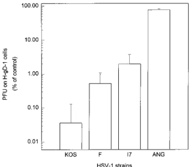
To further explore the differences among HSV-1 strains in sensitivity to gD-mediated interference, strains KOS, 17, F, and ANG were plated at various dilutions on H-gD-1 and H-control cells. The results are shown in Fig. 2, with mean values and standard deviations given for at least three separate determinations. HSV-1 strains F, 17, and ANG infected gD-expressing cells approximately 10, 30, and 1,000 times more efficiently than did KOS. The differences in interference sen-sitivities between KOS and each of the other strains (F, 17, and ANG) were statistically significant.
Our interest was to determine whether the reduced sensitiv-ity to gD-mediated interference exhibited by F, 17, and ANG could be a result of the minor differences in amino acid se-quence among the different forms of gD expressed (4, 8, 10, 16). Recombinant viruses in which the gD genes from HSV-1 strains KOS, F, 17, and ANG were placed in the KOS genetic background were constructed. Genomic DNA from the
gD-FIG. 1. Relative sensitivities of several HSV-1 and HSV-2 strains to inter-ference mediated by gD-1 and gD-2. Plaque assays were performed in triplicate on monolayers of uninduced and induced H-gD-1, H-gD-2, and H-control cells, as described previously (8). Enhanced gD-1 and gD-2 expression was induced by incubation of the cells with medium containing 2mM CdCl2for 6 to 18 h prior
to plating the virus dilutions for the assay (11). Cells were infected with serial 10-fold dilutions of HSV-1 strain KOS, F, or mP or HSV-2 strain G, 186, or 333. All three cell lines, either induced or uninduced, were exposed in parallel to the same dilution series of each virus tested. Cultures having 50 to 500 plaques per flask were counted to determine the virus titer on each cell type. The standard deviation was generally less than 10% of the mean of the three determinations. The mean number of plaques on H-control cells was approximately the same for induced and uninduced cultures, and the virus titers on these cells ranged from 108to 109PFU/ml for all virus stocks. The titer of each virus stock on
gD-expressing cells was divided by the titer determined in parallel on H-control cells and then multiplied by 100 to yield the normalized data shown here.
on November 9, 2019 by guest
http://jvi.asm.org/
[image:2.612.344.524.70.293.2]negative strain KOSgDb(8) was cotransfected with PCR-am-plified gD genes from each virus strain into Vero cells. These transfections into noncomplementing cells selected for binant virus, with gD reconstituted. For each specific recom-binant, three separate PCR amplifications were performed with Vent polymerase (New England Biolabs), and each of the uncloned PCR products was used in separate transfections to generate three replicate recombinant virus pools. The use of a proofreading enzyme for PCR amplification and of unselected replicate populations of PCR product for transfection signifi-cantly reduced the possibility that altered forms of the gD gene would be generated by PCR or that such altered forms would escape detection if they occurred and could alter the pheno-types of the recombinant virus pools. The resulting virus re-combinants were amplified by one round of passage on Hep-2 cells and then counted on H-gD-1 and H-control cells. The interference sensitivity of each recombinant was determined in comparison with control virus stocks obtained by transfection (in triplicate) of genomic DNA from each parental virus (KOS, F, 17, and ANG). Evidence that potential PCR artifacts have not influenced the interference sensitivity determined for re-combinant virus pools comes from the similar and relatively small standard deviations computed for the means of triplicate independent determinations done with recombinant and pa-rental virus pools (Fig. 3). Negative controls included
transfec-tion of KOSgDbDNA alone and of KOSgDbDNA with the
PCR-amplified HSV-1 gene UL-1. In these two cases, no in-fectious virus was obtained.
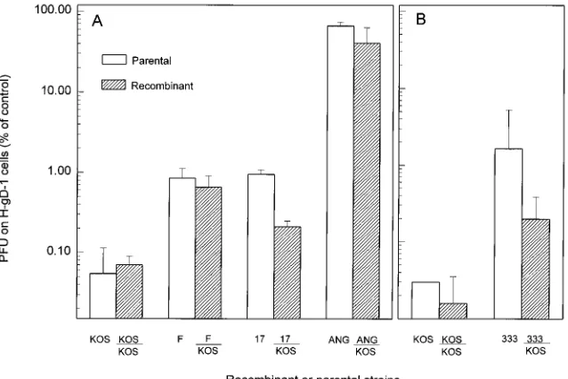
KOSgDbrescued with the KOS gD gene was as sensitive to
interference as virus obtained by transfection with intact KOS DNA (Fig. 3A). Recombinants expressing ANG gD or F gD in the KOS strain background exhibited the level of sensitivity characteristic of the gD donor strain. For the strains KOS, F, and ANG, any small differences in interference sensitivity be-tween the recombinant viruses and the parental strains that donated the gD genes were not statistically significant, whereas the differences between the F/KOS and ANG/KOS
recombi-nant viruses and KOS were statistically significant (P50.029
and P,0.001, respectively). On the other hand, the
interfer-ence sensitivity of recombinants expressing strain 17 gD in the KOS background was intermediate between that of the two parental strains (Fig. 3A). The differences in interference sen-sitivities of the recombinant and each of the parental strains
were statistically significant (P,0.001 for the comparison of
17 and 17/KOS and P,0.029 for the comparison of KOS and
17/KOS). These results show that the gD genes of F and ANG are responsible for the reduced sensitivity or nearly complete resistance, respectively, to gD-mediated interference exhibited by the parental strains. However, for 17, the reduced sensitivity to interference is determined in part by the 17 gD gene but also by another unidentified gene(s).
HSV-2(333) was less sensitive to interference than any of the HSV-1 and HSV-2 strains tested with the exception of ANG (Fig. 1 and 2). To determine if the reduced sensitivity of HSV-2(333) mapped to the gD gene, similar marker transfer exper-iments were performed to introduce the gD-2 gene into the KOS genetic background, except that a cloned recombinant gene was used. A plasmid (pHD20) was engineered, by an approach described previously (8), to carry the gD-2 open reading frame (SpeI-RmaI fragment) from HSV-2(333) DNA flanked by the HSV-1(KOS) sequences normally present up-stream (SmaI-HindIII fragment) and downup-stream (RmaI-SmaI fragment) of the gD-1 open reading frame. This plasmid or a control plasmid (pHD15) carrying the HSV-1(KOS) gD gene
(8) was cotransfected with KOSgDbDNA into Vero cells to
obtain recombinants, as described above. Plaque assays (Fig. 3B) demonstrated that, as with HSV-1(17), the interference sensitivity of the recombinant virus was intermediate between those of the parental strains. The differences between the in-terference sensitivities of the recombinant and parental viruses
were statistically significant (P50.027 for the comparison of
333/KOS with 333 and P50.029 for the comparison of 333/
KOS with KOS). These results indicate that differences in interference sensitivity between HSV-1(KOS) and HSV-2 (333) are determined in part, but not completely, by the gD gene.
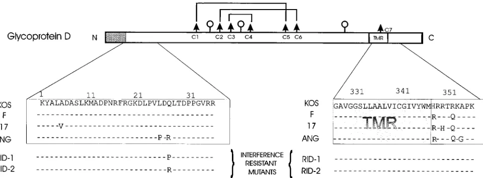
Relevant regions of the amino acid sequences for the HSV-1 gD genes are compared in Fig. 4. KOS gD and F gD differ at only two positions, both in the cytoplasmic domain. Thus, substitutions in this region of gD alone can result in at least 10-fold differences in sensitivity to interference. Excluding the cleaved signal peptide, strain 17 has four amino acid substitu-tions in gD relative to KOS, two being the same as those for F, with two additional substitutions. These additional amino acid changes appear to increase somewhat the interference sensi-tivity of recombinants containing 17 gD relative to recombi-nants containing F gD. The phenotype characteristic of strain 17 is partially but not completely transferred to KOS by the 17 gD gene, indicating that another viral gene must contribute to the phenotype.
[image:3.612.81.273.70.239.2]Strain ANG, which is almost completely resistant to gD-mediated interference, differs from KOS in its gD sequence at seven positions, five of which are illustrated in Fig. 4. Two of the amino acid substitutions in the cytoplasmic domain are the same as those for F gD relative to KOS gD. Interestingly, the two amino acid substitutions near the N terminus of ANG gD (Leu25Pro and Gln27Arg) have been observed individually in different mutants selected for resistance to gD-mediated inter-ference (1, 4, 8). The Gln27Arg substitution accounted fully for the strong interference-resistant phenotype of one mutant de-rived from HSV-1(KOS) (8), whereas the Leu25Pro substitu-tion accounted partially for the interference-resistant pheno-type of mutants derived from HSV-1(F) (1, 4). Thus, the Gln27Arg substitution noted in ANG gD could fully account for the interference-resistant phenotype of HSV-1(ANG) and
FIG. 2. Relative sensitivities of several HSV-1 strains to interference medi-ated by gD-1. Serial dilutions of each of four HSV-1 strains were plmedi-ated on uninduced cells. The titer of each virus stock as determined on H-gD-1 cells was divided by the titer determined in parallel on H-control cells and then multiplied by 100 to yield the normalized data shown here. The values given are the geometric means of three independent experiments, and the error bars indicate standard deviations. By Student’s t test, the mean for HSV-1(KOS) is signifi-cantly different, in pairwise comparisons, from the means obtained for HSV-1 strains F (P50.01), 17 (P50.002), and ANG (P,0.001).
on November 9, 2019 by guest
http://jvi.asm.org/
the ANG/KOS recombinants. HSV-2(333) gD differs from HSV-1(KOS) gD at about 70 positions. The available data do not permit conclusions about the domains of gD-2 that might be responsible for the reduced sensitivity to interference ex-hibited by the 333/KOS recombinants. It should be noted that 333 gD is identical to KOS gD at positions 25 and 27 but differs from KOS gD in the region of the cytoplasmic domain adjacent to the membrane.
The structural determinants of gD required for mediating interference, on the one hand, and for mediating entry of HSV and influencing the sensitivity of virus to interference, on the other hand, can be considered separately and may even be distinct. As discussed above, all forms of alphaherpesvirus gD characterized to date have been shown to mediate interference with homologous and certain heterologous alphaherpesviruses. In some instances, heterologous interference may be more pronounced than homologous interference, at least with the challenge virus strains used. The fact that divergent as well as closely related forms of gD can interfere with infection by a single herpesvirus suggests that the various forms of gD can interact with a common target in cells or virions. The relative interference activities of different variants of cell-associated gD are difficult to assess with cloned transformed cell lines because of variations in the level of gD expressed and in cell factors that may influence cell susceptibility to infection. Def-inition of the gD determinants required for mediating
inter-ference will depend on devising strategies for more controlled expression of various mutant and wild-type forms of gD in uncloned cell populations and for quantitating interference under these conditions.
The roles of gD and other unidentified HSV genes in influ-encing the sensitivity of HSV to interference emerge from this study of different HSV strains and from earlier studies of HSV-1 mutants selected for resistance to gD-mediated inter-ference. Most of the mutants have amino acid substitutions in gD close to the N terminus at positions 25 (Leu25Pro) or 27 (Gln27Pro/Arg) (Fig. 4) (1, 4, 8). Two mutants have Ala185Thr substitutions in gD. Both the Leu25Pro and Ala185Thr substi-tutions appeared to be only partially responsible for the re-duced sensitivity of the mutant viruses to gD-mediated inter-ference, as assessed by plaque formation (1, 23), whereas the Glu27Pro/Arg substitutions alone could confer the interfer-ence-resistant phenotype (8).
[image:4.612.148.467.71.284.2]HSV-1(ANG) is the only wild-type strain of HSV-1 shown to have amino acids other than Leu and Gln at positions 25 and 27 and also shown to be almost completely resistant to gD-mediated interference. HSV-1(ANG) was isolated from a clin-ical specimen that was plated under an agar overlay (19, 20). Because agar contains sulfated polysaccharides that can inhibit HSV infection, the possibility exists that the method of isola-tion selected a variant with altered entry characteristics. Mu-tants with reduced sensitivity to gD-mediated interference
FIG. 3. Relative sensitivities to gD-1-mediated interference of parental viruses and recombinant viruses carrying different alleles of gD in the KOS genomic background. (A) Recombinant viruses in which the gD genes from HSV-1 strains KOS, F, 17, and ANG were placed in the KOS genomic background were constructed. Subconfluent Vero cells in six-well dishes were transfected with 1mg of genomic DNA from a gD-negative mutant, KOSgDb(8), together with 1mg of PCR-amplified gD or gL DNA (as a control) with LipofectAMINE reagent (GIBCO BRL) in OptiMEM serum-free medium according to the manufacturer’s guidelines. In the generation of each recombinant, three separate PCR amplifications were performed, and the PCR products were used in three independent cotransfections with KOSgDbDNA. The primers used to amplify the gD-1 gene had the sequences GAATTCCAGGTCTTCCTCCACTACGC and GGATCCATGTCTGCTTGAGCTC CTGC, and those used for gL-1 amplification had the sequences TGGGTGCGCGGATCCTAGGCGC and AGAGGGAATTCCCTCTCCCCC. Parental virus stocks were produced by transfecting Vero cells as described above with 1mg of parental genomic DNA. Medium on the transfected cells was supplemented with medium 199 containing 10% fetal calf serum at 6 h after transfection and replaced with the same medium at 12 h. At 48 h, the transfected cells were lysed to release progeny virus, which was amplified by one passage on Hep-2 cells. The resulting virus stocks were counted on H-gD-1 and H-control cells. The titer of each viral stock as determined on H-gD-1 cells was divided by the titer determined in parallel on H-control cells and then multiplied by 100 to yield the normalized data shown here. The values shown are the geometric means from three independent determinations and standard deviations. (B) Recombinant viruses in which the gD gene of HSV-2(333) was placed in the KOS genetic background were constructed. Subconfluent Vero cells in six-well dishes were transfected with 1mg of KOSgDbDNA together with 0.5 to 3mg of pHD20, which contains the HSV-2(333) gD open reading frame flanked by HSV-1(KOS) DNA sequences, or pHD15 (8), which contains the HSV-1(KOS) gD open reading frame and flanking sequences. Vero cells were also transfected with genomic DNAs of the parental strains. The DNA samples were prepared for transfection by calcium phosphate coprecipitation as described previously (8). Virus stocks prepared from the transfected cells were counted on H-gD-1 cells and H-control cells as described above. The values shown are as described above and are the geometric means of at least three determinations with standard deviations. For the data presented in both panels, statistical analysis (Student’s t test) was performed to assess the significance of differences in interference sensitivity observed between recombinant and parental strains, with the results summarized in the text.
on November 9, 2019 by guest
http://jvi.asm.org/
have been isolated by methods other than selection for repli-cation on gD-expressing cells, including passage in the pres-ence of the sulfated polysaccharide heparin (21), passage in the presence of a particular anti-gD monoclonal antibody, AP7 (4, 18), and selection for replication on BHK clones found to be resistant to HSV entry (23). The AP7-resistant mutants have Leu25Pro substitutions in gD (1, 4, 18), and the mutant se-lected for replication on the BHK clone has a Ser140Asn substitution in gD (23). This latter mutation contributed only marginally to the interference-resistant phenotype of the mu-tant (1, 23). The mutations responsible for the reduced inter-ference sensitivity of the heparin-selected mutants have not yet been identified, but some of them appear not to map to gD (21a).
It can be concluded that virion-associated gD is a key de-terminant of sensitivity of HSV to gD-mediated interference. Because some amino acid substitutions in gD are only partially responsible for the mutant phenotype, certain other HSV genes must also influence sensitivity, either alone or in com-bination with the appropriate gD allele. Obvious candidates for these other genes include those encoding the other viral glycoproteins that mediate HSV entry, including gB, gC, and gH-gL. The possibility exists that genes encoding tegument proteins may also be relevant, particularly in light of the find-ing that amino acid substitutions in the cytoplasmic or viro-plasmic domain of gD can result in 10-fold differences in sen-sitivity to interference. This domain of gD could be expected to interact with tegument proteins. Alterations in these interac-tions could influence the conformation and function of the gD ectodomain and could also influence the amount of gD incor-porated into virions. Amino acid substitutions in the gD ectodomain, particularly those at Gln-27, have the most pro-found effect and determine whether HSV can clear or bypass the block to entry imposed by cell-associated gD.
The first two authors contributed equally to this work.
We thank Nanette Susmarski for excellent assistance with cell cul-ture and preparation of virus stocks. We are grateful to G. Cohen and
R. Eisenberg for the gift of plasmid pWW65, which was used to construct pRJ4065p.
This work was supported by grants from the National Cancer Insti-tute (CA 21776), the American Cancer Society, and the National Institute for Allergy and Infectious Diseases (AI 36293). H.J.D., M.S.W., and R.M.J. were trainees, supported by grants T32 GM08061, T32 GM08152, and T32 GM07281, respectively.
REFERENCES
1. Brandimarti, R., T. Huang, B. Roizman, and G. Campadelli-Fiume. 1994. Mapping of herpes simplex virus 1 genes with mutations which overcome host restrictions to infection. Proc. Natl. Acad. Sci. USA 91:5406–5410. 2. Brunetti, C. R., R. L. Burke, S. Kornfeld, W. Gregory, F. R. Masiarz, K. S.
Dingwell, and D. C. Johnson.1994. Herpes simplex virus glycoprotein D acquires mannose 6-phosphate residues and binds to mannose 6-phosphate receptors. J. Biol. Chem. 269:17067–17074.
3. Campadelli-Fiume, G., M. Arsenakis, F. Farabegoli, and B. Roizman. 1988. Entry of herpes simplex virus 1 in BJ cells that constitutively express viral glycoprotein D is by endocytosis and results in degradation of the virus. J. Virol. 62:159–167.
4. Campadelli-Fiume, G., S. Qi, E. Avitabile, L. Foa`-Tomasi, R. Brandimarti, and B. Roizman.1990. Glycoprotein D of herpes simplex virus encodes a domain which precludes penetration of cells expressing the glycoprotein by superinfecting herpes simplex virus. J. Virol. 64:6070–6079.
5. Chase, C. C. L., K. Carter-Allen, C. Lohff, and G. J. Letchworth. 1990. Bovine cells expressing bovine herpesvirus 1 (BHV-1) glycoprotein IV resist infection by BHV-1, herpes simplex virus, and pseudorabies virus. J. Virol. 64:4866–4872.
6. Chase, C. C. L., C. Lohff, and G. J. Letchworth. 1993. Resistance and susceptibility of bovine cells expressing herpesviral glycoprotein D homologs to herpesviral infections. Virology 194:365–369.
7. Cohen, G. H., D. Long, J. T. Matthews, M. May, and R. Eisenberg. 1983. Glycopeptides of the type-common glycoprotein gD of herpes simplex virus types 1 and 2. J. Virol. 46:679–689.
7a.Dean, H. J. 1992. Ph.D. dissertation, Northwestern University, Chicago, Ill. 8. Dean, H. J., S. Terhune, M.-T. Shieh, N. Susmarski, and P. G. Spear. 1994. Single amino acid substitutions in gD of herpes simplex virus 1 confer resistance to gD-mediated interference and cause cell type-dependent alter-ations in infectivity. Virology 199:67–80.
9. Eisenberg, R. J., D. Long, R. Hogue-Angeletti, and G. H. Cohen. 1984. Amino-terminal sequence of glycoprotein D of herpes simplex virus types 1 and 2. J. Virol. 49:265–268.
10. Izumi, K. M., and J. G. Stevens. 1990. Molecular and biological character-ization of a herpes simplex virus type 1 (HSV-1) neuroinvasiveness gene. J. Exp. Med. 172:487–496.
[image:5.612.71.551.71.248.2]10a.Johnson, R. M. 1989. Ph.D. dissertation, University of Chicago, Chicago, Ill. FIG. 4. Diagrammatic representation of the gD-1 polypeptide, with comparisons of the amino acid sequences in selected regions for several HSV-1 strains. The gD translation product has a cleavable N-terminal signal peptide (shaded box) and a transmembrane domain (TMR) near the C terminus (9, 15, 29). The three sites for the addition of N-linked glycans (lollipops) are utilized (7, 15, 24). Six of the seven cysteine residues form disulfide linkages as shown (lines joining the arrows) (13). The nucleotide sequences for the KOS, F, 17, and ANG gD genes have been published (4, 8, 10, 16). The amino acid sequences are shown for selected regions as indicated. All amino acid substitutions outside the signal peptide, relative to the KOS sequence, are shown except for two additional amino acid substitutions in the ANG gD gene (Glu52Arg and Ala109Gly). Amino acid substitutions responsible for the interference-resistant phenotype of two mutants (Rid-1 and Rid-2) obtained from KOS (8) are also shown. For this study, we determined the nucleotide sequence of the F gD gene, with the methods and primers described previously for sequencing of the KOS gD gene (8). Our results were the same as those reported previously (4) except for a silent nucleotide substitution (use of a CTG codon for Leu at position 228 instead of a CTC codon). The accession number for the F gD nucleotide sequence deposited with GenBank is L09242.
on November 9, 2019 by guest
http://jvi.asm.org/
11. Johnson, R. M., and P. G. Spear. 1989. Herpes simplex virus glycoprotein D mediates interference with herpes simplex virus infection. J. Virol. 63:819– 827.
12. Lee, W.-C., and A. O. Fuller. 1993. Herpes simplex virus type 1 and pseu-dorabies virus bind to a common saturable receptor on Vero cells that is not heparan sulfate. J. Virol. 67:5088–5097.
13. Long, D., W. C. Wilcox, W. R. Abrams, G. H. Cohen, and R. J. Eisenberg. 1992. Disulfide bond structure of glycoprotein D of herpes simplex virus types 1 and 2. J. Virol. 66:6668–6685.
14. Ludwig, T., H. Munier-Lehmann, U. Bauer, M. Hollinshead, C. Ovitt, P. Lobel, and B. Hoflack.1994. Differential sorting of lysosomal enzymes in mannose 6-phosphate receptor-deficient fibroblasts. EMBO J. 13:3430–3437. 15. Matthews, J. T., G. H. Cohen, and R. J. Eisenberg. 1983. Synthesis and processing of glycoprotein D of herpes simplex virus types 1 and 2 in an in vitro system. J. Virol. 48:521–533.
16. McGeoch, D. J., A. Dolan, S. Donald, and F. J. Rixon. 1985. Sequence determination and genetic content of the short unique region in the genome of herpes simplex virus type 1. J. Mol. Virol. 181:1–13.
17. Mettenleiter, T. C. 1994. Initiation and spread ofa-herpesvirus infections. Trends Microbiol. 2:2–3.
18. Minson, A. C., T. C. Hodgman, P. Digard, D. C. Hancock, S. E. Bell, and E. A. Buckmaster.1986. An analysis of the biological properties of mono-clonal antibodies against glycoprotein D of herpes simplex virus and identi-fication of amino acid substitutions that confer resistance to neutralization. J. Gen. Virol. 67:1001–1013.
19. Munk, K., and D. Donner. 1963. Cytopathischer Effekt und Plaque-Mor-phologie verschiedener Herpes-simplex-Virus-Sta¨mme. Arch. Gesamte Vi-rusforsch. 13:529–540.
20. Munk, K., and G. Ludwig. 1972. Properties of plaque variants of herpes virus hominis strains of genital origin. Arch. Gesamte Virusforsch. 37:308–315.
21. Pertel, P. E., and P. G. Spear. 1994. Variants of herpes simplex virus type 1 selected by passage in soluble heparin (abstract). The 19th International Herpesvirus Workshop, Vancouver, British Columbia, Canada.
21a.Pertel, P. E., and P. G. Spear. Unpublished data.
22. Petrovskis, E. A., A. L. Meyer, and L. E. Post. 1988. Reduced yield of infectious pseudorabies virus and herpes simplex virus from cell lines pro-ducing viral glycoprotein gp50. J. Virol. 62:2196–2199.
23. Roller, R. J., and B. Roizman. 1994. A herpes simplex virus 1 US
11-express-ing cell line is resistant to herpes simplex virus infection at a step in viral entry mediated by glycoprotein D. J. Virol. 68:2830–2839.
24. Sodora, D. L., G. H. Cohen, and R. J. Eisenberg. 1989. Influence of aspar-agine-linked oligosaccharides on antigenicity, processing, and cell surface expression of herpes simplex virus type 1 glycoprotein D. J. Virol. 63:5184– 5193.
25. Sodora, D. L., R. J. Eisenberg, and G. H. Cohen. 1991. Characterization of a recombinant herpes simplex virus which expresses a glycoprotein D lacking asparagine-linked oligosaccharides. J. Virol. 65:4432–4441.
26. Spear, P. G. 1993. Membrane fusion induced by herpes simplex virus, p. 201–232. In J. Bentz (ed.), Viral fusion mechanisms. CRC Press, Inc., Boca Raton, Fla.
27. Spear, P. G. 1993. Entry of alphaherpesviruses into cells. Semin. Virol. 4:167–180.
27a.Susmarski, N., and P. G. Spear. Unpublished data.
28. Tikoo, S. K., D. R. Fitzpatrick, L. A. Babiuk, and T. J. Zamb. 1990. Molec-ular cloning, sequencing, and expression of functional bovine herpesvirus 1 glycoprotein gIV in transfected bovine cells. J. Virol. 64:5132–5142. 29. Watson, R. J., J. H. Weis, S. S. Salstrom, and L. W. Enquist. 1982. Herpes
simplex virus type 1 glycoprotein D gene: nucleotide sequence and expres-sion in Escherichia coli. Science 218:381–383.