ANALYSIS OF 50 CASES OF STROKE IN
YOUNG ADULTS
Dissertation submitted to
THE TAMILNADU DR. M.G.R. MEDICAL UNIVERSITY
for
M.D. Degree in General Medicine (Branch I)
The Tamilnadu
Dr. M.G.R. Medical University
ANALYSIS OF 50 CASES OF STROKE IN
YOUNG ADULTS
Dissertation submitted to
THE TAMILNADU DR. M.G.R. MEDICAL UNIVERSITY
for
M.D. Degree in General Medicine (Branch I)
The Tamilnadu
Dr. M.G.R. Medical University
Chennai March 2009
Coimbatore Medical College
Coimbatore - 641 014DEPARTMENT OF GENERAL MEDICINE
COIMBATORE MEDICAL COLLEGE HOSPITAL
CERTIFICATE
This is to certify that the Dissertation entitled “Analysis of 50 cases of stroke
in young adults” herewith submitted by Dr.N.SENTHILRANI., Post Graduate in
General Medicine, Coimbatore Medical College to the Tamilnadu Dr.M.G.R.Medical
University is a record of a bonafide research work carried out by her under my guidance
and supervision from APRIL 2007 to SEP 2008.
Dr.M.RAMASAMY.M.D Dr.K.UMAKANTHAN.M.D.,
Professor and Unit Chief Professor and Head of Department of Medicine. Department of Medicine.
DEAN
DECLARATION
I solemnly declare that the dissertation titled “Analysis of 50 Cases of Stroke in
Young Adults” was done by me at Coimbatore Medical College & Hospital during the
period from April 2007 to September 2008 under the guidance and supervision of Prof.
Dr. K. Umakanthan M.D., and Prof. M. Ramasamy M.D.
This dissertation is submitted to the Tamilnadu Dr. MGR Medical University
towards the partial fulfillment of the requirement for the award of MD Degree in
General Medicine (Branch I)
Place: Coimbatore Dr. N. SENTHILRANI
ACKNOWLEDGEMENT
I express my regards and gratitude to Dean, Coimbatore Medical College for
permitting and supporting me to conduct this study.
I am indebted to Professor and Head of Department of medicine,
Dr.K.Umakanthan MD, Professor Dr.M.Ramasamy MD, and Unit chiefs – Department
of medicine for their valuable guidance, encouragement, inspirations and support
without which this effort would not have been possible.
I am extremely grateful for the whole hearted support and valuable feedback,
suggestions and guidance from professor and Head of Department of Neuromedicine
Dr.K.Govindarajan MD, DM.
I express my sincere thanks and gratitude to my assistant professors, Dr.S.Usha,
Dr.T.Ravikumar MD, Dr.K.Jamunarani MD, Mr.P.S.Manshur MD, Dr.R.Ramakrishnan
DM, Dr.M.Sacratis DM for their valuable guidance.
I wish to express my thanks to all coPGs of the department for their kind
cooperation.
Last but not the least my gratitude and thanks to the patients and their attenders
for their whole hearted co-operation, without which this study would not have been
CONTENTS
Page No.
1. INTRODUCTION 1
2. AIM OF STUDY 3
3. MATERIALS AND METHODS 4
4. REVIEW OF LITERATURE 7
5. OBSERVATION AND ANALYSIS 42
6. DISCUSSION 52
7. CONCLUSION 56
8. BIBLIOGRAPHY
INTRODUCTION
Stroke is one of the leading causes of morbidity and ranks next only to Coronary
Artery Disease and Malignancy as the leading cause of mortality worldwide. At least 50
percent of the neurological disorders in a general hospital are due to stroke. As
remarked by a renowned neurologist C.M.Fisher, neurology is learnt “Stroke by Stroke”.
Cerebrovascular diseases occur predominately in the middle and late years of life.
The incidence of stroke increases with age; thus the disability affects many people in
their “golden years”, a segment of the population that is growing rapidly in western
countries. Categories of cerebrovascular diseases include ischaemia – infarction and
intracranial haemorrhage . Many of the arterial and cardiac disorders underlying these
diseases are preventable; the morbidity and mortality from cerebrovascular diseases has
been diminishing in recent years, apparently because of better recognition and treatment
of hypertension.
Most cerebrovascular diseases are manifest by the abrupt onset of a focal
neurologic deficit. The deficit may remain fixed or may rapidly improve or
progressively worsen. It is this abrupt onset of a non-occlusive and focal neurologic
deficit that defines a stroke, or cerebrovascular accident (CVA).
Stroke in young adults is uncommon but by no means rare: some 4% of all strokes
occur under the age of 40 years. There are a number of additional causes which should
be specifically considered in young adults beyond those common in adults. These
include neck trauma causing carotid dissection, alcohol intoxication, infarction in
Rhematic heart disease, prolapsing mitral valve or atrial myxoma.
The term stroke denotes the sudden and dramatic development of a focal
neurological deficit due to cerebrovascular disorder. The advent of imaging procedures
such as computerized tomography, magnetic resonance imaging and carotid Doppler
made the evaluation of stroke and its risk factors easier.
However young strokes pose a major socioeconomic challenge, occupational
neurorehabilitational programme of stroke survivors and also have varying aetiology and
prognosis in comparison with stroke elderly also. It is one of the major causes of
AIM OF THE STUDY
1. To evaluate the risk factors of Stroke in young adults.
2. To study the different mode of clinical presentation or types of Stroke in young
adults.
3. To study the pathogenesis of young stroke with the aid of investigations, treatment
outcome and prognosis.
MATERIALS AND METHODS
The study was conducted in Coimbatore Medical College Hospital during the
period of 2007 - 2008. The bed strength of this Hospital is 1020 and about 1200 patients
are cared as inpatients, daily 4500 outpatients come to this hospital for treatment. This
center is rendering medical services to a fairly large size of population nearly 10 lakhs,
cater
ing to the population in and around Coimbatore and nearby areas of Kerala including
Palghat. This being a post-graduate training center, the fulltime services of a team of
qualified neurologist and experienced medical personnel are available round the clock.
This study was possible because of full cooperation and enthusiasm of various
departments like Neurology, Radiology and with available facilities of this college.
The study includes fifty patients of stroke in patients below the age of 40 years
admitted in medical wards and Neurology ward. Ethics committee approval was
obtained for conducting the study. Informed consent was obtained from all the patients.
Patients in the paediatric age group are not included. A carefully elicited history and
repeated clinical examinations were used in ascertaining the temporal profile of the
disease and the probable area of brain that is affected. A detailed history taking was
done for risk factors like Smoking, Alcohol, Rheumatic valvular heart disease,
Tuberculosis and family history of cerebrovascular disease.
A clinical search for extra-cranial carotid artery narrowing, cardiac diseases which
hypertension, diabetes mellitus, Rheumatic valvular heart disease and other risk factors
were made.
Laboratory evaluation and other investigations :
After detailed history and meticulous neuromedical examination including
palpation and auscultation of brachiocephalic and peripheral pulses, the relevance and
priority of any laboratory test in acute stroke should be at the clinician’s judgement. The
value of careful ophthalmoscopic examination of the retina and its vasculature for
disease and embolic fragments needs emphasis.
1. Urine examination for albumin and sugar.
2. Estimation of blood sugar and serum lipid profile levels for detecting diabetes and
hyperlipidemia which are predisposing causes of atherosclerosis.
3. Serological test for syphilis was done in all patients.
4. Haemtological studies like complete hemogram with platelet count, ESR,
bleeding / clotting and prothrombin time to find out conditions like
polycythaemia, anaemia, thrombocytopenia and bleeding disorders were
undertaken.
5. When elevated ESR was present, further test were made to identify the causes like
systemic lupus erythematosus, subacute baterial endocarditis and tuberculous
meningitis.
6. Two dimensional echocardiography for detecting cardiac source of emboli was
7. ECG for detecting rhythm disorders & underlying cardiac disorder were done.
8. CSF analysis was used to determine meningeal infection causing stroke.
9. Carotid angiography was done only in those patients in whom a vascular
malformation, subdural haematoma, intracranial tumour or extracranial vascular
disease was suspected.
10. CT Scan of brain was done to assess the lesion was infartion or haemorrhage and
to locate the site of lesion.
11.HIV testing was done in all patients.
12.Sr.Homocystein level was estimated.
13.MRI Brain / MR Angiogram / MRVenogram – were done.
14.TEE was done.
REVIEW OF LITERATURE
GENERAL CONSIDERATIONS1-8
DEFINITION
Stroke is defined as a focal (or at times global Neurological impairment of sudden
onset, lasting more than 24 hr (or leading to death and of presumed vascular origin)25.
The term Transient Ischaemic Attack (TIA) implies focal neurological deficit with
complete recovery of cerebral function within 24 hrs. Some of the sub types of stroke
include cerebral haemorrhage, cerebral infarction and sub arachnoid haemorrhage.
The normal functioning of the brain is dependant upon a relatively constant
supply of oxygen, glucose and other nutrients derived from the blood perfusing it.
Normal blood flow is brain to 55 – 70 ml/min. If for any reason the blood flow is
critically reduced below 15 ml per 100g per minute, the resulting ischaemia with
hypoxia when sufficiently prolonged, may cause death of neurons and glia (cerebral
infarction).
The mean arterial blood pressure, cerebrovascular and tissue resistance, local
metabolic products (pH, paO2, etc) together with several known and unknown factors
help to maintain the critical threshold of blood flow for energy metabolism. Further
more the blood flow varies in different areas of the brain and auto regulation determines
the regional blood flow to meet local metabolic need.
In regions of cerebral ischaemia there is “Paralysis of auto regulation” and the
agents and other forms of stimuli. The cerebral vasculature in this ischaemic zone
becomes permeable to proteins, and fluid leaks in the vicinity leading to extracellular
oedema. Such events also lead to local haemoconcentration and vascular stasis.
Hence cerebral infarction is not merely the result of ischaemia from occluded
blood vessels but an end result of a series of highly complex ischaemia modifying
events.
BLOOD SUPPLY OF THE BRAIN : (FIG : 1)
At rest the brain which is only 2 percent of total body weight receives 15 percent
of the cardiac output and consumes about 25 percent of the total inspired oxygen. This
rich blood supply is carried by two internal carotid and two vertebral arteries which
anastamose at the base of brain to form the ‘circle of Willis’. The carotid arteries supply
the anterior and the vertebro basilar arterial system supplies the posterior portion of the
Figure : 1 - Base of Brain Showing Circle of Willis
The branches of the internal carotid artery are
a. The Opthalmic artery
b. Anterior cerebral artery
c. Middle cerebral artery
d. Anterior choroidal artery
e. Posterior Communicating artery
The vertebral artery which arises from the subclavian artery, enters foramen
magnum and unites with the opposite vertebral artery at the pontomedullary junction to
form the basilar artery. Vertebral artery gives rise to anterior and posterior spinal
arteries, the posterior inferior cerebellar artery and small penetrating arteries to the
medulla. The basilar artery ascends up to the pontomidbrain junction in the
interpeduncular cistern and divides into the two posterior cerebral arteries. Numerous
small branches penetrate the brainstem and cerebellum. It also gives rise to the anterior
inferior cerebellar artery, the internal auditory artery and the superior cerebellar artery.
The meninges are supplied by branches of the internal carotid, external carotid and
vertebral arteries.
THE COLLATERAL BLOOD SUPPLY OF THE BRAIN
Normally each ICA provides blood to the anterior two thirds of the cerebral
hemisphere on the ipsilateral side. There is little mixing of blood via the posterior
can develop distal to major artery occlusion. The development of collaterals is more
effective if the vessel occlusion occurs insidiously rather than suddenly. But unlike the
normal cerebral blood supply, the functional capacity of the collateral blood supply to
respond to changes in perfusion pressure is limited.
COLLATERAL BLOOD FLOW MAY DEVELOP VIA
1. The circle of Willis. However about 50 percent of circles have one or more
hypoplastic segments (usually one of the communicating arteries) and also since
atheroma commonly affects the circle of Willis, the potential for collateral flow is
not always good.
2. Around the orbit, branches of the ECA anastomose with branches of the
Opthalmic artery if the ICA is severely stenosed.
3. Muscular branches of the vertebral artery in the neck distal to an obstruction of
that artery, receive blood from occipital and ascending pharyngeal of ECA.
4. Leptomeningeal anastomoses on the surface of the brain may develop between
cortical branches of the anterior, middle and posterior cerebral arteries.
5. Dural anastomoses can develop between meningeal branches of the precapillary
bed of the vertebral arteries.
6. Parenchymal anastomoses occasionally develop in the precapillary bed of the
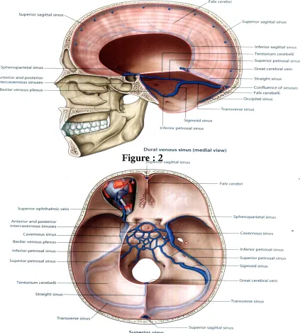
VENOUS DRAINAGE OF THE BRAIN :
[image:17.612.74.497.165.634.2]Figure : 2
Venous blood flow peripherally via the superficial cerebral veins and centrally via
the deep cerebral veins into the venous sinuses which in turn drain into the internal
jugular vein (Fig 2,3). The cerebral veins are thin walled have no valves and the blood
flow is often in the same direction as in the neighbouring arteries. There are numerous
venous connections between the veins and dural sinuses as well as with the venous
system in the meninges, skull, scalp and nasal sinuses so facilitating the propagation of
thrombus or spread of infection between these vessels.
EPIDEMIOLOGY :
This lagged behind coronary heart disease because
1. Stroke is a disorder of late middle age and elderly where other diseases coexist.
2. Stroke pathologically is more diverse and may be due to intracerebral small vessel
disease or embolism from the heart or primary intracerebral haemorrhage.
MORTALITY :
Mortality rises rapidly with age.
INCIDENCE :
Stroke in young adult is surprisingly common. Stroke incidence rises with age
with about 75% of the stroke in adults occur between the age group of 26 to 40 years
and about 25% occurring below the age of 26 years. Ischaemic stroke is much more
GEOGRAPHICAL RACIAL AND SOCIAL INFLUENCES :
Primary intra cerebral haemorrhage is less common in Western countries than in
Japan and China.
In India there is some evidence that stroke is particularly common in young
people. During pregnancy stroke is commonly associated with CVT.
TRENDS IN MORTALITY AND INCIDENCE :
Incidence is declining with assumption that the prevalence of hypertension is less
than it was and early diagnosis and treatment of hypertension may also be responsible.
The prevalence of Rheumatic heart disease also is less than it was earlier.
SEASONAL AND DIURNAL VARIATION :
Stroke incidence and mortality is more in winter, the possible explanation being
the effect of temperature, pollution and higher blood pressure in the winter. The
increased mortality may also be due to complications of stroke such as pneumonia
which is more common in winter.
Cerebral infarction occurs most commonly in the hour or two after waking in the
morning. Subarachnoid haemorrhage is unlikely to occur during sleep. Circadian
changes in physical activity, catecholamine level, blood pressure, blood viscosity and
platelet aggregation may explain.
Intracerebral haemorrhage is more likely to occur during strenuous activity than
ETIOLOGY OF STROKE IN YOUNG ADULTS :
The range of potential etiologies for stroke in young adults is broader than that for
older adults. Like in older adults, stroke in younger adults is typically categorized as
primarily ischaemic or haemorrhagic . Ischaemic etiologies include cardioembolic,
atherosclerotic disease24, and nonatherosclerotic cerebral vasculopathies.
ETIOLOGY OF STROKE IN YOUNG ADULTS :
ISCHAEMIC
26-34
Cardiac disease
LARGE VESSEL DISEASE :
Premature atherosclerosis
Dissection (Spontaneous or traumatic)
Inherited metabolic disease (Homocystinuria, Fabry’s, Pseudoxanthoma elasticum, MELAS syndrome)
Fibromuscular dysplasia
Infection (bacterial, fungal, tuberculosis, syphilis, Lyme’s disease)
Vasculitis (Collagen vascular diseases – systemic lupus erythematosus,
rheumatoid arthritis, Sjogren’s syndrome, polyarteritis nodosa; Takayasu’s
disease, Wegener’s syndrome, cryoglobulinemia, sarcoidosis, inflammatory bowel
disease, isolated central nervous system angitis)
Moyamoya disease
Toxic (Drugs)
SMALL VESSEL DISEASE :
Vasculopathy (infectious, noninfectious, microangiopathy)
HAEMATOLOGIC DISEASE :
Sickle-cell disease
Leukemia
Hypercoagulable states
Disseminated intravascular coagulation
Thrombocytosis
Polycythemia Vera
Paroxysmal nocturnal haemoglobinuria
Thrombotic thrombocytopenic purpura
Venous occlusion
MIGRAINE16
HAEMORRHAGIC
Subarachnoid haemorrhagic (cerebral aneurysm)
Intraparenchymal haemorrhage .
Arteriovenous malformation
Haematological Disease
Drug use
Iatrogenic (peri-procedural)
CARDIAC DISEASE :13, 14
It is the most common cause of stroke in young adult.
It includes :
1. Congenital heart disease
2. Rheumatic valve disease
3. Mitral valve prolapse 9
4. Patent foramen ovale
5. Endocarditis
6. Atrial myxoma
7. Arrhythmias
8. Cardiac Surgery
9. Cardiomyopathy
10.Prosthetic Valves 10
11.Mural thrombi after myocardial infarct17
Of these, rheumatic mitral valvular disease with atrial fibrillation, prosthetic
valves, subacute bacterial endocarditis and intracardiac tumour are universally
recognized causes of emboli. The role of disorders such as non-valvular atrial
prolapse the cerebral symptoms may arise from non-septic or septic emboli and
paroxysmal arrythmias associated with this condition may play a role.
Cardiomyopathies are being increasingly recognized as a cause of embolic stroke in
young adults. Cardiogenic cerebral emboli is the most frequent cause of stroke
recurrence.
CARDIOGENIC EMBOLIC OCCLUSION :
The determination that a stroke is embolic in origin generally results from the
demonstration of an appropriate cardiac abnormality in a patient with a stroke, featuring
characteristics suggestive of an embolus. Clinical features of a stroke which suggest a
possible cardiac embolic origin are –
1) Sudden onset with maximal neurological deficit, appearing immediately.
2) History of multiple episodes of TIA of different pattern. Multifocal cerebral
infarctions especially in a patient with an associated systemic arterial
occlusion, (eg) renal arteries or limb arteries.
PREMATURE ATHEROSCLEROSIS :
Though atherosclerosis is the major cause of stroke in the elderly age group, it
does occur in young patients too, especially when predisposing factors like
hypertension12, diabetes and hyperlipidemia18 are present. Young men who smoke
heavily suffer atherosclerotic brain infarction six times more frequently than
non-smokers11. Non modifiable risk factors include age, race and male sex. Genetics may
DISSECTION OF INTRACRANIAL CEREBRAL ARTERIES :
Dissection of intracranial cerebral arteries most often affect the middle cerebral
and basilar arteries with the clinical presentation of acute infarction. These subintimal
dissections of unknown cause develop in otherwise healthy individuals. Dissection of
the major extracranial cerebral arteries may be spontaneous dissections, occur most
often in patients younger than 40 years.
INHERITED METABOLIC DISEASES :
HOMOCYSTINURIA : 35-37, 49-51
Homocystinuria predisposes to cerebral arterial or less often venous thrombosis as
well as to thromboemboli in other organs. The mechanisms are not delineated but the
estimated risk of stroke at a young age is between 10% and 16%. The diagnosis is
usually made in childhood because of other stigmata such as thin Marfan-like
appearance, malar flush, dislocated ocular lens, bony deformities, mental retardation and
seizures.
FIBROMUSCULAR DYSPLASIA :
Fibromuscular dysplasia is a non-atheromatous vascular disorder, in which there
is intimal and medial fibroplasia of the extracranial internal carotid artery and other large
systemic vessels. The intracranial arteries are usually spared. The disorder is far more
common in women. The cause is not understood.
CEREBRAL & MENINGEAL INFECTIONS :
inflammatory changes in the vessel walls. Meningovascular syphilis and tuberculous
infections are common causes. Pyogenic infections are infrequent causes. Another
infection that may result in cerebral arteritis is mucormycosis. Other rare causes of
cerebral infarction are fungus, typhus, schistosomiasis, falciparum malaria and
trichinosis.
TAKAYASU’S ARTERITIS :
Takayasu’s arteritis (pulsesless disease or aotic arch syndrome) is a giant cell
arteritis that may result in narrowing and thrombosis of large branches of the aortic arch
at their origins and aneurysmal formation, signs of ischaemia of the head and arms
include cataracts, retinal and optic atrophy, transient monocular blindness, focal cerebral
symptoms and hypertension in the legs with intermittent claudication in the arms
(reverse coarctation).
MOYAMOYA DISEASE :
Moyamoya disease is a syndrome of stenosis of the vessels in and around the
circle of Willis, with profuse telangiectatic collacteral vessels at the base of brain that
have the angiographic appearance of a puff of smoke, Microaneurysms may develop and
either infarction or subarachonoid haemorrhage may result.
DRUGS :
Drugs that have been associated with stroke include methamphetamines, LSD,
heroin, oral contraceptives15, and anticoagulants. Methamphetamine induces a
haemorrhages. LSD which is an ergot alkaloid produces arteriospasm and heroin
produces allergic vascular hypersensitivity leading to cerebral infarction. The risk of
stroke with the use of oral contraceptives is increased five to nine fold for thrombosis
and two fold for haemorrhage.
VASCULOPATHY :
Apart from atherosclerosis there are other vascular diseases which can produce
stroke in young. They are –
1) Arteritis due to –
a) Collagen disorders.
b) Infections – Meningovascular syphilis
Tuberculous arteritis
Pyogenic infection
Fungal - Mucormycosis
c) Takayasu’s arteritis
d) Drug induced vasculitis – methamphetamine
e) Irradiation
2) Ruptured saccular aneurysm
3) Moyamoya disease
4) Dissection of intracranial cerebral arteries.
The collagen vascular disease most frequently causing stroke is systemic lupus
erythematosus. There is an increased risk of thrombosis in patients with an
immunoglobulin called “lupus anticoagulant”. The incidence of stroke is less in
polyarteritis nodosa with 13% reportedly experiencing cerebral infarction or
haemorrhage. Stroke is infrequent in scleroderma and rheumatoid arthritis.
SICKLE CELL DISEASE :
Sickle cell disease can cause ischaemic infarction, intracerebral haemorrhage,
venous sinus and cortical vein thrombosis and subarachnoid haemorrhage. The overall
incidence of stroke in sickle cell disease is between 6% to 15%. The risk of cerebral
infarction is greatest in children with sickle cell anaemia. Stroke occurs infrequently in
sickle cell patients older than 20 years, complications are more common in adults. In
addition to small vessel occlusion from intravascular sickling, endothelial proliferation
affects small arteries and arterioles and angiopathy may involve the anterior part of the
circle of Willis. Stroke recurrence is common in sickle cell anaemia and may be avoided
by periodic exchange transfusions aimed at keeping haemoglobin-S levels below 20%.
HYPERCOAGULABLE STATES :
1. Multiple Myeloma
2. Antiphospholipid antibody syndromes
3. Deficiency of antithrombin III or protein S or C
4. Resistance to activated protein C
Hyperviscosity syndrome is most offen associated with multiple myeloma with an
increase in lgG or lgA paraproteins which causes hyperviscosity, small vessel occlusion
and multiple areas of infarction or haemorrhage.
DISSEMINATED INTRAVASCULAR COAGULATION :
Disseminated intravascular coagulation may result in either cerebral haemorrhage
or thrombosis. It involves the consumption of coagulation factors and platelet and may
be associated with carcinoma, disorders of peripartum and postpartum period and sepsis.
POLYCYTHEMIA :
In polycythemia, there is a predispositions of both arterial and venous thrombosis
and retinal vein occlusion. Intracerebral and subarachnoid haemorrhages may occur
occasionally. Paroxysmal nocturnal haemoglobinuria may result in cerebral venous
thrombosis and is suspected in patients with chronic haemolytic anaemia, unexplained
pain and multiple episodes of venous thrombosis at different systemic sites.
PLATELET AND COAGULATION DISORDERS :
Among the platelet and coagulation disorders resulting in stroke are chronic
idiopathic thrombocytopenic purpura, thrombotic thrombocytopenic purpura, idiopathic
thrombocytosis and disseminated intravascular coagulation. Chronic idiopathic
thrombocytopenic purpura is three to four times more common in women than in men
and may occasionally result in intracerebral haemorrhage.
The triad of thrombotic thrombocytopenic purpura includes thrombocytopenic
disorder may start in the second half of pregnancy simulating eclampsia. The small
cerebral vesses demonstrate hyperplasia and platelet thrombi.
In idiopathic thrombocytosis recurrent cerebral haemorrhage, thrombosis or both
are associated with an elevated platelet count, megakaryocytic hyperplasia of the bone
marrow and some-times splenomegaly.
CEREBRAL VENOUS SINUS THROMBOSIS : (Table : 1)
This is one of the most common causes of stroke in young women. About 65% of
nonhaemorrhagic hemiplegias of pregnancy are due to arterial rather than venous
occlusion. Alterations in clotting factors during pregnancy and in the post-partum
period (a decrease in fibrinolysis and an increase in fibrinogen) lead to a
Other causes of stroke during pregnancy and peurperium are –
1. Venous sinus and cortical vein thrombosis.
2. Emboli from mural thrombi of peripartum cardiomyopathy.
3. Intracerebral and subarachnoid haemorrhage due to eclampsia and consumptive
coagulopathies of peripartum period which may occur due to amniotic fluid
embolism, premature separation of placenta, septic abortion, hydatidiform mole,
intrauterine fetal death and uterine rupture.
INFLAMMATORY BOWEL DISEASES :
Chronic inflammatory bowel diseases such as ulcerative colitis and regional
enteritis have been associated with hypercoagulable state and thrombocytosis
predisposing to recurrent retinal artery branch occlusions, cerebral venous and arterial
thrombosis.
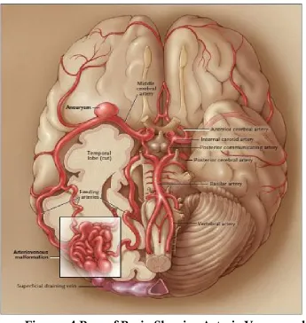
ARTERIOVENOUS MALFORMATIONS OF THE BRAIN :72-79
Arteriovenous malformations of the brain are focal abnormal conglomerations of
dilated arteries and veins within brain parenchyma, in which a loss of normal vascular
organization at the subarteriolar level and a lack of a capillary bed result in abnormal
arteriovenous shunting (fig : 4).
The most common presenting sign of an arteriovenous malformation is
intracerebral haemorrhage (occurring in 42 to 72% of clinically apparent arteriovenous
malformations52-57). Haemorrhage of arteriovenous malformations accounts for
Figure : 4 Base of Brain Showing Arterio Venous malformation
The prevalence of arteriovenous malformation is estimated at approximately
0.01% of the general population, but reported rates range from 0.001% to 0.52%61,62.
The lesions are thought to be congenital in origin. Although occasional cases are
associated with other abnormalities (e.g., Osler-Weber-Rendu disease and the
Sturge-Weber syndrome63,64).
The increased use of advanced imaging technologies has led to the identification
of more arteriovenous malformations, including many that are asymptomatic65.
The overall risk of haemorrhage of arteriovenous malformations is estimated at 2
After an initial haemorrhage, the annual risk of a subsequent haemorrhage has
been reported to range from 4.5 to 34.4% 66-71.
In case series, reported rates of permanent weakness or paralysis, aphasia, and
hemianopsia are 0 to 15%, and most report no deaths.
NEOPLASM CAUSING HAEMORRHAGIC STROKE :
1. Primary tomour in central nervous system
2. Metastatic Tomour
3. Leukaemia
HAEMATOLOGICAL DISORDER ASSOCIATED WITH STROKE INCLUDE :
1. Sickle-cell disease
2. Thrombocytopenia
3. Moyamoya Disease
4. Polycythemia.
5. Paraxysmal nocturnal haemoglobinuria.
6. Disorders of platelets & or blood coagulation.
7. Leukemias & Other Neoplasm.
DRUGS :
1. Warfarin
2. Amphetamines
4. Phenypropanolamine20
TEMPORAL PROFILE OF STROKE :
Depending upon the time course of the disease the strokes are classified as
transient ischaemic attacks, reversible ischaemic neurological deficit, progressing stroke
or stroke in evolution and completed stroke.
TRANSIENT ISCHAEMIC ATTACK (TIA) :
It is a sudden focal neurological dysfunction due to ischaemia of a portion of the
brain and which resolves completely within 24 hours. Most of the episodes last usually
less than 10 minutes but attacks lasting many hours are often documentedly associated
with embolism. They may herald the oncoming vascular catastrophe. Previous history
of TIAs are much more common but very rare in intracerebral haemorrhage. Recurrent
attacks of the same pattern indicates that thrombosis is the possible cause, while
multiple episodes of different pattern indicates embolism 4,5.
Symptoms of TIA vary depending upon the vascular territory involved.
Symptoms due to involvement of carotid system are motor defects (weakness, paralysis
or clumsiness) of the extremity or of both extremities of one side; sensory deficit
(numbers of paraesthesia) of one or both extremities of one side, aphasia, amaurosis
fugax and homonymous hemianopia. Symptoms due to involvement of vertebrobasilar
system are motor defect of any combination of extremities upto quadriplegia or
changing from one side to another in different attacks, sensory defects which are
homonymous hemianopia and ataxia with or without vertigo.
REVERSIBLE ISCHAEMIC NEUROLOGICAL DEFICIT (RIND) :
It is a focal ischaemic event lasting longer than 24 hour but complete resolution of
the deficit within three weeks. These episodes are also referred to as stroke with full
recovery.
PROGRESSING STROKE OR STROKE IN EVOLUTION :
The temporal pattern of stroke is sometimes extended. Disability may increase by
stepwise progression. Sudden deteriorations are interspersed with static intervals. Less
commonly there is slow uninterrupted progression. The full extent of the patient’s
stroke conveys somewhat greater diagnostic uncertainty. A rapidly growing neoplasm or
a subdural haematoma is more often a real differential diagnostic syndromes.
COMPLETED STROKE :
It is the stable focal ischaemic neurological deficit from which recovery occurs
gradually over weeks and months.
SPECIFIC VASCULAR SYNDROMES :
Stroke syndromes are defined not only by their temporal profile but also by the
vascular supply to the area of ischaemic brain.
INTERNAL CAROTID SYNDROME7 :
The cervical portion of the carotid artery is a common site for both severe
atheroma and thrombotic occlusion. About 30 percent of all occlusive lesions may be
difficulties, sensory paraesthesia with or without motor weakness on the opposite side.
Ipsilateral amaurosis fugax, fleeting or semipermanent, alternating with or accompanied
by a contralateral hemiplegia or sensory deficit, is pathognomonic of carotid artery
syndrome, but it is noted in only 15 to 20 percent of the subjects.
The clinical manifestation may be similar to middle cerebral syndrome. Feeble
carotid superficial temporal artery pulsation, dilated pupil and poorly pulsating retinal
vessels (with or without optic atrophy) on the side of suspected carotid lesion and ocular
or cervical bruits on the ipsilateral side may suggest the correct diagnosis.
In subjects with an old or silent occlusive carotid artery lesion on one side, a new
lesion on the other side may prove catastrophic. Here, a physical findings of bilateral
hemiplegia (quadriplegia) with coma can be mistaken for basilar artery syndrome.
MIDDLE CEREBRAL SYNDROME :
The cortical branches supply most of the lateral surface of the cerebral
hemisphere, expect for the regions supplied by the anterior and posterior cerebral
arteries. The areas of supply include the sensory-motor cortex, the motor and sensory
speech centers, auditory area and visual radiation. The penetrating branches
(lenticulo-striate arteries) supply the putamen, globuspallidus, genu and posterior limb of the
internal capsule2,5.
The clinical picture of middle cerebral artery occlusion is variable. Contralateral
hemiplegia, hemianaesthesia with or without homonymous hemianopia and aphasia
superior division results in contralateral hemiparesis with sensory deficit and expressive
aphasia (Broca’s aphasia) whereas Wernicke’s aphasia (sensory aphasia) is frequent with
lesion of the inferior division of dominant side. Monoplegic symptoms can occur with
an occlusive lesion of a single cortical branch.
Several researchers have found that patients with Broca’s aphasia were
significantly younger than those with wernicke’s aphasia. Occlusion of penetrating
branches (lenticulo-striate arteries) has been blamed for a dense sensory motor
hemiplegic syndrome (capsular hemiplegia), but significant sensory loss seldom occurs
with such a occlusion, whereas pure motor hemiplegia is not uncommon.
ANTERIOR CHOROIDAL SYNDROME :
This artery supplies the posterior limb of the internal capsule, which carries the
corticospinal and sensory fibres for the contralateral limb. This syndrome which
represents a true “capsular hemiplegia” (dense hemiplegia hemianaesthesia and
homonymous hemianopia), is rare2,7.
ANTERIOR CEREBRAL SYNDROME :
This cortical branches mainly supply the medial superior surface of the frontal
lobe and the parietal lobe upto the paracentral lobule. The penetrating branches supply
the anterior limb of the internal capsule and part of the head of the caudate nucleus7,8.
Anterior cerebral artery occlusion proximal to the anterior communicating artery,
in subjects with a symmetrical circle of Willis, is frequently asymptomatic. Occlusion
the opposite lower extremity with mild weakness of the opposite shoulder. Mental
changes, rectal and urinary incontinence, gait disturbances, apraxia and grasp and
sucking reflexes may accompany the above findings.
Occlusion of an unpaired anterior cerebral artery (supplying both the
hemispheres) results in a cortical type of paraplegia, with sphincter incontinence and a
mental state in which the patient is alert but mute (akinetic mutism). Aphasia and
hemianopia are never seen.
Occlusion of the penetrating branches and of the Heubner’s artery is frequently
blamed for ataxic tremor of the contralateral limbs (frontal ataxia) Apraxia, idiomotor
dyspraxia of the limbs and gait may also be present.
POSTERIOR CEREBRAL SYNDROME 7,8:
This artery supplies the medial and inferior aspects of the occipital and temporal
lobes. Its branches also supply the mid-brain, cerebral peduncle and most of the
thalamic and subthalamic regions.
Thrombotic occlusion of the posterior cerebral arteries is relatively rare.
Contralateral homonymous hemianopia is a significant finding and this results from
infarction of the primary visual area (calcarine cortex), the central vision is frequently
spared, even in patients with bilateral disease (gun barrel vision). Other manifestations
of visual dysfunction include illusory or distorted vision, visual object agnosia and
various forms of dyslexia without dysgraphia. The pupillary reflexes are well preserved.
and thalamic syndrome (Dejerine Roussy syndrome) may also be present. In the
thalamic syndrome, there is varying degree of sensory loss to all modalities and
spontaneous burning or agonizing pains are frequent (analgia dolorosa). Memory loss
(amnesia) denotes a lesion of the medical temporal cortex. Contralateral involuntary
choreoathetosis or ataxic tremors are rarely observed.
VERTEBRO-BASILAR SYNDROME 7,8 :
After traversing through the bony vertebral canals, both vertebral arteries unite
intracranially to form the basilar trunk. Their short paramedian and long circumferential
branches supply the entire brainstem, cerebellum and the vestibular apparatus.
Ischaemic disorders, therefore manifest by episodes of vertigo, dizziness, diplopia,
dysarthria, dysphasia, incoordination of gait and limbs and bilateral signs of
sensory-motor deficit. Occipital headaches may be present.
Ipsilateral IIIrd nerve palsy (dilated pupil, ptosis and external strabismus) with
contralateral hemiplegia (Weber’s Syndrome) or with crossed cerebellar ataxia (Claude’s
syndrome) is diagnostic of mid-brain localization. Homolateral paralysis of the VIth
and VIIth nerves (internal squint and facial palsy) with contralateral hemiplegia and
hemianaesthesia (Millard-Gubler syndrome) is suggestive of a pontine lesion. Palatal
Paralysis and ataxia of limbs, with impairment of posterior column sensation on same
side of the body together with diminution of pain and thermal sense on the opposite
limbs (Wallenberg’s syndrome) indicate lateral medullary infarction1,5,7.
state with fully preserved consciousness has been described (Locked in syndrome) and
suggests infarction of the basis pontis (sparing the tegmentum), from a midbasilar
occlusion.
Occlusion of isolated cerebellar branches may produce dizziness, nausea,
vomiting, nystagmus and appendicular or trunkal ataxia without sensory-motor deficit in
any limb. Such a syndrome should be differentiated from cerebellar haemorrhage where
emergency surgical decompression proves lifesaving.
LACUNAR SYNDROMES :
Occurs as a result of infarcts in the deep portions of cerebral hemispheres and
brain stem due to occlusion of small perforating branches4. Pure motor hemiplegia is the
most common lacunar syndrome due to an infarct in the posterior limb of internal
capsule. There is no sensory deficit, visual field defect or aphasia.
ARTERITIS :
The clinical features of syphilitic and other forms of arteritis involving various
cerebral arteries are in no way different from the neurovascular syndrome described
under cerebral thrombosis. With specific serological tests, the diagnosis of
meningovascular syphilis is not difficult.
CLINICAL PRESENTATION :
In stroke patients, symptoms and signs are in relation to the arterial circulation
involved. The clinical assessment may be corroborated by diagnostic studies such as,
diagnostic techniques may be, the stroke syndrome continues to be characterised by the
nature and time course of the patient’s clinical findings. A careful analysis of history
may help in differentiating stroke due to thrombosis, embolism or haemorrhage.
ONSET AND PROGRESSION OF STROKE :
Patient’s activity during the onset of stroke indicates the possible cause of stroke.
Intracerebral haemorrhage usually presents abruptly when the patient is awake and is
prone to occur while he or she is engaged in physical exertion. Severe headache may
precede the onset of haemorrhagic stroke by several hours and loss of consciousness is a
usual feature.
In cerebral embolism the full blown picture of stroke evolves within a few
seconds without any warning symptoms. In cerebral thrombosis the focal disability may
occur at any time. The deficit evolves during the period of 1 or 2 days. Loss of
consciousness occurs very rarely though drowsiness is common. Severe headache is
unusual. Sometimes cerebral infarctions due to thrombosis and those due to embolism
may be indistinguishable in terms of the time, course and nature of neurological
findings.
FITS :
Epileptic fits, generalized or focal type may occur at the beginning or during the
evolution of a stroke. Fits may occur in thrombosis, embolism or haemorrhage and it is
not helpful in differentiating these conditions. Fits commonly occur in cerebral venous
SIGNS OF MENINGEAL IRRITATION :
Neck stiffness occurs when blood leaks into CSF in case of intracerebral
haemorrhage or when there is rupture of saccular aneurysm producing subarachnoid
haemorrhage. In our country since tuberculous meningitis is a common disease, it
should be considered in stroke patients with signs of meningitis.
ARTERIAL PULSATIONS AND BRUIT :
A significant reduction in cerebral blood flow can occur due to atherosclerotic
lesion narrowing the internal carotid artery or vertebral artery lumen by more than 60 to
80% of cross sectional area. Severe lesions of this kind are frequently seen in the
internal carotid artery near the carotid sinus and in the vertebral channels coursing over
the forehead and supra-orbital and supratrochlear arterial pulsations on the rim of the
orbit suggest carotid occlusion. An additional sign of carotid occlusion is the presence
of ocular bruit heard over the eye ball. Though vertebral artery is not accessible for
palpation, its occlusion at its origin is suggested by a bruit heard over supraclavicular
OBSERVATION AND ANALYSIS
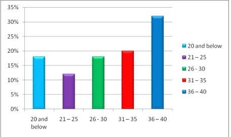
AGE DISTRIBUTION
The age distribution among 50 patients show that stroke in young adult are
common in above 36 years of age.
Table – 2
Age (in Years) No of Cases Percentage
20 and below 9 18%
21 – 25 6 12%
26 - 30 9 18%
31 – 35 10 20%
36 – 40 16 32%
30% of patients are less than 25 years of age
70% of patients are more than 25 years of age.
0% 20% 40% 60% 80% 100% 120%
No of Patients
IN
C
ID
E
N
C
E
Female Male GENDER INCIDENCE OF YOUNG ADULTS WITH STROKE
IN 50 PATIENTS
Table – 3
Gender No of Cases Percentage
Male 32 64%
Female 18 36%
Total 50
It is observed that the incidence of stroke in young adult is higher among male
than female population. (fig : 6)
36%
Figure : 6
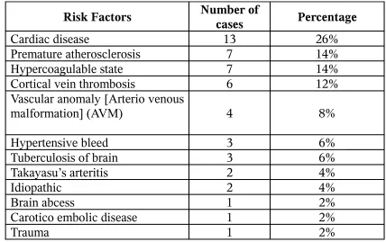
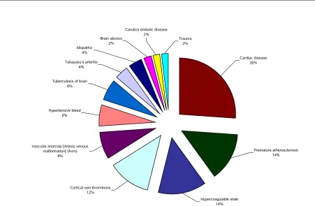
PREVALENCE OF RISK FACTORS IN 50 YOUNG
ADULTS WITH STROKE
Table – 4
Risk Factors Number of cases Percentage
Cardiac disease 13 26%
Premature atherosclerosis 7 14%
Hypercoagulable state 7 14%
Cortical vein thrombosis 6 12%
Vascular anomaly [Arterio venous
malformation] (AVM) 4 8%
Hypertensive bleed 3 6%
Tuberculosis of brain 3 6%
Takayasu’s arteritis 2 4%
Idiopathic 2 4%
Brain abcess 1 2%
Carotico embolic disease 1 2%
Trauma 1 2%
• Most of the patients (26%) who developed stroke are cardiac patients.
• Premature atherosclerosis is the risk factor for 14% of stroke patients.
PREDOMINANT RISK FACTORS IN YOUNG STROKE
Cardiac disease 26%
Premature atherosclerosis 14%
Hypercoagulable state 14% Cortical vein thrombosis
12% Vascular anomaly [Arterio venous
malformation] (Avm) 8% Hypertensive bleed
6% Tuberculosis of brain
6%
Takayasu’s arteritis 4% Idiopathic
4%
Brain abcess 2%
Carotico embolic disease 2%
[image:46.612.61.512.112.407.2]Trauma 2%
0% 10% 20% 30% 40% 50% 60% 70%
Infarction Haemorrhage Others
TYPE OF STROKE
IN
C
ID
E
N
C
E
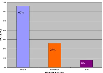
[image:47.612.74.476.351.634.2]TYPE OF STROKE (CLINICAL & CT SCAN & MRI EVALUATION)
Table – 5
Type of Lesion Number of cases Percentage
Infarction 33 66%
Haemorrhage 13 26%
Others 4 8%
Ischaemic – Infarction Stroke is more common than Haemorrhagic
Stroke. (Fig : 8)
66%
26%
Figure : 8
ANALYSIS OF TREATMENT OUTCOME AND PROGNOSIS
TREATMENT OF STROKE INCLUDES :
1) Acute therapies designed to minimise brain infarction and life supportive
measures.
2) Rehabilitative aimed at improving the quality of life.
3) Treatment aimed at preventing recurrence of strokes.
46 patients presented with completed stroke with fixed neurological deficits,
admitted several hours to a few days after the onset of stroke, of which 33 patients had
Brain Infarction & 13 patients had haemorrhagic stroke. Since there were no patients
with stroke in evolution, anticoagulant therapy was not used to minimize brain
infarction. Measures to reduce cerebral edema with intravenous mannitol, oral glycerol,
frusemide and steroids were undertaken in fifty patients.
Anticoagulant therapy with intravenous heparin was started in 10 patients who
had cardioembolic stroke with risk of recurrence. 3 patients with subacute bacterial
endocarditis were treated with appropriate antibiotics. Anticoagulation therapy was not
given to them.
3 patients with tuberculosis of Brain with stroke were treated with rifampicin,
INH, ethambutol and pyrazinamide along with steroids. Blood pressure of the all
hypertensive patients was controlled by oral administration of enalapril. 3 Hypertensive
was used in 6 patient with focal seizures.
Antiplatelet therapy with low dose aspirin and dipyridamole was given in all
patients who presented with thrombotic stroke.
Physical rehabilitative measures to maximize functional recovery were
undertaken in all patients.
Prevention of recurrence of stroke was by long term antiplatelet therapy in those
with thrombotic stroke, oral anticoagulant therapy in those with aseptic cardiogenic
emboli and control of blood pressure in hypertensives. Prophylactic antibiotics before
any minor or major surgical procedures in susceptible cardiac patients minimizes the
risk of subacute bacterial endocarditis.
The prognosis of young adults with ischaemic stroke is better when compared
with older patients. Out of 50 patients, 38 showed stable unchanged course over the first
7 days. 12 patients showed improvement in the first 7 days. None of the patients
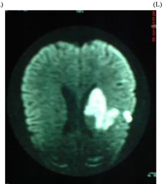
Figure : 9 - C.T. Angiogram Showing Total Occlusion Of (L) Subclavian Artery
Figure : 10 - C.T. Scan Brain showing (L) MCA territory haemorrhage
(R) (L)
DISCUSSION
In this study, 50 young adults with stroke were included.
The incidence in men was 64% compared to 36% in women. This is in agreement
with studies conducted by other centers.
In this study, the peak incidence of stroke was observed in the age group of 36-40
years which was 32%. It was 20% in the age group of 31-35 years. In a study
conducted by P.M.Dalal et al80, the incidence of stroke was maximum in the age group
of 36-40 years which is similar to our study.
In our study 52% of patients were above the age of 30 years, compared is 48%
patients in the above study.
Analysis of Risk factors
:-Hypertension :
Hypertension is one of the common risk factors associated with stroke. The
incidence of hypertension was 18% in this study. In a study conducted by P.G.A.Sander
Cock et al81 this incidence was 32%.
Diabetes Mellitus :
Out of 50 patients studied, 2 patients had diabetes mellitus.
Hyperlipidemia :
7 Patients who had abnormal lipid profile were affected by premature
Transient Ischaemic attack :
Incidence of TIA was 10% in this study. In the study conducted by L.N.Jones the
incidence of TIA was 14%.
Smoking :
In this study, out of 22 male patients, 20 were smokers. The remaining 18 patients
were female non smokers. It became an important risk factor mainly due to premature
atherosclerosis associated with it.
Cordiac Source of Emboli :
Cordiac source of emboli was observed in 26% of cases. In the study conducted
by P.M.Dalal et al80, the incidence of cordiac source of emboli was 20%. In our study,
10% of patients had atrial fibrillation. In the oxford shire community stroke project81,
the incidence of atrial fibrillation was 17%.
7 Patients had RHD, out of which 5 were in AF.
2 Patients had congenital heart disease (TOF).
Alcohol :
In this study, 32% of males were alcoholics, out of them one person had stroke
followed by an alcoholic binge. Similar observation of stroke in young men after
alcoholic binge, was noted by M.R.Wilkins, M.J.Kendall82. 8% of patients who
presented with haemorrhagic stroke had arterio venous malformation in the cerebral
vascular system. 4% of young patients with stroke had Takayasu’s arteritis involving
Cervical arterial Bruit :
Cervical arterial bruit was observed in 2% of cases. In the Dalal et al80 study, it
was observed in 9% of cases.
Uncertain Causes :
Stroke due to undetermined causes was 6% in our study & in oxford shire
community study81, it was 5%. A definite risk factor was present in 70% of cases in our
study but according to other observation it was 80%. More than one risk factors were
noted in 30% of cases.
Serum Homocystein level was raised in 6 patients who had cerebral infarct.
(Normal Range 3.7 – 13.9 µmol/L)
In our study, out of 50 patients, one had Antithrombin – III deficiency.
One patient developed metastatic brain abcess originated from congenital heart
disease (TOF)
Types of Stroke :
In this study
Ischaemic stroke was present in 66% of cases.
Haemorrhagic stroke was present in 26% of cases but according to Harvard
co-operative stroke registry study12, the incidence was 15%.
Cortical vein thrombosis was seen in 12% of cases & all the patients observed
were female in post partum period. Similar observation was made by M.E.
According to Dalal et al study80, which included 93 cases for a period of 5 years,
the incidence of Ischaemic stroke was 80.60% and Haemorrhagic stroke was 12.50%
when compared with this study, the incidence of Ischaemic stroke was 66% and
CONCLUSION
1. The incidence of stroke in young adults is more common in the age group
between 36-40 years & males are more affected (64%) than females (36%).
2. Cardioembolic stroke is the commonest cause of stroke in young adults. Smoking
is the most significant risk factor for stroke in young adults mainly due to the
premature atherosclerosis associated with it.
3. Even though mitral value prolapse is consided as one of the major risk factors for
stroke in young adults, in our study, out of 50 young patients who had stroke only
2 had MVP. But even in those cases MVP was not the cause for stroke.
4. TIA is experienced by 10% of patients.
5. All the patients who suffered from cortical vein thrombosis are females in
puerperal period.
6. Among hypercoagulopathies causing stroke, hyperhomocysteinemia is the
commonest followed by antithrombin III deficiency.
7. In about 6% of cases, no cause could be attributed.
8. The predominant mode of presentation of stroke is middle cerebral territory
involvement. The commonest pathological type is (ischaemic stroke (66%)).
9. In vascular disease, takayasu’s arteritis causes 4% of stroke & arteriovenous
BIBLIOGRAPHY
1. NEUROLOGY IN CLINICAL PRACTICE – PRINCIPLES OF DIAGNOSIS
AND MANAGEMENT – WALTER G.BRADLEY - 5TH EDITION.
2. BRAIN’S DISEASES OF THE NERVOUS SYSTEM - 11th EDITION.
3. PRINCIPLES OF NEUROLOGY – RAYMOND D.ADAMS - 8th EDITION.
4. HARRISION PRINCIPLES OF INTERNAL MEDICINE 17th EDITION –
VOLUME 2.
5. DAVIDSON TEXT BOOK OF MEDICINE – 20th EDITION.
6. OXFORD TEXT BOOK OF MEDICINE – 3rd EDITION.
7. API TEXT BOOK OF MEDICINE - 8th EDITION – VOLUME 2.
8. GRAY’S TEXT BOOK OF ANATOMY – 37th EDITION
9. Martin A. Alpert a study of mvp with stroke. BMJ June 1993 vol.9 no.4 page 311,
312.
10. I.S.Anand, Y.Chandrasekar, prosthetic valve problem JAPI April 91 vol. 39 page
307.
11. Smoking/Donnan et al – smoking as a risk factor for cerebral ischaemia lancet 2
– 643.
12. Hypertension / Mohr JP Caplan LR et al – the Harvard prospective registry
neurology 1978/28 754-762.
13. Nedea s – stroke due to cardiogenic embolism seminars in neurology 1986, 6,
14. Mortality from cardiovascular diease among interregional migrants in England
and Wales D P Strachan, D A Leon, and B Dodgeon BMJ 1995; vol
310:423-427.
15. Case – control study of oral contraceptives and risk of thromboembolic stroke:
results from international study on oral contraceptives and health of young
women Lothar A J Heinemann, Michael A Lewis, Margaret thorogood, Walter O
Spitzer, Irene Guggenmoos-Holzmann, and Rudolf Bruppacher BMJ 1997; 315 :
1502 – 1504.
16. Case-control study of migraine and risk of ischamic stroke in young women
Christophe Tzourio, Alain Tehindrazanarivelo, Serge Iglesias, Annick
Alperovitch, Francois Chedru, Jacques D’Anglejan-chatillon, and Mariegermaine
Bousser BMJ 1995; 310:830-833.
17. Relation between Troponin T concentration and mortality in patients presenting
with an acute stroke : observational study P James, C J Ellis, R M L
Whitlock, A R Mcneil, J Henley, and N E Anderson BMJ 2000;
320: 1502-1504.
18. Influence of total cholesterol, high density lipoprotein cholesterol, and
triglycerides on risk of cerebrovascular disease : the Copenhagen city heart study
E Lindenstrom, G Boysen, and J Nyboe BMJ 1994; 309:11-15.
19. Prognosis after transient monocular blindness associated with carotid-artery
Meldrum H., the north american symptomatic carotid endarterectomy trial
collaborators N EGL J Med 2001; 345: 1084-1090, Oct 11, 2001.
20. Phenylpropanolamine and the risk of haemorrhagic stroke Kernan W.N., Viscoli
C.M., Broderick J.P., Brott T., Feldmann E., Morgenstern L.B., Wilterdink J.L.,
Horwotz R.I., N ENGL J MED 2000; 343: 1826-1832, Dec 21, 2000.
21. Caplan’s stroke : a clinical approach Norris J.N. ENGL J MED 2000; 343:1899,
Dec 21, 2000.
22. Pravastatin therapy and the risk of stroke Brett A. S., Meilof J.F., Uitdehaag B.
M.I., Frechter O., White H.D., Simes R.J., Tonkin A.M., the lipid study group N
ENGL J MED 2000; 343:1894-1896, Dec 21,2000.
23. Adverse cardiovascular and central nervous system events associated with dietary
supplements containing ephedra alkaloids N ENGL J MED 2000;
343:1833-1838, Dec 21, 2000.
24. Stroke in patients with asymptomatic internal-carotid-artery stenosis Oldstein
L.B., Howard G., Cohen S.N., Toole J.F., Tunick P.A., Karonzon I., Inzitari D.,
Eliasziw M., Barnett H.J.M. N ENGL J MED 2000; 343:1420-1421, Nov 9,2000.
correspondence.
25. World Health Organisation the WHO stepwise approach to stroke surveillance –
Geneva : WHO – 2005.
26. Hart R.G.Miller VT : cerebral infarction in young adult : a practical approach
27. Grindal Ab, Cohen RJ, Saul RF, Taylor JR : cerebral infraction in young adult
stroke 1978; 9 : 39-42.
28. Adams HP, Butler MJ, Biller J, Joffol GJ : non haemorrhagic cerebral infarction
in young adults. ARCH NEUROL 1986; 43 : 793 – 796.
29. Radhakrishair K, Ashok PP, Sridharan R, Mousa ME : stroke in the young :
incidence and pattern in Benghazi Libya. ACTA NEUROL SCAND 1986;
73:434-438.
30. Abraham J, Schetty G. Jose CJ : strokes in the young stroke 1971; 2 : 258 -267.
31. Hachinsky V, Norn’s JW : the young stroke, in the acute stroke, Philadelphia, FA
DAVIS, 1985, PP 141 – 164.
32. Snyder BD, Ramirez – Lassepas M : cerebral infarction in young adult : long
term prognosis stroke 1980; 11: 149-153.
33. Stroke vol 19 no 8 August 1988; 982 – 986.
34. Estatisticas DA Saude 1972-1986. Lisboa, Portugal, Instituto National DE
Estatistica, 1987.
35. Mudd SH, Shoubuf, Leuyhl, et al. the natural history of homocystinuria due to
cystathione beta – synthase deficiency. AM I HUM GENET 1985; 37 : 1-3.
36. Hassan A, Hunt BJ, O Sullivan M, et al. Homocysteine is a risk factor for
cerebral small vessal disease, acting via endothelial dysfunction. BRAIN 2004 :
127, 212-9.
Homocystinuria due to cystathione - bete synthase deficiency treated chronically,
a multicantric observational study. ARTERIOSCLER THROMB VASC BIOL
2001; 21 : 2080 - 5.
38. Ringlestein, EB, Zeumerh, Argelou D : the pathogenesis of stroke from internal
carotid artery occlusion. diagnostic and therapeutical implications stroke 1983 :
14 : 867 – 875.
39. Mohr JP, Gautier JC, Hier DB, stein rw : middle cerebral artery, in barnett hjm,
mohr jp, stein bm, yatsu fm (eds) : stroke. new york, churchill, 1986, vol 1, p
409.
40. Sindermann F, Dichgons J, Bergleiter R : occlusion of the middle cerebral artery
and its branches; Angiographic and clinical correlates, BRAIN 1969; 92 : 607 –
620.
41. (1) Basso A, Capitani E, Laiacona M, Luzzatti C : Factors influencing type and
severity of aphasia, CORTEX 1980, 16 : 631 – 636.
(2) DE Renzi E, Faglioni P, Ferrari P : The influence of sex and age on the
incidence and type of aphasia CORTEX 1980 : 16 : 627 – 630.
42. Obler Lk, Albert M, Good Glass H, Benson F : Aphasia type and aging. BRAIN
LARG 1979; 6 : 318 – 322.
43. Kertesz A : Aphasia and associated disorders : taxonomy, localization and
recovery, New York, GRUNE & STRATTON, 1979.
Neurol Neurosurg Psychiatry 1981 : 44 : 377 – 381.
45. Blecic S, Bogousslavsky J. Stroke in young adults. In : Barnett HJM, Mohr JP,
Stein BM, et al, eds. Stroke : pathophysilogy, diagnosis and management. 3rd ed.
NEW YORK, NY : CHURCHILL LIVINGSTONE; 1998:1001-1012.
46. Wiebers DO, Feigin VL, Brown RD. Cerebrovascular disease in children and
young adults. In : Handbook of stroke, Philadelphia, PA : LIPPINCOTT –
RAVEN; 1997:237-243.
47. Stern BJ, Wityk RJ, Stroke in the young. In : Feldmann E, ed. Current diagnosis
in neurology. St. Louis MO : MOSBY; 1994 : 34 – 40.
48. Biller J.strokes in the young in : Toole JF, ed. Cerebrovascular disorders. 5th ed
Philadelphia, PA : LIPPINCOTT WILLIAMS AND WILKINS; 1999 : 283 – 316.
49. Mudd SH, Shovby F, Levy HL, et al. The natural history of Homocystinuria due
to Cystathione beta-synthase deficiency. AM J HUM GENET 1985:37:1-31.
50. Hassan A, Hunt BJ, O’sullivan M, et al. Homocysteine is a risk factor for cerebral
small vessel disease, acting via endothelial dysfunction, BRAIN 2004;
127:212-9.
51. Yap S, Boers GH, Wilcken B, et al. Vascular outcome of patients with
Homocystinuria due to cystathione-beta synthase deficiency treated chronically; a
multicenter observational study. ARTERIOSCLER THROMB VASC BIOL
2001; 21:2080-5.
haemorrhage as a presenting symptom and subtype analysis: a population-based
study of intracranial vascular malformations in OLMSTED COUNTRY,
MINNESOTA. J NEUROSURG 1996; 85:29-32.
53. Apsimon HT, Reef H, Phadke RV, Popovic EA. A population-based study of
brain arteriovenous malformation : Long – term treatment outcomes. Stroke
2002; 33:2794-800.
54. Crawford PM, West CR, Chadwick DW, Shaw MD, arteriovenous malformations
of the brain; natural history in unoperated patients. J NEUROL NEUROSURG
PSYCHIATRY 1986; 49:1-10.
55. Ondra SL, Troupp H, George ED, Schwab K. The natural history of symptomatic
arteriovenous malformations of the brain: a 24 year follow-up assement. J
NEUROSURG 1990;73:387-91.
56. Stapf C, Mast H, Sciacca RR, et al. Predictors of haemorrhage in patients with
untreated brain arteriovenous malformation neurology 2006; 66:1350-5.
57. Stieg PE, Batjer HH, Samson D, eds. Intracranial arteriovenous malformations
NEW YORK : Informa, 2007.
58. Perret G, Nishioka H. Report on the cooperative study of intracranial aneurysms
and subarachnoid haemorrhage . Section vi. Arteriovenous malformations: an
analysis of 545 cases of cranio-cerebral arteriovenous malformations and fistulae
reported to the cooperative study. J NEUROSURG 1966;25:467-90.