0022-538X/96/$04.0010
Copyrightq1996, American Society for Microbiology
Capsid Coding Sequence Is Required for Efficient
Replication of Human Rhinovirus 14 RNA
KEVIN L. MCKNIGHT1ANDSTANLEY M. LEMON1,2*
Departments of Microbiology and Immunology1and Medicine,2The University of
North Carolina at Chapel Hill, Chapel Hill, North Carolina 27599-7030
Received 24 August 1995/Accepted 1 December 1995
Mechanisms by which the plus-sense RNA genomes of picornaviruses are replicated remain poorly defined, but existing models do not suggest a role for sequences encoding the capsid proteins. However, candidate RNA replicons (DP1bgal andDP1Luc), representing the sequence of human rhinovirus 14 virus (HRV-14) with reporter protein sequences (b-galactosidase or luciferase, respectively) replacing most of the P1 capsid-coding region, failed to replicate in transfected H1-HeLa cells despite efficient primary cleavage of the polyprotein. To determine which P1 sequences might be required for RNA replication, HRV-14 mutants in which segments of the P1 region were removed in frame from the genome were constructed. Mutants with deletions involving the 5*-proximal 1,489 nucleotides of the P1 region replicated efficiently, while those with deletions involving the 3* 1,079 nucleotides did not. Reintroduction of the 3* P1 sequence into the nonreplicatingDP1Luc construct resulted in a new candidate replicon,DP1Luc/VP3, which replicated well and expressed luciferase efficiently. Capsid proteins provided intransby helper virus failed to rescue the nonreplicatingDP1Luc genome but were able to package the larger-than-genome-lengthDP1Luc/VP3 replicon. Thus, a 3*-distal P1 capsid-coding se-quence has a previously unrecognizedcis-active function related to replication of HRV-14 RNA.
The picornaviruses comprise a large and diverse group of positive-strand RNA viruses which have a common genetic organization and replication strategy and which are pathogenic in a variety of animal species. In each of these viruses, the dominant feature of the organization of the genome is the presence of a single large open reading frame (ORF), which encodes a multifunctional viral polyprotein. The viral capsid proteins are encoded by a segment of RNA (P1 segment)
located at or near the 59end of the ORF, upstream of the P2
and P3 segments, which encodes a variety of nonstructural proteins involved in replication of the virus (46). In two closely related picornavirus genera which are important causes of dis-ease in humans, enteroviruses (which include the polioviruses)
and rhinoviruses, the P1 segment is located at the extreme 59
end of the ORF, and primary cleavage of the viral polyprotein occurs at the carboxyl terminus of the expressed P1 protein. This structural protein intermediate is subsequently further proteolytically processed into several capsid precursor pro-teins: VP0 (representing VP4 plus VP2), VP3, and VP1 (see Fig. 1). While nontranslated RNA sequences located upstream and downstream of the ORF contain cis-active elements which are critical for replication, and while translation of the P2/P3 coding region appears to be cis active with respect to viral replication (50), the available evidence suggests that the P1 capsid-coding sequence is not required for viral RNA ampli-fication.
The absence of a role for P1 sequences in the replication of poliovirus is supported by the natural occurrence of defective interfering particles with large deletions in this region of the genome (11, 12, 26). In addition, several studies demonstrate that artificially constructed poliovirus genomes with deletions in the P1 region retain all the genetic information necessary for RNA replication (13, 20, 24). Other, more recent studies indi-cate that poliovirus P1 sequences can be replaced by RNA
encoding heterologous proteins without compromising viral RNA replication (3, 9, 36). Such subgenomic P1-deleted po-liovirus genomes have been referred to as replicons or mini-replicons, and those expressing heterologous proteins may be valuable for the study of various aspects of virus multiplication or as general-purpose gene expression vectors (3, 9, 36, 40). Among the poliovirus replicons which have been described, there are several examples of replicating RNAs which contain very large deletions in the P1 region. Some of these deletions are much larger than those present in naturally occurring de-fective interfering poliovirus particles (23, 26, 41), which
usu-ally retain VP4 and VP1 coding sequences near the 59and 39
limits of the P1 segment (10, 20, 26). Despite this, these arti-ficial replicons still retain efficient RNA replication functions (3, 13, 24, 41).
The successful development of poliovirus replicons express-ing heterologous proteins led us to consider the possibility that encapsidated rhinovirus replicons have unique potential for the delivery and expression of foreign genes in human airway cells. The rhinoviruses include approximately 100 serologically distinct pathogens which primarily infect human epithelial air-way cells and which are important causative agents of the common cold (15, 19). The RNA genomes and nonstructural proteins of these viruses share striking similarities with those of poliovirus, as does the structure of the virus capsid (8, 35, 45). However, among several rhinovirus serotypes, some important differences do exist, including the shorter length of the
rhino-virus 59nontranslated RNA (59NTR) (38), sequence
require-ments for substrate recognition by the rhinovirus proteases (8, 49), and utilization of specific cellular receptors (14, 22).
To our knowledge, there are no prior reports describing the construction of rhinovirus replicons or even descriptions of naturally occurring P1-deleted defective interfering human rhi-novirus (HRV) genomes. However, several infectious HRV cDNA clones have been constructed and are available for such studies (17, 28, 32). Here, we describe the successful
construc-tion of a subgenomic HRV-14 replicon, DP1Luc/VP3, which
expresses high levels of a foreign reporter protein, firefly
lu-* Corresponding author. Phone: (919) 2536. Fax: (919) 966-6714.
1941
on November 9, 2019 by guest
http://jvi.asm.org/
ciferase, in cultured cells. The design of this construct involved the direct fusion of the luciferase coding sequence to the
in-ternal ribosomal entry site present in the 59NTR of HRV-14,
resulting in the removal of 1.5 kb of P1 sequence. However, during the course of these experiments, we found that repli-cation of HRV-14 RNA was dependent on the retention of
sequence near the 39end of the P1 segment (VP3-VP1 coding
region). This finding demonstrates an important, previously unrecognized difference in the replication strategies of entero-viruses and rhinoentero-viruses and suggests a novel role for P1 RNA sequences in the replication of rhinoviruses.
MATERIALS AND METHODS
Cells.H1-HeLa cells were obtained from the American Type Culture Collec-tion and maintained in 13minimal essential medium (MEM) supplemented with Earle’s salts (Gibco/BRL, Life Technologies Inc., Grand Island, N.Y.), L-glutamine, and 10% fetal calf serum.
Virus.HRV-14 was recovered following transfection of H1-HeLa cells with RNA transcribed from the infectious cDNA clone, pWR3.26, a generous gift from Roland Rueckert, University of Wisconsin at Madison (28). For prepara-tion of virus stocks, monolayers of H1-HeLa cells in a 100-mm-diameter dish were infected with approximately 107PFU of virus. After removal of the inocula, 5 ml of complete medium was added and the monolayer was incubated at 348C for 24 h, at which time virus was harvested from cytoplasmic extracts as previ-ously described (28, 48). The titer of the resulting virus stock was.108PFU/ml. For plaque assay of HRV-14, H1-HeLa cell monolayers in 60-mm dishes were inoculated with 200ml of virus diluted in 13phosphate-buffered saline (PBS) with 0.1% bovine serum albumin and overlaid 30 min later with MEM containing 5% fetal calf serum 13tryptose-phosphate (Gibco/BRL), and 0.9% agarose. After incubation at 348C for 48 to 72 h, the cell monolayers were stained with crystal violet or neutral red.
Antibodies.A polyclonal rabbit antiserum to HRV-14 was a kind gift from Anne Mosser, University of Wisconsin at Madison. In a microtiter neutralization assay, a 1:500 dilution of this antiserum protected 53104
HeLa cells against infection with 53104
PFU of HRV-14. Polyclonal rabbit antibodies tob -ga-lactosidase (5Prime-3Prime, Inc., Boulder, Colo.) and luciferase (East Acres Biologicals, Southbridge, Mass.) were obtained from commercial sources.
Construction of HRV-14 replicons and P1 segment deletion mutants. Candi-date replicons were constructed from the HRV-14 infectious cDNA, pWR3.26 (28), as follows. Nucleotide positions are based on the HRV-14 sequence pro-vided by Lee et al. (28). To facilitate replicon construction, a shuttle vector (pSPHRV5) was prepared by excising the small SalI-StuI fragment (which con-tains nucleotides [nt] 1 to 851 of HRV-14) from pWR3.26 and inserting this fragment into the SalI and PvuII sites in pSP64 (Promega Corp., Madison, Wis.). The bacterialb-galactosidase and firefly luciferase coding sequences were am-plified by PCR from the plasmids pSVbgal and pGem-luc (Promega), respec-tively, with PCR primers with sequences containing restriction endonuclease sites permitting in-frame fusions with the HRV-14 genome. The PCR primers for
b-galactosidase were 59-GCTCTAGAtgatcatgGTCGTTTTACACGTCG-39and 59-CCGCCCGGGcctaggtcctaaaccataggatttaatgtcacctttTTGACACCAGACC AA-39(where the lowercase bases represent HRV-14 sequences and the italic bases represent unique restriction sites), while the PCR primers for amplification of the luciferase sequence were 59-GCCCGATCCAAtgatcatgGAAGACGCC-39
and 59-CAGTTACATTTTAcctaggtcctaaaccataggatttaatgtcacctttCAATTTGGA CTTTCCG-39. PCR-amplified products containingb-galactosidase sequences were digested with XbaI and SmaI and cloned into unique sites in the vector pSP64 to create the shuttle vector pSPb-gal. The completeb-galactosidase cod-ing sequence was excised from this plasmid by digestion with BclI (partial) and
SmaI and cloned into unique sites of pSPHRV5, creating pSPHRVb-gal which contains theb-galactosidase coding sequence fused to the HRV-14 internal ribosome entry site. PCR-amplified products containing luciferase were digested with BclI and AvrII and similarly cloned into the unique sites pSPHRVb-gal, replacing theb-galactosidase coding sequence with the luciferase sequences and creating the plasmid pSPHRVLuc. Large segments within the PCR-amplified reporter gene sequences were subsequently replaced with plasmid DNA (non-PCR amplified) sequences by using unique sites near the ends of each amplified sequence (ClaI to NdeI forb-galactosidase, and XbaI to ClaI for luciferase). Any remaining PCR-amplified sequences were confirmed by direct sequencing of plasmid DNA. Final construction of full-length candidate replicons was accom-plished by removing the small SalI-AvrII fragments from each of the resulting shuttle plasmids. The SalI-AvrIIb-galactosidase fragment was inserted at unique
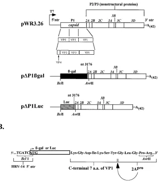
SalI and AvrII sites within pWR3.26 to create pDP1bgal. The SalI-AvrII lucif-erase fragment was inserted into pDP1bgal, replacing the 59NTR–b -galactosi-dase sequence with 59NTR-luciferase sequences to create pDP1Luc. In both of the final constructs, the amino terminus of the reporter sequence was fused in frame to the authentic HRV-14 initiator codon while the carboxyl terminus of the reporter sequence was fused in frame to the carboxyl terminal 7 amino acid
residues of VP1, 30 nt upstream of the unique AvrII site at nt 3206 of the 2Apro coding sequence (see Fig. 1).
Variants of these candidate replicons containing a lethal mutation in the 3Dpol coding sequence were constructed by linearizing the pWR3.26 plasmid with SphI, removing the resulting 39protruding ends with T4 DNA polymerase (Boehring-er-Mannheim Biochemicals, Indianapolis, Ind.), and religating the blunt-ended molecules. This created plasmid pWR3.26(pol2), which contains a frameshift in the HRV-14 polyprotein such that a stop codon terminates translation 26 amino acids upstream of the conserved YGDD motif in 3Dpol. The small SalI-AvrII fragments containing the reporter gene sequences described above were cloned into pWR3.26(pol2) to createDP1bgal(pol2) andDP1Luc(pol2).
A third candidate replicon, pDP1Luc/VP3, which contains sequences coding for part of VP3 and all of VP1, was constructed from pDP1Luc as follows. A PCR product representing the 39end of the luciferase coding sequence was amplified with the primers 59-GGATACCGGGAAAACGCTGGG-39and 59-CCCACCG TACCTAGGccatggACCTTGAAACAAAGCTCCTCCTCCCAATTTGGACTT TCCG-39. In this case, the downstream PCR primer resulted in a fusion of the luciferase coding sequence with a downstream synthetic 3CDprocleavage site, followed by an NcoI site allowing an in-frame fusion within the VP3 sequence of HRV-14. The PCR product was digested with PpuMI (a site 471 nt upstream of the 39end of the luciferase coding region) and AvrII and inserted into pSPHRV-Luc to create pSPHRVpSPHRV-Luc/VP3. Next, the SalI-NcoI fragment was excised from this plasmid and cloned into unique sites in pSPcap (see below). Finally, the small ClaI-AvrII fragment of the resulting clone was inserted into the pDP1Luc replicon to create pDP1Luc/VP3 (see Fig. 7A).
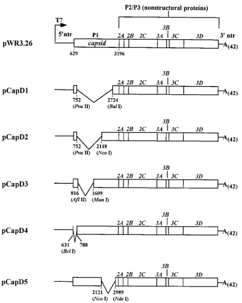
The construction of in-frame deletions within the capsid-coding P1 sequence of pWR3.26 was facilitated by subcloning the entire P1 region (SalI-AvrII frag-ment) in the vector pSP64 to create pSPcap. This plasmid was digested by several different enzymes to create a series of deletion mutants (see Fig. 4A). When necessary, 39ends were filled in by treatment with Klenow enzyme, prior to gel purification and religation. The modified capsid sequences were excised from the pSPcap derivatives by digestion with SalI and AvrII and reinserted into the pWR3.26 vector. To retain the authentic AUG start codon in the pCapd4 construct, the linker 59-GATCATGAAGAACCAGTTAAAGATC-39 was in-serted between the BclI and BglII sites. The initiator AUG codon (boldface type) flanks the first nucleotide of the Thr-54 codon at nt 788 of the HRV-14 genome. Plasmid DNA manipulations were done by standard procedures (47). DNA for use in ligations was purified by the GeneClean II procedure (Bio101, Inc., Vista, Calif.). Each construct was sequenced across regions where replacements took place to confirm the validity of the construction. The deletion constructs were sequenced in the region of the deletion to confirm preservation of the reading frame. DNA sequencing was done by the USBDTaq cycle sequencing system
(United States Biochemical Corp., Cleveland, Ohio).
In vitro transcription.For the production of virus from infectious transcripts, pWR3.26 plasmid DNA was linearized with MluI, which cleaves immediately 39 of the poly(A) tail of the virus genome (28). For the replicon constructs, DNA templates were linearized with SmaI, as theb-galactosidase gene contains nu-merous MluI sites. For experiments comparing replication potential of tran-scripts, other templates were also linearized with SmaI to maintain consistency.
SmaI cleaves the DNA templates 9 nt downstream of the poly(A) sequence. The
infectivity of RNA transcripts derived from pWR3.26 linearized with SmaI was approximately twofold lower than that of transcripts derived from MluI-linear-ized templates (31). RNA transcripts were synthesMluI-linear-ized at 378C for 1 h in 100-ml reaction mixtures containing 1 to 2mg of linearized template DNA, 40 mM Tris-HCl (pH 7.6), 6 mM MgCl2, 5 mM dithiothreitol, 2 mM spermidine-HCl, 100mg of nuclease-free bovine serum albumin per ml, 1.5 mM each nucleoside triphosphate, 5ml of RNasin (Promega), and 200 to 500 U of T7 RNA poly-merase (Promega or New England Biolabs Inc., Beverly, Mass.). To remove template DNA prior to transfection, 5 U of RQ1-DNase (Promega) was added to each reaction mixture and the mixtures were incubated at 378C for an addi-tional 30 min. RNA products were quantified by determining the amount of [a-32P]CTP incorporated into acid-insoluble precipitates. Alternatively, the RNA content was determined by spectrophotometric analysis. Aliquots of tran-scription reaction products were examined by sodium dodecyl sulfate (SDS)-agarose gel electrophoresis in comparison with RNA standards to confirm tran-script integrity. These reaction conditions consistently produced 1 to 3mg of RNA perml.
RNA transfections.H1-HeLa cells were transfected by electroporation as described by Liljestrom et al. (29). Optimal transfection of infectious RNA transcripts was obtained at a setting of 980 V and 25mF with a gene pulser (Bio-Rad, Richmond, Calif.) with the pulse controller unit set at maximum resistance. Cells (0.5 ml at a concentration of 107
cells per ml) were transfected with 10 to 20mg of transcript RNA and subsequently plated into two 60-mm dishes. Specific infectivities derived from monolayers transfected with pWR3.26 transcripts were consistently above 107
PFU/mg of RNA, with,10% of the cells being killed by the transfection procedure alone. RNA transfection of a reporter RNA transcript containing the 59NTR of HRV-14 fused to theb-galactosidase coding sequence, followed by in situ staining forb-galactosidase activity (Pro-mega), indicated that approximately 60 to 70% of cells were successfully trans-fected (31).
Analysis ofb-galactosidase and luciferase enzyme activity.Transfected cells were processed for enzyme assays by the addition of 250 ml of lysis buffer
1942 MCKNIGHT AND LEMON J. VIROL.
on November 9, 2019 by guest
http://jvi.asm.org/
(luciferase assay kit; Promega) per 60-mm dish. The protein content of the cell lysates was determined by the Bradford method (protein assay; Bio-Rad), and lysates were stored at2708C until used in enzymatic analysis. For theb -galac-tosidase assay, lysate containing 50mg of protein was brought to 100ml with lysis buffer and then 100ml of 23assay buffer (200 mM sodium phosphate buffer [pH 7.3], 100 mMb-mercaptoethanol, 20 mM MgCl2, 16.0 mM chlorophenol red
b-D-galactopyranoside substrate [Boehringer-Mannheim Biochemicals]) was added. Reaction mixtures were incubated at 378C for 30 min, and 300ml of 1 M sodium carbonate was added. Then 200ml of each reaction mixture was trans-ferred to a 96-well plate, and the A570was read in a model EL309 microtiter plate reader (BioTek Instruments Inc., Winooski, Vt.). For the luciferase assay, 5ml of lysate (or lysate diluted 1:10 with lysis buffer) was mixed with 100ml of luciferase substrate mixture (Promega). The samples were immediately monitored for light output by using a BioOrbit model 1250 luminometer (LKB-Wallac, Turku, Fin-land). Luciferase activities were normalized to the protein content of individual cell lysates, while theb-galactosidase activities are reported as optical density units at 570 nm per 50mg of lysate protein.
RPA.The RNase protection assay (RPA) was carried out with reagents and protocols supplied with the lysate RNase protection kit (United States Biochemi-cals). Transfected-cell monolayers in 60-mm dishes were lysed by addition of 100
ml of a solution containing 4 M guanidine thiocyanate, 25 mM sodium citrate, and 0.5% sarcosyl. Lysates were stored at2708C until used for hybridization. Riboprobes were prepared from a plasmid (pSPBP) containing HRV-14 genome sequences from nt 5180 to 5446 inserted at the PvuII and XbaI sites in pSP64 (Promega). A bacteriophage SP6 promoter is situated 27 nt upstream of the insert, resulting in production of a 290-nt runoff transcript following linearization of the DNA with BstEII (nt 5183). A 263-nt stretch of this transcript represents HRV-14 minus-strand RNA sequence which is protected from RNase digestion following hybridization to plus strand-virus RNA. A control probe included in hybridization reaction mixtures contained 100 nt of antisense human glyceralde-hyde-3-phosphate dehydrogenase (GAPDH) gene sequence. A plasmid contain-ing this sequence under the control of the bacteriophage promoter T7 produces a 139-nt unprotected runoff transcript (United States Biochemicals). For ribo-probe synthesis, linearized template DNA (200 ng) was mixed with 2ml of 103
transcription buffer (400 mM Tris-HCl [pH 7.6], 60 mM MgCl2, 50 mM dithio-threitol, 20 mM spermidine-HCl), 1ml each of ATP, GTP, and UTP at 10 mM, 1ml of CTP at 0.5mM, 5ml of [a-32P]CTP (50mM, 800 Ci/mmol; Dupont NEN, Wilmington, Del.), 1ml of RNasin (Promega), 2ml (40 U) of RNA polymerase, and diethylpyrocarbonate-treated H2O to 20ml. The reaction mixtures were incubated for 1 h at 378C for T7 polymerase (Promega or New England Biolabs) or 408C for SP6 polymerase (Promega). Two units of RQ1-DNase (Promega) was added, and the reaction mixtures were incubated at 378C for an additional 30 min. The reaction mixtures were brought to 100ml with diethylpyrocarbonate-treated H2O and purified over a Sephadex G-50 RNA spin column (Boehringer-Mannheim Biochemicals). A 106-cpm portion of each probe was added to 45ml of lysate, and hybridization was carried out directly in lysate solutions at 378C for 18 h (overnight) as described by the manufacturer (United States Biochemicals). The hybridization mixture was subsequently diluted 10-fold and digested with RNase and proteinase, and protected RNA fragments were separated by elec-trophoresis in 6% polyacrylamide–6 M urea gels and visualized by autoradiog-raphy.
Metabolic labeling and immunoprecipitation of reporter proteins.After RNA (or mock) transfection, cells were plated in complete medium and incubated at 348C for 1 h. The monolayers were washed five times with PBS and fed with 2 ml of MEM containing 2.5mg of actinomycin D per ml–5% dialyzed fetal calf serum. Following incubation at 348C for 2 h, the monolayers were washed five times with methionine-free MEM and refed with 1 ml of methionine-free MEM containing actinomycin D and fetal calf serum, as above, and 100mCi of [35
S]me-thionine (Dupont NEN). Following incubation for 5 h at 348C, the cultures were washed five times with PBS and incubated at room temperature in lysis buffer (50 mM Tris-HCl [pH 7.5], 150 mM NaCl, 1 mM EDTA, 0.1% Nonidet P-40) for 15 min. The lysate was centrifuged for 2 min in a microcentrifuge, and the super-natant was collected for immunoprecipitations. Immunoprecipitations with rab-bit polyclonal anti-b-galactosidase or anti-luciferase antibodies were carried out as previously described (7). Precipitated complexes were boiled in gel electro-phoresis buffer for 3 min and analyzed by autoradiography following SDS-polyacrylamide gel electrophoresis (PAGE).
RESULTS
Construction of candidate HRV-14 replicons.The HRV-14 genome is a polyadenylated 7,212-nt, positive-sense RNA
mol-ecule that contains a 59NTR of 628 nt, a large ORF encoding
a virus polyprotein of 2,179 amino acids, and a short 39NTR of
47 nt (8). As with other enteroviruses and rhinoviruses, the ORF can be divided into three segments (P1, P2, and P3) based on principal cleavages of the encoded polyprotein (for a review, see reference 27), with the P1 segment encoding the
four capsid proteins. The HRV-14 59NTR contains elements
required for internal entry of ribosomes on uncapped viral RNA (1) and for formation of RNA replication complexes (44).
Candidate HRV-14 replicons were constructed from the plasmid pWR3.26, which contains a full-length cDNA of rhi-novirus type 14 under control of a T7 promoter (28). Since several reports describe replication-competent poliovirus (RNAs with large deletions including almost the entire P1 segment of that virus (3, 13, 24, 41), we initially constructed two plasmids in which the pWR3.26 cDNA was altered to
include the bacterialb-galactosidase (pDP1bgal) or firefly
lu-ciferase (pDP1Luc) coding sequences in lieu of almost the
entire P1 segment of HRV-14 (Fig. 1A). These plasmids were constructed by PCR amplification of the reporter gene se-quences by using oligonucleotide primers with in-frame flank-ing HRV-14 sequences and insertion of the amplified DNA into appropriate restriction sites (BclI and AvrII) in pWR3.26
(see Materials and Methods). In each plasmid, the 59end of
the reporter sequence was fused directly to the authentic ini-tiator AUG of the HRV-14 polyprotein. In an effort to ensure
that recognition and processing at the VP1/2Aprocleavage was
not hampered by the presence of upstream foreign sequences, the reporter sequences were inserted 7 codons upstream of this Tyr-Gly dipeptide (Fig. 1). This configuration thus retained the downstream amino acid residues that have been suggested to be important for autocatalytic processing of the poliovirus
polyprotein by 2Apro (2, 21) and resulted in reporter-DVP1
fusion proteins containing the carboxyl-terminal 7 amino acids
of VP1 (Fig. 1B). The predicted mass of theb-galactosidase
fusion protein was approximately 118 kDa, while that for lu-ciferase was 63 kDa. The HRV-14 RNA transcribed from
pDP1bgal was 462 bases (6.4%) longer than full-length
rhino-virus RNA, whileDP1Luc RNA was 831 bases (11.5%) shorter.
Functional reporter proteins are expressed and processed fromDP1bgal andDP1Luc candidate replicon RNAs.To con-firm that candidate replicon polyproteins were processed as expected and that fusion of the reporter gene sequences had not destabilized upstream RNA secondary structure required for efficient cap-independent initiation of translation under control of the HRV-14 internal ribosomal entry site (1), RNA
was transcribed in vitro from the plasmids pDP1bgal and
pDP1Luc by bacteriophage T7 RNA polymerase. This RNA
was transfected into H1-HeLa cells by an efficient electropo-ration method which results in successful transfection of 60 to 70% of cells (see Materials and Methods). Transfected cells
were pulse-labeled with [35S]methionine from 3 to 8 h
follow-ing transfection. Labeled reporter proteins present in cell ly-sates were immunoprecipitated with rabbit polyclonal
antibod-ies directed againstb-galactosidase or luciferase and subjected
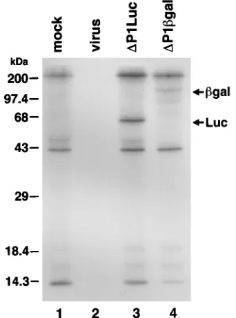
to SDS-PAGE (Fig. 2). Bands containing immunoreactive pro-teins of the expected molecular mass were present in lysates of
cells transfected with eitherDP1Luc (Fig. 2, lane 3) orDP1bgal
(lane 4). These bands were not present in mock-transfected or virus-infected cells (lanes 1 and 2, respectively). Thus, both
candidate replicons expressed a protein product in which 2Apro
accurately processed the reporter-VP1 fusion protein from the remainder of the polyprotein.
Labeled, nonspecifically precipitated proteins were present in lysates of mock-transfected cells and cells transfected with the two candidate replicons (Fig. 2, lanes 1, 3, and 4). The absence of these protein bands in lysates of virus-infected cells
(lane 2) most probably was due to 2Apro shutoff of host cell
protein synthesis. The absence of 2Aproshutoff in the
trans-fected cultures reflects the fact that only 60 to 70% of cells
were successfully transfected. Expression of the b
-galactosi-dase reporter protein fromDP1bgal appeared to be at a lower
on November 9, 2019 by guest
http://jvi.asm.org/
level than luciferase expression fromDP1Luc (compare lanes 3 and 4). This might be explained by different avidities of the antibodies used in the immunoprecipitations. However, minor immunoreactive protein products, which migrated more
rap-idly in SDS-PAGE than the intactb-galactosidase fusion
pro-tein did, were noted in cells transfected with DP1bgal upon
longer exposure of the gel shown in Fig. 2 (31). This suggested the possibility that the somewhat lower expression levels
ob-served withDP1bgal reflect either aberrant translation or
deg-radation of theb-galactosidase reporter protein in vivo.
Lysates of cells transfected with the candidate replicons were also examined for expression of reporter enzyme activity.
Figure 3 depicts the levels of b-galactosidase (Fig. 3A) or
luciferase (Fig. 3B) activities present in H1-HeLa cell lysates prepared at various times following transfection of candidate replicon RNAs. The temporal patterns of reporter protein expression were similar for the two candidate replicons. While measurable quantities of functional enzyme were present in lysates prepared 3 to 12 h following transfection of either
RNA, enzyme activities did not increase over time as expected from previous descriptions of successful poliovirus replicons expressing heterologous reporter proteins (9, 36). The
expres-sion ofb-galactosidase fromDP1bgal appeared to peak at a
later time (9 h posttransfection [Fig. 3A]) than the luciferase
activity fromDP1Luc (3 h [Fig. 3B]). This is probably a result
of the longer half-life ofb-galactosidase than of luciferase (3)
and the potential for accumulation of this reporter product
(31). Furthermore, the functionalb-galactosidase enzyme is a
tetramer, whose assembly may be delayed after translation, while the luciferase enzyme functions as a monomer protein. However, of greater importance, the activities of both reporter proteins declined between 12 and 24 h, suggesting that less RNA was available for translation and hence that there was a failure of RNA replication.
To formally determine whether any of the enzyme activity demonstrated in Fig. 3 could reflect replication of the input
RNAs, a frameshift mutation was created within the 3Dpol
[image:4.612.149.470.101.467.2]coding region of both candidate replicons. This resulted in
FIG. 1. Construction of theDP1bgal andDP1Luc candidate replicons. (A) Full-length HRV-14 cDNA contained in pWR3.26 is under control of a T7 promoter (28). TheDP1bgal andDP1Luc replicons contain theb-galactosidase or luciferase sequences inserted in frame and replacing HRV-14 sequences between nt 628 and 3176. (B) The 59and 39in-frame fusions of the reporter sequences. At the 59terminus, the reporter sequences are inserted at a BclI site which overlaps the authentic AUG initiator for the HRV-14 polyprotein. At the 39terminus, the reporter sequence is fused in frame to the carboxyl-terminal 7 amino acids of VP1, just upstream of the 2Aprocleavage site. Constructions were done as described in Materials and Methods.
1944 MCKNIGHT AND LEMON J. VIROL.
on November 9, 2019 by guest
http://jvi.asm.org/
pol2 mutant replicons, DP1bgal(pol2) and DP1Luc(pol2),
which encoded a truncated version of the viral 3Dpol RNA
polymerase lacking the conserved YGDD motif. There was no reduction in the expressed enzyme activities following
trans-fection of these 3Dpol-mutated RNAs into H1-HeLa cells
com-pared with the reporter enzyme activities present following transfection with intact candidate replicons (31). These results indicated that the expression of reporter enzyme activity was due to translation of the input RNA only and provided further
evidence that the DP1bgal and DP1Luc candidate replicons
were not capable of replication.
Analysis ofDP1bgal andDP1Luc RNA replication by RPA.
To directly ascertain whether the candidate DP1bgal and
DP1Luc replicon RNAs were undergoing amplification
follow-ing transfection of H1-HeLa cells, the HRV-14 RNA content of cell lysates was quantified by RPA at various time intervals following transfection. For these measurements, equivalent amounts of cell lysate were hybridized with an excess of radio-labeled virus-specific RNA probe and then extensively digest-ed with RNase, as describdigest-ed in Materials and Methods. A second probe complementary to the cellular mRNA for human GAPDH was included in each hybridization mixture to moni-tor the cellular RNA content of the lysates. As shown in Fig. 4,
the amount of protected, virus-specificDP1bgal (Fig. 4A) or
DP1Luc (Fig. 4B) replicon RNA increased minimally or not at
all over the 24 h following transfection. While minimal in-creases in the RNA signal were often observed between 0 and 6 h following transfection of the replicon RNAs, similar minor
increases were also observed with the 3Dpol mutant RNAs
(Fig. 4A), indicating that this was an artifact of the system and was not indicative of RNA replication. The apparent failure of these candidate replicons to sustain efficient RNA replication was consistent with the results of reporter enzyme assays (Fig.
3), which failed to show an increase in enzyme activity indic-ative of increasing abundance of a replicating, messenger-ac-tive replicon RNA.
In contrast, a striking, sustained increase in the HRV-14 hybridization signal was always obtained following transfection of infectious WR3.26 RNA (shown in both Fig. 4A and 4B). In these experiments, cultures transfected with WR3.26 RNA were maintained in medium containing a concentration of neutralizing anti-HRV-14 antibody sufficient to prevent sec-ond-cycle infection of any nontransfected cells. Therefore, the RNA signal observed in these cultures should closely mimic what would be expected with a successful HRV-14 replicon RNA. The higher-molecular-weight bands that are HRV-14 specific and present only at late time points in cells transfected with the replicating virus RNA are of unknown origin.
Al-FIG. 2. Processing of reporter proteins expressed fromDP1bgal andDP1Luc. 2Apro
[image:5.612.92.263.74.304.2]processing of b-galactosidase–VP1 and luciferase-VP1 fusion proteins resulted in immunoreactive proteins with predicted masses of 118 and 63 kDa (arrows) in methionine-labeled cells transfected withDP1bgal RNA andDP1Luc RNA. Similar proteins were absent from mock-transfected or virus-infected cell extracts. Cell lysates were subjected to immunoprecipitation with anti-b -galac-tosidase or anti-luciferase antibodies followed by electrophoresis on an SDS– 12% polyacrylamide gel and autoradiography.
FIG. 3. Enzyme activity produced following transfection of candidate repli-cons. Cytoplasmic extracts were prepared at 0, 3, 6, 9, 12, and 24 h following transfection withDP1bgal orDP1Luc RNAs. (A) Cells were transfected with
DP1bgal RNA (h) or mock transfected (■). Lysate containing 50mg of protein was analyzed forb-galactosidase activity as described in Materials and Methods. (B) Cells were transfected withDP1Luc RNA (E) or mock transfected (F), and 5ml of cell lysate was analyzed for luciferase activity. The luciferase activity was normalized for the protein content of lysates. Results shown represent means (6range) of duplicate transfections.
on November 9, 2019 by guest
http://jvi.asm.org/
[image:5.612.326.543.253.652.2]though surprising, these larger-than-probe-sized bands appear to represent either the presence of stable duplexes which sur-vive the denaturing gel conditions or the unexpected extension of probe molecules acting as primers and annealing to positive-strand RNAs present in the cell lysate (31).
We considered the possibility that the replicon plasmid DNAs acquired a spurious mutation during subcloning and passage in Escherichia coli and that this accounts for the lack of RNA replication. To exclude this possibility, we reconstructed
a replicating virus genome by replacing the 59NTR–b
-galacto-sidase sequence (the small SalI-AvrII fragment) in pDP1bgal
with the analogous 59NTR-P1 sequence of HRV-14. Because
the HRV-14 59NTR/P1 sequence used in this reconstruction
was derived from a pol2virus cDNA mutant, pWR3.26(pol2),
the replicating RNAs which were recovered from the recon-structed clone must contain the P2-P3 segments of the
DP1bgal replicon. This result thus confirmed the integrity of
the P2-P3 segments in pDP1bgal. To ensure that there were no
adventitious mutations in the 59NTR, this region of pDP1bgal
was sequenced in its entirety.
Taken together, these results provide strong evidence that subgenomic HRV-14 RNAs in which foreign RNA sequences
replace all but the 3921 nt of the P1 segment of HRV-14 are
not capable of efficient replication. This lack of replication capacity is not likely to be due to a unique feature of the inserted RNA sequence, as it was observed with two distinctly different foreign sequences inserted into the P1 region, one longer and one shorter than the excised P1 sequence.
Deletion of capsid sequences compromises HRV-14 RNA replication.The lack of replication of the candidateDP1bgal
andDP1Luc replicons, despite retention of an active internal
ribosomal entry site and processing of the expected
reporter-VP1 fusion protein at the reporter-VP1/2Aprojunction, suggested that
sequences within the P1 region are necessary for efficient HRV-14 RNA replication. To test this hypothesis, we con-structed a series of five HRV-14 mutants which contained various in-frame deletions within the P1 segment (Fig. 5). The genomes of these mutants are similar to the genomes of pur-posely constructed poliovirus defective interfering particles that have been described previously (20, 24). We monitored amplification of RNAs derived from these plasmids following their transfection into H1-HeLa cells. As shown in Fig. 6, increasing amounts of HRV-14 RNA were present in lysates prepared at various intervals over the 24 h following transfec-tion of three of the deletransfec-tion mutants (CapD2, CapD3, and CapD4). Replication of the viable mutant with the largest deletion (CapD2 [Fig. 6]) appeared to proceed somewhat more rapidly than replication of RNA from pWR3.26, the full-length cDNA clone, while the increases in viral RNA seen with the other two deletion mutants were very similar to the intact RNA.
In contrast, the remaining two deletion mutant RNAs, CapD1 and CapD5, were not amplified following transfection, as evidenced by the lack of increasing amounts of protected RNA fragments in the RPA (Fig. 6). These RNAs thus
resem-ble the nonviaresem-ble DP1bgal and DP1Luc candidate replicons
(Fig. 4). As shown in Fig. 5, the two replication-incompetent deletion mutants have a common deleted region, extending from nt 2121 within the VP3 coding region to nt 2724 within the VP1 coding region. It is unlikely that these deletion mu-tants fail to replicate because of aberrant processing at the VP1/2A site. The CapD1 and CapD5 transcripts encode dif-ferent lengths of residual VP1 sequences, but in both cases the retained VP1 segment is considerably longer than the 7
resi-FIG. 4. RPA analysis ofDP1bgal andDP1Luc RNA replication. Cell lysates were prepared at the indicated times following transfection with each candidate replicon RNA or WR3.26 infectious RNA. Virus-specific RNA was detected in lysates by hybridization with an HRV-specific probe (HRV) followed by RNase treatment and gel electrophoresis as described in Materials and Methods. An internal control probe (GAPDH) allowed monitoring of the cellular RNA content of each lysate. The extreme right lane was loaded with aliquots of each probe representing 1:100 the amount used in individual hybridization reactions. Protected fragments that are specific for virus (263 nt) and for GAPDH (100 nt) are indicated by arrows. (A) Transfection ofDP1bgal,DP1bgal(pol2), and WR3.26 RNA transcripts; cell lysates were prepared at 0, 3, 6, 9, 12, and 24 h following transfection. (B) Transfection ofDP1Luc and WR3.26 RNAs prepared at 0, 6, 10, and 24 h following transfection. mock, lysate of mock-transfected cells. All WR3.26 transfected cell cultures were maintained in medium containing neutralizing anti-HRV-14 antibody.
1946 MCKNIGHT AND LEMON J. VIROL.
on November 9, 2019 by guest
http://jvi.asm.org/
[image:6.612.72.540.70.323.2]dues present inDP1bgal andDP1Luc, which do undergo effi-cient processing at this cleavage site (Fig. 2). Sequencing of plasmid DNA confirmed that the deletions in these constructs were in frame, as designed. Thus, these data strongly support
the conclusion that a 603-nt sequence located near the 39end
of the P1 segment (nt 2121 to 2724) contains an element or encodes a protein that is necessary for efficient replication of HRV-14 RNA.
Addition of sequence from the 3* end of the P1 segment restores replication competence to theDP1Luc replicon.Since efficient RNA replication of P1 deletion mutants was obtained
only when sequences within the 39region of the P1 segment
were retained (Fig. 6), we determined whether the presence of this sequence would restore replication competence to the
DP1Luc candidate replicon. This was accomplished by
insert-ing into pDP1Luc the sequence spanning the region from nt
2117 (the NcoI site in VP3) to the 39end of the P1 segment of
HRV-14 (see Materials and Methods for details of this
con-struction). A synthetic 3CDpro recognition and cleavage site
(Ala-X-X-Gln/Gly) was linked to the carboxyl-terminal lucif-erase sequence (Fig. 7A). A -Gly-Gly-Gly- hinge was placed
between the luciferase sequence and this 3CDprorecognition
site to provide greater flexibility at the artificial cleavage site.
Processing by 3CDproat this site should result in a luciferase
protein with only 7 additional carboxyl-terminal amino acid residues and an expected mass of about 63 kDa. The design for
this synthetic 3CDprocleavage site was based on 3CDpro
rec-ognition sites found in the HRV-14 polyprotein (8, 28) and on
the design of a similar functional synthetic poliovirus 3CDpro
cleavage site reported recently by others (4, 30). The synthetic cleavage site includes a Gln-Gly dipeptide with an Ala residue at the P4 position, which has been found to be important for
efficient 3CDproprocessing in poliovirus (6, 16). The resulting
candidate replicon was designatedDP1Luc/VP3.
RNA transcribed from pDP1Luc/VP3 was tested for its
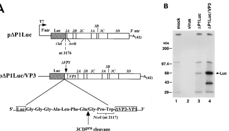
abil-ity to produce properly processed luciferase protein by pulse-labeling transfected H1-HeLa cells, and then immunoprecipi-tating luciferase as described above. SDS-PAGE demonstrated an immunoreactive protein of the expected mass in cells
trans-fected with DP1Luc/VP3 RNA (Fig. 7B, lane 4) but not in
mock-transfected or rhinovirus-infected cells (lanes 1 and 2,
respectively). Lack of processing at the synthetic 3CDpro
[image:7.612.58.299.69.370.2]cleav-age site would have resulted in a fusion protein consisting of the luciferase enzyme and 70 additional residues of VP3 (ap-proximately 70 kDa). Immunoreactive proteins of this size
FIG. 5. Subgenomic HRV-14 constructs with P1 segment deletions. Shown is a schematic diagram of the 5 HRV-14 in-frame P1 segment deletion mutants created as described in Materials and Methods. The nucleotide positions flank-ing the termini of each deletion are indicated. For the pCapD4 deletion, a linker was inserted between the BclI and BglII sites to create a deletion beginning at the AUG initiator codon and extending to the first nucleotide of the Thr-54 codon at nt 788.
FIG. 6. Replication of P1-deleted mutant RNAs in H1-HeLa cells. Lysates were prepared at 0, 6, 10, or 24 h following transfection of cells with the indicated RNA and processed for RPA as described in Materials and Methods. See the legend to Fig. 4 for details.
on November 9, 2019 by guest
http://jvi.asm.org/
[image:7.612.134.486.513.706.2]were not apparent in DP1Luc/VP3-transfected cells (lane 4), indicating efficient trans recognition and utilization of the
syn-thetic 3CDpro cleavage site within the DP1Luc/VP3
polypro-tein. The abundance of the luciferase protein was significantly
greater in cells transfected with DP1Luc/VP3 RNA than in
cells transfected withDP1Luc (compare lanes 3 and 4). Since
equivalent amounts of replicon RNA were transfected in this experiment, this provided preliminary evidence for replication
of theDP1Luc/VP3 RNA.
To confirm thatDP1Luc/VP3 RNA was replicating, HRV-14
RNA was quantified by RPA of lysates derived from trans-fected cells (Fig. 8). Control transfections carried out
simulta-neously included WR3.26 (full-length HRV-14) and DP1Luc
RNAs. Cell lysates were prepared as before, at various points in time following transfection. Increasing quantities of virus-specific RNA were evident in lysates of cells transfected with
DP1Luc/VP3 (Fig. 8). The RNA levels were similar to those
produced by intact HRV-14 transcripts in the same experi-ment. As before, no significant increase in the RNA signal was
observed in cells transfected with DP1Luc RNA. Thus, the
addition of P1 sequences extending from nt 2117 to the P1/P2 junction resulted in restoration of replication competence to
the candidateDP1Luc replicon.
We compared the luciferase activity expressed in cells
trans-fected withDP1Luc/VP3 with that expressed following
trans-fection withDP1Luc (Fig. 9). Although theD
P1Luc/VP3-trans-fected cells contained approximately 5-fold less luciferase
activity thanDP1Luc-transfected cells did (Fig. 9) 3 to 6 h after
transfection, this activity increased exponentially in DP1Luc/
VP3- but not inDP1Luc-transfected cells (Fig. 9) between 6
and 12 h. Thus, levels of luciferase at 12 and 24 h were over
100-fold greater than those present in DP1Luc-transfected
cells. This pattern of reporter protein expression is in
agree-ment with previous reports of successful poliovirus replicons that express foreign proteins (3, 9, 36). The early delay in
luciferase expression from DP1Luc/VP3 compared with
DP1Luc (Fig. 9) may reflect sequestration of input RNA in
replication complexes, reducing the availability of the input RNA for translation.
Capsid sequences provided intransdo not rescue nonrepli-cating candidate HRV-14 replicons.To determine whether the
replication defect inDP1Luc could be complemented in trans,
cell monolayers which had been previously transfected with replicon RNAs were infected with HRV-14 at high multiplicity of infection. Under these conditions, the replicating virus ge-nomes should provide an abundance of the capsid proteins expressed by the P1 segment. If the capsid proteins (VP1 or
VP3 fragment expressed by DP1Luc/VP3) are important for
RNA replication and can function in trans, a nonreplicating
RNA such asDP1Luc might be rescued and undergo normal
RNA replication. For this experiment, cells were transfected
withDP1Luc orDP1Luc/VP3 RNAs and infected 6 h later with
HRV-14 at multiplicity of infection of 10 PFU per cell. Mono-layers were assayed for luciferase enzyme activity 18 h later. As shown in Table 1, the enzyme activity was not increased in
DP1Luc-transfected cells that were infected with HRV-14 but
was instead decreased (42% of control transfected cells). These data thus suggest that the requirement for P1 sequences in RNA replication cannot be provided in trans. The decrease in luciferase activity observed with HRV-14 infection probably reflects competition between the viral and candidate replicon RNAs for cellular translation factors, as well as increased cell death related to viral replication.
Compared withDP1Luc, the fractional decrease in luciferase
activity was substantially greater following virus infection of
[image:8.612.86.539.66.326.2]cells which had been transfected previously with theDP1Luc/
FIG. 7. Organization of theDP1Luc/VP3 replicon. (A) HRV-14 sequences between nt 2118 and 3176 were reinserted inDP1Luc to createDP1Luc/VP3, as illustrated. A synthetic 3CDprorecognition and cleavage site was placed at the carboxyl terminus of the luciferase sequence. (B) 3CDproprocessing of luciferase from theDP1Luc/VP3 polyprotein. An immunoreactive protein (arrow) of the predicted size was present in [35S]Met-labeled cells transfected withDP1Luc/VP3 RNA but absent in mock-transfected or virus-infected cell extracts. Luciferase generated from cells transfected withDP1Luc RNA is shown for comparison. Cell lysates were immunoprecipitated with rabbit antibody to luciferase and subjected to electrophoresis on an SDS–10% polyacrylamide gel. The minor immunoreactive band (at about 40 kDa) which migrated more rapidly in theDP1Luc/VP3 immunoprecipitation is likely a result of aberrant initiation or premature termination of translation.
1948 MCKNIGHT AND LEMON J. VIROL.
on November 9, 2019 by guest
http://jvi.asm.org/
VP3 replicon (4% of uninfected control cells; Table 1). This decrease could not be related to removal of RNA from the pool of translating RNAs as a result of packaging of the rep-licon, because a similar decrease was observed following co-transfection with CapD2 RNA (Fig. 5), which does not encode a complete set of capsid proteins (Table 1). This decrease in
luciferase expression is reflected by reduced DP1Luc/VP3
RNA replication, probably owing to competition from a more efficiently replicating CapD2 or HRV-14 RNA (31). The
ap-parent difference in the replication efficiency of these RNAs could be related to differences in their length (7,389 nt for
DP1Luc/VP3 versus 7,212 nt for HRV-14 and 5,846 nt for
CapD2). Importantly,DP1Luc/VP3 and CapD2 RNAs contain
similar HRV-14 P1 sequences (Fig. 5 and 7A).
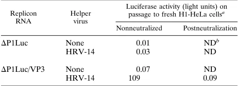
DP1Luc/VP3 RNA is encapsidated during HRV-14 infection.
To determine whether DP1Luc/VP3 replicon RNA could be
[image:9.612.143.467.73.341.2]packaged by capsid proteins provided in trans, transfected cell monolayers were infected with HRV-14 as described above. Virus harvests were prepared 18 h postinfection. Prior to in-oculation onto fresh H1-HeLa cells, this first-passage virus was treated with 1 mg of RNase A per ml to remove any free replicon RNA. As shown in Table 2, cell lysates prepared from monolayers inoculated with this virus harvest contained small but readily detectable amounts of luciferase enzyme activity. The expression of this luciferase activity could be completely
FIG. 8. RPA analysis ofDP1Luc/VP3 RNA replication. Cell lysates were prepared at 0, 3, 6, 9, 12, and 24 h following transfection of the indicated RNA. See the legend to Fig. 4 for details.
[image:9.612.71.288.499.693.2]FIG. 9. Luciferase activity in cells transfected withDP1Luc/VP3 RNA. Cy-toplasmic extracts were prepared at 0, 3, 6, 9, 12, and 24 h following transfection withDP1Luc/VP3 RNA (F) orDP1Luc RNA (h) or mock transfection (■). Results shown represent means (6range) of duplicate transfections.
TABLE 1. Luciferase activity in H1-HeLa cells transfected with candidate replicon RNAs and coinfected with HRV-14
or cotransfected with CapD2 RNA
Replicon RNA
Helper
genomea Luciferase activity (light units) (%)b
DP1Luc None 164 (100)
HRV-14 69 (42)
DP1Luc/VP3 None 19,985 (100)
HRV-14 802 (4)
CapD2 781 (4)
a
Helper genome supplied by infection with HRV-14 6 h following replicon transfection or by simultaneous cotransfection of replicon and CapD2 RNAs.
b
Luciferase values represent means of duplicate cultures assayed 24 h follow-ing transfection (percent luciferase expression in the absence of helper genome is given in parentheses).
on November 9, 2019 by guest
http://jvi.asm.org/
[image:9.612.316.556.603.686.2]blocked by pretreatment of the virus harvest with neutralizing antibody to HRV-14. In contrast, no luciferase activity was
present in cells inoculated with virus harvested fromD
P1Luc-transfected cultures that were infected with HRV-14 or from
DP1Luc/VP3-transfected cultures that were not infected with
helper virus (Table 2). These results confirm thatDP1Luc/VP3
replicon RNA is packaged by helper virus during infection. However, no evidence for survival of the replicon RNA could be obtained on subsequent passage of the virus-replicon
mix-ture (31), indicating that the packagedDP1Luc/VP3 genomes
were not stable in the helper virus population.
DISCUSSION
We have shown that an HRV replicon RNA,DP1Luc/VP3,
in which sequences encoding the firefly luciferase protein
re-placed the 59 1,488 nt of the P1 capsid-coding sequence of
HRV-14, replicated efficiently following its transfection into H1-HeLa cells (Fig. 8). The luciferase activity expressed from
DP1Luc/VP3 RNA was dependent on replication of the RNA
genome (Fig. 9). In contrast, a similar candidate replicon,
DP1Luc, in which all but 21 nt of the HRV-14 P1 segment were
removed from the genome, failed to undergo efficient RNA replication (Fig. 4B) and expressed only very low levels of luciferase (Fig. 3B). While it is not possible to exclude a very
low level of replication ofDP1Luc or the structurally related
DP1bgal candidate replicons, these RNAs did not undergo
amplification that was detectable in a sensitive RPA following transfection of HeLa cells (Fig. 4). Intracellular RNA levels remained similar to those present following transfection with candidate replicon RNAs containing a lethal frameshift
muta-tion within the 3DpolRNA polymerase coding region. These
results were consistent with the analysis of a series of sub-genomic HRV-14 RNAs containing various in-frame deletions within the P1 segment of the genome, which suggested that the inclusion of sequence between nt 2121 and 2724 was essential for replication of subgenomic HRV-14 RNAs (Fig. 6). How-ever, the minimal sequence within this 603-nt segment which is required for replication has yet to be determined.
Inefficient or aberrant processing of the HRV-14 polypro-tein at the P1/P2 junction seems unlikely to account for the
lack of replication ofDP1bgal,DP1Luc, or the capsid sequence
deletion mutants CapD1 and CapD5. Pulse-labeling
experi-ments demonstrated efficient processing of the 2Apro-directed
cleavage at the carboxyl terminus of the Luc-VP1 fusion
pro-tein expressed byDP1Luc (Fig. 2). Similarly, an
immunoreac-tiveb-galactosidase protein of the expected size was present
in cells transfected withDP1bgal (Fig. 2). These observations
indicate the retention of autocatalytic processing of the poly-protein of these replication-defective candidate replicons by
2Apro and are consistent with the retention of proteolytic
ac-tivity when poliovirus 2Aprowas similarly fused to heterologous
sequences (2, 34, 41). Processing of the remaining P2/P3 por-tion of the replicon polyprotein was not examined, because it is
expected to proceed normally after the primary 2Aprocleavage.
Although we did not examine the P1/P2 cleavage in cells trans-fected with the capsid deletion mutants shown in Fig. 5, the replication-defective mutants CapD1 and CapD5 encode poly-proteins with carboxyl-terminal fragments of VP1 that are
sub-stantially longer than the 7 residues present in theDP1Luc and
DP1bgal polyproteins and are thus likely to be effectively
pro-cessed.
That P1 sequences appear to be required for efficient repli-cation of HRV-14 RNA contrasts sharply with reports of po-liovirus replicons which replicate well despite large deletions in the P1 region (3, 13, 24, 41). However, it is interesting that naturally occurring poliovirus defective interfering genomes
generally retain sequence at the 39end of the P1 segment (20,
23, 26), although the downstream P1 sequences retained by these genomes are significantly shorter than the P1 sequence
reinserted in DP1Luc/VP3 and encode only a truncated VP1
protein. The fact that candidate rhinovirus replicons which lacked almost all of the P1 segment failed to replicate indicates an important difference between these two otherwise quite closely related viruses.
The required P1 sequence could function in several different ways during the replication of HRV-14 RNA. The VP1 protein
is expressed as an intact protein byDP1Luc/VP3 and in each of
the replication-competent P1 deletion mutants. The analogous poliovirus VP1 protein is closely associated with the mem-brane-bound RNA replication complex in poliovirus-infected cells (37). While this association has been assumed to reflect the well-documented role of this protein in encapsidation of RNA (25), an accessory function associated with RNA repli-cation would be consistent with the tendency of picornavirus proteins to fulfill multiple functions in viral replication. VP1 provided in trans by replicating HRV-14 helper virus did not result in the rescue and replication of nonviable RNAs such as
DP1Luc (Table 1). While this does not rule out a cis-acting role
for VP1 in viral RNA replication, as is known to be the case for several of the nonstructural proteins of poliovirus (43, 50), this observation does argue against a trans-acting role for any of the capsid proteins of HRV-14 in RNA replication. Further exper-iments are required to determine whether the essential P1 sequence must be expressed as protein to support viral repli-cation. The construction of dicistronic HRV-14 replicons may be helpful in addressing this question (33).
[image:10.612.57.298.93.181.2]It is also possible that this region of the P1 segment acts directly as RNA in facilitating the replication of the HRV-14 genome. While such a P1 segment function is not suggested by any current model of picornavirus RNA replication, the exis-tence of a cis-acting replication signal located within this re-gion of the viral genome would act to suppress the evolution of defective interfering particles. Such an element would prevent the amplification of RNA molecules sustaining large P1 dele-tions that might, because of their shorter length, be otherwise favored for replication. Interestingly, internally located, cis-acting RNA replication elements are not without precedent among plus-strand RNA viruses. Efficient synthesis of RNA-3 of brome mosaic virus, a positive-sense plant virus with a tri-partite genome, requires the presence of internal sequences which are located within an intercistronic region of this RNA (18, 39, 42). These internal sequences may facilitate assembly
TABLE 2. Encapsidation of candidate replicon RNAs by HRV-14 helper virus infection
Replicon RNA
Helper virus
Luciferase activity (light units) on passage to fresh H1-HeLa cellsa
Nonneutralized Postneutralization
DP1Luc None 0.01 NDb
HRV-14 0.03 ND
DP1Luc/VP3 None 0.07 ND
HRV-14 109 0.09
aLuciferase expression in H1-HeLa cells infected with RNase-digested lysates of cells which had been transfected with replicon RNAs and infected with helper virus as indicated. Lysates (first-passage virus harvests) were inoculated onto fresh cells with or without prior incubation with HRV-14 neutralizing anti-body.
bND, not determined.
1950 MCKNIGHT AND LEMON J. VIROL.
on November 9, 2019 by guest
http://jvi.asm.org/
of an active brome mosaic virus RNA-dependent RNA poly-merase complex required for negative-strand RNA-3 synthesis in yeast cells (42), but they may not be absolutely required for minus-strand initiation in plants, because a low level of RNA-3 replication proceeds in their absence (18). Recent work also has revealed the presence of an internal cis-acting replication
signal located between 3.1 and 3.9 kb of the 59end of a murine
coronavirus RNA genome (24a). Similar to the HRV artificial defective interfering genomes described in this report, this sequence was found to be required for efficient replication of coronavirus defective interfering genomes.
Studies with the bacteriophage Qb replicase have
demon-strated a requirement for binding of the replicase complex to two internal sites on the plus-strand RNA (5). Both the viral replicase and a host factor bind to these internal sites, with
binding of the host factor possibly acting to bring the 39end of
the genome into close proximity to the internal sites, thereby allowing initiation of minus-strand RNA synthesis (5). It is not difficult to imagine a similar event in the replication of picor-navirus RNAs, although there are no prior data to suggest this. The existence and nature of possible RNA secondary struc-tures within the relevant region of the rhinovirus P1 coding segment are not known, but alignment of available rhinovirus sequences indicates the presence of several strong areas of nucleotide sequence conservation (31). Multiple studies indi-cate that the P1 segment of poliovirus is not required for efficient RNA replication, but there are no data which would exclude the possibility of such a cis-acting RNA replication element within the P2/P3 segments of poliovirus. Interestingly, Novak and Kirkegaard identified a region within the P2-P3 segments of the poliovirus genome which must undergo trans-lation in cis for replication of the RNA to proceed (34a). It is intriguing to speculate that the replication element we have identified in HRV-14 may be analogous to this ‘‘cis-translation required,’’ element of poliovirus but located within a different region of the genome.
We found clear evidence for encapsidation of theDP1Luc/
VP3 RNAs following infection of previously transfected cells with the HRV-14 helper virus (Table 2). However, luciferase expression was very low when helper virus harvests were used to infect fresh H1-HeLa cells, and in situ visualization of lu-ciferase expression demonstrated only very small numbers of infected cells (31). No luciferase activity was noted on further passage of the helper-replicon mixture. At present, it is not clear whether this reflects the inhibition of replicon replica-tion by the helper viral genome or restricreplica-tions on packaging of the longer-than-genome-length RNA. Additional experiments were carried out in which the time of HRV-14 infection was incrementally delayed up to 12 h after replicon transfection, when substantial amounts of newly replicated RNA should have been available for encapsidation. However, we observed no significant increase in encapsidation of replicon RNA over that shown in Table 2 (31). Although these results suggest that the low levels of replicon encapsidation may be related to the size or structure of RNA preferred by the HRV-14 packaging mechanism, further studies are required to determine the mag-nitude of any restriction to encapsidation.
The luciferase expressed by theDP1Luc/VP3 replicon
pro-vides a simple and sensitive marker for HRV-14 RNA repli-cation. Since the pathogenesis of HRV infections is not well understood, this or other replicons may be useful in studying certain aspects of HRV pathogenesis, including the cell types within the respiratory mucosa that are infected by HRV and the routes by which the virus disseminates through the epithe-lium of the upper airway during infection. Furthermore, the unique tropism of HRV for cells of the respiratory mucosa
offers the possibility that HRV replicons expressing heterolo-gous antigens or functional proteins will ultimately prove use-ful as vaccine delivery systems or for targeted gene therapy.
ACKNOWLEDGMENTS
We thank Wai-Ming Lee and Roland Rueckert for their kind gift of the HRV-14 cDNA, pWR3.26. Also, much appreciation goes to Anne Mosser for her gift of the anti-HRV-14 neutralizing antibody and to Ann Palmenberg for providing us with the rhinovirus sequence align-ments. We are also grateful to John Newbold for his critical review of the manuscript.
This work was supported by grants from the U.S. Public Health Service (HL51818 and AI32599) and the Cystic Fibrosis Foundation (S880).
REFERENCES
1. Alsaadi, S., S. Hassard, and G. Stanway. 1989. Sequences in the 59 non-coding region of human rhinovirus 14 RNA that affect in vitro translation. J. Gen. Virol. 70:2799–2804.
2. Alvey, J. C., E. E. Wyckoff, S. F. Yu, R. Lloyd, and E. Ehrenfeld. 1991. cis- and
trans-cleavage activities of poliovirus 2A protease expressed in Escherichia coli. J. Virol. 65:6077–6083.
3. Andino, R., G. E. Rieckhof, P. L. Achacoso, and D. Baltimore. 1993. Polio-virus RNA synthesis utilizes an RNP complex formed around the 59-end of viral RNA. EMBO J. 12:3587–3598.
4. Andino, R., D. Silvera, S. D. Suggett, P. L. Achacoso, C. J. Miller, D. Baltimore, and M. B. Feinberg.1994. Engineering poliovirus as a vaccine vector for the expression of diverse antigens. Science 265:1448–1451. 5. Barrera, I., D. Schuppli, J. M. Sogo, and H. Weber. 1993. Different
mech-anisms of recognition of bacteriophage Qbplus and minus strand RNAs by Qbreplicase. J. Mol. Biol. 232:512–521.
6. Blair, W. S., and B. L. Semler. 1991. Role for the P4 amino acid residue in substrate utilization by the poliovirus 3CD proteinase. J. Virol. 65:6111– 6123.
7. Brown, E. A., S. P. Day, R. W. Jansen, and S. M. Lemon. 1991. The 59 nontranslated region of hepatitis A virus: secondary structure and elements required for translation in vitro. J. Virol. 65:5828–5838.
8. Callahan, P. L., S. Mizutani, and R. J. Colonno. 1985. Molecular cloning and complete sequence determination of the RNA genome of human rhinovirus type 14. Proc. Natl. Acad. Sci. USA 82:732–736.
9. Choi, W. S., R. Pal-Ghosh, and C. D. Morrow. 1991. Expression of human immunodeficiency virus type 1 (HIV-1) gag, pol, and env proteins from chimeric HIV-1-poliovirus minireplicons. J. Virol. 65:2875–2883. 10. Cole, C. N. 1975. Defective interfering (DI) particles of poliovirus. Prog.
Med. Virol. 20:180–207.
11. Cole, C. N., and D. Baltimore. 1973. Defective interfering particles of po-liovirus. II. Nature of the defect. J. Mol. Biol. 76:325–343.
12. Cole, C. N., D. Smoler, E. Wimmer, and D. Baltimore. 1971. Defective interfering particles of poliovirus. I. Isolation and physical properties. J. Virol. 7:478–485.
13. Collis, P. S., B. J. O’Donnell, D. J. Barton, J. A. Rogers, and J. B. Flanegan. 1992. Replication of poliovirus RNA and subgenomic RNA transcripts in transfected cells. J. Virol. 66:6480–6488.
14. Colonno, R. J., R. L. Lafemina, C. M. Dewitt, and J. E. Tomassini. 1990. The major-group rhinoviruses utilize the intracellular adhesion molecule 1 ligand as a cellular receptor during infection, p. 257–261. In M. A. Brinton and F. X. Heinz (ed.), New aspects of positive-strand RNA viruses. American Society for Microbiology, Washington, D.C.
15. Couch, R. B. 1990. Rhinoviruses, p. 607–629. In B. N. Fields and D. M. Knipe (ed.), Virology. Raven Press, New York.
16. Dewalt, P. G., M. A. Lawson, R. J. Colonno, and B. L. Semler. 1989. Chimeric picornavirus polyproteins demonstrate a common 3C proteinase substrate specificity. J. Virol. 63:3444–3452.
17. Duechler, M., T. Skern, D. Blaas, B. Berger, W. Sommergruber, and E. Kuechler.1989. Human rhinovirus serotype 2: in vitro synthesis of an infec-tious RNA. Virology 168:159–161.
18. French, R., and P. Ahlquist. 1987. Intercistronic as well as terminal se-quences are required for efficient amplification of brome mosaic virus RNA3. J. Virol. 61:1457–1465.
19. Gwaltney, J. M., Jr. 1982. The rhinoviruses, p. 491–517. In E. A. Evans (ed.), Viral infection of humans: epidemiology and control. Plenum Publishing Corp., New York.
20. Hagino-Yamagishi, K., and A. Nomoto. 1989. In vitro construction of polio-virus defective interfering particles. J. Virol. 63:5386–5392.
21. Hellen, C. U. T., C.-K. Lee, and E. Wimmer. 1992. Determinants of substrate recognition by poliovirus 2A proteinase. J. Virol. 66:3330–3338.
22. Hofer, F., M. Gruenberger, H. Kowalski, H. Machat, M. Huettinger, E.
on November 9, 2019 by guest
http://jvi.asm.org/
Kuechler, and D. Blass.1994. Members of the low density lipoprotein re-ceptor family mediate cell entry of a minor-group common cold virus. Proc. Natl. Acad. Sci. USA 91:1839–1842.
23. Kajigaya, S., H. Arakawa, S. Kuge, T. Koi, N. Imura, and A. Nomoto. 1985. Isolation and characterization of defective-interfering particles of poliovirus. Sabin 1 strain. Virology 142:307–316.
24. Kaplan, G., J. Lubinski, A. Dasgupta, and V. R. Racaniello. 1985. In vitro synthesis of infectious poliovirus RNA. Proc. Natl. Acad. Sci. USA 82: 8424–8428.
24a.Kim, Y.-N., and S. Makino. 1995. Characterization of murine coronavirus defective interfering RNA internal cis-acting replication signal. J. Virol. 69: 4963–4971.
25. Kirkegaard, K. 1990. Mutations in VP1 of poliovirus specifically affect both encapsidation and release of viral RNA. J. Virol. 64:195–206.
26. Kuge, S., I. Saito, and A. Nomoto. 1986. Primary structure of poliovirus defective-interfering particle genomes and possible generation mechanisms of the particles. J. Mol. Biol. 192:473–487.
27. Lawson, M. A., and B. L. Semler. 1990. Picornavirus protein processing-enzymes, substrates, and genetic regulation, p. 49–87. In V. R. Racaniello (ed.), Picornaviruses. Springer-Verlag KG, Berlin.
28. Lee, W.-M., S. S. Monroe, and R. R. Rueckert. 1993. Role of maturation cleavage in infectivity of picornaviruses: activation of an infectosome. J. Virol. 67:2110–2122.
29. Liljestrom, P., S. Lusa, D. Huylebroeck, and H. Garoff. 1991. In vitro mu-tagenesis of a full-length cDNA clone of Semliki Forest virus: the small 6,000-molecular-weight membrane protein modulates virus release. J. Virol. 65:4107–4113.
30. Mattion, N. M., P. A. Reilly, S. J. DiMichele, J. C. Crowley, and C. Weeks-Levy.1994. Attenuated poliovirus strain as a live vector: expression of re-gions of rotavirus outer capsid protein VP7 by using recombinant Sabin 3 viruses. J. Virol. 68:3925–3933.
31. McKnight, K. L., and S. M. Lemon. Unpublished data.
32. Mizutami, S., and R. J. Colonno. 1985. In vitro synthesis of an infectious RNA from cDNA clones of human rhinovirus type 14. J. Virol. 56:628–632. 33. Molla, A., S. K. Jang, A. V. Paul, Q. Reuer, and E. Wimmer. 1992. Cardio-viral internal ribosomal entry site is functional in a genetically engineered dicistronic poliovirus. Nature (London) 356:255–257.
34. Molla, A., A. V. Paul, M. Schmid, S. K. Jang, and E. Wimmer. 1993. Studies on dicistronic polioviruses implicate viral proteinase 2Aproin RNA replica-tion. Virology 196:739–747.
34a.Novak, J. E., and K. Kirkegaard. 1994. Coupling between genome transla-tion and replicatransla-tion in an RNA virus. Genes Dev. 8:1726–1737.
35. Palmenberg, A. C. 1989. Sequence alignments of picornaviral capsid pro-teins, p. 211–241. In B. Semler and E. Ehrenfeld (ed.), Molecular aspects of picornavirus infection and detection. American Society for Microbiology, Washington, D.C.
36. Percy, N., W. S. Barclay, M. Sullivan, and J. W. Almond. 1992. A poliovirus replicon containing the chloramphenicol acetyltransferase gene can be used
to study the replication and encapsidation of poliovirus RNA. J. Virol. 66: 5040–5046.
37. Pfister, T., D. Egger, and K. Bienz. 1995. Poliovirus subviral particles asso-ciated with progeny RNA in the replication complex. J. Gen. Virol. 76:63– 71.
38. Pilipenko, E. V., V. M. Blinov, and V. I. Agol. 1990. Gross rearrangements within the 59-untranslated region of the picornaviral genomes. Nucleic Acids Res. 18:3371–3375.
39. Pogue, G. P., and T. C. Hall. 1992. The requirement for a 59stem-loop structure in brome mosaic virus replication supports a new model for viral positive-strand RNA initiation. J. Virol. 66:674–684.
40. Porter, D. C., D. C. Ansardi, W. S. Choi, and C. D. Morrow. 1993. Encap-sidation of genetically engineered poliovirus minireplicons which express human immunodeficiency virus type 1 Gag and Pol proteins upon infection. J. Virol. 67:3712–3719.
41. Porter, D. C., D. C. Ansardi, and C. D. Morrow. 1995. Encapsidation of poliovirus replicons encoding the complete human immunodeficiency virus type 1 gag gene by using a complementation system which provides the P1 capsid protein in trans. J. Virol. 69:1548–1555.
42. Quadt, R., M. Ishikawa, M. Janda, and P. Ahlquist. 1995. Formation of brome mosaic virus RNA-dependent RNA polymerase in yeast requires coexpression of viral proteins and viral RNA. Proc. Natl. Acad. Sci. USA 92: 4892–4896.
43. Richards, O. C., and E. Ehrenfeld. 1990. Poliovirus RNA replication, p. 89–119. In V. R. Racaniello (ed.), Picornaviruses. Springer-Verlag KG, Ber-lin.
44. Rohll, J. B., N. Percy, R. Ley, D. J. Evans, J. W. Almond, and W. S. Barclay. 1994. The 59-untranslated regions of picornavirus RNAs contain indepen-dent functional domains essential for RNA replication and translation. J. Virol. 68:4384–4391.
45. Rossmann, M. G., E. Arnold, J. W. Erickson, E. A. Frankenberger, J. P. Griffith, H.-J. Hecht, J. E. Johnson, G. Kamer, M. Luo, A. G. Mosser, R. R. Rueckert, B. Sherry, and G. Vriend.1985. Structure of a human common cold virus and functional relationship to other picornaviruses. Nature (Lon-don) 317:145–153.
46. Rueckert, R. R., and E. Wimmer. 1984. Systematic nomenclature of picor-navirus proteins. J. Virol. 50:957–959.
47. Sambrook, J., E. F. Fritsch, and T. Maniatis. 1989. Molecular cloning: a laboratory manual, 2nd ed. Cold Spring Harbor Laboratory, Cold Spring Harbor, N.Y.
48. Sherry, B., and R. R. Rueckert. 1985. Evidence for at least two dominant neutralization antigens on human rhinovirus 14. J. Virol. 53:137–143. 49. Sommergruber, W., H. Ahorn, A. Zo¨phel, I. Maurer-Fogy, F. Fessl, G.
Schnorrenberg, H.-D. Liebig, D. Blaas, E. Kuechler, and T. Skern.1992. Cleavage specificity on synthetic peptide substrates of human rhinovirus 2 proteinase 2A. J. Biol. Chem. 267:22639–22644.
50. Wimmer, E., C. U. T. Hellen, and X. Cui. 1993. Genetics of poliovirus. Annu. Rev. Genet. 27:353–436.
1952 MCKNIGHT AND LEMON J. VIROL.