0022-538X/95/$04.0010
Copyrightq1995, American Society for Microbiology
Involvement of the V1/V2 Variable Loop Structure in the
Exposure of Human Immunodeficiency Virus Type 1
gp120 Epitopes Induced by Receptor Binding
RICHARD WYATT,
1JOHN MOORE,
2MOLLY ACCOLA,
1ELIZABETH DESJARDIN,
1JAMES ROBINSON,
3ANDJOSEPH SODROSKI
1,4*
Division of Human Retrovirology, Dana-Farber Cancer Institute, and Department of Pathology, Harvard
Medical School,
1and Department of Cancer Biology, Harvard School of Public Health,
4Boston,
Massachusetts 02115; Aaron Diamond AIDS Research Center, New York University School
of Medicine, New York, New York 10016
2; and Department of Pediatrics, University
of Connecticut Health Sciences Center, Farmington, Connecticut 06030
3Received 7 February 1995/Accepted 13 June 1995
The binding of human immunodeficiency virus type 1 (HIV-1) to the cellular receptor CD4 has been
suggested to induce conformational changes in the viral envelope glycoproteins that promote virus entry.
Conserved, discontinuous epitopes on the HIV-1 gp120 glycoprotein recognized by the 17b, 48d, and A32
antibodies are preferentially exposed upon the binding of soluble CD4 (sCD4). The binding of the 17b and 48d
antibodies to the gp120 glycoprotein can also be enhanced by the binding of the A32 antibody. Here we
constructed HIV-1 gp120 mutants in which the variable segments of the V1/V2 and V3 structures were deleted,
individually or in combination, while the 17b, 48d, and A32 epitopes were retained. The effects of the variable
loop deletions on the function of the HIV-1 envelope glycoproteins and on the exposure of epitopes induced by
sCD4 or A32 binding to the monomeric gp120 glycoprotein were examined. The variable-loop-deleted envelope
glycoproteins were able to mediate virus entry, albeit at lower efficiencies than those of the wild-type
glyco-proteins. Thus, the V1/V2 and V3 variable sequences contribute to the efficiency of HIV-1 entry but are not
absolutely required for the process. Neither the V1/V2 nor V3 loops were necessary for the increase in exposure
of the 17b/48d epitopes induced by binding of the A32 monoclonal antibody. By contrast, induction of the 17b,
48d, and A32 epitopes by sCD4 binding apparently involves a movement of the V1/V2 loops, which in the
absence of CD4 partially mask these epitopes on the native gp120 monomer. The results obtained with a
mutant glycoprotein containing a deletion of the V1 loop alone indicated that the contribution of the V2 loop
to these phenomena was more significant than that of the V1 sequences. These results suggest that the V1/V2
loops, which have been previously implicated in CD4-modulated, postattachment steps in HIV-1 entry,
con-tribute to CD4-induced gp120 conformational changes detected by the 17b, 48d, and A32 antibodies.
Human immunodeficiency virus type 1 (HIV-1) is the
etio-logic agent of AIDS (3, 27). The entry of HIV-1 into target
cells, like that of other retroviruses, is mediated by the viral
envelope glycoproteins. The gp120 exterior envelope
glycopro-tein binds to the CD4 receptor, while the gp41 transmembrane
envelope glycoprotein serves to anchor the tetrameric
enve-lope glycoprotein complex in the viral membrane (1, 18, 41, 46,
48, 63). Following receptor binding, the envelope glycoproteins
mediate the fusion of viral and target cell membranes, which is
critical for successful entry of the viral core into the cell (73).
Most of the surface-exposed elements of the mature
enve-lope glycoprotein complex are contained on the gp120 exterior
envelope glycoprotein (53, 67). When the gp120 glycoproteins
derived from different HIV-1 isolates are compared, five
con-served regions (C1 to C5) and five variable regions (V1 to V5)
can be identified (57, 71). Efficient CD4 binding is dependent
on discontinuous elements derived from the third (aspartic
acid 368 and glutamic acid 370) and fourth (tryptophan 427
and aspartic acid 457) conserved regions (15, 42, 44, 58).
Con-served, discontinuous epitopes overlapping the CD4 binding
site serve as targets for neutralizing antibodies (34, 40, 61, 72,
78, 81, 82).
Intramolecular disulfide bonds in the gp120 glycoprotein
result in the inclusion of the first four variable regions (V1 to
V4) in large loop-like structures (45). Antibody mapping
stud-ies indicate that, of the linear epitopes on the gp120
glycopro-tein, those located in the V2 and V3 regions constitute the
most exposed elements on the HIV-1 multimeric envelope
glycoprotein complex (53). Both V2 and V3 loops can serve as
targets for neutralizing antibodies (26, 33, 38, 49, 54, 62, 64,
69). Changes in V2 and V3 loop amino acids have been shown
to affect the membrane fusion process and can influence the
tropism of HIV-1 isolates for primary macrophages and
T-lymphocyte lines (4, 7, 10–12, 24, 28, 29, 31, 36, 37, 59, 68, 75,
84, 85). Amino acid alterations in the fourth conserved (C4)
region, which has been suggested to interact with the V3 loop
(55, 56, 76, 89), can also affect membrane fusion events
in-volved in HIV-1 entry (77).
Analogy with the influenza virus hemagglutinin (86) suggests
that, after receptor binding, conformational changes in the
HIV-1 envelope glycoproteins may have to occur to allow
exposure of the gp41 glycoprotein to the target cell membrane.
Mutagenic analysis has defined gp41 regions important for
membrane fusion, and the locations of these regions are
con-sistent with models proposed for the orthomyxoviruses (8, 20,
23, 32, 42). Unlike the orthomyxoviruses, however, HIV-1 does
* Corresponding author. Mailing address: Jimmy Fund Building, Room 824, Dana-Farber Cancer Institute, 44 Binney St., Boston, MA 02115. Phone: (617) 632-3371. Fax: (617) 632-4338. Electronic mail address: [email protected].
5723
on November 9, 2019 by guest
http://jvi.asm.org/
not require a decrease in pH to trigger the membrane fusion
process (73). Instead, it has been suggested that CD4 binding
serves as a trigger for fusion-related conformational changes.
Such a model is consistent with the observation that a soluble
form of the CD4 receptor (sCD4) (19, 21, 35, 70, 83) can
enhance the infection of HIV-2 (14), simian immunodeficiency
virus (SIVagm) (2), and some isolates of HIV-1 (74).
The binding of sCD4 has been shown to induce
conforma-tional changes in the HIV-1 envelope glycoproteins. For
T-cell-line-passaged HIV-1 isolates, sCD4 binding results in the
shedding of the gp120 glycoprotein from the envelope complex
(6, 30, 50). This observation, combined with the report that the
HIV-1 gp41 glycoprotein alone could mediate fusion (60),
sug-gested a model in which CD4 binding exposed the gp41
glyco-protein by removing the gp120 glycoglyco-protein from the envelope
glycoprotein complex. More recent data, however, have cast
doubt on both the relevance of gp120 shedding to the
mem-brane fusion process (4, 17, 25, 51, 52, 77, 80) and on the
reproducibility of cell fusion mediated by the isolated gp41
glycoprotein (47).
The uncertain relevance of gp120 shedding to HIV-1 entry
has prompted a search for other markers of fusion-related
conformational changes. A recent analysis of the effects of
CD4 amino acid changes on HIV-1 entry suggests that
confor-mational changes within the gp120-CD4 binding site itself are
unlikely to be required for promoting subsequent
fusion-re-lated events in virus entry (5). Instead, regions of the gp120
glycoprotein outside the CD4 binding site, some of which have
been reported to undergo conformational changes upon CD4
binding (39, 65, 66, 79), might be better candidates for
struc-tures involved in conformational changes related to virus entry.
CD4 binding has been reported to increase the accessibility of
the V3 loop to proteases, whereas the binding of some
neu-tralizing antibodies to the V2 loop is decreased upon CD4
binding (49, 65, 66, 69). Discontinuous epitopes recognized by
the 17b and 48d neutralizing antibodies are more exposed after
CD4 binding (79). Mutagenic analysis of the HIV-1 gp120
glycoprotein has suggested that the conserved 17b and 48d
epitopes are dependent upon the stem of the V1/V2 stem-loop
structure, the fourth (C4) conserved region, and amino acids
near or within the CD4 binding site (79).
A recent study of HIV-1 variants adapted to replicate on
CD4 mutants with lower gp120 binding affinities suggested that
the fusion-related functions of the V2, V3, and C4 regions of
the gp120 glycoprotein could be modulated by CD4 binding
(13). The involvement of the gp120 V2 and V3 loops in fusion
events specifically modulated by CD4 binding prompted us to
examine whether these variable structures might be involved in
some of the conformational changes in the HIV-1 gp120
gly-coprotein induced by receptor binding. The 17b and 48d
neu-tralization epitopes, which are more exposed on both
mono-meric gp120 and oligomono-meric envelope glycoproteins of HIV-1
following sCD4 binding (79), were studied. The A32 human
monoclonal antibody was also included in this study, because
its binding to the monomeric gp120 glycoprotein is also
en-hanced by sCD4 binding and because A32 antibody binding to
the gp120 glycoprotein results in enhanced binding of the 17b
and 48d antibodies (63a).
MATERIALS AND METHODS
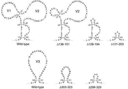
Construction of mutatedenv genes.The mutants were constructed in the HXBc2 strain of HIV-1 (22) by site-directed mutagenesis as previously described (43, 58) and cloned into the expressor plasmid pSVIIIenv. TheD136-151 mutant contains a deletion of residues 136 to 151, with the sequence Gly-Ala replacing the missing sequences (see Fig. 1). (In the numbering system used, 1 represents the initiator methionine.) TheD128-194 mutant contains a deletion of amino
acids 128 to 194, with a Gly-Ala-Gly sequence replacing the missing V1/V2 loops. TheD303-323 mutant contains a deletion of residues 303 to 323, with a Gly-Ala sequence replacing the missing V3 loop amino acids. TheD128-194/303-323 mutant contains both deletions and substitutions associated with the individual D128-194 andD303-323 mutants. TheDV1/V2 andDV3 mutants, which contain more-extensive deletions of the respective variable loop structures, were previ-ously characterized (88) and are herein designatedD121-203 andD298-329, respectively. The pSVIIIenvDKS plasmid, which contains an out-of-frame dele-tion within the env gene, was used as a negative control for some of the exper-iments.
Expression of mutant envelope glycoproteins.COS-1 cells were transfected with the expressor plasmid DNA by the DEAE-dextran technique as described previously (16). Forty-eight hours following transfection, the cells were labeled in cysteine-free medium with 10% heat-inactivated fetal calf serum (HIFCS) and 40 mCi of [35S]cysteine (Dupont, NEN Research Products) per ml for 16 h. Cells
were washed with phosphate-buffered saline (PBS) and lysed in Nonidet P-40 buffer (0.5% Nonidet P-40, 0.5 M NaCl, 10 mM Tris-HCl; pH 7.5). Envelope glycoproteins were precipitated from the lysates with either a mixture of sera from AIDS patients or monoclonal antibodies (1mg/ml final concentration) as described previously (58, 81). Cell supernatants were precipitated with the same antibodies (0.66mg/ml) in the absence of detergent and washed with PBS and 2% HIFCS. sCD4 (Agmed) (10mg/ml) was added to some of the cell lysates and supernatants prior to immunoprecipitation. Precipitates were analyzed on so-dium dodecyl sulfate-polyacrylamide gels.
ELISA determination of mutant gp120 recognition by antibodies.COS-1 cells were transfected with 10mg of pSVIIIenv DNA expressing wild-type or mutant HXBc2 envelope glycoproteins and a Tat-expressing plasmid, pSVTat. Seventy-two hours after transfection, cell supernatants were collected and frozen. For analysis of antibody recognition, various amounts of the supernatants (1 to 100 ml), supplemented with Tris-buffered saline–10% HIFCS to a total volume of 100 ml, were incubated in wells of Immulon II enzyme-linked immunosorbent assay (ELISA) plates (Dynatech, Ltd.) coated with sheep antibody D7324 to the carboxyl-terminal 15 amino acids of gp120. The amount of each mutant glyco-protein captured was estimated with the 133/290 monoclonal antibody (57a) directed against C1 gp120 residues 61 to 70. On the basis of this determination, the volume of each supernatant added to the ELISA plate was adjusted, so that equal amounts of each mutant glycoprotein were captured. The accuracy of this adjustment was verified by examining the amount of bound 133/290 antibody over a range (0.03 to 10mg/ml) of antibody concentrations. For all experiments, 133/290 antibody was diluted in TMSS buffer (Tris-buffered saline containing 2% nonfat milk powder, 20% sheep serum) and reacted with the captured gp120 glycoprotein, and bound antibody was detected with alkaline phosphatase-con-jugated rabbit anti-mouse immunoglobulin (Accurate Chemicals) and the AMPAK system (DaKo Diagnostics).
The CD4 binding abilities of the captured mutant glycoproteins were deter-mined by incubating CD4-immunoglobulin G (IgG) (Genentech) (9) diluted in TMSS buffer at various concentrations (0.02 to 6mg/ml) with the captured gp120 glycoprotein, followed by detection with alkaline phosphatase-conjugated goat anti-human immunoglobulin (Accurate Chemicals) and the AMPAK system.
The binding of the 17b, 48d, and A32 antibodies in the absence or presence of 5mg of sCD4 per ml was determined as described above for the binding of CD4-IgG. The 17b and 48d antibodies were tested over a concentration range of 0.01 to 3mg/ml, while the A32 antibody was tested over a range of 0.03 to 10 mg/ml.
The effects of binding the A32 antibody to the captured gp120 glycoprotein on the binding of 17b and 48d antibodies were determined with biotinylated 17b and 48d antibodies in the absence or presence of 3mg of the A32 antibody per ml. The bound biotinylated antibody was detected with streptavidin-conjugated al-kaline phosphatase and the AMPAK system.
For some of the experiments, the envelope glycoproteins were captured on ELISA plates with the 133/290 antibody. The 133/290 antibody was coated onto the ELISA plates at 20mg/ml, and the amount of captured envelope glycoprotein was normalized by using biotinylated 21h antibody, which recognizes an epitope overlapping the CD4 binding site (78). The binding of biotinylated 17b and 48d antibodies to the captured envelope glycoproteins in the absence or presence of 5 mg of sCD4 per ml was monitored with alkaline phosphatase-conjugated streptavidin and the AMPAK system described above.
All ELISA datum points shown in Fig. 3 to 5 represent the means of duplicate determinations; in a typical experiment, variation was less than 15% of the mean optical density value. Experiments were repeated at least twice with comparable results.
sCD4-induced shedding of exterior envelope glycoproteins. Transfected COS-1 cells were labeled with [35
S]cysteine as described above and incubated with 0, 10, or 30mg of sCD4 in 1 ml of medium with 2% HIFCS for 90 min at 378C. The medium was then precipitated with a mixture of sera from AIDS patients. Precipitates were washed and analyzed as described above.
Functional activities of envelope glycoprotein mutants.The abilities of enve-lope glycoproteins expressed in transfected COS-1 cells to induce the formation of syncytia following cocultivation with SupT1 CD4-positive lymphocytes were measured as follows. For each envelope construct, a 100-mm-diameter dish of subconfluent COS-1 cells was transfected with 10mg of plasmid DNA by the DEAE-dextran method as previously described (80). The next day, the cells were
on November 9, 2019 by guest
http://jvi.asm.org/
detached from the plate by incubation with trypsin-EDTA. The COS-1 cells were suspended in a volume of 12 ml of Dulbecco modified Eagle medium (DMEM) containing 10% HIFCS (DMEM1HIFCS). Two milliliters of the suspension was added to one well of a six-well tissue culture plate. Approximately 24 h later, SupT1 cells were suspended in DMEM1HIFCS at a concentration of 106
/ml. The medium was removed from the COS-1 cells and replaced with 2 ml of the SupT1 cell suspension. The next day, trituration of the medium above the COS-1 monolayer in the cocultivation was performed to detach the syncytia. The syn-cytia and suspended SupT1 cells were diluted 1:10 in PBS, and 1 ml of the dilution mixture was added to a 24-well plate to score syncytia.
The abilities of the envelope glycoproteins to complement the entry of the env-deleted provirus HXBDenvCAT into Jurkat lymphocytes were assessed as described previously (31, 80). The pSVIIIenvDKS plasmid, which contains a deletion and frameshift mutation within the env gene, was used as a negative control in these experiments.
RESULTS
Effects of variable loop deletions on the HIV-1 envelope
glycoproteins.
To study the contribution of the V1/V2 and V3
variable loops to the induction of conformational changes in
the HIV-1 gp120 glycoprotein by CD4 binding, gp120 mutants
in which large portions of these regions were deleted but which
retained the CD4-induced epitopes were constructed. Previous
studies showed that deletion of the complete V1/V2 stem-loop
structure disrupted the 17b and 48d epitopes and that
recog-nition of these mutant glycoproteins was not restored by CD4
binding (79). Other data suggested that the conserved stem of
the V1/V2 structure might contribute to the 17b and 48d
epitopes (79). Complete deletion of the V3 loop also disrupted
the binding of 17b and 48d antibodies, but recognition of this
mutant could be restored by sCD4 binding (79). These results
suggested that the deletion of the V3 loop disrupted local
gp120 conformation and thus compromised the integrity of the
17b and 48d epitopes but that V3 sequences did not directly
contribute to the formation of either epitope. Thus, new gp120
mutants in which conserved regions in the stem of the V1/V2
structure or at the base of the V3 loop were preserved were
created. These mutants are shown in Fig. 1 and are compared
with the previously reported mutants. The
D
128-194 mutant
contains a complete deletion of the V1/V2 variable structures
but retains the conserved stem at the base of the V1/V2
vari-able loops. The
D
136-151 mutant contains a deletion affecting
only the V1 loop. The
D
303-323 mutant contains a deletion of
the variable tip of the V3 loop, retaining the more conserved
residues near the base of the loop. The
D
128-194/303-323
mu-tant contains a combined deletion of the variable portions of
the V1/V2 and V3 loops.
The wild-type and mutant glycoproteins were expressed
transiently in COS-1 cells, which were radiolabeled and used
for immunoprecipitation by either monoclonal antibodies or a
mixture of sera from AIDS patients. Since the AIDS patient
sera recognize a number of different epitopes, precipitation
with the mixture of sera allows an assessment of the relative
steady-state levels of the wild-type and mutant glycoproteins.
Figure 2A shows that the processing of the precursor forms of
most mutant envelope glycoproteins was comparable to that of
the wild-type envelope glycoproteins. The complete deletion of
the V1/V2 stem-loop structure (
D
121-203) resulted in a
[image:3.612.86.530.73.383.2]de-crease in the efficiency of precursor processing, as previously
reported (88). The processed mutant exterior envelope
glyco-proteins were observed in the COS-1 supernatants (Fig. 2B).
Compared with the level for the wild-type gp120 glycoprotein,
FIG. 1. Mutant envelope glycoproteins. The predicted sequence of each of the mutant glycoproteins in the region of the deletion is shown. The amino acid numbers
corresponding to the cysteine residues are shown.
on November 9, 2019 by guest
http://jvi.asm.org/
the levels of the exterior glycoprotein in cell supernatants were
greater for the mutants containing V3 deletions (
D
298-329 and
D
303-323). This result is consistent with the mild decrease in
gp120-gp41 association previously observed for mutant
glyco-proteins with insertions or deletions in the V3 loop (42, 88).
Precipitation of the radiolabeled supernatants with the 17b
and 48d monoclonal antibodies in the absence of detergent was
performed (Fig. 2B). As was previously reported, deletion of
the complete V1/V2 stem-loop (
D
121-203) resulted in a loss of
both 17b and 48d recognition that was not altered by the
addition of sCD4, which binds efficiently to the
D
121-203
mu-tant (58, 88). By contrast, deletion of only the V1 loop (
D
136-151) or of only the V1/V2 variable loops (
D
128-194) resulted in
retention of the 17b and 48d epitopes. Figure 2B also
illus-trates that the complete deletion of the V3 loop (
D
298-329)
significantly decreased recognition by the 17b antibody and
completely disrupted recognition by the 48d antibody;
how-ever, the binding of sCD4 restores the integrity of these
epitopes on the mutant glycoprotein. By contrast, a deletion of
only the variable tip of the V3 loop (
D
303-323) resulted in a
glycoprotein that was precipitated by both 17b and 48d
anti-bodies in the absence of sCD4. Similarly, a mutant with
dele-tions of both V1/V2 and V3 variable loops (
D
128-194/303-323)
was recognized by both the 17b and 48d antibodies. Thus, in
the context of the soluble gp120 glycoprotein, retention of the
conserved elements of the V1/V2 and V3 loops preserved the
integrity of the 17b and 48d epitopes.
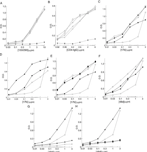
sCD4 induction of the 17b and 48d epitopes.
The addition of
sCD4 to the transfected COS-1 cell lysates increased the
effi-ciency with which the wild-type gp160 and gp120 glycoproteins
were precipitated by the 17b antibody (Fig. 2A). Similar results
were obtained for the
D
136-151 and
D
303-323 mutants
con-taining deletions of the V1 and V3 loops, respectively. When
the V1 and V2 loops were deleted, either alone (
D
128-194) or
in combination with the V3 loop (
D
128-194/303-323), the
pre-cipitation of the envelope glycoproteins by the 17b antibody in
the absence of sCD4 was significantly greater than that of the
wild-type envelope glycoproteins. The addition of sCD4 to the
cell lysates containing the
D
128-194 or
D
128-194/303-323
mu-tant did not result in further increases in the efficiency of
precipitation by the 17b antibody, despite the ability of these
mutant glycoproteins to bind CD4 efficiently (see below).
These results suggest that the presence of the V1/V2 variable
loops exerts a negative effect on the recognition of the
enve-lope glycoproteins by the 17b antibody and that CD4 binding
relieves that negative effect.
To determine more precisely the effects of the variable loop
deletions on 17b and 48d recognition in the absence and
pres-ence of sCD4, we used an assay in which the gp120
glycopro-tein was captured on an ELISA plate by an antibody directed
against the gp120 carboxyl terminus. Supernatants derived
from COS-1 transfections were used as a source of the
enve-lope glycoproteins, and the amounts of the various enveenve-lope
glycoproteins captured on the ELISA plate were normalized
with an antibody (133/290) that recognizes a well-conserved
linear C1 epitope (57a) present in all of the mutant
glycopro-teins (Fig. 3A). As shown in Fig. 3B, all of the captured
enve-lope glycoproteins were able to bind CD4-IgG (9) with roughly
comparable affinities. Thus, the abilities of the mutant
glyco-proteins to bind CD4 were not significantly altered by the
variable loop deletions.
The binding of both the 17b and 48d antibodies to the
captured wild-type gp120 glycoprotein was enhanced by the
addition of sCD4 (Fig. 3C and F). The recognition of the
D
128-194 mutant glycoprotein, which contains a deletion of the
[image:4.612.66.283.71.631.2]V1 and V2 loops, by the 17b and 48d antibodies in the absence
FIG. 2. Precipitation of wild-type and mutant glycoproteins. (A) Precipita-tion of labeled COS-1 cell lysates by a mixture of sera from AIDS patients or by the 17b antibody is shown for the wild-type and mutant envelope glycoproteins. In some lanes (1lanes), 10mg of sCD4 per ml was added to the lysate prior to immunoprecipitation. In each of the lanes, the more slowly migrating band represents the precursor glycoprotein, while the faster-migrating band represents the processed exterior envelope glycoprotein. (B) Precipitation of labeled enve-lope glycoproteins from detergent-free supernatants of transfected COS-1 cells by a mixture of sera from AIDS patients or by the 17b and 48d monoclonal antibodies. In some lanes (1lanes), 6.6mg of sCD4 per ml was added to the supernatant prior to immunoprecipitation.
on November 9, 2019 by guest
http://jvi.asm.org/
of sCD4 was greater than or equal to that of the wild-type
gp120 glycoprotein in the presence of sCD4. Addition of sCD4
did not further enhance the binding of the 17b antibody and
even slightly decreased the binding of the 48d antibody to the
D
128-194 mutant (Fig. 3C and F). Deletion of only the V1 loop
(
D
136-151) affected neither basal recognition by either
anti-body nor the enhancement of antianti-body binding by sCD4
rela-tive to those of the wild-type gp120 glycoprotein (data not
shown). In the ELISA format, deletion of the V3 loop (
D
303-323) disrupted 48d recognition more than 17b recognition, but
addition of sCD4 increased the binding of both antibodies to
the
D
303-323 glycoprotein (Fig. 3D and G). Thus, in some
contexts, deletion of the variable portion of the V3 loop affects
the 48d, but not the 17b, epitope. When both the V1/V2 and
V3 loops were deleted (
D
128-194/303-323), the basal
[image:5.612.69.547.72.565.2]recogni-tion of the mutant glycoprotein by the 17b antibody was
en-hanced, while that by the 48d antibody was decreased relative
to those observed for the wild-type gp120 glycoprotein (Fig. 3E
FIG. 3. Effects of variable loop deletions on antibody recognition and CD4 binding. The binding of antibodies or CD4-IgG to the wild-type or mutant envelope glycoproteins captured on an ELISA plate via a carboxyl-terminus-directed antibody is shown. Binding of the 133/290 antibody (A), CD4-IgG (B), 17b antibody (C to E), and 48d antibody (F to H) is shown for the wild-type (E,F),D128-194 (h,■),D303-323 (Ç,å), andD128-194/303-323 ({,}) envelope glycoproteins. The open symbols and broken lines indicate that the experiment was performed in the absence of sCD4, while the closed symbols and solid lines indicate the presence of 5mg of sCD4 per ml. In some experiments, the binding to wells to which no envelope glycoprotein was added (X) is shown. O.D., optical density.
on November 9, 2019 by guest
http://jvi.asm.org/
and H). The addition of sCD4 did not enhance the binding of
either antibody to the
D
128-194/303-323 mutant. These results
and the results shown in Fig. 2A suggest that the V1/V2 loop
structure masks the 17b and 48d epitopes on the gp120
glyco-protein and that sCD4 enhancement of 17b and 48d antibody
binding is functionally equivalent to removing this masking
effect. The data also indicate that the V1 and V3 loops are
dispensable for the masking of the 17b/48d epitopes and for
the enhancement of 17b/48d antibody binding in the presence
of sCD4.
The increased basal recognition of the
D
128-194 mutant
compared with that of the wild-type gp120 glycoprotein by the
17b and 48d antibodies was also seen when the glycoproteins
were captured on the ELISA plates with an antibody (133/290)
directed against the first conserved (C1) region. The addition
of sCD4 enhanced 17b and 48d binding to the wild-type gp120
glycoprotein captured in this manner (data not shown). These
results indicate that the masking effects of the V1/V2 loops on
the 17b and 48d epitopes and the induction by sCD4 binding
do not depend upon the capture of the gp120 glycoprotein by
an antibody directed against the carboxyl terminus.
sCD4 induction of the A32 epitope on the gp120
glycopro-tein.
The binding of sCD4 to the captured HIV-1 gp120
gly-coprotein results in enhanced binding of the 17b and 48d
antibodies, as shown above and as previously reported (79).
sCD4 binding also results in smaller increases in the binding of
the A32 human monoclonal antibody to the wild-type gp120
glycoprotein (Fig. 4). When both V1/V2 and V3 loops were
deleted from the gp120 glycoprotein (
D
128-194/303-323), the
basal level of binding of the A32 antibody was increased
rela-tive to that of the wild-type gp120 glycoprotein (Fig. 4C). For
the
D
128-194/303-323 mutant, sCD4 binding did not result in
an increase in the binding of the A32 antibody (Fig. 4C). These
results indicate that the V1/V2 and V3 variable loops
contrib-ute to the masking of the A32 epitope on the gp120 monomer
and that sCD4 binding is equivalent to demasking the A32
epitope by variable loop removal. Most of this effect is due to
the V1/V2 loops, since an increase in the basal level of binding
and lack of induction by sCD4 were also seen for the
D
128-194
mutant (Fig. 4A). By contrast, the V3 loop-deleted mutant
(
D
303-323) exhibited basal levels of A32 antibody binding and
sCD4 induction comparable to those of the wild-type gp120
glycoprotein (Fig. 4B).
Induction of the 17b and 48d epitopes by binding of the A32
antibody.
The binding of the A32 antibody to the captured
wild-type and mutant envelope glycoproteins resulted in an
increase in the binding of the 48d and 17b antibodies (Fig. 5
and data not shown). Although deletion of the V3 loop (
D
303-329) resulted in a decrease in recognition by the 48d antibody,
as previously seen, both the V1/V2 and V3 loops could be
deleted without abrogating the ability of A32 to increase 48d
binding (Fig. 5). Similarly, the increased binding of the 17b
antibody to the gp120 glycoprotein in the presence of the A32
antibody was not dependent on the presence of the V1/V2 or
V3 loops (data not shown).
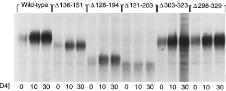
[image:6.612.81.536.70.230.2]Effects of variable loop deletions on sCD4-induced shedding
of the gp120 glycoprotein.
Previous studies indicated that the
sCD4-induced shedding of the gp120 glycoprotein from the
HIV-1 envelope glycoprotein complex depended upon
high-affinity binding of sCD4 and also upon gp120 regions not
FIG. 4. Effects of variable loop deletions on sCD4-induced exposure of the A32 epitope. The binding of the A32 antibody to the wild-type (E,F),D128-194 (h, ■),D303-323 (Ç,å), andD128-194/303-323 ({,}) envelope glycoproteins captured on the ELISA plate is shown. The open symbols and broken lines indicate that the experiment was performed in the absence of sCD4, while the closed symbols and solid lines indicate that the experiment was performed in the presence of 5mg of sCD4 per ml. O.D., optical density.
FIG. 5. Effects of variable loop deletions on A32 antibody-induced exposure of the 48d epitope. The binding of the biotinylated 48d antibody to the wild-type (E,F),D128-194 (h,■),D303-323 (Ç,å), andD128-194/303-323 ({,}) enve-lope glycoproteins captured on an ELISA plate is shown. The open symbols and broken lines indicate that the experiment was performed in the absence of the A32 antibody, while the closed symbols and solid lines indicate that the experi-ment was performed in the presence of 3mg of the A32 antibody per ml. O.D., optical density.
on November 9, 2019 by guest
http://jvi.asm.org/
[image:6.612.354.513.500.657.2]contributing directly to CD4 binding (77, 80, 88). In particular,
complete deletion of the V1/V2 stem-loop structure (
D
121-203) did not decrease CD4 binding ability but completely
ab-rogated gp120 shedding in response to sCD4 binding (88). To
examine whether the V1/V2 variable loops or the conserved
stem is critical for the shedding process, the ability of the
D
128-194 mutant to shed the gp120 glycoprotein as a result of
sCD4 binding was examined. Figure 6 shows that the wild-type
HXBc2 envelope glycoproteins demonstrated gp120 shedding
in response to incubation at 37
8
C with sCD4. Although no
gp120 shedding was observed for the
D
121-203 mutant, as
previously reported (88), the
D
136-151,
D
128-194, and
D
303-323 mutant glycoproteins all exhibited gp120 shedding in
re-sponse to sCD4 comparable to that seen for the wild-type
envelope glycoproteins. These results indicate that the V1/V2
variable loops are not required for sCD4-induced shedding but
that components in the conserved V1/V2 stem contribute to
the shedding process. As was previously seen for larger V3
deletions (88), the
D
303-323 mutant shed efficiently in
re-sponse to sCD4, verifying that the V3 loop is completely
dis-pensable for sCD4-induced gp120 shedding.
Effects of variable loop deletions on envelope glycoprotein
function.
To determine the effects of the variable loop
dele-tions on the function of the HIV-1 envelope glycoproteins, the
wild-type and mutant glycoproteins were expressed in COS-1
cells and tested for the abilities to induce the formation of
syncytia and to mediate virus entry. Compared with the
wild-type glycoproteins, the mutant glycoproteins with deletions of
the V1 loop (
D
136-151), the V1/V2 loop (
D
128-194), or the V3
loop (
D
303-323) exhibited relative abilities to complement
vi-rus entry into Jurkat lymphocytes of 34, 30, and 11%,
respec-tively. Of these mutants, only the
D
136-151 and
D
128-194
en-velope glycoproteins exhibited detectable levels of syncytia.
Syncytium formation is typically more sensitive to amino acid
changes in the HIV-1 envelope glycoproteins than is virus
entry (8, 31, 80). Additional mutant glycoproteins containing
more-extensive deletions involving the entire V3 (
D
298-329)
loop or combinations of V1/V2 and V3 deletions were included
in these assays for comparative purposes. These mutants did
not exhibit detectable ability to mediate syncytium formation,
consistent with the lower abilities of these mutants to mediate
virus entry into Jurkat lymphocytes (Table 1) (88).
DISCUSSION
The observed enhancement of HIV-2 and SIVagm envelope
glycoprotein-mediated fusion events by sCD4 suggested that
receptor binding might trigger positive events in virus entry not
related to virus attachment to the target cell (2, 14). More
recently, sCD4 enhancement of infection by certain strains of
HIV-1 has been observed (74), indicating that
receptor-medi-ated activation may apply to the primate immunodeficiency
viruses in general. Several effects of CD4 binding on the
con-formation of the HIV-1 envelope glycoproteins have been
de-scribed, although not all of these changes appear to represent
necessary events in virus entry (25, 30, 39, 50, 65, 66, 69, 79).
Understanding the molecular basis of CD4-induced changes in
the viral envelope glycoproteins will be critical in distinguishing
between functionally relevant and irrelevant events. The
in-creased exposure of the 17b and 48d epitopes represents an
attractive candidate for a functionally relevant CD4-induced
conformational change, since these epitopes are neutralization
targets (79) and may be located in gp120 regions important for
virus entry.
The discontinuous regions recognized by the 17b and 48d
antibodies are among the most conformation-sensitive epitopes
on the native HIV-1 gp120 glycoprotein. These epitopes, for
example, are completely disrupted in some buffers containing
ionic detergents that do not affect the integrity of other
dis-continuous epitopes, such as those overlapping the CD4
bind-ing site (79). Perhaps as a result of this sensitivity to subtle
changes in gp120 conformation, some of the effects of
dele-tions in the variable loops on the 17b and 48d epitopes are
dependent upon the context in which binding of the antibodies
to the gp120 glycoprotein is measured. The binding of other
ligands, for example, may restrict the number of conformations
available to gp120 mutants, which in turn can affect the degree
to which certain changes disrupt the 17b and 48d epitopes.
sCD4 binding has been shown to restore 17b or 48d
recogni-tion of several mutant gp120 glycoproteins that, as free
mole-cules in solution, were unable to be precipitated by these
an-tibodies (Fig. 2) (81). In this study, the variable-loop-deleted
gp120 glycoproteins were examined in two contexts, as free
molecules in solution and as glycoproteins captured on ELISA
plates by an antibody directed against the gp120 carboxyl
ter-minus. Perhaps related to the effects of binding the capture
antibody to the gp120 glycoprotein, some differences in the
effects of variable loop deletions on the 17b and 48d epitopes
were seen in these two assay systems. First, the studies
per-formed using envelope glycoproteins in solution confirm that
the highly conserved stem in the V1/V2 stem-loop structure
can influence the integrity of the 17b and 48d epitopes.
[image:7.612.69.296.72.164.2]Re-FIG. 6. Effects of variable loop deletions on shedding of the mutant envelope glycoproteins. The amounts of envelope glycoprotein precipitated from the su-pernatants of labeled COS-1 cells expressing the wild-type and mutant glycopro-teins in the absence (0) or presence of 10 or 30mg of sCD4 per ml are shown.
TABLE 1. Functional properties of the HIV-1 envelope glycoprotein mutants
Envelope glycoprotein
Syncytium-forming abilitya(%)
Replication complementationb
(%)
Wild-type 100 100
D136-151 14 34
D128-194 4 30
D303-323 ,1 11
D298-329 ,1 6
D128-194/303-323 ,1 6
D121-203/298-329 ,1 4
a
Syncytium-forming ability with SupT1 target cells was measured as described in Materials and Methods and is normalized to that of the wild-type envelope glycoproteins (100%). The syncytia induced by the D136-151 and D128-194 mutants were smaller than those formed by the wild-type envelope glycoproteins.
b
Complementation of virus entry into Jurkat lymphocytes was performed as described previously (31, 80). The CAT activity observed with the pSVIIIenvDKS control plasmid was subtracted from the CAT activity seen for each of the plasmids expressing the wild-type mutant envelope glycoproteins, and the result-ant value was normalized to that observed for the wild-type envelope glycopro-teins (100%). The values shown are from a typical experiment. The relative complementation ability obtained from three independent experiments differed from the values shown by less than 5%.
on November 9, 2019 by guest
http://jvi.asm.org/
[image:7.612.315.555.91.192.2]tention of this stem preserves recognition of a V1/V2
variable-loop-deleted glycoprotein by both antibodies, whereas deletion
of the complete V1/V2 stem-loop structure (
D
121-203 in Fig.
2B) resulted in a complete loss of precipitation by both 17b and
48d antibodies. By contrast, when the gp120 glycoprotein is
captured on an ELISA plate with an antibody directed against
the C terminus, the complete deletion of the V1/V2 structure
(
D
119-205 or
D
121-203) disrupted recognition by the 48d
an-tibody but not by the 17b anan-tibody (data not shown). This
result indicates that these are subtle differences in the 17b and
48d epitopes; the V1/V2 stem is not required for the binding of
the 17b antibody in all contexts, whereas the V1/V2 stem
appears to be more critical for 48d binding. A second example
in which the context influenced the results was found in the
effects of V3 loop deletions on recognition of the gp120
gly-coprotein by the 17b and 48d antibodies. Deletion of the entire
V3 loop (
D
298-329) resulted in a significant loss of
precipita-tion of the deleted glycoprotein from detergent-free soluprecipita-tions
by both 17b and 48d antibodies. The binding of sCD4 to this
V3-deleted mutant fully restored the ability to be precipitated
by both 17b and 48d antibodies (Fig. 2B). The more
conser-vative deletion of the V3 loop (
D
303-323) did not affect the
ability of either the 17b or 48d antibody to precipitate the
mutant glycoprotein in solution (Fig. 2B). The same
conserva-tive deletion, however, considerably decreased recognition by
the 48d, but not the 17b, antibody, when the mutant
glyco-protein was captured on the ELISA plate with the
C-terminus-directed antibody (Fig. 3D and G). These results suggest that,
in some contexts, the 48d epitope is more sensitive to changes
in the V3 loop than is the 17b epitope. This observation is
consistent with the greater effect of V3-directed antibodies on
the binding of the 48d antibody than on the binding of the 17b
antibody (63a). Apparently, critical components of the 48d
epitope can be altered by changes in the V3 loop.
The more conservative nature of the deletions in the major
gp120 variable loops made in this study compared with those in
a previous study (88) allowed retention of the integrity of the
17b and 48d epitopes in most contexts. This, in turn, allowed us
to investigate the contribution of the variable loops to changes
in the exposure of the 17b, 48d, and A32 epitopes induced by
the binding of sCD4. A model derived from these studies is
presented in Fig. 7. Removal of the V1/V2 loops (
D
128-194)
resulted in an increased exposure of the 17b and 48d epitopes
to an extent even greater than that seen upon sCD4 binding to
the wild-type gp120 glycoprotein. The binding of sCD4
re-sulted in no further increase in exposure of the 17b and 48d
epitopes on the V1/V2-deleted mutant glycoprotein.
There-fore, the V1/V2 variable loops appear to mask the 17b/48d
epitopes on the monomeric gp120 glycoprotein and the major
effect of sCD4 binding is to demask these epitopes by inducing
conformational changes in the V1/V2 structure. Selective
re-moval of the V1 loop alone (
D
136-151) did not increase the
exposure of the 17b and 48d epitopes, and the binding of sCD4
to this mutant glycoprotein resulted in an increased exposure
of both epitopes. This result suggests that the V2 loop, but not
the V1/V2 stem, is sufficient to mask the 17b and 48d epitopes
and may be more important than the V1 loop in mediating
sCD4-induced changes. Previous studies have suggested that
sCD4-induced movement of the HIV-1 gp120 V2 loop could
result in selective masking of some conformation-dependent
V2 epitopes (49, 54, 69). Furthermore, the V2 tip has been
implicated in fusion-related virus entry functions modulated by
CD4 binding (13, 75). The potential relevance of the exposure
of the 17b and 48d epitopes as markers for functionally
impor-tant CD4-induced alterations in the HIV-1 envelope
glycopro-teins is supported by the association of the V2 loop with both
processes.
The induction by sCD4 of the exposure of the A32 epitope
was also dependent upon the gp120 major variable loops (Fig.
7). When the V1/V2 and V3 loops were removed (
D
128-194/
303-323), a demasking of the A32 epitope on the monomeric
gp120 glycoprotein was observed. As was seen for the 17b and
48d epitopes, sCD4 binding did not result in further exposure
of the A32 epitope. While the smaller magnitude of
sCD4-induced exposure of the A32 epitope compared with those of
the 17b and 48d epitopes makes a precise comparison among
the mutants more difficult, it appears that the V1/V2 structure
rather than the V3 loop plays the major role in this induction
as well. The A32 epitope is a poorer neutralization target than
are the 17b and 48d epitopes, probably because the former
epitope is not as exposed on the oligomeric envelope
glyco-proteins (87). The exposure of the A32 epitope on the
oligo-meric envelope glycoproteins in the presence of CD4 and the
functional relevance of such exposure will be the subject of
future studies.
On the monomeric HIV-1 gp120 glycoprotein, the binding
of the A32 antibody increases the subsequent binding of the
17b and 48d antibodies. Our results indicate that this induction
occurs via a mechanism that is not dependent upon the
pres-ence of the V1/V2 or V3 loops. Therefore, the increases in
exposure of the 17b and 48d epitopes induced by sCD4 and by
the A32 antibody proceed via different mechanisms.
Examination of the functional abilities of the mutant
glyco-proteins revealed that the large variable segments of the V1/V2
and V3 loops are not absolutely required for HIV-1 entry,
although they contribute to the efficiency of this process.
Syn-cytium-forming ability was more dramatically affected than was
virus entry, as has been previously seen for other HIV-1
enve-lope glycoprotein mutants with defects in membrane fusion (8,
31, 75, 80). The decreased functional activities of the
variable-loop-deleted glycoproteins are probably not due solely to
ef-fects on exposure of the 17b and 48d epitopes, since the
V1-deleted mutant is not apparently altered in this parameter yet
exhibits partially attenuated virus entry and cell-cell fusion.
The retention of some function by the mutants with
more-conservative variable loop deletions contrasts with the loss of
function observed for mutants with more-extensive V1/V2 or
V3 truncations (Table 1) (88). This result suggests that some of
the loss of function associated with the latter mutants may arise
from secondary effects of the deletions on conserved structures
near or within the 17b and 48d epitopes. While the existence of
neutralization escape mutants (79) indicates that the
preser-FIG. 7. Model for variable-loop-mediated masking of the 17b and A32 epitopes and for the effects of sCD4 binding.2sCD4, in the absence of sCD4; 1sCD4, sCD4 binding.
on November 9, 2019 by guest
http://jvi.asm.org/
vation of the 17b and 48d epitopes per se is not essential for
HIV-1 entry, conserved structures overlapping these epitopes
may represent critical components in post-receptor binding
events.
Previous studies indicated that complete deletion of the
V1/V2 stem-loop structure (
D
121-203) resulted in a mutant
glycoprotein that bound CD4 at a level comparable to that of
the wild-type envelope glycoprotein but did not exhibit
sCD4-induced shedding of the exterior envelope glycoprotein (88).
Here we show that retention of the V1/V2 stem in the
D
128-194 mutant allows sCD4-induced gp120 shedding to occur
ef-ficiently. Thus, even with the V1/V2 loops deleted, CD4
bind-ing mediates changes in the conformation of the HIV-1
envelope glycoproteins through a mechanism dependent upon
the conserved V1/V2 stem. The relevance of such changes to
HIV-1 entry remains uncertain. The existence (4, 77) of
fusion-defective gp120 mutants that shed the exterior glycoprotein
efficiently upon CD4 binding, such as V3 loop-deleted
mu-tants, makes it highly unlikely that dissociation from the gp120
glycoprotein represents the sole postattachment function of
the gp120 glycoprotein during the virus entry process. The
existence of fusion-competent mutant glycoproteins that do
not shed the gp120 glycoprotein in response to sCD4 provides
a strong argument against the necessity of shedding in HIV-1
entry (77). Nonetheless, it remains possible that some overlap
exists between conformational changes in the gp120
glycopro-tein related to shedding and those related to virus entry.
Fu-ture strucFu-ture-function analysis of the HIV-1 envelope
glyco-proteins should help to define these relationships and to
increase our understanding of the mechanism of virus-cell fusion.
ACKNOWLEDGMENTS
We thank Lorraine Rabb and Jan Welch for manuscript preparation and Amy Emmert for artwork.
This work was supported by grants from the National Institutes of Health (AI 31783, AI36082, and AI24030) and by Center for AIDS Research grants to the Dana-Farber Cancer Institute and the Aaron Diamond AIDS Research Center (AI28691 and AI27742, respective-ly). The Dana-Farber Cancer Institute is also the recipient of a Cancer Center grant from the National Institutes of Health (CA 06516). This work was made possible by gifts from the late William McCarty-Cooper, from the G. Harold and Leila Y. Mathers Charitable Foun-dation, and from the Aaron Diamond Foundation. R.W. is a fellow of the American Foundation for AIDS Research.
REFERENCES
1. Allan, J., T. H. Lee, M. F. McLane, J. Sodroski, W. Haseltine, and M. Essex. 1983. Identification of the major envelope glycoprotein product of HTLV-III. Science 228:1091–1094.
2. Allan, J., J. Strauss, and D. Buck. 1990. Enhancement of SIV infection with soluble receptor molecules. Science 247:1084–1088.
3. Barre-Sinoussi, F., J. C. Chermann, F. Rey, M. T. Nugeyre, S. Chamaret, J.
Gruest, C. Dauget, C. Axler-Bin, F. Vezinet-Brun, C. Rouzioux, W. Rozen-baum, and L. Montagnier.1983. Isolation of a T-lymphocyte retrovirus from a patient at risk for acquired immunodeficiency syndrome (AIDS). Science
220:868–871.
4. Berger, E., J. Sisler, and P. Earl. 1992. Human immunodeficiency virus type 1 envelope glycoprotein molecules containing membrane fusion-impairing mutations in the V3 region efficiently undergo soluble CD4-stimulated gp120 release. J. Virol. 66:6208–6212.
5. Brand, D., K. Srinivasan, and J. Sodroski. 1995. Determinants of human immunodeficiency virus type 1 entry in the CDR2 loop of the CD4 glycop-rotein. J. Virol. 69:166–171.
6. Bugelski, P., H. Ellens, T. Hart, and R. Kirsh. 1991. Soluble CD4 and dextran sulfate mediate release of gp120 from HIV-1: implications for clin-ical trials. J. Acquired Immune Defic. Syndr. 4:923–924.
7. Cann, A. J., M. J. Churcher, M. Boyd, W. O’Brien, J.-Q. Zhao, J. Zack, and
I. S. Y. Chen.1992. The region of the envelope gene of human immunode-ficiency virus type 1 responsible for determination of cell tropism. J. Virol.
66:305–309.
8. Cao, J., L. Bergeron, E. Helseth, M. Thali, H. Repke, and J. Sodroski. 1993. Effects of amino acid changes in the extracellular domain of the human immunodeficiency virus type 1 gp41 envelope glycoprotein. J. Virol. 67:2747– 2755.
9. Capon, D., S. Chamow, J. Mordenti, S. Marsters, T. Gregory, H. Mitsuya, R.
Byrn, C. Lucas, F. Wurm, J. Groopman, and D. Smith.1989. Designing CD4 immunoadhesins for AIDS therapy. Nature (London) 337:525–531. 10. Cheng-Mayer, C., M. Quiroga, J. W. Tung, D. Dina, and J. A. Levy. 1990.
Viral determinants of human immunodeficiency virus type 1 T-cell or mac-rophage tropism, cytopathogenicity, and CD4 antigen modulation. J. Virol.
64:4390–4398.
11. Chesebro, B., J. Nishio, S. Perryman, A. Cann, W. O’Brien, I. S. Y. Chen,
and K. Wehrly.1991. Identification of human immunodeficiency virus enve-lope gene sequences influencing viral entry into CD4-positive HeLa cells, T-leukemia cells, and macrophages. J. Virol. 65:5782–5789.
12. Chesebro, B., K. Wehrly, J. Nishio, and S. Perryman. 1992. Macrophage-tropic human immunodeficiency virus isolates from different patients exhibit unusual V3 envelope sequence homogeneity in comparison with T-cell-tropic isolates: definition of critical amino acids involved in cell tropism. J. Virol. 66:6547–6554.
13. Choe, H.-R., and J. Sodroski. 1995. Adaptation of human immunodeficiency virus type 1 to cells expressing a binding-deficient CD4 mutant (lysine 46 to aspartic acid). J. Virol. 69:2801–2810.
14. Clapham, P. R., A. McKnight, and R. A. Weiss. 1992. Human immunodefi-ciency virus type 2 infection and fusion of CD4-negative human cell lines: induction and enhancement by soluble CD4. J. Virol. 66:3531–3537. 15. Cordonnier, A., L. Montagnier, and M. Emerman. 1989. Single amino acid
changes in HIV envelope affect viral tropism and receptor binding. Nature (London) 340:571–574.
16. Cullen, B. R. 1987. Use of eukaryotic expression technology in the functional analysis of cloned genes. Methods Enzymol. 152:64–73.
17. Daar, E., X. L. Li, T. Moudgil, and D. Ho. 1990. High concentrations of recombinant soluble CD4 are required to neutralize primary human im-munodeficiency virus type 1 isolates. Proc. Natl. Acad. Sci. USA 87:6574– 6578.
18. Dalgleish, A. G., P. C. L. Beverley, P. R. Clapham, D. H. Crawford, M. F.
Greaves, and R. A. Weiss.1984. The CD4 (T4) antigen is an essential component of the receptor for the AIDS retrovirus. Nature (London) 312: 763–767.
19. Deen, K., J. S. McDougal, R. Inacker, G. Folena-Wassterman, J. Arthos, J.
Rosenberg, P. Maddon, R. Axel, and R. Sweet.1988. A soluble form of CD4 (T4) protein inhibits AIDS virus infection. Nature (London) 331:82–84. 20. Dubay, J., S. Roberts, B. Brody, and E. Hunter. 1992. Mutations in the
leucine zipper of the human immunodeficiency virus type 1 transmembrane glycoprotein affect fusion and infectivity. J. Virol. 66:4748–4756. 21. Fisher, R., J. Bertonis, W. Meier, V. Johnson, D. Costopoulos, T. Liu, R.
Tizard, B. Walker, M. Hirsch, R. Schooley, and R. Flavell.1988. HIV infection is blocked in vitro by recombinant soluble CD4. Nature (London)
331:76–78.
22. Fisher, R., E. Collati, L. Ratner, R. Gallo, and F. Wong-Staal. 1985. A molecular clone of HTLV-III with biological activity. Nature (London) 316: 262–265.
23. Freed, E., D. Myers, and R. Risser. 1990. Characterization of the fusion domain of the human immunodeficiency virus type 1 envelope glycoprotein gp41. Proc. Natl. Acad. Sci. USA 87:4650–4654.
24. Freed, E., D. Myers, and R. Risser. 1991. Identification of the principal neutralizing determinant of human immunodeficiency virus type 1 as a fusion domain. J. Virol. 65:190–194.
25. Fu, Y.-K., T. K. Hart, Z. L. Jonak, and P. J. Bugelski. 1993. Physiochemical dissociation of CD4-mediated syncytium formation and shedding of human immunodeficiency virus type 1 gp120. J. Virol. 67:3818–3825.
26. Fung, M. S. C., C. R. Y. Sun, W. L. Gordon, R.-S. Liou, T. W. Chang, W. N. C.
Sun, E. S. Daar, and D. D. Ho.1992. Identification and characterization of a neutralization site within the second variable region of human immuno-deficiency virus type 1 gp120. J. Virol. 66:848–856.
27. Gallo, R. C., S. Z. Salahuddin, M. Popovic, G. M. Shearer, M. Kaplan, B. F.
Haynes, T. J. Palker, R. Redfield, J. Oleske, B. Safai, G. White, P. Foster, and P. D. Markham.1984. Frequent detection and isolation of cytopathic retroviruses (HTLV-III) from patients with AIDS and at risk for AIDS. Science 224:500–503.
28. Groenink, M., A. C. Anderweg, R. A. M. Fouchier, S. Broersen, R. C. M. van
der Jagt, H. Schuitemaker, R. E. Y. de Goede, M. L. Bosch, H. G. Huisman, and M. Tersmette.1992. Phenotype-associated env gene variation among eight related human immunodeficiency virus type 1 clones: evidence for in vivo recombination and determinants of cytotropism outside the V3 domain. J. Virol. 66:6175–6180.
29. Groenink, M., R. Fouchier, S. Broersen, C. Baker, M. Koot, A. van’t Woot,
H. Huisamn, F. Miedama, M. Tersmette, and H. Schuitemaker.1993. Re-lation of phenotype evolution of HIV-1 to envelope V2 configuration. Sci-ence 260:1513–1516.
30. Hart, T., R. Kirsch, H. Ellens, R. Sweet, D. Lambert, S. Petteway, J. Leary,
and P. Bugelski.1991. Binding of soluble CD4 proteins to HIV-1 and
on November 9, 2019 by guest
http://jvi.asm.org/
infected cells induces release of envelope glycoprotein gp120. Proc. Natl. Acad. Sci. USA 88:2189–2193.
31. Helseth, E., M. Kowalski, D. Gabuzda, U. Olshevsky, W. Haseltine, and J.
Sodroski.1990. Rapid complementation assays measuring replicative poten-tial of human immunodeficiency virus type 1 envelope glycoprotein mutants. J. Virol. 64:2416–2420.
32. Helseth, E., U. Olshevsky, D. Gabuzda, B. Ardman, W. Haseltine, and J.
Sodroski.1990. Changes in the transmembrane region of the human immu-nodeficiency virus type 1 gp41 envelope glycoprotein affect membrane fu-sion. J. Virol. 64:6314–6318.
33. Ho, D. D., M. S. C. Fung, Y. Cao, X. L. Li, C. Sun, T. W. Chang, and N.-C.
Sung.1991. Another discontinuous epitope on glycoprotein gp120 that is important in human immunodeficiency virus type 1 neutralization is identi-fied by a monoclonal antibody. Proc. Natl. Acad. Sci. USA 88:8949–8952. 34. Ho, D. D., J. A. McKeating, X. L. Li, T. Moudgil, E. S. Daar, N.-C. Sun, and
J. E. Robinson.1991. Conformational epitope on gp120 important in CD4 binding and human immunodeficiency virus type 1 neutralization identified by a human monoclonal antibody. J. Virol. 65:489–493.
35. Hussey, R., N. Richardson, M. Kowalski, N. Brown, H. Change, R. Siliciano,
T. Dorfman, B. Walker, J. Sodroski, and E. Reinherz.1988. A soluble CD4 protein selectively inhibits HIV replication and syncytium formation. Nature (London) 331:78–81.
36. Hwang, S. S., T. J. Boyle, H. K. Lyerly, and B. R. Cullen. 1991. Identification of the envelope V3 loop as the primary determinant of cell tropism in HIV-1. Science 253:71–74.
37. Ivanoff, L., D. Looney, C. McDanal, J. Morris, F. Wong-Staal, A. Langlois,
S. Petteway, and T. Matthews. 1991. Alteration of HIV-1 infectivity and neutralization by a single amino acid replacement in the V3 loop domain. AIDS Res. Hum. Retroviruses 7:595–603.
38. Javaherian, K., A. J. Langlois, C. McDanal, K. L. Ross, L. I. Eckler, C. L.
Jellis, A. T. Profy, J. R. Rusche, D. P. Bolognesi, S. D. Putney, and T. J. Matthews.1989. Principal neutralizing domain of the human immunodefi-ciency virus type 1 envelope protein. Proc. Natl. Acad. Sci. USA 86:6768–6772. 39. Kang, C.-Y., K. Hariharan, M. R. Posner, and P. Nara. 1993. Identification of a new neutralizing epitope conformationally affected by the attachment of CD4 to gp120. J. Immunol. 151:449–457.
40. Kang, C.-Y., P. Nara, S. Chamat, V. Caralli, T. Ryskamp, N. Haigwood, R.
Newman, and H. Kohler.1991. Evidence for non-V3-specific neutralizing antibodies that interfere with gp120/CD4 binding in human immunodefi-ciency virus-infected humans. Proc. Natl. Acad. Sci. USA 88:6171–6175. 41. Klatzmann, D., E. Champagne, S. Chamaret, J. Gruest, D. Guetard, T.
Hercend, J. C. Gluckman, and L. Montagnier.1984. T-lymphocyte T4 mol-ecule behaves as the receptor for human retrovirus LAV. Nature (London)
312:767–768.
42. Kowalski, M., J. Potz, L. Basiripour, T. Dorfman, W. G. Goh, E. Terwilliger,
A. Dayton, C. Rosen, W. Haseltine, and J. Sodroski.1987. Functional regions of the human immunodeficiency virus envelope glycoproteins. Science 237: 1351–1355.
43. Kunkel, T., J. Roberts, and R. Zakour. 1987. Rapid and efficient site-specific mutagenesis without phenotype selection. Methods Enzymol. 154:367–382. 44. Lasky, L., G. Nakamura, D. Smith, C. Shimisaki, E. Patzer, and P. Berman. 1987. Delineation of a region of the human immunodeficiency virus type 1 gp120 glycoprotein critical for interaction with the CD4 receptor. Cell 50: 975–985.
45. Leonard, C., M. Spellman, L. Riddle, R. Harris, J. Thomas, and T. Gregory. 1990. Assignment of intrachain disulfide bonds and characterization of po-tential glycosylation sites of the type 1 human immunodeficiency virus en-velope glycoprotein (gp120) expressed in Chinese hamster ovary cells. J. Biol. Chem. 265:10373–10382.
46. Maddon, P., A. Dalgleish, J. S. McDougal, P. Clapham, R. Weiss, and R.
Axel.1986. The T4 gene encodes the AIDS receptor and is expressed in the immune system and the brain. Cell 47:333–348.
47. Marcon, L., and J. Sodroski. 1994. gp120-independent fusion mediated by the human immunodeficiency virus type 1 gp41 envelope glycoprotein: a reassessment. J. Virol. 68:1977–1982.
48. McDougal, J. S., M. Kennedy, J. Sligh, S. Cort, A. Mowie, and J. Nicholson. 1986. Binding of the HTLV-III/LAV to T41T cells by a complex of the 100 K viral protein and the T4 molecule. Science 231:382–385.
49. McKeating, J., C. Shotton, J. Cordell, S. Graham, P. Balfe, N. Sullivan, M.
Charles, M. Page, A. Bolmstedt, S. Olofsson, S. Kayman, Z. Wu, A. Pinter, C. Dean, J. Sodroski, and R. Weiss.1993. Characterization of neutralizing monoclonal antibodies to linear and conformation-dependent epitopes within the first and second variable domains of human immunodeficiency virus type 1 gp120. J. Virol. 67:4932–4944.
50. Moore, J., J. McKeating, R. Weiss, and Q. Sattentau. 1990. Dissociation of gp120 from HIV-1 virions induced by soluble CD4. Science 250:1139–1142. 51. Moore, J. P., L. C. Burkly, R. I. Connor, Y. Cao, R. Tizard, D. D. Ho, and
R. A. Fisher. 1993. Adaptation of two primary human immunodeficiency virus type 1 isolates resistant to growth in transformed T cell line correlates with alterations in the responses of their envelope glycoproteins to soluble CD4. AIDS Res. Hum. Retroviruses 9:529–539.
52. Moore, J. P., J. A. McKeating, Y. Huang, A. Ashkenazi, and D. D. Ho. 1992.
Virions of primary human immunodeficiency virus type 1 isolates resistant to soluble CD4 (sCD4) neutralization differ in sCD4 binding and glycoprotein gp120 retention from sCD4-sensitive isolates. J. Virol. 66:235–243. 53. Moore, J. P., Q. J. Sattentau, R. Wyatt, and J. Sodroski. 1994. Probing the
structure of the human immunodeficiency virus surface glycoprotein gp120 with a panel of monoclonal antibodies. J. Virol. 68:469–484.
54. Moore, J. P., Q. J. Sattentau, H. Yoshiyama, M. Thali, M. Charles, N.
Sullivan, S.-W. Poon, M. S. Fung, F. Traincard, M. Pinkus, G. Robey, J. E. Robinson, D. D. Ho, and J. Sodroski.1993. Probing the structure of the V2 domain of the human immunodeficiency virus type 1 surface glycoprotein gp120 with a panel of eight monoclonal antibodies: human immune response to the V1 and V2 domains. J. Virol. 67:6136–6151.
55. Moore, J. P., M. Thali, B. A. Jameson, F. Vignaux, G. K. Lewis, S.-W. Poon,
M. Charles, M. S. Fung, B. Sun, P. J. Durda, L. Åkerblom, B. Wahren, D. D. Ho, Q. J. Sattentau, and J. Sodroski.1993. Immunochemical analysis of the gp120 surface glycoprotein of human immunodeficiency virus type 1: probing the structure of the C4 and V4 domains and the interaction of the C4 domain with the V3 loop. J. Virol. 67:4785–4796.
56. Morrison, H., F. Kirchhoff, and R. Desrosiers. 1993. Evidence for the co-operation of gp120 amino acids 322 and 448 in SIVmac entry. Virology
195:167–174.
57. Myers, G., J. Berzofsky, B. Korber, R. Smith, and G. Pavlakis. 1992. Human retroviruses and AIDS. A compilation and analysis of nucleic acid and amino acid sequences. Los Alamos National Laboratory, Los Alamos, N.Mex. 57a.Niedrig, M., H.-P. Harthus, J. Hinkula, M. Bro¨ker, H. Bickhard, G. Pauli,
H. R. Gelderblom, and B. Wahren.1992. Inhibition of viral replication by monoclonal antibodies directed against human immunodeficiency virus gp120. J. Gen. Virol. 73:2451–2455.
58. Olshevsky, U., E. Helseth, C. Furman, J. Li, W. Haseltine, and J. Sodroski. 1990. Identification of individual human immunodeficiency virus type 1 gp120 amino acids important for CD4 receptor binding. J. Virol. 64:5701– 5707.
59. Page, K., S. Stearns, and D. Littman. 1992. Analysis of mutations in the V3 domain of gp160 that affect fusion and infectivity. J. Virol. 66:524–533. 60. Perez, L. G., M. A. O’Donnell, and E. B. Stephens. 1992. The transmembrane
glycoprotein of human immunodeficiency virus type 1 induces syncytium formation in the absence of the receptor binding glycoprotein. J. Virol.
66:4134–4143.
61. Posner, M., T. Hideshima, T. Cannon, M. Mukherjee, K. Mayer, and R.
Byrn.1991. An IgG human monoclonal antibody which reacts with HIV-1 gp120, inhibits virus binding to cells, and neutralizes infection. J. Immunol.
146:4325–4332.
62. Putney, S., T. Matthews, W. G. Robey, D. Lynn, M. Robert-Guroff, W.
Mueller, A. Langlois, J. Ghrayeb, S. Petteway, K. Weinhold, P. Fischinger, F. Wong-Staal, R. Gallo, and D. Bolognesi.1986. HTLV-III/LAV-neutralizing antibodies to an E. coli-produced fragment of the virus envelope. Science
234:1392–1395.
63. Robey, W. G., B. Safai, S. Oroszlan, L. Arthur, M. Gonda, R. Gallo, and P. J.
Fischinger.1985. Characterization of envelope and core structural gene products of HTLV-III with sera from AIDS patients. Science 228:593–595. 63a.Robinson, J., J. Sodroski, and J. Moore. Unpublished observations. 64. Rusche, J. R., K. Javaherian, C. McDanal, J. Petro, D. L. Lynn, R. Grimaila,
A. Langlois, R. C. Gallo, L. O. Arthur, P. J. Fischinger, D. P. Bolognesi, S. D. Putney, and T. J. Matthews.1988. Antibodies that inhibit fusion of human immunodeficiency virus-infected cells bind a 24-amino-acid sequence of the viral envelope gp120. Proc. Natl. Acad. Sci. USA 85:3198–3202.
65. Sattentau, Q., and J. Moore. 1991. Conformational changes induced in the human immunodeficiency virus envelope glycoprotein by soluble CD4 bind-ing. J. Exp. Med. 174:407–415.
66. Sattentau, Q., J. Moore, F. Vignaux, F. Traincard, and P. Poignard. 1993. Conformational changes induced in the envelope glycoproteins of human and simian immunodeficiency viruses by soluble receptor binding. J. Virol.
67:7383–7393.
67. Sattentau, Q., S. Zolla-Pazner, and P. Poignard. Epitope exposure on func-tional, oligomeric gp41 molecules. Submitted for publication.
68. Shioda, T., J. A. Levy, and C. Cheng-Mayer. 1991. Macrophage and T cell-line tropisms of HIV-1 are determined by specific regions of the enve-lope gp120 gene. Nature (London) 349:167–169.
69. Shotton, C., C. Arnold, Q. Sattentau, J. Sodroski, and J. McKeating. 1995. Identification and characterization of monoclonal antibodies specific for polymorphic antigenic determinants within the V2 region of the human immunodeficiency virus type 1 envelope glycoprotein. J. Virol. 69:222–230. 70. Smith, D., R. Byrn, S. Marsters, T. Gregory, J. Groopman, and D. Capon. 1987. Blocking of HIV-1 infectivity by a soluble secreted form of the CD4 antigen. Science 238:1704–1707.
71. Starcich, B. R., B. H. Hahn, G. M. Shaw, P. D. McNeely, S. Modrow, H. Wolf,
W. P. Parks, S. F. Josephs, R. C. Gallo, and F. Wong-Staal.1986. Identifi-cation and characterization of conserved and variable regions of the enve-lope gene of HTLV-III/LAV, the retrovirus of AIDS. Cell 45:637–648. 72. Steimer, K. S., C. J. Scandella, P. V. Skiles, and N. L. Haigwood. 1991.
Neutralization of divergent HIV-1 isolates by conformation-dependent hu-man antibodies to gp120. Science 254:105–108.