Imperfect Symmetry of Sp1 and Core Promoter Sequences Regulates
Early and Late Virus Gene Expression of the Bidirectional BK
Polyomavirus Noncoding Control Region
Tobias Bethge,a*Elvis Ajuh,aHans H. Hirscha,b,c
Transplantation & Clinical Virology, Department of Biomedicine (Haus Petersplatz), University of Basel, Basel, Switzerlanda
; Division of Infection Diagnostics, Department of Biomedicine (Haus Petersplatz), University of Basel, Basel, Switzerlandb
; Infectious Diseases & Hospital Epidemiology, University Hospital Basel, Basel, Switzerlandc
ABSTRACT
Rearrangements or point mutations in the noncoding control region (NCCR) of BK polyomavirus (BKPyV) have been associated with higher viral loads and more pronounced organ pathology in immunocompromised patients. The respective alterations af-fect a multitude of transcription factor binding sites (TFBS) but consistently cause increased expression of the early viral gene region (EVGR) at the expense of late viral gene region (LVGR) expression. By mutating TFBS, we identified three phenotypic groups leading to strong, intermediate, or impaired EVGR expression and corresponding BKPyV replication. Unexpectedly, Sp1
TFBS mutants either activated or inhibited EVGR expression when located proximal to the LVGR (sp1-4) or the EVGR (sp1-2),
respectively. We now demonstrate that the bidirectional balance of EVGR and LVGR expression is dependent on affinity, strand
orientation, and the number of Sp1 sites. Swapping the LVGR-proximal high-affinitySP1-4with the EVGR-proximal
low-affin-itySP1-2in site strand flipping or inserting an additionalSP1-2site caused a rearranged NCCR phenotype of increased EVGR
expression and faster BKPyV replication. The 5=rapid amplification of cDNA ends revealed an imperfect symmetry between the
EVGR- and LVGR-proximal parts of the NCCR, consisting ofTATAandTATA-like elements, initiator elements, and
down-stream promoter elements. Mutation or deletion of the archetypal LVGR promoter, which is found in activated NCCR variants,
abrogated LVGR expression, which could be restored by providing large T antigen (LTag) intrans. Thus, whereas Sp1 sites
con-trol the initial EVGR-LVGR expression balance, LTag expression can override inactivation of the LVGR promoter and acts as a key driver of LVGR expression independently of the Sp1 sites and core promoter elements.
IMPORTANCE
Polyomaviridaecurrently comprise more than 70 members, including 13 human polyomaviruses (PyVs), all of which share a bidirectional genome organization mediated by the NCCR, which determines species and host cell specificity, persistence, repli-cation, and virulence. Here, we demonstrate that the BKPyV NCCR is fine-tuned by an imperfect symmetry of core promoter
elements centered aroundTATAandTATA-like sequences close to the EVGR and LVGR, respectively, which are governed by the
directionality and affinity of two Sp1 sites. The data indicated that the BKPyV NCCR is poised toward EVGR expression, which can be readily unlatched by a simple switch affecting Sp1 binding. The resulting LTag, which is the major EVGR protein, drives viral genome replication, renders subsequent LVGR expression independently of archetypal promoter elements, and can over-come enhancer/promoter mutations and deletions. The data are pivotal for understanding how human PyV NCCRs mediate sec-ondary host cell specificity, reactivation, and virulence in their natural hosts.
T
he Polyomaviridae comprise more than 70 polyomavirus (PyV) members, including at least 13 human PyV (HPyV) species (1). HPyV seroprevalence data obtained using the major capsid protein Vp1 as pentamers or virus-like particles indicate that HPyVs infect 50% to 90% of the general human population without known specific signs or symptoms of disease (2–7). As information about the virology and pathology of the 13 human PyVs is only emerging (8,9), we focused on BK polyomavirus (BKPyV) as a model (10). After primary infection in early child-hood, BKPyV persists latently in the renourinary tract with peri-ods of low-level shedding into urine (4,11,12). Immunosuppres-sion, which is commonly administered posttransplantation, has been shown to relax the adaptive antiviral immune control (13– 16) and to permit high-level replication with median urine BKPyV loads of over 7 log10copies/ml that become apparent as decoy cell shedding in urine (15–20). Progression to BKPyV disease is most consistently encountered as nephropathy in 1 to 15% of kidney transplant recipients (21–23) or as hemorrhagic cystitis in 5 to20% of allogeneic hematopoietic stem cell transplant recipients (24–26).
BKPyV disease involves viral genotypes indistinguishable from those of archetype BKPyV strains shed asymptomatically in the urine of healthy individuals (27–29), arguing that impaired adap-tive immune control is necessary and sufficient. However,
immu-Received23 May 2016Accepted22 August 2016
Accepted manuscript posted online31 August 2016
CitationBethge T, Ajuh E, Hirsch HH. 2016. Imperfect symmetry of Sp1 and core promoter sequences regulates early and late virus gene expression of the bidirectional BK polyomavirus noncoding control region. J Virol 90:10083–10101.
doi:10.1128/JVI.01008-16.
Editor:L. Banks, International Centre for Genetic Engineering and Biotechnology Address correspondence to Hans H. Hirsch, [email protected].
*Present address: Tobias Bethge, Kantonsspital Aarau, Aarau, Switzerland.
Copyright © 2016, American Society for Microbiology. All Rights Reserved.
on November 7, 2019 by guest
http://jvi.asm.org/
nosuppressive drug-specific effects (20, 30) and viral determi-nants (31,32) may also contribute to the evolution, kinetics, and severity of disease. At the core of what distinguishes archetype BKPyV strains with a low replicative capacity from the highly rep-licative strains found in kidney transplant patients are changes in the noncoding control region (NCCR) of the BKPyV genome (33). Akin to all PyVs, the BKPyV NCCR consists of an approxi-mately 400-bp-long sequence which is sandwiched between the early viral gene region (EVGR) and the late viral gene region (LVGR), altogether yielding the double-stranded BKPyV DNA genome of 5,100 bp (34,35). The NCCR containscis-acting elements coordinat-ing the timcoordinat-ing and consecutive steps of EVGR expression, viral ge-nome replication, and LVGR expression (33,36).
The archetype BKPyV (ww) found in the urine of healthy im-munocompetent individuals (29, 37,38) is defined by a linear ww-NCCR sequence architecture arbitrarily denoted O143, P68, Q39, R63, and S63 (where the subscript numbers represent the number of nucleotides), which conveys rather slow replication in primary human renal tubular epithelial and urothelial cells (31, 39). In contrast, rearranged NCCRs (rr-NCCRs) associated with high blood viral loads and severe renal allograft pathology carry partial duplications and/or deletions of the archetypeww-NCCR sequence and give rise to virus strains with elevated levels of EVGR expression, faster viral replication, and more pronounced cyto-pathic effects in cell culture (31,32,40).
Although practically all emergingrr-NCCR BKPyV variants appear as unique sequences, we noted some common themes. These consist of an unaltered EVGR-proximal region covering the O143block with the origin of replication and the central P68block. Insertion strains carry complete or partial duplications of the P68 block, while deletion strains are characterized by the absence of the LVGR-proximal Q39and R63blocks and, rarely, the S63block (31,32,40,41).
In a detailed point mutational analysis in which more than 28 different transcription factor binding sites (TFBS) were inacti-vated without altering the overall archetypeww-NCCR architec-ture and length, we identified a hierarchy of TFBS resulting in three phenotypic groups: group 1 TFBS mutations resulted in EVGR activation at the expense of LVGR expression and fast rep-lication rates, similar to the findings for clinicalrr-NCCR variants. Group 2 TFBS mutations showed an intermediate phenotype with respect to EVGR expression and replication rates, whereas group 3 TFBS mutations reduced both EVGR and LVGR expression and permitted little or no viral replication (32).
Among the various factors, Sp1 appeared to have an outstand-ing role, since the BKPyV NCCR contained at least three Sp1 sites, two of which appeared to have fundamentally different effects on BKPyV expression and replication. Whereas inactivation of the SP1-4site proximal to the LVGR resulted in strong EVGR expres-sion, inactivation of theSP1-2site proximal to the EVGR abro-gated both EVGR and LVGR expression. This effect of the inacti-vatingsp1-2mutant also overrode group 1 activation, including the one seen with thesp1-4mutant, suggesting a key role of Sp1 in the functionality of the NCCR as a whole.
In the present study, we investigated the role of these two key Sp1 sites and their relationship to core promoter elements in greater detail. Our results demonstrate that the BKPyV NCCR is fine-tuned by an imperfect symmetry of two core promoters with TATAandTATA-like elements at the EVGR and the LVGR, re-spectively. The bidirectional EVGR and LVGR expression is
gov-erned by the host cell transcription factor Sp1 and involves both DNA strand directionality and binding affinity. The data indicate that the BKPyV NCCR is poised toward EVGR expression, which can be readily discharged by a simple switch in Sp1 binding. The large T antigen (LTag), which is the major EVGR protein product, then drives genome replication and subsequent LVGR expression independently of Sp1 and TATA-like elements, thereby being able to overcome a range of deletions seen in the LVGR promoter of natural BKPyV variants. We discuss the implications of our results as a model for the novel human PyVs and the control of bidirec-tional gene expression.
MATERIALS AND METHODS
Cell culture.Human primary renal proximal tubule epithelial cells (RPT-ECs; PCS-400-010; ATCC, Manassas, VA, USA) were grown in epithelial cell medium (EpiCM; catalog number 4101; ScienCell Research Labora-tory, Carlsbad, CA, USA) supplemented with epithelial cell growth sup-plement (EpiCGS; catalog number 4152; ScienCell Research Laboratory, Carlsbad, CA, USA) and 2% fetal bovine serum (FBS; catalog number 0010; ScienCell Research Laboratory, Carlsbad, CA, USA). HEK293 cells (CRL1573; ATCC, Manassas, VA, USA), HEK293T cells (CRL3216; ATCC, Manassas, VA, USA), and HEK293TT cells (catalog number 0508000; NCI-Frederick Repository Service, Frederick, MD, USA) were propagated in Dulbecco’s modified Eagle’s medium (DMEM), high-glu-cose formulation (DMEM-H; catalog number D5671; Sigma-Aldrich, St. Louis, MO, USA), containing 8% FBS (catalog number S0113; Biochrome AG, Berlin, Germany). Medium for cultivation of HEK293TT cells was supplemented with 400g/ml hygromycin B (Calbiochem, San Diego, CA, USA). COS-7 cells (CRL1651; ATCC, Manassas, VA, USA) were grown in DMEM-H, containing 5% FBS. All cultures were supplemented with 2 mML-glutamine (catalog number K0302; Biochrome AG, Berlin, Germany).
FACS-based, bidirectional reporter assay.The bidirectional reporter construct used to measure expression of the early (red fluorescent protein [RFP]) and late (green fluorescent protein [GFP]) virus gene regions has been described before (31). Furthermore, we described a fluorescence-activated cell sorter (FACS)-based quantification method for this reporter construct (32). While the use of flow cytometry for the reporter assay enables a convenient means of visualization of expression, at the same time, accurate and reproducible quantification is achieved. In particular, the gating for the same number of transfected cells from each sample as a method of normalization overcomes the problem of different transfection efficiencies between different plasmid preparations, mutants, or cell lines. HEK293, HEK293T, or HEK293TT cells were seeded in 12-well plates and transfected at 70 to 80% confluence with the Lipofectamine 2000 reagent (catalog number 11668-019; Invitrogen, Carlsbad, CA) at a ratio of 3:1 (3
l reagent, 1g plasmid DNA) in Opti-MEM medium (Gibco, Grand Island, NY, USA) according to the manufacturer’s instructions. Medium was replaced with DMEM-H–10% FBS on the next morning. At 48 h posttransfection, the cells were rinsed once with phosphate-buffered sa-line (PBS)–2.5 mM EDTA and then detached, suspended, and transferred to 5-ml polystyrene round-bottom FACS tubes (BD, Franklin Lakes, NJ, USA) with DMEM (without phenol red) and 1% fetal calf serum (FCS). Directly before each measurement, DAPI (4= ,6-diamidino-2-phenylin-dole; catalog number D8417; Sigma-Aldrich, St. Louis, MO, USA) was added (final concentration, 1 ng/ml) as a dead cell marker, and the cells were resuspended. FACS measurements were carried out on a Fortessa cytometer (BD, Franklin Lakes, NJ, USA). For GFP, excitation was at 488 nm (blue laser) and emission was at 530/30 nm; for RFP, excitation was at 561 nm (yellow-green laser) and emission was at 586/15 nm; and for DAPI, excitation was at 405 nm (violet laser) emission was at 450/50 nm. In order to calculate the weighted mean fluorescence intensity (MFI) for red (early) and green (late) expression, the cell number (N) and mean fluorescence (I) of quadrants Q1 (red cells), Q2 (red and green cells), and Bethge et al.
on November 7, 2019 by guest
http://jvi.asm.org/
Q4 (green cells) were inserted into the following formulas: MFI (red)⫽ [(NQ1·IQ1)⫹(NQ2·IQ2)]/(NQ1⫹NQ2⫹NQ4) and MFI (green)⫽
[(NQ2·IQ2)⫹(NQ4·IQ4)]/(NQ1⫹NQ2⫹NQ4).
The early expression of all constructs was normalized to that of the Dunlop strain (set as 100%), and late expression was normalized to that of the archetypeww(1.4) strain (set as 100%). The mean values and standard deviations were calculated with GraphPad Prism software (version 6 for Mac OS).
EMSA.Electrophoretic mobility shift assay (EMSA) analysis of TFBS was performed as described previously (42). Briefly, 10 g of nuclear extract from RPTECs was mixed with binding buffer (20 mM PIPES [pi-perazine-N,N=-bis(2-ethanesulfonic acid)], pH 6.8; 50 mM NaCl; 1 mM dithiothreitol [DTT]; 0.25 mg/ml bovine serum albumin; 100M ZnSO4;
0.05% NP-40; 4% Ficoll) and approximately 5 fmol of32P-labeled,
du-plexed oligonucleotide in a final volume of 20l. The oligonucleotides with theSP1-2andSP1-4binding sites were described before (32). Nu-clear extracts were prepared as described by Schreiber et al. (43) by scrap-ing cells with ice-cold PBS off 10-cm dishes and collectscrap-ing them in Eppen-dorf tubes. After centrifugation they were swollen in hypotonic buffer (10 mM HEPES, pH 7.9; 10 mM KCl; 0.1 mM EDTA; 2.5 mM DTT) and lysed by addition of NP-40 (final concentration, 0.5%). After centrifugation of the nuclei, proteins were extracted in nuclear extract buffer (20 mM HEPES, pH 7.9; 25% glycerol; 400 mM NaCl; 1 mM EDTA; 2.5 mM DTT). Competing, unlabeled oligonucleotides were used as described in the legend toFig. 2. After incubating the mixture on ice for 30 min, samples were separated on a native 4% polyacrylamide gel with 0.25⫻ TBE (Tris-borate-EDTA) as the running buffer. The detection ofdecay was carried out with a Fujifilm FLA-7000 image plate reader.
ChIP.Chromatin immunoprecipitation (ChIP) was performed ac-cording to the manufacturer’s instructions (Magnify chromatin immu-noprecipitation system; Life Technologies/Thermo Fisher Science). Briefly, HEK293TT cells were transfected in 10-cm dishes with the BKPyV wwreporter construct and incubated for 48 h. The chromatin was linked directly in the dish for 10 min with 10% formaldehyde. The cross-linking was quenched with 0.5 M glycine solution. Cells were harvested with ice-cold PBS, collected by centrifugation, and lysed. For the chroma-tin fragmentation, lysates were treated with micrococcal nuclease (20 min at 37°C; NEB) and sonicated with 30 cycles of 30-s pulses and 30 s of cooling in between each pulse (Sonicator Q700; QSonica). The ChIP an-tibodies were Sp1 D4C3 (ChIP-validated rabbit monoclonal antibody; Cell Signaling) (44) and a rabbit control antibody included with the kit. For PCR analysis by HotStarTaq (Qiagen) and a quantitative PCR (qPCR) SYBR green assay (PowerUp SYBR green; Thermo Fisher Scien-tific), the following primers were validated and used: primer NCCR-f (TCAGAAAAAGCCTCCACACC) and primer NCCR-r (CTTTGGCCA GTTTCCACTTC). qPCR data were analyzed by the⌬⌬CTthreshold cycle
(CT) method as described before usingww-NCCR input and background
control ChIP for normalization.
5=RACE analysis of early and late transcripts.To characterize the 5= ends of viral transcripts and, hence, to identify the transcription start sites, we performed 5=rapid amplification of cDNA ends (5=-RACE) proce-dures according to the manufacturer’s instructions (5=RACE system for rapid amplification of cDNA ends; Invitrogen/Thermo Fisher Scientific). Briefly, we isolated total RNA from RPTECs (RNeasy minikit; Qiagen) which were infected with either archetype virus or the Dunlop strain and reverse transcribed the mRNA by using a mixture of random hexamers and oligo(dT) primers (SuperScript II; Invitrogen/Thermo Fisher Scien-tific). The resulting cDNAs of viral origin were specifically amplified by using the following gene-specific primers (GSP): sTag-GSP1 (5=-AAGGG CAGTGCACAGAAG-3=), sTag/LTag (5=-AATCAGGCTGATGAGCTAC C-3=), LTag-GSP1 (5=-TCTTCAGGCTCTTCTGG-3=), LTag-GSP2 (5=-T GGTGTKGAGTGTTGAGA-3=), agno-GSP1 (5=-TACAGCAGGTAAAG CAGTG-3=), agno-GSP2 (5=-TCCCGTCTACACTGTCTT-3=), VP2-GSP1 (5=-TGATAGAGGCCTACAGTG-3=), VP1-GSP1 (5=-AAGGCCT
CTCCACTGTTG-3=), and VP1-GSP2 (5=-CCAGCACTCAACTGGATA AG-3=).
In silicoanalysis of the BKPyV NCCR.To search for potential tran-scription factor binding sites within the BKPyV NCCR and to evaluate the effect of mutations on the binding sites, we used the MatInspector soft-ware tool (Genomatix, Munich, Germany). Additionally, to analyze the presence of core promoter elements, we used the search tool ElemeNT (http://lifefaculty.biu.ac.il/gershon-tamar/index.php/resources) (45).
Transfection of BKPyV genome into COS-7 cells and infection of RPTECs.Transfection of BKPyV genomes into cells was initiated by cut-ting BKPyV plasmid DNA with BamHI and religacut-ting the diluted DNA as described previously (46). Transfection of religated BKPyV genomic DNA into COS-7 cells was performed when the cells were at 90 to 95% conflu-ence in 6-well plates using Lipofectamine 2000 (catalog number 11668-019; Invitrogen, Carlsbad, CA, USA) at a reagent/DNA ratio of 3:1 accord-ing to the manufacturer’s instructions. At 6 h after transfection, the medium was replaced with DMEM-H containing 5% FCS. At 7 days post-transfection, COS-7 cell supernatants were harvested and the number of DNase-protected genomic equivalents (GEq) per milliliter was quantified by quantitative reverse transcription-PCR (see the next paragraph). For the infection of RPTECs with COS-7 cell supernatants, cells were seeded at 75,000 cells per well of a 24-well plate in 0.5 ml of supplemented EpiCM. After 24 h, when the cells were at a confluence of approximately 50%, RPTECs were exposed to 200l of the corresponding virus preparations adjusted to 2⫻107GEq for each construct at 37°C for 2 h, followed by
removal, washing, and replacement with EpiCM supplemented with 0.5% FCS.
BKPyV viral loads by real-time PCR.BKPyV loads were quantified after DNA extraction from 200-l cell culture supernatants with a QIAamp DNA blood minikit (Qiagen, Hombrechtikon, Switzerland). The real-time PCR protocol for detection of BKPyV DNA samples targets the BKPyV large T antigen-coding sequence and has been described be-fore (47).
Western blotting.Lysis of HEK293, HEK293T, and HEK293TT cells was done in NP-40 buffer (50 mM Tris-HCl, pH 8.0; 150 mM NaCl; 1% NP-40; 1⫻protease inhibitors [Roche]). Equal volumes of cell lysates, adjusted to 10g total protein, were separated by SDS-PAGE and elec-trotransferred onto a 0.45-m-pore-size polyvinylidene difluoride mem-brane (catalog number IPFL00010; Millipore/Merck, Darmstadt, Ger-many). The membrane was blocked with Odyssey blocking buffer (catalog number 927-40000; LI-COR, Lincoln, NE, USA) diluted 1:2 in Tris-buff-ered saline (TBS). The membrane was then incubated with the following primary antibodies: polyclonal rabbit anti-LTag (1:10,000; C. H. Rinaldo, University of North Norway, Tromsø, Norway) and monoclonal mouse antiactin (1:5,000; Abcam, Cambridge, England) in Odyssey blocking buffer. After washing 5 times with TBS– 0.1% Tween 20, the membrane was incubated in the following secondary antibodies: goat anti-rabbit im-munoglobulin antibody conjugated with IRDye 800CW (1:10,000; cata-log number 926-32211; LI-COR) and donkey anti-mouse immunoglob-ulin antibody conjugated with Alexa Fluor 680 (1:15,000; catalog number A10038; Invitrogen). Protein detection and quantification were done with a LI-COR Odyssey CLx system.
RESULTS
Swapping of Sp1 sites within the NCCR causes
rearrangement-like activation of EVGR expression.To measure the early and late
promoter activity of the BKPyV NCCR, we transfected a bidirec-tional reporter construct into human embryonic kidney HEK293 cells, which allows monitoring of EVGR expression by the red fluorescence of DsRed and monitoring of LVGR expression by the green fluorescence of enhanced green fluorescent protein (31,32). Using this reporter assay, the strain Dunlop NCCR, as a represen-tative ofrr-NCCRs bearing duplications and deletions, showed strong EVGR expression (red) but only a low level of LVGR ex-pression (green) (Fig. 1A). The Dunlop NCCR contains an intact
on November 7, 2019 by guest
http://jvi.asm.org/
FIG 1Swapping ofSP1-2 andSP1-4in the BKPyV NCCR affects early and late promoter activity similarly to major rearrangements. (A to D) (Left) Schematic representation of NCCRs tested with the FACS-based, bidirectional reporter assay. All NCCRs were cloned into the previously described reporter vector pHRG, in which RFP serves as a marker for early expression and GFP serves as a marker for late expression (31). The Gardner isolate-derived laboratory strain Dunlop is characterized by triplication of the P-block sequences and deletion of Q- and R-block sequences. The BKPyV archetype NCCR124is used as a reference. Horizontal black lines, double-stranded DNA of the NCCR; black box, conserved TATA box; gray box, TATA-like element; yellow triangles labeled 2,SP1-2Sp1 transcription factor binding site; yellow triangles labeled 4,SP1-4Sp1 transcription factor binding site; black number 4, Sp1-binding site with archetype sequence; red number 4, Sp1-binding site carrying one or more mutations; yellow highlight, the respective Sp1 upper-strand sequence with respect to the direction of early and late transcription (black arrows). (Middle two columns) The indicated NCCR reporter constructs were transfected into HEK293 cells, and fluorescence images for RFP (early) and GFP (late) expression were taken at 2 days posttransfection. (Right) Representative flow cytometry measurements of the indicated NCCRs.xaxis, GFP fluorescence;yaxis, RFP fluorescence. For each measurement, we gated for 5,000 transfected (fluorescent) cells in quadrants Q1, Q2, and Q4. (E) For quantification, the normalized MFI (in percent) was calculated as described in Materials and Methods. Early expression was normalized to that of strain Dunlop (red MFI⫽100%), and late expression was normalized to that of NCCR124(wwarchetype) (green MFI⫽100%). Quantification results are from at least 3 independent replicates.
on November 7, 2019 by guest
http://jvi.asm.org/
[image:4.594.41.546.36.599.2]SP1-2site upstream of the TATA box but bears two incomplete duplications of the P block, adding oneSP1-2site at a more distant upstream location, while LVGR proximal sequences, including SP1-4, are deleted (Fig. 1A, left, with the yellow triangles labeled 2 indicatingSP1-2). In contrast, the archetype BKPyVwwstrain has all three Sp1 sites in the archetype position (ww-NCCR124) and showed a low level of EVGR expression (red) but strong LVGR expression (green) (Fig. 1B, left,wwarchetype NCCR124, with the yellow triangles labeled 2 and 4 indicatingSP1-2andSP1-4, respectively). As reported previously (32), point mutations, like the naturally occurringSP1-4variant 5CG (which has a C-to-G transition at position 5) from a patient with BKPyV-associated hemorrhagic cystitis (48), are sufficient to cause an activation of EVGR expression compared to the archetypeww-NCCR, at the expense of LVGR expression [NCCR12(4-5CG) inFig. 1CandE]. Given the striking effects of rather subtle Sp1 site mutations with respect to EVGR and LVGR expression (32), we investigated whether or not the different Sp1 sites might have different se-quence-encoded properties. We noted thatSP1-2andSP1-4have inverse orientations relative to one another, which is possible be-cause Sp1 sites are composed of nonpalindromic sequences. In a first approach, the earlySP1-2site and the lateSP1-4site were simply swapped and placed in reverse orientation to maintain the archetypal strand orientation, yielding NCCR14r2r(where the lettersr indicate inverted SP1-4 and SP1-2 sequences, respec-tively) (Fig. 1DandE; the C-rich sequence of the EVGR-proximal Sp1 site and the C-rich sequence of the LVGR-proximal Sp1 site are highlighted in yellow). Compared to the archetype, NCCR 14r2rcaused a strong increase of EVGR expression and decreased LVGR, similar to the phenotype produced by therr-NCCR of the Dunlop strain (Fig. 1A,D, andE). Quantification using the mean fluorescence intensities (MFI) demonstrated that the increase in EVGR expression corresponded to 80 to 100% of the Dunlop NCCR signals (Fig. 1E). At the same time, the level of LVGR ex-pression was reduced to about 10% of the archetype level. We concluded that maintenance of the Sp1 sites but simple swapping ofSP1-2andSP1-4within the archetypal NCCR was sufficient to invert the bidirectional expression pattern of the archetype pro-moter/enhancer system.
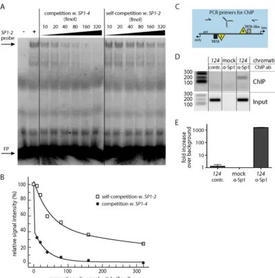
The LVGRSP1-4has a higherin vitroSp1-binding affinity
than the EVGRSP1-2.Although Sp1 has been shown to bind to
bothSP1-2andSP1-4 in vitro(32), there are differences in the respective sequence compositions. WhileSP1-4represents a per-fect Sp1 consensus sequence,SP1-2carries deviations by A/T base pairs (Fig. 1BtoD, sequences indicated in yellow). To examine whether the sequence differences were associated with different binding affinities, an EMSA-based competition assay was em-ployed. 32P-radiolabeled, duplexed oligonucleotides carrying SP1-2were incubated with nuclear extract from primary human renal proximal tubule epithelial cells (RPTECs) and mixed with increasing concentrations of unlabeled oligonucleotides contain-ingSP1-2 orSP1-4 (Fig. 2A). As shown, competition with the unlabeled SP1-4 oligonucleotides decreased the EMSA signal more readily than competition with the unlabeledSP1-2 oligonu-cleotides: while 10 fmol of theSP1-4oligonucleotide was enough to reduce theSP1-2signal to about 30% of the signal for the ar-chetype, self-competition required approximately 30 times higher competitor concentrations (Fig. 2B). This finding indicates that theSP1-4binding site has a higher affinity for Sp1 binding than SP1-2, as expected from comparison of their sequences with the
perfect Sp1 consensus sequence. Together, the combined data suggest that the affinity of Sp1 binding to the intact localSP1-2 andSP1-4sites determined the higher activity of the LVGR pro-moter in the archetypeww-NCCR. To complement thein vitro data, a chromatin immunoprecipitation (ChIP) assay was per-formed inww-NCCR reporter construct-transfected cells (Fig. 2C andD). ChIP with the Sp1 antibody yielded an enrichment of about 1,600-fold compared to that achieved with a control anti-body, demonstrating the strong binding of Sp1 to the BKPyV NCCRin vivo(Fig. 2E).
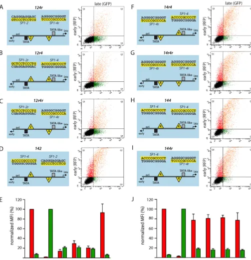
Strand inversion of Sp1 sites disrupts the archetype
EVGR-LVGR expression balance.To examine the potential
contribu-tions of the nonpalindromic Sp1 sites to the directionality of gene expression,SP1-2andSP1-4were flipped in place, yielding the NCCRs124r,12r4, and12r4r, as well as swapped with respect to their original archetype position (NCCR142) (Fig. 3AtoD). All NCCR inversion mutants showed impaired LVGR expression with a reduction in the level of expression to about 20% of the archetype level. Of note, all of these constructs except142still carried an intact high-affinitySP1-4site in the LVGR promoter region, including NCCR12r4, in whichSP1-4 was even main-tained in its original strand orientation (Fig. 3BandE). At the same time, all inversion mutants showed a moderate level of EVGR activation compared to the archetype, whereby the NCCR 12r4had the strongest expression in the range of 25% of the level seen for the Dunloprr-NCCR. These observations indicate that the relative Sp1 site orientation was critical for regulating bidirec-tional expression, in addition to affinity. Importantly, the Sp1 sites were able to convey an orientation-dependent directionality that seemed to be functionally as important as the local Sp1-binding affinity or deletion ofSP1-4. Interestingly, NCCR142yielded a high EVGR expression level similar to that of Dunlop and the initially tested14r2rmutant (Fig. 3DandEand1DandE). We concluded that, when placing the high-affinitySP1-4site in the proximity of the EVGR, its orientation relative to the remaining NCCR did not matter anymore. This notion was further corrob-orated by another set of mutants, where the low-affinitySP1-2was replaced with the high-affinitySP1-4, such that the EVGR and LVGR proximal positions contained Sp1 sites with equal affinities in all possible combinations of site orientations (Fig. 3F to J, NCCRs14r4,14r4r,144, and144r). All of the resulting mutants showed an equally high level of EVGR expression in the range of 80% of that of strain Dunlop, coupled to a profound loss of LVGR expression. Thus, a high-affinity Sp1 site in the proximity of the EVGR provided a strong activation, which in no case permitted rescue of the archetype expression pattern by an Sp1 site in an LVGR of similar sequence, affinity, or orientation (Fig. 3J). Con-versely, the data further support the notion that the bidirectional fine-tuning of NCCR expression depends on the modulation of Sp1 affinity at the EVGR site.
Early promoter activity correlates with the Sp1-binding
af-finity of the early Sp1 site.To examine the role of Sp1 affinity in
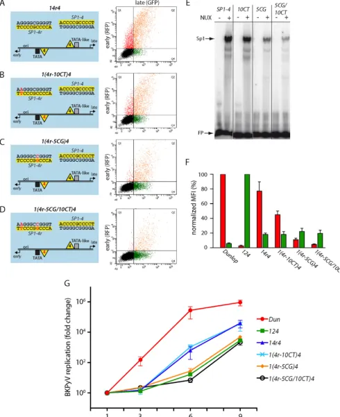
the critical EVGR position, the mutant14r4was used for further study by introducing point mutations that had been identified in patient variants (48) and previously shown by us to lower the Sp1-binding affinity (32). By slightly lowering the affinity of the EVGR-proximal Sp1 site with the mutant1(4r-10CT)4(where 10CT indicates the exchange of C to T in position 10), the expres-sion of EVGR decreased to about half the level seen for mutant 14r4(or 30 to 50% of that for the Dunlop reference strain) (Fig. 4B
on November 7, 2019 by guest
http://jvi.asm.org/
andF). The intermediate-affinity mutant1(4r-5CG)4allowed ap-proximately 15% of the level of14r4EVGR expression (10% of that of Dunlop), while the double mutant showed only 5% of the level of expression by Dunlop (a⬎75% reduction compared to that for the14r4mutant) (Fig. 4C,D, andF). In contrast to the gradual decrease in the level of EVGR expression, the late pro-moter activity remained at the same low level (about 20% of the archetype level of expression). Hence, just lowering the affinity of the EVGR-proximal Sp1 site was not sufficient to restore a high level of LVGR expression. The data suggest that, in addition to affinity, particularities of theSP1-2sequence might play a role for achieving a high level of LVGR activity.
To confirm the mechanistic relevance of these findings, the NCCR mutants were placed into a BKPyV Dunlop backbone and analyzed in a viral replication assay. To minimize potential
effects of EVGR expression and, hence, LTag levels, infectious supernatants were generated in COS-7 cells constitutively ex-pressing simian virus 40 (SV40) LTag, as reported previously (32), and were used to infect RPTECs. Supernatants from RPTECs were then taken at 1, 3, 6, and 9 days postinfection (dpi), and viral loads were quantified by qPCR (Fig. 4G). As expected, the Dunlop strain had the highest replication rate of the tested strains, reaching supernatant viral loads of more than 1010genome equivalents (GEq)/ml at 6 dpi, which corre-sponded to a more than 200,000-fold increase compared to the input (1 dpi). Importantly, the recombinant virus strains14r4 and1(4r-10CT)4were functional and showed accelerated rep-lication compared to that of the archetype virus, reaching levels of replication 1,000-fold that of the archetype virus at 6 dpi and about 40,000-fold that of the archetype virus at 9 dpi,
respec-FIG 2SP1-2andSP1-4differ in Sp1 affinity in EMSA competition assays. (A) Representative EMSA result after competition of the radiolabeledSP1-2probe with (w.) unlabeledSP1-4or unlabeledSP1-2(self-competition) at the indicated concentrations. Lane⫺, binding mix without nuclear extract; lane⫹, binding mix without competition; FP, free probe. (B) Plot of quantitative readout of EMSA signals.xaxis, concentration of competing oligonucleotide;yaxis, signal strength relative to that of the uncompetedSP1-2signal, set to 100%. (C) Scheme of NCCR with primer binding sites for ChIP assay. (D) Semiquantitative analysis of ChIP assay results by agarose gel electrophoresis. Mock, untransfected HEK293TT cells; contr., ChIP with rabbit negative-control antibody (ab) supplied with the kit. Numbers on the left are denote DNA fragment sizes (in base pairs). (E) Quantification of ChIP assay result by qPCR with SYBR green. Calculation was carried out by⌬⌬CTmethod (with normalization to theww-NCCR input and the background control).
Bethge et al.
on November 7, 2019 by guest
http://jvi.asm.org/
[image:6.594.95.493.64.466.2]tively. However, the weakened affinity in the 1(4r-10CT)4 strain seen in the reporter assay did not translate into measur-able differences in replication kinetics, suggesting that the EVGR-proximal Sp1 affinity was still strong enough to drive
EVGR expression in the context of viral infection in cell cul-ture. In contrast, the viruses carrying the stronger mutations 1(4r-5CG)4and1(4r-5CG/10CT)4did not seem to allow rapid viral replication (Fig. 4G). Thus, the data indicate that the Sp1
FIG 3Position, orientation, and affinity of Sp1 sites determine the directionality of EVGR and LVGR expression. (A to D) (Left) Schematic representation of NCCRs tested with the FACS-based, bidirectional reporter assay in HEK293 cells. All mutant NCCRs have the length and architecture of the BKPyV archetype NCCR124but carry inverted Sp1 sites, as indicated with the letterr(e.g., NCCR124rhas an invertedSP1-4sequence). (Right) Representative flow cytometry measurements are shown next to the schematic representations, which are described in the legend toFig. 1. (E) Quantification of the constructs listed in panels A to D using the normalized MFI from at least 3 independent replicates, as described in the legend toFig. 1. (F to I) Schematic representation of NCCRs, where the archetypeSP1-2was replaced with the sequence of the high-affinitySP1-4. (J) Quantification of the constructs listed in panels F to I using the normalized MFI from at least 3 independent replicates, as described in the legend toFig. 1.
on November 7, 2019 by guest
http://jvi.asm.org/
[image:7.594.43.546.67.587.2]FIG 4Lowering of the Sp1 affinity in the EVGR-proximal position of NCCR14r4reduces EVGR expression and viral replication. (A to D) Schematic representation (left) and representative FACS plot (right) of the indicated NCCRs tested in the bidirectional reporter assay in HEK293 cells. The numbering of the point mutants is relative to the first position of theSP1-4site in the archetype orientation [e.g., NCCR1(4r-10CT)4contains the exchange of C to T in position 10, while the lateSP1-4site is unaltered]. (E) EMSA analysis of binding affinity in the absence and presence of nuclear extracts (NUX) confirms that the inserted point mutations lower thein vitroaffinity of the respective variants. (F) Quantification of the constructs listed in panels A to D using the normalized MFI from at least 3 independent replicates, as described in the legend toFig. 1. (G) Mutant BKPyV replication in primary human RPTECs using infectious supernatants generated by transfection of COS-7 cells. The cell culture supernatants were taken at 1, 3, 6, and 9 days postinfection (xaxis), and DNase-protected viral loads were quantified by qPCR. Results were normalized to the amount of input virus (day 1) and are shown as the fold change (yaxis). Dun, strain Dunlop.
on November 7, 2019 by guest
http://jvi.asm.org/
[image:8.594.46.537.38.640.2]affinity of the EVGR-proximal site was also reflected in the level of BKPyV replication, although the infection assay did not achieve the same nuanced resolution observed by the flow cy-tometry assay interrogating the bidirectional reporter con-structs.
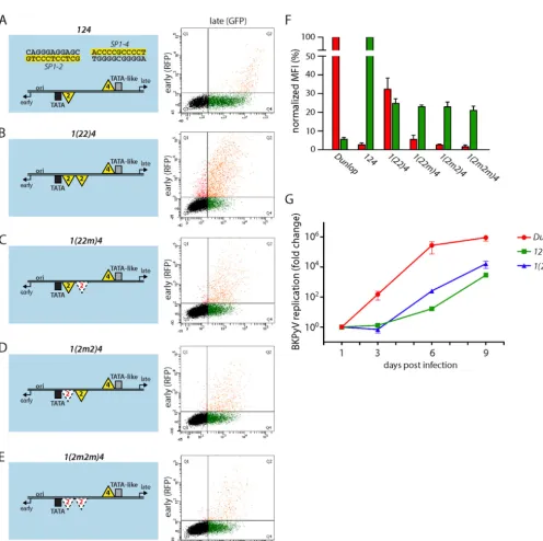
EVGR expression can be enhanced by low-affinity Sp1 site
duplication.Given the frequently observed duplications in the
vicinity of the EVGR of rearranged NCCRs, including one of the
NCCRs of the Dunlop strain, we examined expression in another set of NCCR mutants which carried a secondSP1-2site in the EVGR-proximal position, NCCR1(22)4(Fig. 5AandB). This additional binding site was sufficient to increase the level of EVGR expression 15-fold compared to that for the archetype, reaching about 30% of the level for Dunlop (Fig. 5F). To exclude a potential distance or length effect, we also examined inactivated Sp1 sites yielding the NCCRs1(22m)4,1(2m2)4, and1(2m2m)4, in which
FIG 5Addition of a second low-affinitySP1-2site increases the level of EVGR expression. (A to E) The indicated mutant NCCRs inserted into the bidirectional reporter constructs (left) were transfected into HEK293 cells, and representative FACS plots are shown (right). (F) Quantification of the constructs listed in panels A to E using the normalized MFI from at least 3 independent replicates, as described in the legend toFig. 1. Note that theyaxis was divided for better visibility of the low range. (G) Mutant BKPyV replication in primary human RPTECs using infectious supernatants generated by transfection of COS-7 cells. Cell culture supernatant was taken at 1, 3, 6, and 9 days postinfection (xaxis), and DNase-protected viral loads were quantified by qPCR. Results were normalized to the amount of input virus (day 1) and are shown as the fold change (yaxis).
on November 7, 2019 by guest
http://jvi.asm.org/
[image:9.594.43.539.56.550.2]the proximalSP1-2site, the distalSP1-2site, or bothSP1-2sites were mutated to sequences that no longer bound Sp1 (Fig. 5C,D, andE). These additional NCCR mutants showed reduced EVGR expression compared to the1(22)4mutant, in line with the notion that Sp1 binding and not any inserted sequence mediated an ele-vated level of EVGR expression. Together, these findings confirm thatSP1-2duplications found in parts of the P block of some patient isolates (31) as well as the Dunlop strain contribute to activation of the EVGR phenotype. Interestingly, all the experi-mentally designed insertions of the low-affinitySP1-2diminished LVGR expression to a similar degree, again suggesting that some additional factors in the NCCR sequence might play a role. In the viral replication assay, recombinant BKPyV bearing NCCR1(22)4 propagated slightly faster than the archetype NCCR 124virus, which is in line with the results of the bidirectional reporter assay (Fig. 5G).
Characterizing the early and late core promoter elements of
the archetype NCCR.Given the strong effects of Sp1 sites on
EVGR and LVGR expression through position, orientation, and affinity, we addressed potential cooperating components in their close proximity. Analysis of the NCCR sequence in the vicinity of theSP1-2and theSP1-4sites identified a canonicalTATAbox upstream of the EVGR start codon but only potentialTATA-like elements upstream of the LVGR start codon which deviated from a perfectTATAbox by several mismatches (49). The actual tran-scriptional start site (TSS) for the BKPyV NCCR EVGR was de-rived by 5=rapid amplification of cDNA ends (5=-RACE) analysis (Fig. 6A), which also revealed a sequence consistent with loci of initiator elements (Inr) (Fig. 6A). The positions of the Inr ele-ments allowed the deduction of other core promoter eleele-ments (CPEs), which were located within the canonical distances up-stream and downup-stream of the identified TSSs. The web-based search tool ElemeNT (45) confirmed the position of the early core promoter, which is located close to the O-block-P-block bound-ary and which is defined by the conservedTATAbox and by a prominent Inr 31 bp downstream (Fig. 6A). In addition, a down-stream promoter element (DPE) was identified at the typical dis-tance of 28 to 32 nucleotides downstream from the Inr TSS. When the LVGR promoter region of the archetype NCCR was analyzed, a similar composition with a dominant Inr and potential DPEs was found (Fig. 6B), but in contrast to the EVGR promoter with its conserved TATA box, two potentialTATA-like elements were lo-cated upstream of the Inr/major TSS (Fig. 6B, plot below the se-quence). Of note, this major TSS was in line with a previous map-ping of the BKPyV late promoter by a primer extension assay (50). One of theTATA-like elements had two mismatches compared to the sequence of a perfectTATAbox but showed the canonical distance of 30 nucleotides to the major TSS (TATA-like element 1; Fig. 6B), while the other had three mismatches and was located further upstream (TATA-like element 2). An additional potential CPE was identified as a transcription factor IIB (TFIIB) recogni-tion element upstream (BREu), which is typically located directly upstream of a TATA box. Thereby, the promoter elements of EVGR and LVGR presented an imperfect symmetry of a classical TATAand aTATA-like promoter. Interestingly, in the case of the LVGR promoter, theBREuwas adjacent toTATA-like element 2 and consisted of a GC-rich consensus sequence which partially overlappedSP1-4(Fig. 6BandD). This overlap of the LVGRBREu and high-affinity SP1-4 raised intriguing questions about the functional implications with respect to the regulation of LVGR
expression through high-affinity Sp1 binding. It was therefore of interest to first examine deletion-bearingrr-NCCRs. As shown inFig. 6C, the Dunloprr-NCCR is characterized by a deletion of the Q35and R63blocks, which removes theTATA-like elements as well asSP1-4, and the dominant Inr, thereby disrupting the aforementioned imper-fect symmetry of the late and the early sides of the archetype NCCR. The 5=-RACE analysis of the strain Dunlop NCCR revealed an overall dispersion of TSS but a slight overrepresentation of TSS at two po-tential Inr elements located in a canonical distance to the DPEs of 28 to 33 nucleotides (Fig. 6C, plot below the sequence). The results in-dicate that the loss of the LVGR CPEs could be partially compensated for by multiple initiator elements, which were found to be located throughout the triplicate P-block sequences and which acted in con-junction with the conserved DPEs of the S block. Together, the data indicate that the basal expression pattern of the archetype BKPyV NCCR involved an imperfect symmetry centering around aTATA box andTATA-like elements, which are modulated by juxtaposed Sp1-binding sites. Disruption of this basal symmetry by LVGR-prox-imal deletions was compensated for by the next-best fitting of analo-gous sequences.
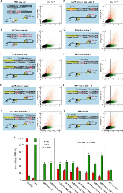
TheTATAbox is essential for both early and late expression.
To better understand the contribution of the respective core pro-moter elements for EVGR and LVGR expression, we designed a set of constructs in which we either removed defined sequence ele-ments or changed them to obtain optimized consensus sequences but, again, without altering the overall length of the archetype NCCR. When we mutated the EVGRTATAbox (Fig. 7A,TATA box-out, where “out” indicates that the CPEs were removed) or replaced it with aTATA-like element similar to the one at the LVGR (Fig. 7B,TATA-like-in early, where “in” indicates that the CPEs were inserted), not only was EVGR expression almost com-pletely abolished but also LVGR expression was reduced to 23% of that of the archetype NCCR (Fig. 7K). These observations dem-onstrate the central importance of theTATAbox not only for EVGR but also for the NCCR as a whole. In contrast, mutating the LVGRTATA-like elements individually or together (Fig. 7CtoE, TATA-like-out late 1,TATA-like-out late 2, andTATA-like-out late 1⫹2) caused an increase in the level of EVGR expression to 10 to 15% of that of the archetype, while the level of LVGR expression was reduced to 20 to 25% of that of the archetype (Fig. 7K). Thus, effective LVGR expression from the late core promoter seemed to be equally dependent on both TATA-like elements, although the TATA-like element 1 sequence better matched a perfectTATA box and was situated within the canonical distances to the major TSS (Fig. 6B). When we mutatedSP1-4, in addition to TATA-like element 1 (Fig. 7F,TATA-like-out late 1/sp1-4), LVGR expression went down by another 10%, suggesting a cooperative effect of the TATA-like element and the Sp1-binding site on the late promoter activity (Fig. 7K). Interestingly, changing theTATA-like elements individually or together to perfect TATA boxes did not increase the level of LVGR expression compared to that of the archetype NCCR (Fig. 7GtoI,TATAbox-in late 1,TATAbox-in late 2, and TATAbox-in late 1⫹2). Instead, the distance to the major Inr seemed to gain importance when a perfectTATAbox sequence was inserted into the position ofTATA-like element 1, as the level of LVGR expression then reached 35% of that of the archetype, while that of the corresponding construct with theTATAbox-in late 2 reached only 23% of that of the archetype (Fig. 7K). Hence,
the insertion of consensus TATA boxes in both positions
diminished the LVGR promoter activity compared to that of the Bethge et al.
on November 7, 2019 by guest
http://jvi.asm.org/
archetype, but the canonical positioning of theTATAbox could partlycompensate for this adverse effect.The single insertions showed an activation of EVGR expression of about 5-fold com-pared to that of the archetype (11% of that of the Dunlop strain),
and the double mutantTATAbox-in late 1⫹2 showed EVGR expression in the range of 5% of that of Dunlop.
As mentioned above, the high-affinitySP1-4overlaps a poten-tial BREu. Both share a GC-rich consensus sequence, and the
FIG 6Experimental and computational core promoter analysis indicates the imperfect symmetry of the BKPyV archetype NCCR. The arbitrary sequences of the O, P, Q, R, and S blocks of the NCCR are shown on top. Selected nucleotide sequences enlarged with core promoter elements (CPE) are highlighted in the color of the respective sequence block, except for Sp1 sites, which are highlighted in yellow. Inr, initiator element; DPE, downstream promoter element (sequence in the dashed box highlighted in blue);BREu, TFIIB recognition element upstream, overlapping (dashed box) withSP1-4highlighted in yellow; red nucleotides, mismatches compared to the CPE consensus sequence; vertical black lines, sequence block boundaries; horizontal black lines, distance (in nucleotides [nt]) from CPEs; dashed boxes, overlapping CPEs. The plot below the sequence shows 5=-RACE data, used to determine the transcription start site (TSS), with each point corresponding to the base pair position in the sequence presented above and theyaxis showing the number of 5=reads for the respective position. (A) Analysis of early core promoter located at the O block-P block boundary. (B) Analysis of the archetype late core promoter in the Q, R, and S blocks. *, position with the known polymorphism A/G, with G introducing an additional mismatch into theTATA-like element. (C) Analysis of Dunlop late core promoter with deletions removing the Q and R blocks. (D) Relevant CPEs with sequence logos, the consensus sequence in the IUPAC ambiguity code, and the typical position (Pos.) relative to the TSS (position⫹1) (45). R, A/G; W, A/T; S, C/G; Y, C/T; V, A/C/G; N, any nucleotide.
on November 7, 2019 by guest
http://jvi.asm.org/
[image:11.594.49.529.72.551.2]FIG 7Mutating the EVGR TATA box or the LVGRTATA-like elements increases early expression. (A to J) (Left) The indicated mutant NCCRs inserted into the bidirectional reporter constructs were transfected into HEK293 cells. Red letters, mutations compared to the archetype sequence. CPEs were either removed (indicated “out”) or inserted (indicated “in”). (Right) Representative FACS plots. (K) Quantification of the constructs listed in panels A to J using the normalized MFI from at least 3 independent replicates, as described in the legend toFig. 1.
on November 7, 2019 by guest
http://jvi.asm.org/
[image:12.594.88.497.36.677.2]SP1-4site carries only one mismatch compared to the sequence of a perfectBREusite (Fig. 6D). Therefore, a point mutation was introduced to create a perfect consensus BREu site (Fig. 7J, BREu-in late), which at the same time inactivated Sp1 binding, as shown for the previously analyzedSP1-4point mutations (Fig. 1). The results show that introduction of a perfect BREu consensus site did not increase the level of LVGR expression but, rather, reduced it to a level of about 15% of that of the archetype. At the same time, EVGR expression was increased only moderately to about 14% of that of Dunlop or 7-fold of that of the archetype. This finding indicates that theSP1-4site is more important than the potential BREu for driving LVGR expression.
LTag can drive LVGR expression independently of
arche-typal late CPEs.LTag is a major product of EVGR expression and
an important regulator of the viral life cycle with respect to viral DNA replication and LVGR expression. We therefore compared the effects of critical NCCR mutants in HEK293, HEK293T, and HEK293TT cells, which produce no, small, or large amounts of SV40 LTag intrans, respectively (Fig. 8A). One essential function of LTag for the viral life cycle is to bind to the viral origin of replication (in the O block) and to facilitate, together with cellular factors, the replication of the viral genome (Fig. 8B). Interestingly, for almost all NCCR mutants tested, LTag was able to boost LVGR expression in a concentration-dependent manner. Despite varia-tions in the absolute fluorescence intensities (Fig. 8CandD), most NCCR reporter constructs showed similar relative increases in the levels of LVGR expression in HEK293T and HEK293TT cells com-pared to the levels in their HEK293 cell counterparts (indicated by a dashed line, set to 1 for early and late fluorescence and each construct in HEK293 cells;Fig. 8E). The Dunlop strain, for exam-ple, which is characterized by a very low level of late expression in HEK293 cells, expressed 4 times more LVGR in HEK293T cells and 13 times more LVGR in HEK293TT cells than in HEK293 cells. At the same time, the level of EVGR expression of Dunlop stayed at the same level in the three cell lines. Since the LTag-mediated increase of LVGR expression was also observed with the Dunlop NCCR, which carries a large deletion of the archetypal late promoter elements (Fig. 6C), we concluded that LTag rendered LVGR expression independent of the expression of most arche-typal late CPEs. This is in line with the finding that theTATA-like elements are dispensable, as observed with the constructTATA -like-out late 1, which showed an 8-fold increase in HEK293T cells and an 18-fold increase in HEK293TT cells compared with that in HEK293 cells. On the other hand, the highest absolute intensity (24,400 arbitrary units;Fig. 8D) and the highest relative increase (20-fold; Fig. 8E) in HEK293TT cells were recorded with the TATAbox-in late 1 construct, which carried a perfectTATAbox in a canonical distance to the major late Inr (Fig. 7G). We con-cluded that although the activating effect of LTag on LVGR ex-pression could occur without the archetypal late promoter, there still seemed to be a benefit from the presence of a perfectTATA box and the high-affinitySP1-4site. The latter becomes visible when the levels of expression in the constructs NCCR14r2rand 14r4, both of which carry the high-affinitySP1-4site in the early promoter but differ in the affinity of the late Sp1 site, are com-pared. While swapping of NCCR 14r2r increased the level of LVGR expression by only 6-fold in HEK293TT cells, LVGR ex-pression of NCCR14r4was boosted by 11-fold. However, LVGR expression of the archetype NCCR 124 was already high in HEK293 cells, which explains why the absolute and relative
in-creases in HEK293T and HEK293TT cells were only moderate (4-fold). Intriguingly, LTag activation of LVGR expression also worked in the absence of the otherwise essential and conserved
TATAbox when it was compared in the EVGR mutantsTATA
box-out (not shown) andTATA-like-in early (Fig. 8CtoE). The only exception to this LTag-mediated increase in LVGR expression was in the mutant NCCRBREu-in late, where a point mutation in the high-affinitySP1-4site created a perfect BREu site. Here, the increase in the level of LVGR expression was only 1.3-fold (HEK293T cells) and 1.8-fold (HEK293TT cells). Since both the SP1-4 mutant (not shown) and the double mutantTATA-like-out late 1/sp1-4(Fig. 8E) were activated by LTag, we can only speculate at this point that the binding of TFIIB to the late promoter might pose an obstacle to LTag-mediated LVGR expression. Finally, we observed that EVGR expres-sion remained similar for most constructs in this comparative trans-fection study, suggesting that the previously reported negative feedback of LTag on EVGR expression might be offset by increasing reporter construct replication. Notable exceptions to this observation were two constructs which had very low levels of EVGR expression in HEK293 cells, namely, the archetype NCCR124and theTATAbox mutantTATA-like-in early. Here, few changes in early expression occurred, and these were probably attributable to plasmid copy num-ber changes or an only subtle stimulation of the early promoter by LTag, which resulted in large relative increases in the expression levels.
DISCUSSION
The NCCR determines within only 400 bp essential functions of PyV biology, including viral persistence and the appropriate tim-ing of virus gene expression, genome replication, and virion as-sembly. Whereas other similarly sized viruses, like members of the Papillomaviridaeor proviral copies of theRetroviridae, use unidi-rectional promoter organization, PyVs have effectively evolved a complex bidirectional expression modality to sequentially role out a strand- and orientation-specific set of virus gene products from their NCCRs. Notably, these steps of the PyV life cycle are host specific and, within a given host, restricted to certain cells, arguing that cellular differentiation and activation states must be readily sensed and interpreted by PyV-specific NCCRs.
As the archetype NCCR of BKPyV is stably maintained in the general population, we were intrigued by the rapid real-time emergence of BKPyV variants with rearranged NCCRs in immuno-suppressed kidney transplant patients and their functional link to increased EVGR expression, accelerated viral replication, higher blood viral loads, and more advanced pathology (31). Given the di-verse array of affected TFBS in therr-NCCRs, we recently introduced defined point mutations that excluded the potential effects of overall length and architecture on NCCR function and identified three phe-notypic groups of TFBS (32): group 1 and group 2 mutations caused a strong and an intermediate level of activation of EVGR expression and viral replication, respectively, similar to that caused by natural BKPyV NCCR variants associated with disease, whereas group 3 mu-tations permitted only reduced or no NCCR function (32). Impor-tantly, Sp1 emerged as a key regulatory factor, being able to mediate either extreme of the group 1 mutant (e.g.,SP1-4) or the group 3 mutant (e.g.,SP1-2) phenotype.
In the present study, we demonstrate that Sp1 can exert these differential effects as a result of the position, orientation, and af-finity of two Sp1 TFBS located on the LVGR- and the EVGR-proximal sides of the archetype NCCR. Swapping of the
on November 7, 2019 by guest
http://jvi.asm.org/
Bethge et al.
on November 7, 2019 by guest
http://jvi.asm.org/
finitySP1-2site and high-affinitySP1-4site (e.g., NCCR14r2r), introducing affinity-lowering mutations into the high-affinity SP1-4site [e.g., NCCR12(4-5CG)], flipping the Sp1 site orienta-tion (e.g., NCCR124r), and creating various permutations and combinations thereof perturbed the EVGR-LVGR balance and in most cases led to a significant increase in the level of EVGR ex-pression. In a key experiment, we made use of the construct NCCR 14r4, which carries the same high-affinitySP1-4sequences in the respective EVGR- and LVGR-proximal positions, followed by in-troducing affinity-lowering mutations that caused a stepwise de-cline in the level of the activated EVGR phenotype. In another approach, the low-affinitySP1-2site was duplicated, showing that two low-affinity Sp1 sites were better than one or none, overall supporting the role of local Sp1-binding affinity. Importantly, the mutant NCCRs were functional in recombinant BKPyVs, yielding elevated viral replication compared to that of archetype BKPyV in RPTECs, as predicted. Thus, the nonpalindromic nature of the Sp1-binding sequence and the deviations from its consensus se-quence affected the directionality of expression and regulated the delicate EVGR-LVGR balance of the BKPyV archetype NCCR.
We also obtained evidence that the NCCR balance of the Sp1 sites acts in conjunction with core promoter elements (CPE), showing an imperfect rotation symmetry of a developmental pro-moter on the EVGR and a housekeeping propro-moter on the LVGR side of the BKPyV archetype NCCR. In general, most eukaryotic promoters can be assigned to (i) developmental or regulated pro-moters, which are characterized by a focused transcriptional start, that need to be activated by a stimulus and contain either aTATA box or a DPE and (ii) housekeeping or tissue-restricted promoters that are constitutively active and show multiple, dispersed tran-scriptional start sites, that areBREuenriched, typically without a TATAbox, and that contain a CpG island (51,52). Indeed, the EVGR side carries downstream of the Sp1 site a perfectTATAbox
and one majorInr/TSS in a canonical distance of 31 bp, followed by a consensus downstream promoter element (DPE) at a distance of 28 bp (53,54). Thus, our results indicate that the EVGR pro-moter of the BKPyV archetype NCCR fulfills the criteria of a reg-ulated promoter with a perfectTATAbox, a focused TSS, and a low level of basal activity. The LVGR promoter is less easily clas-sified, since it bears the high-affinitySP1-4partially overlapping a potentialBREu, followed by twoTATA-like elements, a majorInr besides some dispersed TSS, and potential DPEs. We demonstrate here that the archetype BKPyV LVGR promoter shows a mixed focused-dispersed activity, which changes to a fully dispersed type of activity in the BKPyV Dunlop NCCR due to the deletion of the archetypal late promoter, extending the initial observation in hy-brid NCCRs (50). The LVGR promoter is reminiscent of the pre-viously describedTATA-less housekeeping promoters described in the model ofSaccharomyces cerevisiae, which essentially contain TATA-like elements (49). It is currently unclear whetherTATA -like elements bindTATA-binding protein (TBP). One hypothesis is that they do but, compared to the results obtained with perfect TATAboxes, that binding leads to the assembly of altered preini-tiation complexes (PIC), which—at least in yeast— have been shown to be Taf1/TFIID enriched (49). Alternatively, these kinds of sequences might be binding sites for paralogs of TBP, like TRF2, thereby leading to differential PIC formation and transcriptional activity. Accordingly, the imperfect symmetry of Sp1 sites,TATA elements,Inr, and DPEs in the BKPyV NCCR seems to allow the maintenance of the archetypal focus on the LVGR side while re-pressing EVGR expression. At the same time, this organization permits an effective bidirectional system that is highly poised to shift the transcriptional balance from LVGR to EVGR expression upon differential stimulation and mutation (32). A summary model of our findings is depicted inFig. 9. It should be noted that a second layer of regulation that involves BKPyV microRNAs
FIG 8LTag expression intransincreases the level of LVGR expression in a concentration-dependent manner and can drive late expression independently of core promoter elements. (A) Western blot showing the different LTag expression levels by HEK293, HEK293T, and HEK293TT cells and the level of actin expression as a loading control. Numbers on the right are molecular masses (in kilodaltons). (B) Schematic view of LTag-binding sites (sequences with a red background) in the O block of the BKPyV NCCR. (C) Representative flow cytometry of HEK293, HEK293T, and HEK293TT cells transfected with the indicated BKPyV NCCR bidirectional reporter constructs. (D) For quantification of EVGR and LVGR expression in the transfected cells, the normalized MFI (in percent) was calculated as described in Materials and Methods. The level of EVGR expression was normalized to that of strain Dunlop (red MFI⫽100%), and the level of LVGR expression was normalized to that of NCCR124(wwarchetype) (green MFI⫽100%). Quantification was based on at least 3 independent replicates. (E) Relative change in the levels of EVGR and LVGR expression by the indicated reporter constructs in HEK293T and HEK293TT cells using HEK293 cells as a reference (for which the level of expression was set equal to 1 and is indicated by a horizontal dashed line).
on November 7, 2019 by guest
http://jvi.asm.org/
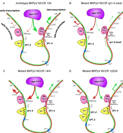
[image:15.594.137.451.65.246.2]FIG 9Sp1, TATA, and downstream promoter element (DPE) core promoter elements forming an imperfect symmetry underlying bidirectional EVGR and LVGR expression of the BKPyV NCCR. (A) BKPyV archetype NCCR124predominantly drives LVGR expression as a result of the high-affinitySP1-4(the three triangular indentations represent the zinc finger binding) and TATA-like elements, whereas EVGR expression is regulated by the low-affinitySP1-2site (two triangular indenta-tions) and theTATAbox. The Sp1 protein is depicted in yellow, with the three small triangles symbolizing zinc fingers. Pink ovals depict TBP (TATAbox-binding protein) or TRF2 (TBP-related factor 2), with the latter potentially being recruited byTATA-like elements. A low level of early expression is due to less efficient recruitment of RNA polymerase II (Pol II) to the early promoter (red dashed arrows), and a high level of late transcription is due to recruitment of a major fraction of RNA polymerase II to the late promoter (fat green arrows). Inr, initiator element. For simplicity, other components, such as TFIIB and the TFIID complex or transcription factors like NF1, are not depicted (see reference32). (B) Mutant BKPyV NCCR124carrying anSP1-4sequence with a point mutation (mut) that abrogated Sp1 affinity and thereby the recruitment of polymerase II to the late promoter, leading to a decrease in the level of LVGR expression (green dashed arrows). The low-affinitySP1-2 now permits preferential recruitment of RNA polymerase II to the early promoter, causing increased levels of EVGR expression (red arrows). (C) Mutant BKPyV NCCR 14r4in which the low-affinitySP1-2sequence was replaced with a high-affinitySP1-4sequence in the EVGR promoter. An increased fraction of RNA polymerase II is recruited to the early tandem of theTATAbox andSP1-4, giving rise to high levels of EVGR expression (fat red arrows), while the unaltered late promoter is disfavored (green arrows). (D) Mutant BKPyV NCCR1(22)4carrying a duplication of the low-affinitySP1-2site in the early promoter increases RNA polymerase II recruitment and EVGR expression (red arrows), while the unaltered late promoter is disfavored (green arrows).
Bethge et al.
on November 7, 2019 by guest
http://jvi.asm.org/
[image:16.594.43.537.61.597.2](miRNAs) also governs the effective control of EVGR expression. These miRNAs are transcribed in the late direction and target the EVGR transcripts, thus mediating repression of LTag expression (55). Thereby, persistent archetype BKPyV infection maintains a double-stitched mechanism to ensure tight control of LTag ex-pression below the radar of the immune system (55,56). In turn, activating rearrangements not only switch on EVGR expression but also overcome miRNA suppression, an alteration that requires the absence of a functional immune response (31).
The late DPEs located at the start of the S block are maintained in most patient isolates (31,32), thus keeping the DPEs in place in rr-NCCR BKPyV variants lacking archetype late promoter se-quences. Interestingly, these LVGR promoter deletions signifi-cantly increased EVGR expression at the expense of LVGR expres-sion, but the associated increases in viral replication and progeny still depend on the corresponding expression of the LVGR struc-tural gene products, e.g., Vp1, Vp2, and Vp3. We approached this issue by also providing two levels of SV40 LTag intransin the HEK293 cell derivatives HEK293T and HEK293TT when trans-fecting selected NCCR mutants, thereby complementing the re-sults of recombinant BKPyV infection in primary human RPT-ECs. The HEK293, HEK293T, and HEK293TT cell transfection experiments indicated that LTag expression intranscan overcome the adverse effects of deletingSP1-4or the TATA-like elements in the LVGR promoter. These results are in line with those of a pre-vious study reporting that the SV40 LTag is able to rescue a crip-pled promoter function which was lost due to the lack of a TBP-associated factor (TAF), TAF(II)250 (57). TAFs and TBP have been shown to form the TFIID complex and target the core pro-moter region by binding to DPEs, Inr, and TATA elements (52, 58). Our data suggest that LTag is able to activate the LVGR pro-moter by executing a TAF-like function within the TFIID com-plex, helping to recognize the remaining CPEs, namely, the DPEs of the S63block and the dispersedInrin the P68block of the NCCR. As mentioned earlier, few mutants also showed an increase in the level of EVGR expression, suggesting a small contribution of LTag to early promoter activity and/or an increase in episomal plasmid copy number viaori-binding functions. The NCCRBREu-in late mutant was a notable exception with respect to LVGR activation, as it showed little change in its level of expression in HEK293T and HEK293TT cells, suggesting a possible negative interference with LTag-mediated activation. This mutation abrogated Sp1 binding, while it introduced a perfect consensusBREu, thus indicating that theSP1-4site is more important than the potentialBREufor driv-ing LVGR expression.
This point mutational dissection of the bidirectional BKPyV NCCR may have implications for other HPyVs which show an architecture and an organization similar to those of BKPyV but which show a different TFBS composition and tissue specificity (59). In fact, NCCR rearrangements are best known from JC polyomavirus (JCPyV) variants from patient suffering from pro-gressive multifocal leukoencephalopathy (46,60–62). Following duplication of the early promoter and deletion of the late pro-moter sequences, the JCPyVrr-NCCRs demonstrate activation of EVGR at the expense of LVGR similar to those of BKPyV (46). These alterations also broaden the host cell range, which has par-ticular relevance for neurotropism and pathology (34, 63,64). Given the power of the bidirectional reporter construct to predict PyV NCCR expression and viral replication, our approach may
help to close the current knowledge gaps in viral host cell specific-ity and replication of the dozen novel human PyVs.
In conclusion, this study provides a novel view of the BKPyV NCCR by revealing an imperfect symmetry of the EVGR and LVGR promoter characteristics that are functionally balanced by the position, orientation, and affinity of Sp1 and core promoter elements. As there is currently no antiviral treatment available for BKPyV or the closely related JCPyV, this extended mechanistic understanding of the BKPyV NCCR might be utilized to design new therapeutic strategies targeting viral persistence and reactiva-tion. Inhibition of EVGR promoter activation or the interaction of LTag with the LVGR promoter might open up the possibility of blocking viral infection while maintaining sufficient immunosup-pression in transplant recipients.
ACKNOWLEDGMENTS
We thank Rainer Gosert, Julia Manzetti, Gunhild Unterstab, and Marion Wernli of the research group Transplantation & Clinical Virology in Basel, Switzerland, as well as Christine Hanssen Rinaldo from the University Hospital North Norway/Arctic University in Tromsø, Norway, for sup-port and helpful discussions.
FUNDING INFORMATION
This work was funded by an appointment grant of the University of Basel to Hans H. Hirsch. The funders had no role in study design, data collec-tion and interpretacollec-tion, or the decision to submit the work for publica-tion.
REFERENCES
1.Polyomaviridae Study Group of the International Committee on Tax-onomy of Viruses, Calvignac-Spencer S, Feltkamp MC, Daugherty MD, Moens U, Ramqvist T, Johne R, Ehlers B.2016. A taxonomy update for the family Polyomaviridae. Arch Virol161:1739 –1750.http://dx.doi.org /10.1007/s00705-016-2794-y.
2.DeCaprio JA, Garcea RL.2013. A cornucopia of human polyomaviruses. Nat Rev Microbiol11:264 –276.http://dx.doi.org/10.1038/nrmicro2992. 3.Rinaldo CH, Hirsch HH.2013. The human polyomaviruses: from
or-phans and mutants to patchwork family. APMIS121:681– 684.http://dx .doi.org/10.1111/apm.12125.
4.Kardas P, Leboeuf C, Hirsch HH.2015. Optimizing JC and BK polyoma-virus IgG testing for seroepidemiology and patient counseling. J Clin Virol
71:28 –33.http://dx.doi.org/10.1016/j.jcv.2015.07.305.
5.Kean JM, Rao S, Wang M, Garcea RL.2009. Seroepidemiology of human polyomaviruses. PLoS Pathog5:e1000363.http://dx.doi.org/10.1371 /journal.ppat.1000363.
6.Gossai A, Waterboer T, Nelson HH, Michel A, Willhauck-Fleckenstein M, Farzan SF, Hoen AG, Christensen BC, Kelsey KT, Marsit CJ, Pawlita M, Karagas MR.2016. Seroepidemiology of human polyomaviruses in a US population. Am J Epidemiol183:61– 69.http://dx.doi.org/10.1093/aje /kwv155.
7.Nicol JT, Robinot R, Carpentier A, Carandina G, Mazzoni E, Tognon M, Touze A, Coursaget P.2013. Age-specific seroprevalences of Merkel cell polyomavirus, human polyomaviruses 6, 7, and 9, and trichodysplasia spinulosa-associated polyomavirus. Clin Vaccine Immunol20:363–368.
http://dx.doi.org/10.1128/CVI.00438-12.
8.van der Meijden E, Kazem S, Dargel CA, van Vuren N, Hensbergen PJ, Feltkamp MC.2015. Characterization of T antigens, including middle T and alternative T, expressed by the human polyomavirus associated with trichodysplasia spinulosa. J Virol 89:9427–9439. http://dx.doi.org/10 .1128/JVI.00911-15.
9.Tsang SH, Wang R, Nakamaru-Ogiso E, Knight SA, Buck CB, You J.
2016. The oncogenic small tumor antigen of Merkel cell polyomavirus is an iron-sulfur cluster protein that enhances viral DNA replication. J Virol
90:1544 –1556.http://dx.doi.org/10.1128/JVI.02121-15.
10. Tsai B, Qian M.2010. Cellular entry of polyomaviruses. Curr Top Mi-crobiol Immunol343:177–194.http://dx.doi.org/10.1007/82_2010_38. 11. Gardner SD, Field AM, Coleman DV, Hulme B. 1971. New human