with Host KAP1 To Facilitate Establishment of Viral Latency
Rui Sun, Deguang Liang, Yuan Gao, Ke Lan
Unit of Tumor Virology, Key Laboratory of Molecular Virology and Immunology, Institut Pasteur of Shanghai, Chinese Academy of Sciences, Shanghai, China
ABSTRACT
Kaposi’s sarcoma-associated herpesvirus (KSHV) typically displays two different phases in its life cycle, the default latent phase
and the lytic phase. There is a short period of lytic gene expression in the early stage of KSHV primary infection. The factors
in-volved in the shutdown process of lytic gene expression are poorly identified. It has been shown that the latency-associated
nu-clear antigen (LANA) encoded by KSHV plays an important role in the establishment of viral latency. In screening, we identified
a host protein, Krüppel-associated box domain-associated protein 1 (KAP1), that bound to LANA. We validated the interaction
between LANA and KAP1
in vivo
and
in vitro
, as well as their colocalization in the nucleus. We mapped out that LANA
inter-acted with both the N- and C-terminal domains of KAP1. Based on the interface of LANA-KAP1 interaction determined, we
proved that LANA recruited KAP1 to the RTA promoter region of the KSHV genome. We revealed that KAP1 was involved in
transcriptional repression by LANA. We found multiple cooccupation sites of LANA and KAP1 on the whole KSHV genome by
chromatin immunoprecipitation for sequencing (ChIP-seq) and demonstrated that LANA-recruited KAP1 played a critical role
in the shutdown of lytic gene expression during the early stage of KSHV primary infection. Taken together, our data suggest that
LANA interacts with KAP1 and represses lytic gene expression to facilitate the establishment of KSHV latency.
IMPORTANCE
Our study revealed the mechanism of transcriptional repression by LANA during KSHV primary infection, providing new
in-sights into the process of KSHV latency establishment.
K
aposi’s sarcoma-associated herpesvirus (KSHV), also called
human herpesvirus 8, belongs to the subfamily
Gammaher-pesviridae
. It is the most recently identified oncogenic DNA
her-pesvirus. KSHV is related to Kaposi’s sarcoma, primary effusion
lymphoma, and multicentric Castleman’s disease (MCD), which
cause severe illness in AIDS patients (
1–3
). As a member of the
herpesvirus family, KSHV typically displays two different phases
in its life cycle, the default latent phase and the lytic phase (
4
,
5
).
No infectious viral particles are produced, and only limited gene
expression is detected during the default latent phase (
6
,
7
). The
virus can be reactivated from the latent phase with specific stimuli,
such as valproic acid, butyrate, or hypoxia (
8–10
). Replication
transcriptional activator (RTA), the switch protein encoded by
KSHV, is able to trigger the reactivation; however, its expression is
tightly restricted during the latent phase (
11–13
). The
establish-ment of KSHV latency is a key step for viral infection. There is a
short period of lytic gene expression at the early stage of KSHV
primary infection (
14–16
). Lytic gene expression begins early and
is then quickly shut down in the human foreskin fibroblast,
pri-mary human dermal microvascular endothelial cell, and pripri-mary
human umbilical vein endothelial cell infection systems (
14
,
15
).
The process of lytic gene expression shutdown remains largely
unknown, and the factors involved have not been fully identified.
It has been shown that the latency-associated nuclear antigen
(LANA) encoded by KSHV plays an important role in the
estab-lishment of KSHV latency (
13
,
17–20
).
The LANA protein is essential for the replication and
persis-tence of the viral episome during latent infection (
21–23
).
Delet-ing LANA from the KSHV genome results in the loss of the viral
episome and enhanced lytic gene expression (
18
,
19
). At the early
stage of KSHV primary infection, lytic gene expression is shut
down, along with increasing LANA expression (
14
,
20
). LANA can
inhibit RTA expression by repressing its promoter (
13
,
17
). The
same phenomenon is also seen in rhesus monkey rhadinovirus,
which suggests a conserved mechanism of viral latency control in
the herpesvirus family (
24
). However, the mechanism by which
LANA represses gene expression remains less clear. It has been
reported that LANA can accumulate heterochromatin
compo-nents on the terminal repeat (TR) of the KSHV genome to repress
viral gene expression and recruit Dnmt3a or other repressors to
silence host gene expression (
25–27
), but how LANA represses the
transcriptional activity of the RTA promoter remains largely
un-known. Both the N- and C-terminal domains of LANA can
medi-ate repression of gene expression (
28
,
29
). In an artificial system, it
has been shown that the N-terminal domain can interact with the
mSin3A complex to repress gene expression (
30
), yet little is
known about the C-terminal domain of LANA.
In screening, we identified a host protein, Krüppel-associated
box domain-associated protein 1 (KAP1), that binds to LANA.
KAP1 is also known as tripartite-motif-containing protein 28
(TRIM28) and transcription intermediary factor 1

(TIF1

) (
31
).
KAP1 functions as a transcriptional repressor and can change
epi-genetic state by recruiting a histone deacetylase (HDAC) and
Received27 February 2014 Accepted9 April 2014 Published ahead of print16 April 2014 Editor:L. Hutt-Fletcher
Address correspondence to Ke Lan, [email protected].
Copyright © 2014, American Society for Microbiology. All Rights Reserved.
doi:10.1128/JVI.00596-14
on November 7, 2019 by guest
http://jvi.asm.org/
methyltransferase complex (
31–33
). KAP1 can be recruited to the
genome as a scaffold protein by KRAB (Krüppel-associated box)
or non-KRAB zinc finger protein (
31
). The repression mediated
by the KRAB-KAP1 complex can exert a long-range effect on the
genome by spreading H3K9me3 and heterochromatin protein 1

(HP1

) (
34
). Also, the KRAB-KAP1 complex can mediate the
repression of episomal gene expression of adeno-associated viral
and nonintegrated lentiviral vectors, which implies a functional
role in the herpesvirus family (
35
). A previous study has
demon-strated that KAP1 regulates KSHV latency by association with
transcriptional promoters in the viral genome, which is
modu-lated by the viral protein kinase (vPK) (
36
). However, how KAP1
is recruited to the KSHV genome is completely unknown.
In this study, we found and validated direct interaction
be-tween LANA and KAP1, as well as their colocalization in the
nucleus. We mapped out that LANA can interact with both the
N-and C-terminal domains of KAP1. Based on the interface
deter-mined, we proved that LANA can recruit KAP1 to the KSHV
genome to repress gene expression. We found multiple
cooccupa-tion sites of LANA and KAP1 on the whole KSHV genome and
demonstrated that LANA-recruited KAP1 plays a critical role in
the shutdown of lytic gene expression during the early stage of
KSHV primary infection. Our results indicate that LANA interacts
with the KAP1 protein and represses lytic gene expression to
fa-cilitate the establishment of KSHV latency.
MATERIALS AND METHODS
Cells, plasmids, and antibodies.KSHV-positive B lymphoma cell lines (BCBL1, JSC-1, and BC-3) were maintained in RPMI 1640 medium con-taining 10% fetal bovine serum (FBS) and antibiotics (penicillin and streptomycin). Human embryonic kidney (HEK) 293T and HeLa cell lines were cultured in Dulbecco’s modified Eagle’s medium supplemented with 10% FBS and antibiotics (penicillin and streptomycin). The HeLa-shKAP1 stable cell line was a kind gift from Chuangui Wang (East China Normal University, Shanghai, China). All cell lines were grown at 37°C supplemented with 5% CO2.
A DNA construct expressing Strep-Flag-tagged LANA was generated by cloning full-length LANA into a modified pCDH-puro vector, which was introduced into a fragment encoding tandem Strep-tag II and Flag epitopes at the BamHI site. A plasmid encoding Flag-tagged KAP1 was a kind gift from Xiajun (John) Li (Black Family Stem Cell Institute, New York, NY, USA). Plasmid pCAGGS-HA-LANA was described previously (37). The LANA truncated constructs glutathioneS-transferase (GST)-LANA-N (amino acids [aa] 1–340) and GST-LANA-C (aa 1022–1162) were described previously (38). GST-fused full-length KAP1 and its trun-cated constructs (⌬RBCC,⌬PB, RBCC, M, and PB) were obtained by cloning the corresponding fragments into the 5=EcoRI and 3=XhoI sites of pGEX-4T-1 vector. Hemagglutinin (HA)-tagged KAP1 truncated con-structs (⌬RBCC,⌬PB, and M) were constructed by cloning the corre-sponding fragments into the pCMV-HA vector in frame at the 5=EcoRI and 3=XhoI sites. All primers are listed inTable 1. The luciferase reporter plasmid with the RTA promoter (⬃3 kb), pGL2-RTAp, was described previously (13). Plasmid pGL3p-TR was generated by subcloning the TR fragment from plasmid pBSpuroA3 (a kind gift from Subhash C. Verma, University of Nevada, Reno, NV, USA) into the pGL3p vector at the NotI site.
Anti-HA and anti-Flag M2 monoclonal antibodies were purchased from Sigma. Anti-KAP1 rabbit monoclonal antibody (no. 4124) was pur-chased from Cell Signaling Technology. Anti-KAP1 mouse monoclonal antibody (chromatin immunoprecipitation [ChIP] grade; ab22553) was purchased from Abcam. Anti-acetyl-histone H3 (anti-AcH3) rabbit poly-clonal antibody (no. 06-599) was purchased from Merck Millipore. Anti-LANA mouse monoclonal antibody produced by 1B5 hybridoma was
made in our laboratory (antigen source for immunization, LANA aa 900 to 1162).
TAP-MS.Tandem affinity purification-mass spectrometry (TAP-MS) was used to identify LANA protein complexes. The procedures were de-scribed previously (39). Strep-Flag-tagged LANA was overexpressed in 293T cells. Specifically, Strep-Tactin Sepharose (IBA) and Flag M2 Sep-harose (Sigma) were utilized to purify native protein complexes.
Immunoprecipitation (IP) and Western blotting.Cells were lysed in radioimmunoprecipitation assay (RIPA) buffer (50 mM Tris-HCl, pH 7.4, 150 mM NaCl, 0.5% Triton X-100, 1 mM phenylmethylsulfonyl flu-oride [PMSF]) for 15 min. The cells were centrifuged at 12,000⫻gat 4°C for 30 min to remove cell debris. Five percent of the cell lysates were kept as inputs, and the remainder were precleared with protein A- or protein G-coupled Sepharose (Life Technologies) for 2 h at 4°C and then immu-noprecipitated with the corresponding antibodies for 6 to 12 h. For the tagged protein, the cell lysates were directly incubated with Strep-Tactin Sepharose for 6 to 10 h. The immunoprecipitates were washed four times with RIPA buffer and then boiled in SDS loading buffer for Western blot analysis. For Western blotting, protein samples were analyzed by SDS-PAGE and transferred onto nitrocellulose membranes, followed by blocking and probing with the indicated antibodies for detection.
Protein purification andin vitrobinding assay (GST pulldown). Escherichia colistrain BL21(DE3) expressing GST or GST fusion proteins was grown in Luria broth (LB) medium at 37°C to exponential phase and cultured overnight at 16°C after induction with isopropyl-thiogalactopy-ranoside. Cells were harvested and resuspended in ice-cold phosphate-buffered saline (PBS), followed by sonication lysis (Sonics; cycle, 3-s on/ 5-s off pulses; amplitude, 35%). The cell lysates were centrifuged at 12,000 ⫻gto obtain the supernatant, which was combined with Sepharose 4B-glutathione resin (GE Healthcare) for affinity purification, according to the manufacturer’s instructions.In vitro-translated biotin-labeled pro-teins produced by a TNT coupled transcription/translation system
(Pro-TABLE 1Primers for PCR amplification and analysis
Primer name Sequence
GST-KAP1-FL-F CGGAATTCATGGCGGCCTCCGCGGCGGCAG GST-KAP1-FL-R CCGCTCGAGTCAGGGGCCATCACCAGGGCCAC CAG GST-KAP1-RBCC-KO-F CGGAATTCAAGATGATTGTGGATCCCGTGGAG GST-KAP1-PB-KO-R CCGCTCGAGTCAGCAAATGGTGGCACTGTCATC CAG GST-KAP1-RBCC-R CCGCTCGAGTCAGAGGGCCCGGTGCAGCTGG AAGTAG GST-KAP1-PB-F CGGAATTCCGTGTCTGCCAGAAGCCAGGCGATC HA-KAP1-F CGGAATTCGGATGGCGGCCTCCGCGGCGGCAG HA-KAP1-RBCC-KO-F CGGAATTCGGAAGCGCCTTGGGGACAAACA HA-KAP1-R CGCCTCGAGTCAGGGGCCATCACCAGGGC HA-KAP1-PB-KO-R CGCCTCGAGTCAACTGTCATCCAGGGTTCCCG ChIP-TR-F GGGGGACCCCGGGCAGCGAG ChIP-TR-R GGCTCCCCCAAACAGGCTCA
ChIP-RTAp-F (RTAp8-F) TCCCCTTCTCCACCGTCA ChIP-RTAp-R (RTAp8-R) TCCGCAATGTCAGGTTCCAC
ChIP-ZNF554_F CGGGGAAAAGCCCTATAAAT ChIP-ZNF554_R TCCACATTCACTGCATTCGT ChIP-GAPDH_F TACTAGCGGTTTTACGGGCG ChIP-GADPH_R TCGAACAGGAGGAGCAGAGAGCGA qRT-RTA-F CAGTAATCACGGCCCCTTGA qRT-RTA-R AGACCCGGCGTTTATTAGTACGT qRT-K2-F ACCCTTGCAGATGCCGG qRT-K2-R GGATGCTATGGGTGATCGATG qRT-K15-F CCCAATGTATTCGGGTATGC qRT-K15-R TCCCACCATCAACCCTTAAA qRT-GAPDH-F AGCCACATCGCTCAGACAC qRT-GAPDH-R GCCCAATACGACCAAATCC ChIP-K7-F CTCCATAAGCCCGCAGAACA ChIP-K7-R GCGCTGCTTTCCTTTCACAT ChIP-vIRF2-F CTCTGGGACTTGGGAGCAAA ChIP-vIRF2-R TCTTAACCGCCACCCAATCC ChIP-ORF63-F GGTCAAGACCACATGGGGAG ChIP-ORF63-R AAAGAGCGGGCTTCTCACAA ChIP-LANA-F GATCGCAGACACTGAAACGC ChIP-LANA-R AGGCCAGCAAACCCACTTTA
on November 7, 2019 by guest
http://jvi.asm.org/
[image:2.585.298.546.77.375.2]mega) or nuclear extracts were incubated with purified GST fusion pro-tein-bound beads in RIPA buffer for 6 to 8 h. After washing with RIPA buffer four times, the pulled-down products were analyzed with a Tran-scend chemiluminescent translation detection system (Promega) or by Western blotting.
Immunofluorescence assay (IFA).Cells were fixed with 4% parafor-maldehyde for 30 min, followed by permeabilization with 0.1% Triton X-100 for 10 min. After blocking with 5% normal goat serum (Life Tech-nologies) for 1 h, the cells were incubated with anti-LANA mouse mono-clonal antibody and anti-KAP1 rabbit monomono-clonal antibody at a 1:50 di-lution for 1 h and then further stained with goat anti-mouse IgG conjugated with Alexa Fluor 488 (Life Technologies) and goat anti-rabbit IgG conjugated with Alexa Fluor 555 (Life Technologies) at a 1:1,000 dilution for 1 h. Cell nuclei were stained with 4= ,6-diamidino-2-phenylin-dole (DAPI) (Sigma). Coverslips were mounted with anti-fade mounting medium (Beyotime) and photographed using a digital camera and soft-ware (Leica).
ChIP.The ChIP protocol was adapted from the Rockland website with some modifications. Cells were cross-linked in the medium with 1% formaldehyde for 30 min (LANA and KAP1) or 5 min (acetyl-histone H3) at room temperature and quenched with 0.125 M glycine. After cross-linking, the cells were washed with PBS twice and resuspended in 1 ml buffer A (10 mM Tris-HCl, pH 7.4, 10 mM NaCl, 3 mM MgCl2, 0.2% Triton X-100, 1 mM dithiothreitol, 0.5 mM EDTA, 0.2 mM PMSF) for 10 min at 4°C. The extracted nuclei were pelleted by centrifugation at 1,300⫻gfor 5 min. The nuclei were lysed in SDS lysis buffer (50 mM HEPES, 1 mM EDTA, 1% SDS, 1 mM PMSF) for 10 min on ice. The lysates were subjected to sonication to obtain 200- to 500-bp fragments of DNA (Sonics; cycle, 2 6-s pulses; amplitude, 30 to 35%) and then centri-fuged at 12,000⫻gat 4°C for 10 min to obtain the supernatants. The supernatants were diluted 1:10 with RIPA buffer. Samples were precleared with pretreated protein A or G beads (1 mg/ml bovine serum albumin [BSA], 1 mg/ml sperm DNA, 20% beads) for 2 h at 4°C. A small fraction of the supernatants were kept as input, and the remainder were divided into groups according to the experiment. The aliquots were incubated with pretreated protein A or G beads and the corresponding antibody over-night at 4°C. After extensive washing with RIPA buffer, wash buffer (20 mM Tris-HCl, pH 8.0, 1 mM EDTA, 250 mM LiCl, 0.5% NP-40, 1 mM PMSF) and TE buffer (10 mM Tris-HCl, pH 8.0, 1 mM EDTA) (four times each), the beads were resuspended in TE buffer. The resuspended beads were subjected to RNase A and proteinase K digestion, and the cross-linking was reversed at 65°C for 8 to 10 h. DNA was recycled with a DNA purification kit (Tiangen).
RNA interference.KAP1 knockdown was achieved with short hairpin RNA (shRNA) (target sequence, GCGTCCTGGCACTAACTCA), as pre-viously described (40). The knockdown effect of KAP1 cannot be detected at the protein level in a transient-transfection assay (we tested at least five reported shRNAs, and none of them worked); thus, the stable HeLa-shKAP1 cell line (a kind gift from Chuangui Wang) was utilized in this study.
Luciferase assay.All the procedures were described in the manual of the luciferase assay system (Promega). A dual luciferase reporter assay was not used because LANA may affect the expression of the control plasmid (29). The luciferase reporter plasmid was transfected into cells, along with the plasmid expressing HA-LANA, and the total amount of DNA was normalized with empty vector in the transfection. Cells were harvested at 36 h posttransfection for the luciferase assay.
KSHV virion purification and primary infection.KSHV virions were acquired by the induction of iSLK.219 cells with doxycycline as previously described (41). The primary infection of HeLa-Ctrl and HeLa-shKAP1 cells was achieved by centrifugation at 1,250⫻gat 30°C for 2 h after adding concentrated virus to the medium.
qPCR.Quantitative PCR (qPCR) was used to determine the relative quantities of RNA (cDNA) and DNA. Total RNA was extracted from harvested cells using TRIzol reagent (Life Technologies), and cDNA was
obtained by reverse transcription with a genomic DNA (gDNA) eraser RT kit (TaKaRa). Total DNA (host and viral genomes) was extracted from harvested cells using the genome DNA extraction kit (Life Technologies). qPCR was performed with a SYBR green Real-Time PCR master mix kit (Toyobo). Reaction mixtures contained 5l master mix plus Rox passive reference dye, 1M each primer, and 4l diluted sample. All primers are listed inTable 1. The program set on the 7900HT sequence detection system (Life Technologies) was 95°C for 5 min, followed by 40 cycles at 95°C for 15 s, 58°C for 20 s, and 72°C for 30 s. Values for the relative quantification were calculated by the⌬⌬CTmethod. Melting curve
anal-ysis was performed to verify the specificity of the products, and each sample was tested in triplicate.
ChIP-seq.The procedures for ChIP for sequencing (ChIP-seq) were similar to those for ChIP described above, except for the pretreated beads, to which sperm DNA was not added. More cells were needed for ChIP-seq, and 108BC-3 cells were harvested for the experiment. The lysates were sonicated to obtain DNA fragments of⬃200 bp. The enriched DNA was subjected to library preparation for high-throughput sequencing. The ChIP-seq library was prepared according to the Illumina instructions. The size selection range for the library was 100 to 400 bp.
Bioinformatics analysis of ChIP-seq data.The ChIP-seq data were aligned with the human genome (hg19) and the KSHV genome (HQ404500plus 35 copies of the TR [U75699.1]) using Bowtie2 (42); only one mismatch was allowed. The output files were subjected to peak calling with MACS (model-based analysis of ChIP-seq), as described pre-viously (43). The data aligned to the human genome were analyzed to obtain a model, because the KSHV genome is too small to build a model for analysis. All the parameters were default, except that the parameter of the fold range was changed to 8 to 30. The defaultPvalue cutoff for peak detection was 10⫺5. However, thedvalues (distances between the modes of the Watson and Crick peaks) in the calculated models of LANA and KAP1 were both⬍60 bp, so the analysis was done without the MACS model (⫺nomodel), according to the protocol (43). The parameters were customized to analyze data aligned to the KSHV genome. Because of the smaller size of the KSHV genome, the IgG group was not used as a control for its strong bias, and the parameters were set as follows: --bw (bandwidth)⫽ 100, --nomodel (without the MACS model), --shiftsize (shift tags by an arbi-trary number)⫽50, --nolambda (use of a global lambda).Pvalue cutoffs at 10⫺3and 10⫺5were both analyzed. The results were visualized with IGV software (44). For data validation, we chose the more reliable results (P⬍ 10⫺5).
RESULTS
LANA forms a complex with KAP1
in vivo
and
in vitro
.
LANA
works like a scaffold protein in the nucleus, with many
protein-protein interactions (
38
,
45
). To study the function of the LANA
protein systematically, we used TAP-MS to identify proteins
in-teracting with LANA. Strep-Flag-tagged LANA was expressed in
HEK 293T cells. The cell lysates were subjected to affinity
purifi-cation with streptavidin beads, followed by IP with Flag M2 beads,
and the purified eluates were analyzed by MS. A few novel proteins
were identified by peptide correlation with the International
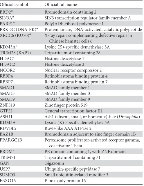
Pro-tein Index (IPI) database. The results are summarized in
Table 2
.
KAP1 (an
⬃
110-kDa band was observed) (
Fig. 1A
) was one of the
identified binding partners of LANA. The interaction between
LANA and KAP1 was first confirmed by coimmunoprecipitation
(co-IP). Flag-tagged KAP1 was transfected into HEK 293T cells,
along with Strep-Flag-tagged LANA. After affinity purification
with streptavidin beads, the purified proteins, along with input
samples, were detected by Western blotting with anti-Flag and
anti-KAP1 antibodies. KAP1 was coimmunoprecipitated with
LANA (
Fig. 1B
). The reverse co-IP experiment was also used to
test the interaction. HA-tagged LANA and Flag-tagged KAP1 were
utilized. After IP with Flag M2 beads, the immunoprecipitated
on November 7, 2019 by guest
http://jvi.asm.org/
proteins, along with input samples, were detected by Western
blotting with anti-Flag and anti-HA antibodies. LANA was
coim-munoprecipitated with KAP1 (
Fig. 1C
). The interaction between
endogenous LANA and KAP1 was verified in JSC-1 cells (
Fig. 1D
).
To determine whether this interaction was direct, we investigated
the interaction by
in vitro
binding assay.
In vitro
-translated
biotin-KAP1 was incubated with purified GST, GST-fused LANA-N (aa 1
to 340), and GST-fused LANA-C (aa 1022 to 1162) beads. KAP1
bound to the C-terminal domain of LANA instead of the
N-ter-minal domain (
Fig. 1E
). Taken together, these results demonstrate
that LANA forms a complex with KAP1
in vivo
and
in vitro
.
LANA is colocalized with KAP1 in the nucleus.
To address
whether LANA and KAP1 were in the same nuclear compartment
of naturally KSHV-infected cells, we performed an
immunofluo-rescence assay in BCBL-1 and JSC-1 cells. BCBL-1 and JSC-1 cells
were fixed and probed with mouse antibody against LANA and
rabbit antibody against KAP1, followed by incubation with
appro-priate secondary antibodies. LANA was distributed in a nuclear
punctate pattern, and KAP1 was abundantly diffused in the
nu-cleus (
Fig. 2
). LANA colocalized with KAP1 in the nuclei of
BCBL-1 (
Fig. 2
, top) and JSC-1 (bottom) cells.
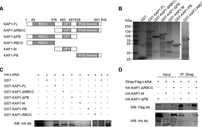
[image:4.585.41.289.77.397.2]LANA interacts with both N- and C-terminal domains of
KAP1.
KAP1 is a member of the TRIM protein family, with typical
structural features. It has a ring finger, B-box, zinc finger,
coiled-coil (RBCC) domain at its N terminus; a plant homeodomain and
bromodomain (PB) at its C terminus; and a domain for HP1
bind-ing in the middle (
31
). To explore the function of the interaction
between LANA and KAP1, we determined the interface of
LANA-KAP1 interaction by GST pulldown and co-IP. A series of
GST-fused, truncated KAP1 constructs were generated in the GST
pull-down assay (
Fig. 3A
and
B
). The purified, GST-fused, truncated
TABLE 2LANA-interacting proteins identified in TAP-MSOfficial symbol Official full name
BRD2a Bromodomain containing 2
SIN3Aa SIN3 transcription regulator family member A PARP1a Poly(ADP-ribose) polymerase 1
PRKDC (DNA-PK)a Protein kinase, DNA-activated, catalytic polypeptide XRCC6 (KU70)a X-ray repair complementing defective repair in
Chinese hamster cells 6 KDM3Aa Lysine (K)-specific demethylase 3A TRIM28 (KAP1) Tripartite motif containing 28 HDAC1 Histone deacetylase 1 HDAC2 Histone deacetylase 2 NCOR2 Nuclear receptor corepressor 2 RBBP4 Retinoblastoma binding protein 4 RBBP7 Retinoblastoma binding protein 7 SMAD1 SMAD family member 1 SMAD3 SMAD family member 3 SMAD9 SMAD family member 9 ZNF519 Zinc finger protein 519 GTF2I General transcription factor IIi
ASH1L Ash1 (absent, small, or homeotic)-like (Drosophila) KDM5A Lysine (K)-specific demethylase 5A
RUVBL2 RuvB-like AAA ATPase 2
BAZ1B Bromodomain adjacent to zinc finger domain 1B PPARGC1B Peroxisome proliferator-activated receptor gamma,
coactivator 1 beta
PRDM1 PR domain containing 1, with ZNF domain TRIM71 Tripartite motif containing 71
GAN Gigaxonin
USP7 Ubiquitin-specific peptidase 7 SUMO3 Small ubiquitin-related modifier 3 FBXO16 F-box-only protein 16
aPreviously reported LANA-interacting protein.
FIG 1LANA forms a complex with KAP1in vivoandin vitro. (A) TAP. A plasmid expressing Strep-Flag-tagged LANA was transfected into HEK 293T cells. The equivalent empty vector was transfected as a control (Con). Cell lysates were subjected to affinity purification with streptavidin beads, followed by IP with Flag M2 beads. The purified eluates were resolved by SDS-PAGE and visualized by silver staining. The bands corresponding to LANA and KAP1 are indicated. (B and C) Co-IP of LANA and KAP1 in HEK 293T cells. (B) Flag-tagged KAP1 was transfected into cells, along with Strep-Flag-tagged LANA or empty-vector controls. After affinity purification with streptavidin beads, the purified proteins, along with input samples, were detected by Western blotting (WB) with anti-Flag and anti-KAP1 antibodies (Ab). (C) HA-tagged LANA was transfected into cells, along with Flag-tagged KAP1or empty-vector controls. After IP with Flag M2 beads, the purified proteins, along with input samples, were detected by Western blotting with anti-HA and anti-Flag antibodies. (D) Co-IP of endogenous LANA and KAP1 in JSC-1 cells. JSC-1 cell lysates were subjected to IP with anti-LANA (␣-LANA) antibody or mouse IgG controls. Purified proteins, along with input samples, were detected by Western blotting with anti-LANA and anti-KAP1 antibodies. (E)In vitrointeraction between LANA and KAP1. Purified GST, GST-fused LANA-N (aa 1 to 340), and GST-fused LANA-C (aa 1022 to 1162) beads were incubated with equivalentin vitro-translated (IVT) biotin-KAP1, and pulled-down proteins were subjected to Transcend chemiluminescent translation detection. The asterisks indicate the specific bands of IVT biotin-KAP1.
on November 7, 2019 by guest
http://jvi.asm.org/
[image:4.585.103.484.434.608.2]KAP1 beads were incubated with cell lysates containing
HA-tagged LANA. The pulled-down proteins and input samples were
detected by Western blotting with anti-HA antibody. As shown in
Fig. 3C
, HA-tagged LANA was pulled down by FL,
KAP1-⌬
RBCC, KAP1-
⌬
PB, KAP1-PB, and KAP1-RBCC, but not GST
and KAP1-M; thus, we concluded that LANA interacted with both
the N- and C-terminal domains of KAP1. This conclusion was
supported by co-IP, and only HA-tagged KAP1-
⌬
RBCC and
KAP1-
⌬
PB were coimmunoprecipitated with Strep-Flag-tagged
LANA, but not KAP1-M (
Fig. 3D
).
We established that LANA interacted with the N- and
C-ter-minal domains of KAP1. KAP1 does not bind directly to DNA,
and the RBCC domain is mainly responsible for its recruitment
(
31
). Transcriptional repression by LANA of the KSHV and
cel-FIG 2LANA colocalizes with KAP1 in the nucleus. BCBL-1 (top row) and JSC-1 (bottom row) cells were fixed and probed with mouse antibody against LANA and rabbit antibody against KAP1, followed by incubation with goat anti-mouse IgG conjugated with Alexa Fluor 488 (green) and goat anti-rabbit IgG conjugated with Alexa Fluor 555 (red). Significant colocalized dot signals are indicated by arrowheads. Magnification: oil lens,⫻63; zoom,⫻2. Magnified views of the boxed areas are shown in the insets.FIG 3LANA interacts with both N- and C-terminal domains of KAP1. (A) Domains of KAP1 and truncated constructs used in this study. (B) Purified GST-fused KAP1 truncated constructs for GST pulldown assay. Purified GST-fused KAP1 truncated constructs were subjected to SDS-PAGE and Coomassie blue staining. (C) GST pulldown assay. Purified GST and GST-fused KAP1 truncated constructs were incubated with equivalent cell lysates containing HA-tagged LANA, and the pulled-down proteins were subjected to Western blotting with anti-LANA antibody. (D) Co-IP of LANA and KAP1 truncated constructs in HEK 293T cells. HA-tagged KAP1 truncated constructs were transfected into HEK 293T cells, along with Strep-Flag-tagged LANA. After affinity purification with streptavidin beads, the purified proteins, along with input samples, were detected by Western blotting with anti-Flag and anti-HA antibodies. The asterisks indicate the major bands.
on November 7, 2019 by guest
http://jvi.asm.org/
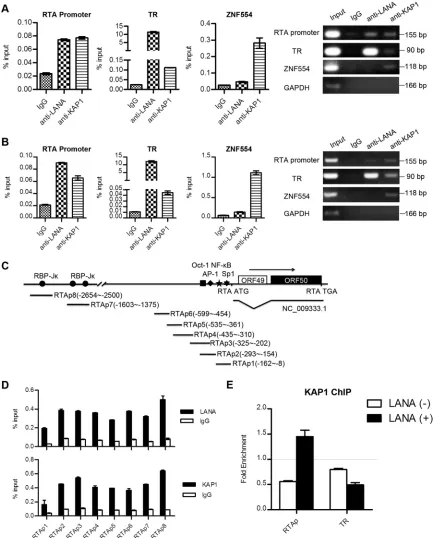
[image:5.585.113.468.433.657.2]FIG 4LANA recruits KAP1 to the RTA promoter region. (A and B) ChIP assays of LANA and KAP1 in BCBL-1 (A) and JSC-1 (B) cells. The immunoprecipitated DNA in the ChIP assays was subjected to qPCR (left) and DNA gel analysis (right). Binding of LANA and KAP1 at the RTA promoter and TR regions was determined using corresponding primers. The ZNF554 region was detected as a positive control for KAP1 binding. ChIP-qPCR data were normalized by the percent input method (signals obtained from ChIP were divided by signals obtained from an input sample). The data are presented as means⫾standard deviations (SD). (C) Illustration of the primer sets at the RTA promoter used in the ChIP assays. (D) Binding of LANA and KAP1 at the RTA promoter region as determined by the primers shown in panel C. ChIP-qPCR data were normalized by the percent input method. (E) ChIP assays of KAP1 in the presence (⫹) or absence (⫺) of LANA expression. Plasmid pGL2-RTAp containing the RTA promoter or pGL3p-TR containing the TR fragment was transfected into cells with LANA expression plasmid or empty-vector controls. Cells were collected for ChIP assay at 36 h posttransfection. ChIP-qPCR data were normalized by the fold enrichment method (ChIP signals were divided by the IgG signals). The data are presented as means and SD.
on November 7, 2019 by guest
http://jvi.asm.org/
[image:6.585.75.508.67.605.2]lular genomes has been reported (
13
,
17
,
24–30
). Combined with
the repressive role of KAP1 and a previous report that KAP1 is
associated with the KSHV genome (
36
), we hypothesized that
LANA might recruit KAP1 to the KSHV genome to repress gene
expression.
LANA recruits KAP1 to the RTA promoter region.
LANA
strongly binds to the TR region and has relatively weak binding at
other regions of the KSHV genome, such as the RTA promoter
(
46–48
). To determine whether LANA recruited KAP1 to the
KSHV genome, we examined the binding of LANA and KAP1 at
the TR and RTA promoter regions by ChIP assay. We performed
ChIP with anti-LANA and anti-KAP1 antibodies, as well as the
control IgG in KSHV-positive BCBL-1 and JSC-1 cell lines. The
purified DNA eluate was quantitated by qPCR and gel analysis,
and the results are shown in
Fig. 4A
(BCBL-1) and B (JSC-1).
LANA bound specifically to the TR region at a high level, while it
had relatively weak binding in the RTA promoter region. KAP1
bound to the TR region more weakly than LANA and bound
weakly to the RTA promoter. KAP1 strongly bound to the 3
=
end
of the ZNF554 exon, which was a positive control. LANA binds to
the RTA promoter through interaction with recombination signal
binding protein-J
(RBP-J
), specificity protein 1 (Sp1), and
his-tone H2A/B (
17
,
49
,
50
). To explore further the specific binding
sites of KAP1 at the RTA promoter, we designed eight pairs of
primers at the RTA promoter to determine the binding (
Fig. 4C
).
Both LANA and KAP1 showed comprehensive binding
through-out the RTA promoter (
Fig. 4D
). These results demonstrate that
LANA and KAP1 cooccupy the TR and RTA promoter regions of
the KSHV genome.
To demonstrate the role of LANA in KAP1 recruitment, that is,
whether the recruitment of KAP1 to the TR and RTA promoter
regions was dependent on LANA, we determined the binding of
KAP1 in the presence or absence of LANA expression. Plasmid
pGL2-RTAp, containing the RTA promoter, or pGL3p-TR,
con-taining a TR fragment, was transfected into cells with or without
the LANA expression plasmid. Cells were collected for ChIP assay
at 36 h posttransfection.
Figure 4E
shows that enrichment of
KAP1 at the RTA promoter region was detected only in cells with
LANA expression. However, we did not obtain the enrichment
signal for KAP1 in the TR region, even in the presence of LANA.
Therefore, our results indicate that LANA recruits KAP1
specifi-cally to the RTA promoter region.
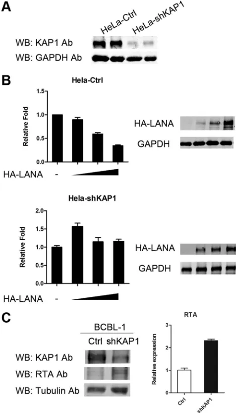
KAP1 is involved in transcriptional repression by LANA.
Al-though we proved that LANA recruits KAP1 to the RTA promoter
region, we still did not know whether the binding of KAP1 is
functional, so we determined whether KAP1 was involved in
tran-scriptional repression by LANA. We performed a luciferase
re-porter assay in HeLa-Ctrl and HeLa-shKAP1 stable cell lines.
Ex-pression of KAP1 was reduced significantly by shRNA in
HeLa-shKAP1 cells (
Fig. 5A
). The reporter plasmid pGL2-RTAp, which
contained the RTA promoter, was transfected into HeLa-Ctrl and
HeLa-shKAP1 cells, along with HA-LANA expression plasmid.
Figure 5B
, top, shows that LANA repressed transcriptional activity
of the RTA promoter in HeLa-Ctrl cells in a dose-dependent
man-ner. In contrast, transcriptional repression by LANA was
abro-gated in HeLa-shKAP1 cells (
Fig. 5B
, bottom). Therefore, we
con-cluded that KAP1 is involved in transcriptional repression by
LANA. To confirm the result, we also knocked down KAP1 in
BCBL-1 cells. The expression of RTA was elevated when KAP1 was
knocked down, as shown in
Fig. 5C
. However, very few virions
were detected in the supernatants of KAP1 knockdown BCBL-1
cells (data not shown). Knocked-down KAP1 alone did not
reac-tivate KSHV, suggesting other restriction factors are involved in
the maintenance of KSHV latency. Another explanation for this
result is that the effect of KAP1 knockdown by shRNA in BCBL-1
cells may not be strong enough for us to see an increase in virus
release in the supernatant.
LANA and KAP1 have multiple cooccupation sites on the
KSHV genome.
To determine whether LANA recruited KAP1 to
other regions of the KSHV genome, we mapped all the
genome-FIG 5KAP1 is involved in transcriptional repression by LANA. (A) Knocked-down KAP1 in shKAP1 cells. (B) Luciferase reporter gene assay in HeLa-Ctrl and HeLa-shKAP1 cells. pGL2-RTAp reporter plasmid (0.2g) was trans-fected into HeLa-Ctrl cells, along with 0, 0.5, 1, or 2g pCAGGS-HA-LANA and HeLa-shKAP1 cells and 0, 1, 2, or 4g pCAGGS-HA-LANA due to lower LANA expression in HeLa-shKAP1 cells. The total transfected DNA was nor-malized with the pCAGGS empty vector. Promoter activity is presented as the fold relative to the control (without LANA expression). The data are presented as means and SD. Multiple independent experiments were performed in trip-licate. (C) Knocked-down KAP1 in BCBL-1 cells. The expression of RTA was detected by Western blotting (left) and qPCR (right).on November 7, 2019 by guest
http://jvi.asm.org/
[image:7.585.300.538.68.484.2]on November 7, 2019 by guest
http://jvi.asm.org/
wide binding sites of LANA and KAP1 by seq. The
ChIP-enriched DNA with anti-LANA and anti-KAP1 antibodies in the
KSHV-positive cell line BC-3 was analyzed by deep sequencing.
Sequence reads for each sample were mapped to the KSHV
ge-nome (
HQ404500
plus 35 copies of the TR) and human genome
(hg19) using Bowtie2, and then the aligned file was subjected to
peak calling using MACS. The peak models of LANA and KAP1
built by MACS based on the human genome alignment indicated
that both LANA and KAP1 had broad peaks (
Fig. 6A
); thus, we
applied the
⫺
nomodel parameter to MACS analysis. The peaks of
LANA and KAP1 binding (
P
⬍
10
⫺3) on the whole KSHV genome
are shown in
Fig. 6B
. The first
⬃
28-kb region of the KSHV
ge-nome failed to show an enrichment signal for LANA and KAP1
due to possible deletion or large sequence variation in the KSHV
genome of BC-3 cells, which was mentioned previously (the
KSHV genome in BC-3 cells is smaller than in BC1 and BC2 cells
by Southern blotting) (
51
). The most significant peaks of LANA
binding are grouped in the TR region, and smaller peaks are
scat-tered throughout the genome, which is consistent with previous
results (
46
). The annotated information on LANA peaks (
P
⬍
10
⫺5) is shown in
Table 3
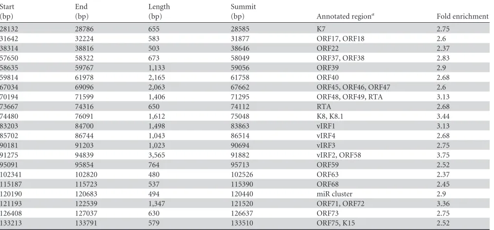
. The peaks of KAP1 binding identified
on the viral genome were small. The enrichment signal at the TR
region, as determined by real-time PCR, was not detected for the
alignment of 35 copies of the TR. The annotated information on
KAP1 peaks (
P
⬍
10
⫺5) is shown in
Table 4
. The peaks of KAP1
covered regions of lytic genes, as well as latent genes, and most of
them coincided with peaks of LANA (
Fig. 6C
), which implies that
LANA may be important for KAP1 recruitment to the KSHV
ge-nome, or vice versa. The incompletely overlapping peaks of LANA
and KAP1 indicated that KAP1 could also be recruited to the
KSHV genome by an alternative mechanism. To validate the
iden-tified peaks, we determined the binding by qPCR.
Figure 6D
shows that LANA and KAP1 cooccupied the newly identified
re-gions. The ChIP-seq demonstrated that LANA and KAP1 had
multiple cooccupation sites on the KSHV genome, indicating that
LANA-recruited KAP1 functions widely on the genome to control
gene expression. Additionally, we validated the possible deletion
or large sequence variation in the KSHV genome of BC-3 cells by
PCR (
Fig. 6E
).
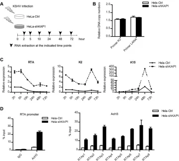
LANA-recruited KAP1 is crucial for establishment of KSHV
latency.
To determine whether LANA-recruited KAP1 was
in-volved in establishment of KSHV latency, we observed the
ef-fect of KAP1 knockdown on viral gene expression and the
change in early epigenetic modification during KSHV primary
infection. HeLa-Ctrl and HeLa-shKAP1 cells were
de novo
in-fected with rKSHV.219 at the same multiplicity of infection,
and lytic gene expression was determined (
Fig. 7A
). To ensure
the correctness of the gene expression analysis, we examined
the relative KSHV genome copy numbers in HeLa-Ctrl and
HeLa-shKAP1 cells at 24 h postinfection. The KSHV genome
FIG 6LANA and KAP1 had multiple cooccupation sites on the KSHV genome. (A) Peak models of LANA and KAP1 built by MACS based on the human genome alignment. (B) Illustration of LANA and KAP1 binding sites on the whole KSHV genome. The ChIP-seq data on LANA and KAP1 were aligned to the KSHV genome (HQ404500plus 35 copies of the TR [U75699.1]) and subjected to peak calling with MACS (P⬍10⫺3). The output files (peaks, summits, and wigs) were
visualized in IGV software. The identified peaks are shown in the peak diagrams. The summit diagrams show the sites with the highest scores in the peaks. The wig diagrams show the general binding information for the whole KSHV genome. The region marked by the asterisk covers⬃28 kb of the KSHV genome that failed to show the enrichment signal of LANA and KAP1, due to possible deletion or large sequence variation in the KSHV genome of BC-3 cells. (C) Diagram of regions occupied by LANA (red) and KAP1 (blue) (P⬍10⫺5). (D) Validation of ChIP-seq results by qPCR. ChIP-qPCR data were normalized by fold
[image:9.585.41.551.78.296.2]enrichment (ChIP signals were divided by the IgG signals). GAPDH was a negative control. The data are presented as means and SD. (E) Validation of possible deletion or large sequence variation in the KSHV genome of BC-3 cells by PCR.
TABLE 3LANA peaks on the KSHV genome (P⬍10⫺5)
Start (bp)
End (bp)
Length (bp)
Summit
(bp) Annotated regiona Fold enrichment
27817 29077 1,261 28560 K7b,c 2.84
67657 68464 808 68229 ORF45c, ORF46 2.29
68520 69146 627 68954 ORF46, ORF47 2.48
70231 71720 1,490 71184 ORF48, ORF49, RTAc 2.38
74228 75089 862 74648 K8 2.43
84336 85004 669 84749 vIRF1c 2.43
85733 86710 978 86107 vIRF4c 2.06
91484 92543 1,060 92254 vIRF2b,c 2.71
92574 94219 1,646 92812 vIRF2, ORF58 2.66
94224 94774 551 94648 ORF58 2.02
95128 95792 665 95301 ORF59 2.16
101668 102468 801 101863 ORF63 2.2
119130 119716 587 119420 miR clusterc 2.52
120199 120921 723 120477 miR clusterc 2.48
121227 122643 1,417 121480 ORF71, ORF72b 2.38 122655 123664 1,010 123396 ORF72, ORF73b 2.52
124866 127084 2,219 125469 ORF73b,c 2.94
132811 134190 1,380 133702 ORF75, K15b,c 2.29
136572 137001 430 136840 TRb,c 6.10
aPeaks (start to end) were annotated to genes in the KSHV genome (HQ404500) that could be promoters, coding sequences, or untranslated regions. b
Previously reported LANA peak on KSHV genome by ChIP-seq.
cPreviously reported LANA peak on KSHV genome by ChIP-on-chip.
on November 7, 2019 by guest
http://jvi.asm.org/
copy numbers in HeLa-Ctrl and HeLa-shKAP1 cells were
sim-ilar, as assayed by both K2 and LANA primer sets (
Fig. 7B
).
Expression of a limited number of lytic genes can be detected
during KSHV primary infection (
14
). We determined the
ex-pression of the RTA, K2, and K15 genes. The results of the lytic
gene expression at each time point are shown in
Fig. 7C
. As
expected, RTA expression was highest at the beginning (as early
as 2 h postinfection) but soon decreased. It was almost
com-pletely stopped at 24 h postinfection in HeLa-Ctrl cells,
al-though it remained high in HeLa-shKAP1 cells, even at 72 h
postinfection. RTA expression was significantly higher in
HeLa-shKAP1 cells at each time point. Similar to that of the
RTA gene, K2 gene expression was also higher in HeLa-shKAP1
cells and remained at a high level at 72 h postinfection. The
expression pattern of the K15 gene differed from that of the
RTA and K2 genes, which is consistent with a previous study
(
14
). It showed a slight increase over time until 48 h
postinfec-tion. K15 expression achieved a more robust peak at 48 h
postinfection in HeLa-shKAP1 cells and was not shut down at
72 h postinfection. The effect of KAP1 knockdown on viral
gene expression during KSHV
de novo
infection indicates that
KAP1 is involved in the shutdown of lytic genes. KAP1
re-presses gene expression via epigenetic change of histone
acet-ylation or methacet-ylation (
31–33
). The epigenetic modification of
the RTA promoter is mainly subject to the regulation of histone
acetylation (
17
,
52
), and therefore, we determined the AcH3
level in the RTA promoter region at the early stages of KSHV
primary infection (
Fig. 7D
). The AcH3 level was significantly
upregulated in HeLa-shKAP1 cells at 48 h postinfection. The
upregulated AcH3 level disrupted shutdown of RTA gene
ex-pression. The elevated AcH3 level in HeLa-shKAP1 cells also
validated the higher RTA expression. Therefore, we conclude
that LANA-recruited KAP1 is crucial for the rapid
establish-ment of KSHV latency.
DISCUSSION
The establishment of KSHV latency is a rapid and efficient process
during primary infection. Lytic gene expression is shut down,
while latent gene expression is sustained, which implies a process
of differential gene expression. The mechanism of this process
remains largely unclear, but previous studies on the LANA protein
provide some clues. The LANA protein is crucial for the
establish-ment of viral latency, and thus, insight from LANA sheds light
on the process. In this study, we showed that LANA interacted
with the KAP1 protein and repressed lytic gene expression to
fa-cilitate the establishment of KSHV latency.
[image:10.585.41.549.78.315.2]Multiple proteins have been shown to interact with LANA
(
26
,
30
,
38
,
45
,
48–50
,
53–69
). In our TAP-MS screening, a few
novel proteins that were related to transcriptional regulation
and posttranslational modification were identified, such as
KAP1, NCOR2, retinoblastoma-associated protein (RBBP),
SMAD, USP7, and SUMO3. Previously reported
LANA-inter-acting proteins, like BRD2 (
54
), SIN3A (
30
), PARP1 (
67
),
PRKDC (DNA-PK) (
68
), XRCC6 (KU70) (
68
), and KDM3A
(
48
), were also found in this study (
Table 2
). We validated the
interaction between LANA and KAP1
in vivo
and
in vitro
. We
found that KAP1 interacted with the C-terminal domain of
LANA directly. We mapped out that LANA interacted with
both the N- and C-terminal domains of KAP1. Recent studies
by different groups have resolved the structure of the
nal domain of LANA and have demonstrated that the
C-termi-nal domain works in an oligomeric state (
70–72
). The
N-ter-minal domain of KAP1 (RBCC) has been reported to function
as a homo-oligomer, which is recruited by the KRAB domain to
the genome (
73
). Thus, our results imply that KAP1 is recruited
by the oligomeric LANA to the genome. Coincidentally, when
this paper was in preparation, Cai et al. reported that LANA
does not directly interact with KAP1 but with sumoylated
TABLE 4KAP1 peaks on the KSHV genome (P⬍10⫺5)Start (bp)
End (bp)
Length (bp)
Summit
(bp) Annotated regiona Fold enrichment
28132 28786 655 28585 K7 2.75
31642 32224 583 31877 ORF17, ORF18 2.6
38314 38816 503 38646 ORF22 2.37
57650 58322 673 58049 ORF37, ORF38 2.83
58635 59767 1,133 59056 ORF39 2.9
59814 61978 2,165 61758 ORF40 2.68
67034 69096 2,063 67662 ORF45, ORF46, ORF47 2.6 70194 71599 1,406 71295 ORF48, ORF49, RTA 3.13
73667 74316 650 74112 RTA 2.68
74480 76091 1,612 75048 K8, K8.1 3.44
83203 84700 1,498 83863 vIRF1 3.13
85702 86744 1,043 86514 vIRF4 2.68
90181 91203 1,023 90694 vIRF3 2.75
91275 94839 3,565 91882 vIRF2, ORF58 3.75
95091 95854 764 95713 ORF59 2.52
102341 102820 480 102526 ORF63 2.37
115187 115723 537 115390 ORF68 2.45
120190 120683 494 120440 miR cluster 2.9
121193 122539 1,347 121520 ORF71, ORF72 3.36
126408 127037 630 126637 ORF73 2.75
133213 133791 579 133510 ORF75, K15 2.52
aPeaks (start to end) were annotated to genes in the KSHV genome (HQ404500) that could be promoters, coding sequences, or untranslated regions.
on November 7, 2019 by guest
http://jvi.asm.org/
KAP1 through the SUMO-interacting motif (SIM) in the
N-terminal domain of the LANA protein (
74
). However, our
re-sults demonstrated that LANA directly interacted with both the
N- and C-terminal domains of KAP1. We think that these
con-flicting results were caused by the different
in vitro
binding
assays. We also explored the possible function of the
interac-tion between LANA and the C-terminal domain of KAP1, but
the result was negative, as LANA did not affect the sumoylation
of KAP1 (data not shown).
In the ChIP assay, we demonstrated the cooccupation by
LANA and KAP1 at the RTA promoter and in the TR region of the
KSHV genome. However, the binding of LANA and KAP1 in the
RTA promoter region was weak, which is likely due to their
indi-rect binding to DNA. The binding of LANA in the TR region was
stronger than the binding of KAP1, which suggests a weak
inter-action between LANA and KAP1 in the TR region. This is possibly
true, because DNA binding may promote higher oligomerization
of LANA (
70
), which partially disrupts the interaction between
LANA and KAP1. The recruitment assay demonstrated that
LANA recruited KAP1 to the RTA promoter region, but not the
TR region (
Fig. 4E
). KAP1 did not bind to the TR region in
tran-siently transfected cells, which was different from the observation
in PEL cell lines. The transient transfection could be regarded as a
process mimicking KSHV primary infection, and thus, the result
indicated that KAP1 bound to the RTA promoter, but not the TR
region, at the early stage of KSHV primary infection. The binding
of KAP1 in the TR region might be a later event after the
estab-lishment of KSHV latency. We unexpectedly demonstrated that
both LANA and KAP1 showed comprehensive binding in the RTA
promoter region, which implies that the repressive role of LANA
may not be limited to one or two sites in the RTA promoter region.
This result also verified the recent finding that LANA and KAP1
can cooccupy the HIF-1
␣
binding sites at the RTA promoter (
74
).
We revealed that KAP1 was involved in transcriptional
repres-sion by LANA in a reporter gene assay, which is consistent with the
result in the recent publication by Cai et al. (
74
). The expression
level of transfected LANA was lower in HeLa-shKAP1 cells, which
suggests that KAP1 stabilizes LANA in the nucleus. KAP1 is an
abundant protein expressed in the nucleus, with a possible long
half-life (difficult to knock down by transient RNA interference).
FIG 7LANA-recruited KAP1 is crucial for establishment of KSHV latency. (A) Illustration ofde novoKSHV infection in HeLa-Ctrl and HeLa-shKAP1 cells. (B) Relative KSHV genome DNA copy numbers in cells. Cells were collected for DNA extraction at 24 h postinfection, and the relative quantity was determined by qPCR. Two primer sets were used. The data were normalized against GAPDH. The data are presented as means and SD. (C) Kinetics of lytic gene expression during primary infection. Cells were collected for RNA extraction at the indicated time points and reverse transcribed to cDNA. The relative quantity was determined by qPCR. The data were normalized against GAPDH. The data are presented as means⫾SD. Shown is a representative result ofde novoinfection experiments. The completede novoinfection experiments were done twice independently and showed the same tendency. For some individual time points, the results were repeated at least three times. (D) ChIP assay of AcH3 at the RTA promoter region. Cells were collected for AcH3 ChIP assay at 48 h postinfection. ChIP-qPCR data were normalized by the percent input method (signals obtained from ChIP were divided by signals obtained from an input sample). The data are presented as means and SD.on November 7, 2019 by guest
http://jvi.asm.org/
[image:11.585.112.473.65.390.2]It has also been shown that LANA has a long half-life, exceeding 24
h (
75
). KAP1 is a SUMO intramolecular E3 ligase, and LANA is
subject to sumoylation modification (
74
,
76–78
). Thus, it is
rea-sonable to speculate that KAP1 is responsible for the sumoylation
of LANA and that the degradation of LANA may be regulated by
its sumoylation.
We identified multiple cooccupation sites of LANA and KAP1
on the whole KSHV genome by ChIP-seq. In the previous study,
ChIP-seq and ChIP-on-chip methods were both applied to study
the binding sites of LANA on the KSHV genome (
46–48
). Besides
the strong binding sites of LANA in the TR region, only a few
binding sites (K7, vIRF2, open reading frame 73 [ORF73], and
K15) on the KSHV genome were reported across studies. The
ChIP-seq data on LANA in BC-3 cells were consistent with
previ-ous ChIP-seq data in BCBL-1 cells (
Table 3
), except that more
peaks were identified in this study, such as the well-studied RTA
promoter region (
17
,
50
). Most of the identified KAP1 peaks
co-incided with LANA peaks, implying that LANA may be important
for the recruitment of KAP1 to the KSHV genome. A previous
study only selectively detected the binding of KAP1 in the
pro-moter regions of the KSHV genome (
36
). However, we found that
the cooccupation sites of LANA and KAP1 were not limited to the
promoter regions. KAP1 can repress transcriptional activity, not
only in the promoter regions, but also in the coding sequence
regions, which produces an advantage for LANA in controlling
gene expression (
31
,
79
). We found cooccupation by LANA and
KAP1 in the latent gene regions. Latent gene expression should be
maintained at an appropriate level to evade immune surveillance,
and thus, the interaction between LANA and KAP1 may also
reg-ulate the expression of latent genes.
Our results demonstrated that LANA-recruited KAP1 is
cru-cial for KSHV primary infection. Lytic gene expression is
en-hanced and cannot be shut down in HeLa-shKAP1 cells in time.
The enhanced expression can be detected at an early time point,
suggesting that KAP1 is involved in an immediate-early step
dur-ing primary infection. However, knockdown of KAP1 does not
change the expression pattern of viral genes. Expression of the K15
gene in HeLa-shKAP1 cells remains a slowly increasing process at
the beginning, implying that another mechanism is responsible
for selective lytic gene expression. KAP1 represses gene expression
by changing the epigenetic state of the genome. It can recruit both
the histone methyltransferase and deacetylase complexes (
32
,
33
).
Here, we found upregulation of the AcH3 level in the RTA
pro-moter region in HeLa-shKAP1 cells, which suggests that KAP1
utilizes the deacetylase complex to repress gene expression.
In-deed, many of the identified host proteins interacting with LANA,
along with KAP1, are members of the deacetylase complex, such as
HDAC1, HDAC2, and RBBP. Previous studies have demonstrated
that transcriptional repression by LANA can be interrupted by
deacetylase inhibitors (
9
,
17
,
24
). Therefore, the interaction
be-tween LANA and KAP1 could be important for the latent KSHV
genome to maintain a hypoacetylated state.
In conclusion, our study revealed the mechanism of
transcrip-tional repression by LANA during KSHV primary infection,
pro-viding new insights into the process of KSHV latency
establish-ment.
ACKNOWLEDGMENTS
This work was supported by grants from the key project of the Natural Science Foundation of China (81230037) and the National Basic Research
Program of China (2011CB504805 and 2012CB519002) to K.L. and the Science Foundation of China for the Youth (81101478) to Y.G.
We thank Chuangui Wang (East China Normal University, Shanghai, China) and Subhash C. Verma (University of Nevada, Reno, NV) for their plasmids and cell lines. We acknowledge the Omics Core, CAS-MPG Part-ner Institute for Computational Biology, Shanghai Institutes for Biologi-cal Sciences, Chinese Academy of Sciences, for ChIP-seq service.
REFERENCES
1.Chang Y, Cesarman E, Pessin MS, Lee F, Culpepper J, Knowles DM, Moore PS. 1994. Identification of herpesvirus-like DNA sequences in AIDS-associated Kaposi’s sarcoma. Science266:1865–1869.http://dx.doi .org/10.1126/science.7997879.
2.Cesarman E, Chang Y, Moore PS, Said JW, Knowles DM.1995. Kaposi’s sarcoma-associated herpesvirus-like DNA sequences in AIDS-related body-cavity-based lymphomas. N. Engl. J. Med.332:1186 –1191.http://dx .doi.org/10.1056/NEJM199505043321802.
3.Soulier J, Grollet L, Oksenhendler E, Cacoub P, Cazals-Hatem D, Babinet P, d’Agay MF, Clauvel JP, Raphael M, Degos L, Sigaux F.1995. Kaposi’s sarcoma-associated herpesvirus-like DNA sequences in multi-centric Castleman’s disease. Blood86:1276 –1280.
4.Boshoff C, Chang Y.2001. Kaposi’s sarcoma-associated herpesvirus: a new DNA tumor virus. Annu. Rev. Med.52:453– 470.http://dx.doi.org/10 .1146/annurev.med.52.1.453.
5.Ye F, Lei X, Gao SJ.2011. Mechanisms of Kaposi’s sarcoma-associated herpesvirus latency and reactivation. Adv. Virol.2011:193860.http://dx .doi.org/10.1155/2011/193860.
6.Sarid R, Flore O, Bohenzky RA, Chang Y, Moore PS.1998. Transcrip-tion mapping of the Kaposi’s sarcoma-associated herpesvirus (human herpesvirus 8) genome in a body cavity-based lymphoma cell line (BC-1). J. Virol.72:1005–1012.
7.Dittmer DP.2003. Transcription profile of Kaposi’s sarcoma-associated herpesvirus in primary Kaposi’s sarcoma lesions as determined by real-time PCR arrays. Cancer Res. 63:2010 –2015. http://cancerres .aacrjournals.org/content/63/9/2010.long.
8.Shaw RN, Arbiser JL, Offermann MK. 2000. Valproic acid induces human herpesvirus 8 lytic gene expression in BCBL-1 cells. AIDS14:899 – 902.http://dx.doi.org/10.1097/00002030-200005050-00021.
9.Yu Y, Black JB, Goldsmith CS, Browning PJ, Bhalla K, Offermann MK.
1999. Induction of human herpesvirus-8 DNA replication and transcrip-tion by butyrate and TPA in BCBL-1 cells. J. Gen. Virol.80:83–90. 10. Davis DA, Rinderknecht AS, Zoeteweij JP, Aoki Y, Read-Connole EL,
Tosato G, Blauvelt A, Yarchoan R.2001. Hypoxia induces lytic replica-tion of Kaposi sarcoma-associated herpesvirus. Blood 97:3244 –3250.
http://dx.doi.org/10.1182/blood.V97.10.3244.
11. Lukac DM, Renne R, Kirshner JR, Ganem D.1998. Reactivation of Kaposi’s sarcoma-associated herpesvirus infection from latency by ex-pression of the ORF 50 transactivator, a homolog of the EBV R protein. Virology252:304 –312.http://dx.doi.org/10.1006/viro.1998.9486. 12. Sun R, Lin SF, Gradoville L, Yuan Y, Zhu F, Miller G.1998. A viral gene
that activates lytic cycle expression of Kaposi’s sarcoma-associated herpes-virus. Proc. Natl. Acad. Sci. U. S. A.95:10866 –10871.http://dx.doi.org/10 .1073/pnas.95.18.10866.
13. Lan K, Kuppers DA, Verma SC, Robertson ES.2004. Kaposi’s sarcoma-associated herpesvirus-encoded latency-sarcoma-associated nuclear antigen inhib-its lytic replication by targeting Rta: a potential mechanism for virus-mediated control of latency. J. Virol.78:6585– 6594.http://dx.doi.org/10 .1128/JVI.78.12.6585-6594.2004.
14. Krishnan HH, Naranatt PP, Smith MS, Zeng L, Bloomer C, Chandran B.2004. Concurrent expression of latent and a limited number of lytic genes with immune modulation and antiapoptotic function by Kaposi’s sarcoma-associated herpes-virus early during infection of primary endothelial and fibroblast cells and subse-quent decline of lytic gene expression. J. Virol.78:3601–3620.http://dx.doi.org/10 .1128/JVI.78.7.3601-3620.2004.
15. Yoo SM, Zhou FC, Ye FC, Pan HY, Gao SJ.2005. Early and sustained expression of latent and host modulating genes in coordinated transcriptional program of KSHVproductiveprimaryinfectionofhumanprimaryendothelialcells.Virology
343:47–64.http://dx.doi.org/10.1016/j.virol.2005.08.018.
16. Gao SJ, Deng JH, Zhou FC. 2003. Productive lytic replication of a recombinant Kaposi’s sarcoma-associated herpesvirus in efficient pri-mary infection of pripri-mary human endothelial cells. J. Virol.77:9738 – 9749.http://dx.doi.org/10.1128/JVI.77.18.9738-9749.2003.
on November 7, 2019 by guest
http://jvi.asm.org/
17. Lu F, Day L, Gao SJ, Lieberman PM.2006. Acetylation of the latency-associated nuclear antigen regulates repression of Kaposi’s sarcoma-associated herpesvirus lytic transcription. J. Virol.80:5273–5282.http: //dx.doi.org/10.1128/JVI.02541-05.
18. Ye FC, Zhou FC, Yoo SM, Xie JP, Browning PJ, Gao SJ.2004. Disrup-tion of Kaposi’s sarcoma-associated herpesvirus latent nuclear antigen leads to abortive episome persistence. J. Virol.78:11121–11129.http://dx .doi.org/10.1128/JVI.78.20.11121-11129.2004.
19. Li Q, Zhou F, Ye F, Gao SJ.2008. Genetic disruption of KSHV major latent nuclear antigen LANA enhances viral lytic transcriptional program. Virology379:234 –244.http://dx.doi.org/10.1016/j.virol.2008.06.043. 20. Lan K, Kuppers DA, Verma SC, Sharma N, Murakami M, Robertson
ES.2005. Induction of Kaposi’s sarcoma-associated herpesvirus latency-associated nuclear antigen by the lytic transactivator RTA: a novel mech-anism for establishment of latency. J. Virol.79:7453–7465.http://dx.doi .org/10.1128/JVI.79.12.7453-7465.2005.
21. Ballestas ME, Chatis PA, Kaye KM.1999. Efficient persistence of extrach-romosomal KSHV DNA mediated by latency-associated nuclear antigen. Sci-ence284:641– 644.http://dx.doi.org/10.1126/science.284.5414.641. 22. Hu J, Garber AC, Renne R.2002. The latency-associated nuclear antigen
of Kaposi’s sarcoma-associated herpesvirus supports latent DNA replica-tion in dividing cells. J. Virol.76:11677–11687.http://dx.doi.org/10.1128 /JVI.76.22.11677-11687.2002.
23. Si H, Verma SC, Lampson MA, Cai Q, Robertson ES.2008. Kaposi’s sarcoma-associated herpesvirus-encoded LANA can interact with the nuclear mitotic ap-paratus protein to regulate genome maintenance and segregation. J. Virol.82:
6734–6746.http://dx.doi.org/10.1128/JVI.00342-08.
24. DeWire SM, Damania B.2005. The latency-associated nuclear antigen of rhesus monkey rhadinovirus inhibits viral replication through repression of Orf50/Rta transcriptional activation. J. Virol.79:3127–3138.http://dx .doi.org/10.1128/JVI.79.5.3127-3138.2005.
25. Sakakibara S, Ueda K, Nishimura K, Do E, Ohsaki E, Okuno T, Yamanishi K.2004. Accumulation of heterochromatin components on the terminal repeat sequence of Kaposi’s sarcoma-associated herpesvirus mediated by the latency-associated nuclear antigen. J. Virol.78:7299 – 7310.http://dx.doi.org/10.1128/JVI.78.14.7299-7310.2004.
26. Shamay M, Krithivas A, Zhang J, Hayward SD.2006. Recruitment of the de novo DNA methyltransferase Dnmt3a by Kaposi’s sarcoma-associated herpesvirus LANA. Proc. Natl. Acad. Sci. U. S. A.103:14554 –14559.http: //dx.doi.org/10.1073/pnas.0604469103.
27. Di Bartolo DL, Cannon M, Liu YF, Renne R, Chadburn A, Boshoff C, Cesarman E.2008. KSHV LANA inhibits TGF-beta signaling through epigenetic silencing of the TGF-beta type II receptor. Blood111:4731– 4740.http://dx.doi.org/10.1182/blood-2007-09-110544.
28. Schwam DR, Luciano RL, Mahajan SS, Wong L, Wilson AC. 2000. Carboxy terminus of human herpesvirus 8 latency-associated nuclear an-tigen mediates dimerization, transcriptional repression, and targeting to nuclear bodies. J. Virol.74:8532– 8540.http://dx.doi.org/10.1128/JVI.74 .18.8532-8540.2000.
29. Garber AC, Shu MA, Hu J, Renne R.2001. DNA binding and modula-tion of gene expression by the latency-associated nuclear antigen of Kapo-si’s sarcoma-associated herpesvirus. J. Virol.75:7882–7892.http://dx.doi .org/10.1128/JVI.75.17.7882-7892.2001.
30. Krithivas A, Young DB, Liao G, Greene D, Hayward SD.2000. Human herpesvirus 8 LANA interacts with proteins of the mSin3 corepressor complex and negatively regulates Epstein-Barr virus gene expression in dually infected PEL cells. J. Virol.74:9637–9645.http://dx.doi.org/10 .1128/JVI.74.20.9637-9645.2000.
31. Iyengar S, Farnham PJ.2011. KAP1 protein: an enigmatic master regu-lator of the genome. J. Biol. Chem.286:26267–26276.http://dx.doi.org/10 .1074/jbc.R111.252569.
32. Schultz DC, Friedman JR, Rauscher FJ, III.2001. Targeting histone deacetylase complexes via KRAB-zinc finger proteins: the PHD and bro-modomains of KAP-1 form a cooperative unit that recruits a novel iso-form of the Mi-2alpha subunit of NuRD. Genes Dev.15:428 – 443.http: //dx.doi.org/10.1101/gad.869501.
33. Schultz DC, Ayyanathan K, Negorev D, Maul GG, Rauscher FJ, III.
2002. SETDB1: a novel KAP-1-associated histone H3, lysine 9-specific methyltransferase that contributes to HP1-mediated silencing of euchro-matic genes by KRAB zinc-finger proteins. Genes Dev.16:919 –932.http: //dx.doi.org/10.1101/gad.973302.
34. Groner AC, Meylan S, Ciuffi A, Zangger N, Ambrosini G, Denervaud N, Bucher P, Trono D.2010. KRAB-zinc finger proteins and KAP1 can
mediate long-range transcriptional repression through heterochromatin spreading. PLoS Genet. 6:e1000869. http://dx.doi.org/10.1371/journal .pgen.1000869.
35. Barde I, Laurenti E, Verp S, Groner AC, Towne C, Padrun V, Aebischer P, Trumpp A, Trono D.2009. Regulation of episomal gene expression by KRAB/KAP1-mediated histone modifications. J. Virol. 83:5574 –5580.
http://dx.doi.org/10.1128/JVI.00001-09.
36. Chang PC, Fitzgerald LD, Van Geelen A, Izumiya Y, Ellison TJ, Wang DH, Ann DK, Luciw PA, Kung HJ.2009. Kruppel-associated box domain-associated protein-1 as a latency regulator for Kaposi’s sarcoma-associated herpesvirus and its modulation by the viral protein kinase. Cancer Res.69:5681–5689.http://dx .doi.org/10.1158/0008-5472.CAN-08-4570.
37. Jin Y, He Z, Liang D, Zhang Q, Zhang H, Deng Q, Robertson ES, Lan K.2012. Carboxyl-terminal amino acids 1052 to 1082 of the latency-associated nuclear antigen (LANA) interact with RBP-Jkappa and are re-sponsible for LANA-mediated RTA repression. J. Virol.86:4956 – 4969.
http://dx.doi.org/10.1128/JVI.06788-11.
38. Kaul R, Verma SC, Robertson ES.2007. Protein complexes associated with the Kaposi’s sarcoma-associated herpesvirus-encoded LANA. Virol-ogy364:317–329.http://dx.doi.org/10.1016/j.virol.2007.03.010. 39. Gloeckner CJ, Boldt K, Ueffing M.2009. Strep/FLAG tandem affinity
purifica-tion (SF-TAP) to study protein interacpurifica-tions. Curr. Protoc. Protein Sci. Chapter 19:Unit 19.20.http://dx.doi.org/10.1002/0471140864.ps1920s57.
40. Hu C, Zhang S, Gao X, Gao X, Xu X, Lv Y, Zhang Y, Zhu Z, Zhang C, Li Q, Wong J, Cui Y, Zhang W, Ma L, Wang C. 2012. Roles of Kruppel-associated box (KRAB)-associated co-repressor KAP1 Ser-473 phosphorylation in DNA damage response. J. Biol. Chem.287:18937– 18952.http://dx.doi.org/10.1074/jbc.M111.313262.
41. Myoung J, Ganem D.2011. Generation of a doxycycline-inducible KSHV producer cell line of endothelial origin: maintenance of tight latency with efficient reactivation upon induction. J. Virol. Methods174:12–21.http: //dx.doi.org/10.1016/j.jviromet.2011.03.012.
42. Langmead B, Salzberg SL.2012. Fast gapped-read alignment with Bowtie 2. Nat. Methods9:357–359.http://dx.doi.org/10.1038/nmeth.1923. 43. Zhang Y, Liu T, Meyer CA, Eeckhoute J, Johnson DS, Bernstein BE,
Nusbaum C, Myers RM, Brown M, Li W, Liu XS.2008. Model-based analysis of ChIP-Seq (MACS). Genome Biol.9:R137.http://dx.doi.org/10 .1186/gb-2008-9-9-r137.
44. Robinson JT, Thorvaldsdottir H, Winckler W, Guttman M, Lander ES, Getz G, Mesirov JP.2011. Integrative genomics viewer. Nat. Biotechnol.
29:24 –26.http://dx.doi.org/10.1038/nbt.1754.
45. Shamay M, Liu J, Li R, Liao G, Shen L, Greenway M, Hu S, Zhu J, Xie Z, Ambinder RF, Qian J, Zhu H, Hayward SD.2012. A protein array screen for Kaposi’s sarcoma-associated herpesvirus LANA interactors links LANA to TIP60, PP2A activity, and telomere shortening. J. Virol.
86:5179 –5191.http://dx.doi.org/10.1128/JVI.00169-12.
46. Lu F, Tsai K, Chen HS, Wikramasinghe P, Davuluri RV, Showe L, Domsic J, Marmorstein R, Lieberman PM.2012. Identification of host-chromosome binding sites and candidate gene targets for Kaposi’s sarco-ma-associated herpesvirus LANA. J. Virol.86:5752–5762.http://dx.doi .org/10.1128/JVI.07216-11.
47. Campbell M, Chang PC, Huerta S, Izumiya C, Davis R, Tepper CG, Kim KY, Shevchenko B, Wang DH, Jung JU, Luciw PA, Kung HJ, Izumiya Y.2012. Protein arginine methyltransferase 1-directed methylation of Kaposi sarcoma-associated herpesvirus latency-sarcoma-associated nuclear antigen. J. Biol. Chem.287:
5806–5818.http://dx.doi.org/10.1074/jbc.M111.289496.
48. Kim KY, Huerta SB, Izumiya C, Wang DH, Martinez A, Shevchenko B, Kung HJ, Campbell M, Izumiya Y.2013. Kaposi’s sarcoma-associated herpesvirus (KSHV) latency-associated nuclear antigen regulates the KSHV epigenome by association with the histone demethylase KDM3A. J. Virol.87:6782– 6793.http://dx.doi.org/10.1128/JVI.00011-13.
49. Barbera AJ, Chodaparambil JV, Kelley-Clarke B, Joukov V, Walter JC, Luger K, Kaye KM.2006. The nucleosomal surface as a docking station for Kaposi’s sarcoma herpesvirus LANA. Science311:856 – 861.http://dx .doi.org/10.1126/science.1120541.
50. Lan K, Kuppers DA, Robertson ES.2005. Kaposi’s sarcoma-associated herpes-virus reactivation is regulated by interaction of latency-associated nuclear antigen with recombination signal sequence-binding protein Jkappa, the major down-stream effector of the Notch signaling pathway. J. Virol.79:3468–3478.http://dx .doi.org/10.1128/JVI.79.6.3468-3478.2005.
51. Arvanitakis L, Mesri EA, Nador RG, Said JW, Asch AS, Knowles DM, Cesarman E.1996. Establishment and characterization of a primary effu-sion (body cavity-based) lymphoma cell line (BC-3) harboring Kaposi’s
on November 7, 2019 by guest
http://jvi.asm.org/
sarcoma-associated herpesvirus (KSHV/HHV-8) in the absence of Ep-stein-Barr virus. Blood88:2648 –2654.
52. Toth Z, Maglinte DT, Lee SH, Lee HR, Wong LY, Brulois KF, Lee S, Buckley JD, Laird PW, Marquez VE, Jung JU.2010. Epigenetic analysis of KSHV latent and lytic genomes. PLoS Pathog.6:e1001013.http://dx.doi .org/10.1371/journal.ppat.1001013.
53. Krithivas A, Fujimuro M, Weidner M, Young DB, Hayward SD.2002. Protein interactions targeting the latency-associated nuclear antigen of Kaposi’s sarcoma-associated herpesvirus to cell chromosomes. J. Virol.
76:11596 –11604.http://dx.doi.org/10.1128/JVI.76.22.11596-11604.2002. 54. Platt GM, Simpson GR, Mittnacht S, Schulz TF.1999. Latent nuclear antigen of Kaposi’s sarcoma-associated herpesvirus interacts with RING3, a homolog of the Drosophila female sterile homeotic (fsh) gene. J. Virol.
73:9789 –9795.
55. Watanabe A, Higuchi M, Fukushi M, Ohsawa T, Takahashi M, Oie M, Fujii M.2007. A novel KRAB-Zinc finger protein interacts with latency-associated nuclear antigen of Kaposi’s sarcoma-latency-associated herpesvirus and activates transcription via terminal repeat sequences. Virus Genes
34:127–136.http://dx.doi.org/10.1007/s11262-006-0048-x.
56. Chen W, Dittmer DP.2011. Ribosomal protein S6 interacts with the latency-associated nuclear antigen of Kaposi’s sarcoma-associated herpes-virus. J. Virol.85:9495–9505.http://dx.doi.org/10.1128/JVI.02620-10. 57. Paudel N, Sadagopan S, Balasubramanian S, Chandran B.2012.
Kapo-si’s sarcoma-associated herpesvirus latency-associated nuclear antigen and angiogenin interact with common host proteins, including annexin A2, which is essential for survival of latently infected cells