Copyright © 2003, American Society for Microbiology. All Rights Reserved.
Restriction of Human Immunodeficiency Virus Type 1 Rev Function
in Murine A9 Cells Involves the Rev C-Terminal Domain
Sandra M. P. Marques,
1,2Jean-Luc Veyrune,
2† Ram R. Shukla,
1,2‡ and Ajit Kumar
1,2*
Graduate Program in Genetics1and Department of Biochemistry and Molecular Biology,2
The George Washington University, Washington, D.C. 20037 Received 1 July 2002/Accepted 3 December 2002
The human immunodeficiency virus type 1 (HIV-1) Rev and human T-cell leukemia virus type 1 (HTLV-1) Rex proteins are essential for the expression of viral structural proteins and productive infection. Both contain a nuclear export signal (NES) in their C-terminal domain and a nuclear localization signal (NLS) in their N-terminal domain. The NES and NLS are necessary for shuttling between nucleus and cytoplasm and are therefore indispensable for the transport of unspliced and singly spliced viral transcripts. HIV-1 Rev function is restricted in A9 cells, a murine fibroblast cell line, whereas HTLV-1 Rex is functional in these cells. Immunofluorescence studies with RevGFP fusion protein demonstrate normal import and export of Rev in A9 cells. To ascertain which domains of Rev are necessary for the restriction of Rev function in A9 cells, we studied a chimeric construct in which the NES domain of Rev was exchanged with Rex C-terminal amino acids 79 to 95, the Rev1-79/Rex79-95 chimera, which restored Rev function in A9 cells. In addition, overexpression of a truncated Rev containing the Rev C-terminal domain in the presence of wild-type Rev, led to restoration of Rev function in A9 cells. These results suggest that the C-terminal domain of HIV-1 Rev plays an important role in restricting Rev function in murine cells.
The human immunodeficiency virus type 1 (HIV-1) Rev function is essential for the cytoplasmic transport of unspliced and singly spliced viral transcripts and thus is required for the expression of viral structural proteins and the establishment of productive infection (12). Rev function appears to be essential in RNA splicing, transport, transcript stabilization, and the translation steps (1, 8, 21). Rev protein binds specifically to the Rev response element (RRE) present in theenvRNA region of HIV-1 (23). Rev interacts directly with an RNA loop struc-ture within the RRE through an arginine-rich RNA binding motif (23). This region in the N-terminal end of the Rev pro-tein comprising amino acids 33 to 46 also constitutes a nuclear localization signal (NLS) (6, 20). In addition to the NLS, the Rev protein contains in its C-terminal region a nuclear export signal (NES) encompassing amino acids 73 to 84 (9, 35). The presence of both NLS and NES allows Rev to shuttle between the nucleus and the cytoplasm of HIV-1-infected cells (25). The ability to shuttle between cellular compartments is crucial for HIV-1 Rev function and mutations in the C-terminal NES domain that render the Rev protein unable to shuttle produce atrans-dominant phenotype (20). Thetrans-dominant mutant Rev protein (RevTD) can multimerize with the wild-type Rev protein and inhibit its nuclear export (34).
The prototypic Rev NES consists of a short stretch of hy-drophobic amino acids, primarily leucines (4, 17). Leucine-rich NESs have been identified in several other proteins, including the human T-cell leukemia virus type 1 (HTLV-1) Rex protein
(27). Rev and Rex have limited amino acid homology, yet HTLV-1 Rex protein performs functions analogous to those of HIV-1 Rev (13). The Rex protein also shuttles between the nucleus and the cytoplasm of infected cells, and through bind-ing to the Rex response element, it allows expression of the incompletely spliced mRNAs that encode the HTLV-1 Gag, Pol, and Env structural proteins (10). Several cellular proteins specifically interact with the Rev NES, RRE RNA, or the Rev-RRE complex and likely influence HIV-1 Rev function. For example, the Rev NES domain was shown to bind the nuclear export receptor CRM1/exportin 1 (26). Other proteins involved in RNA splicing or processing have been reported to bind to the RRE or the Rev-RRE complex (28).
An important goal in AIDS research has been the develop-ment of small animal models for HIV-1 infection that simulate the stages of HIV disease in humans (5). Although several murine models for studies of AIDS pathogenesis have been reported, these models lack long-term, productive viral repli-cation presumably due to blocks at several steps of viral gene expression (24). A goal of this study is to characterize post-transcriptional block in Rev function that prevents productive viral replication in A9 cells, a murine fibroblast cell line. We have shown that in A9 cells, HIV-1 Rev function is restricted, whereas HTLV-1 Rex protein is functional (33). In this study, we rescued Rev function in A9 cells using either a Rev-Rex chimeric protein or expression of the C-terminal domain of Rev in conjunction with the Rev protein. The results suggest that the C-terminal domain of Rev mediates the restriction of Rev function in A9 cells, presumably by its interaction with murine Rev inhibitory factors.
MATERIALS AND METHODS
Plasmids.pCMVGag2RRE (2) was provided by Frank Maldarelli, National Institute of Allergy and Infectious Diseases, National Institutes of Health (NIH), Bethesda, Md. pRSVRev1-79/Rex79-95 (17), pRSVRev, and the reporter
con-* Corresponding author. Mailing address: Department of Biochem-istry and Molecular Biology, The George Washington University, Washington, DC 20037. Phone: (202) 994-2919. Fax: (202) 994-8974. E-mail: [email protected].
† Present address: Unite´ de Therapie Cellulaire, Hoˆpital Saint Eloi, Montpellier, France.
‡ Present address: United States Patent and Trademark Office, Crystal City, Va.
3084
on November 8, 2019 by guest
http://jvi.asm.org/
struct pCMV128 (14) were provided by Thomas J. Hope (University of Illinois at Chicago). pRSVRev1-79 was derived from pRSVRev1-79/Rex79-95 digested
withBglII to remove the Rex sequences and religated. pRSVRex79-95 contains
amino acids 79 to 95 of Rex, and pRSVCRev61-116 contains amino acids 61 to 116 of Rev protein. The constructs used for indirect immunofluorescence, pcsRevGFP and pcsRevTDGFP, were provided by George N. Pavlakis, National Cancer Institute, Frederick, Md.
Cell culture and transfection.HeLa and A9 cells were maintained as described
previously (33). Cells were plated at approximately 2⫻105cells per
35-mm-diameter dish 24 to 48 h before transfection and transfected with 2 to 4g of
DNA, using Superfect (Qiagen) according to the manufacturer’s instructions, or
10g of DNA by the calcium phosphate method. Cells were harvested 48 h after
transfection and lysed as previously described (33). Protein concentration of the cell lysates was determined by a Bradford assay. For indirect immunofluores-cence studies, HeLa and A9 cells were cultured on coverslips and transfected
with 1g of pcsRevGFP or pcsRevTDGFP in the presence or absence of 1g
of pCMV128.
CAT assay.The amount of protein used for chloramphenicol acetyltransferase (CAT) assay was normalized to the transfection efficiency, which in turn was
determined by cotransfecting a-galactosidase expression plasmid, pCMV, or
a green fluorescent protein (GFP) expression plasmid, pEGFPN1 (Clontech). Fluorescence-activated cell sorter analysis using Cell Quest Program was used to
quantify GFP, and a luminometer was used to quantify-galactosidase enzyme
product (Tropix). CAT assays were performed as previously described (33). Re-sults are expressed as fold increase in CAT activity normalized to the control val-ue (pCMV128 reporter construct alone) from three to five independent assays.
Immunoprecipitation and immunoblot analysis.For immunoprecipitation, A9
cells transfected with 5 g of pCMV128 alone or cotransfected with both
pCMV128 (5g) and pRSVRev (10g) were washed with phosphate-buffered
saline (PBS) and lysed in buffer containing 100 mM KPO4and 0.2% Triton
X-100 (pH 7.8). Cell lysates (1 ml) were preincubated with 100l of protein
G-Sepharose beads (Bio-Rad) for 1 h and cleared at 12,000⫻g for 20 s.
Following the preclearing step, the supernatants (500l) were incubated with a
pool of two mouse anti-Rev monoclonal antibodies, Ab2 and Ab4 (11) (kindly provided by Jonathan Karn, MRC Center, Cambridge, England) for 1 h and
precipitated with 50l of protein G-Sepharose beads in lysis buffer. The
result-ing immunoprecipitates were centrifuged for 20 s at 12,000⫻gand washed three
times with lysis buffer followed by a final wash with 50 mM Tris (pH 8.0). All manipulations were performed at 4°C. The precipitated proteins were separated on a denaturing 4 to 20% gradient acrylamide gel (Bio-Rad) along with prestained protein markers (Invitrogen), transferred to polyvinylidene difluoride membranes (Bio-Rad), and probed for Rev protein (using the same pool of antibodies [at a 1:1,000 dilution] employed in the immunoprecipitation proce-dure). Purified HIV-1 Rev (NIH AIDS Research and Reference Reagent Pro-gram) was used as a positive control.
For Gag immunoblotting, cell lysates were resolved by sodium dodecyl sulfate-10% polyacrylamide gel electrophoresis, transferred to nitrocellulose mem-branes, and probed with a rabbit polyclonal HIV-1 p25/p24 Gag antiserum (NIH AIDS Research and Reference Reagent Program). The antigen-antibody com-plexes were detected by chemiluminescence (Amersham).
Export and import of Rev.Cells were washed with PBS 24 h after transfection
and treated with cycloheximide (25g/ml) (Sigma) and/or actinomycin D (4g/
ml) (Sigma) for 3 h at 37°C. The cells were then washed three times in 1⫻PBS
and fixed with cold methanol at 4°C for 5 min. Fixed cells were washed three
times with cold 1⫻PBS and treated with acetone for 4 min at⫺20°C. The
permeabilized cells were then washed with 1⫻PBS and blocked with 0.3%
bovine serum albumin in 1⫻PBS for 1 h at room temperature. HeLa cells were
incubated with a rabbit antinucleolin antibody (16) at a dilution of 1:500 (pro-vided by Raymond Petryshyn, Children’s Research Institute, Washington, D.C.), and A9 cells were incubated with a mouse antifibrillarin MAb72B9 antibody (30) at a dilution of 1:500 (provided by Joseph Gall, Carnegie Institution, Baltimore, Md.). After 1 h of incubation with primary antibody at 37°C, cells were washed and incubated with Texas red-conjugated anti-rabbit and anti-mouse secondary antibodies at a dilution of 1:1,000 (Amersham). The immunofluorescence of the stained cells was examined with a Bio-Rad MRC 1024 confocal laser scanning microscope using Bio-Rad Lasersharp MRC 1024 software.
RESULTS
Rev-dependent Gag protein expression is restricted in A9 cells.In an earlier report, using the HIV-1 Env-CAT reporter, pCMV128, we showed that A9 cells did not support Rev
func-tion, although HTLV-1 Rex was functional in these cells (33). To determine whether the Rev-dependent expression of both intron-containing and intron-lacking transcripts was restricted in A9 cells, HeLa and A9 cells were transfected with a gag
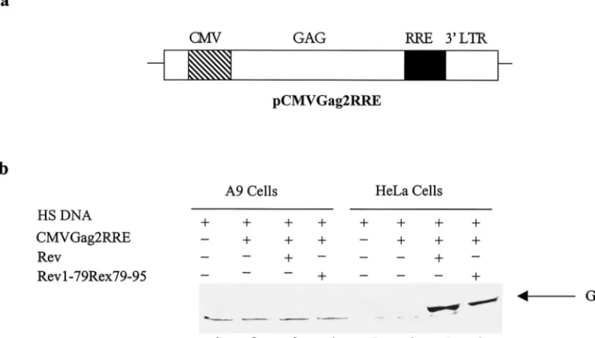
expression vector, pCMVGag2RRE, and evaluated for the cy-toplasmic expression of Gag protein in response to Rev or the Rev1-79/Rex79-95 chimera. The Gag expression vector con-sists of HIV-1 gag p17, p24, and a portion of p9 coding se-quences, linked to the RRE sequence (2). Expression of Gag protein in this reporter system is dependent upon binding of Rev to the RRE in the unspliced transcript. The Rev function was monitored by detecting the presence of 44-kDa unproc-essed Gag protein in extracts from A9 and HeLa cells. As expected, in HeLa cells in the absence ofgagexpression plas-mid or the reporter alone, there was no detectable Gag ex-pression. However, in the presence of Rev or the Rev1-79/ Rex79-95 chimera, a 44-kDa unprocessed Gag protein was detected in HeLa cell extracts. On the other hand, A9 cells showed no Gag protein expression with Rev or the Rev 1-79/ Rex79-95 chimera (Fig. 1b, compare lanes 3 and 4 to lanes 7 and 8). Thus, we conclude that the restriction of Rev func-tion in A9 cells is not limited to the HIV-1 env reporter (pCMV128). Importantly, substitution of C-terminal Rev with amino acids 79 to 95 of Rex did not rescue the inhibition of Gag expression in A9 cells.
Rev-Rex fusion protein rescues Rev function in A9 cells.We showed earlier that nuclear proteins from HeLa cells formed specific ribonucleoprotein complexes with RRE RNA, whereas nuclear proteins from A9 cells lacked this ability (32). We also reported that Rev-mediated expression of the reporter con-struct pCMV128 was restricted in A9 cells (33). The reporter pCMV128 is derived from the second intron of HIV-1 ex-pressed under the cytomegalovirus major immediate-early pro-moter with the CAT gene inserted within the intron upstream of the RRE (14). Since HIV-1 Rev and HTLV-1 Rex proteins serve analogous functions (13), we attempted to clarify which domain of the Rev protein is required to restrict Rev function in A9 cells, utilizing a Rev-Rex chimeric construct in which amino acids 1 to 79 of Rev were fused in frame with Rex amino acids 79 to 95, pRSVRev1-79/Rex79-95.
A9 and HeLa cells were transfected with either the pCMV128 reporter alone or in the presence of the pRSVRev or pRSVRev1-79/Rex79-95 expression vector. HeLa cells co-transfected with pCMV128 and pRSVRev yielded on average a 27-fold increase in CAT activity compared to cells trans-fected with the pCMV128 reporter plasmid alone (Fig. 2b, lanes 4 and 5, and c). Cotransfection of pCMV128 and the Rev-Rex chimera (pRSVRev1-79/Rex79-95) in HeLa cells re-sulted in, on average, a 17-fold increase in CAT activity (Fig. 2b, lane 6, and c). On the other hand, A9 cells cotransfected with pCMV128 and pRSVRev showed no significant increase in CAT activity compared to cells transfected with pCMV128 alone (Fig. 2b, lanes 1 and 2, and c), confirming our previous observation (33). However, when A9 cells were cotransfected with pCMV128 and pRSVRev1-79/Rex79-95, a 19-fold in-crease in CAT activity was observed (Fig. 2b, lane 3, and c).
The production of Rev protein in A9 and HeLa cells was confirmed by dot blot analysis of extracts prepared from cells transfected with 1, 2, or 5 g of Rev expression plasmids (results not shown) and by immunoprecipitation followed by
on November 8, 2019 by guest
http://jvi.asm.org/
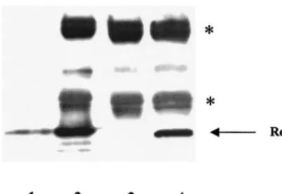
Western blot analysis (Fig. 3). The results shown in Fig. 3 make two important points that HIV-1 Rev is expressed and stable in A9 cells, thus excluding the possibility that restriction of Rev function in A9 cells is due to a block in the production or the instability of Rev protein in these cells. Furthermore, the block in Rev-mediated expression of pCMV128 (the envreporter) can be rescued in A9 cells by the Rev/Rex (Rev1-79/Rex79-95) chimera, which is not the case with the Gag reporter (pCMVGag2RRE), arguing that the nucleocytoplasmic trans-port of Env and Gag mRNA may be differentially regulated in murine cells.
Restriction of Rev function in A9 cells is not due to impaired localization or shuttling of Rev protein.HIV-1 Rev function requires proper utilization of Rev’s NLS and NES (25). To verify whether an impaired nucleocytoplasmic shuttling of HIV-1 Rev in A9 cells is responsible for the restriction of Rev function, fusion constructs of either wild-type Rev or
trans-dominant mutant Rev and GFP (pcsRevGFP and pcsRevTDGFP, respectively) were employed. In order to char-acterize the intracellular transport of Rev, cells were trans-fected with RevGFP or the RevTDGFP plasmids in the pres-ence or abspres-ence of the reporter, pCMV128, and with or without incubation with the translation and transcription in-hibitors cycloheximide (25 g/ml) and actinomycin D (4g/ ml). In A9 and HeLa cells, both RevTDGFP and RevGFP localized primarily to the nucleolus. Upon drug treatment, RevTDGFP remained in the nucleolus in both cell types. On the other hand, in both A9 and HeLa cells, the wild-type RevGFP was able to exit the cell nucleus (data not shown). Even though RevGFP does not support Rev function in A9
cells, its intracellular localization is similar to that in HeLa cells, suggesting that the mechanism of restriction of Rev func-tion in A9 cells is not related to impaired localizafunc-tion or shut-tling of Rev protein.
Overexpression of the Rev C-terminal domain rescues Rev function in A9 cells.The above data suggest that the restriction of HIV-1 Rev function in A9 cells involves a host cell factor(s) which requires the C-terminal domain of Rev. Such factor(s) may bind the C-terminal domain of Rev and either fail to mediate Rev function or actively inhibit Rev function. In the latter case, overexpression of Rev C-terminal domain se-quences might squelch the host factor(s) responsible for inhib-iting Rev function in A9 cells. To test the hypothesis, HeLa and A9 cells were transfected with Rev expression plasmid (pRSVCRev), comprising the C-terminal amino acids 61 to 116 of the Rev protein along with pCMV128 reporter plus the Rev expression vector, pRSVRev. The ratio of C-terminal Rev (CRev)/Rev expression vectors used ranged from 0.5:1 to 4:1 (Fig. 4).
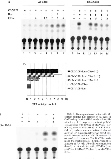
[image:3.603.86.509.78.318.2]HeLa cells cotransfected with the reporter (pCMV128) and the C-terminal Rev expression plasmid (pRSVCRev) showed no increase in CAT activity over background levels (Fig. 4a, lanes 9 and 11), whereas cotransfection of the reporter and the Rev expression plasmid (pRSVRev) yielded on average 15-fold increase in CAT activity over background levels (Fig. 4a, lane 10). When both Rev and increasing concentrations of CRev were transfected in HeLa cells along with the reporter plasmid, a decrease in CAT activity was observed (Fig. 4a, lanes 12 to 16), presumably due to the titration of host cell Rev
FIG. 1. HIV-1 Rev-dependent Gag expression in HeLa and A9 cells. (a) Diagram depicting pCMVGag2RRE. CMV, cytomegalovirus major immediate-early promoter; GAG, HIV-1 gag p17, p24, and part of p9 open reading frame (protease gene not included); RRE, HIV-1 RRE; 3⬘ LTR, the HIV-1 long terminal repeat polyadenylation signal. (b) Gag expression in HeLa and A9 cells. A9 and HeLa cells were transfected with herring sperm DNA (HS DNA) (lanes 1 and 5), pCMVGag2RRE (2g) alone (lanes 2 and 6) or with pRSVRev (1g) (lanes 3 and 7) or pRSVRev1-79Rex79-95 (1g) (lanes 4 and 8). The position of the Gag protein is indicated by the arrow.
on November 8, 2019 by guest
http://jvi.asm.org/
cofactors that interact with the carboxyl end of the Rev pro-tein.
On the other hand, at CRev/Rev ratios of 1.5:1 and greater, a significant increase in CAT activity was observed in A9 cells compared to cells with the reporter alone (Fig. 4a, lanes 4 to 8, and b). The results suggest a squelching of Rev inhibitory factor(s) in A9 cells with affinity for the Rev carboxyl-terminal sequences. In other experiments, A9 cells cotransfected with the reporter and increasing amounts of either Rev or CRev expression plasmid (ranging from 1 to 6 g) showed no in-crease in CAT activity above background levels, suggesting that the restriction of Rev function in A9 cells is not dependent on the amount of Rev and is not rescued by increasing amounts of CRev without the presence of full-length Rev protein (data not shown).
DISCUSSION
A greater understanding of immune dysfunction during HIV-1 infection is compromised by the absence of a suitable animal model system (24). Mice provide an attractive model for studying HIV-1 pathogenesis given the unparalleled under-standing of their immune function, the ease with which they can be genetically manipulated, and the expanding repertoire of mice with genetically defined immunological defects (15). However, previous efforts to develop a transgenic murine model for HIV-1 infection have been hindered by specific blocks to virus replication in murine cells (24).
Several possible reasons for the restriction of HIV-1 Rev function in murine cells have been proposed. One would ex-pect that a combination of one or more of these mechanisms
FIG. 2. Rev-Rex chimera rescues CAT reporter activity in A9 cells. (a) Diagram depicting pCMV128. CMV, cytomegalovirus major immediate-early promoter; SD, HIV-1 splice donor site; SA, HIV-1 splice acceptor site; 3⬘LTR, HIV-1 3⬘long terminal repeat. (b) HIV-1 Rev-dependent CAT activity in A9 and HeLa cells. Both cell lines were transfected with 1g of reporter construct pCMV128 alone (lanes 1 and 4) or cotransfected with 500 ng of pRSVRev (lanes 2 and 5) or 500 ng of pRSVRev1-79/Rex79-95 (lanes 3 and 6). CAT enzyme activity was determined as described in Materials and Methods. (c) Quantification of CAT assay results. Graphed results are expressed as fold increase in the pCMV128 reporter CAT activity in cells cotransfected with pRSVRev or pRSVRev1-79/Rex79-95 compared to cells transfected with pCMV128 alone. Values are averages ⫾standard errors (error bars) from five independent transfections.
on November 8, 2019 by guest
http://jvi.asm.org/
may contribute to the loss of Rev function in different murine cells. For example, our previous results suggested a lack of cellular factors in A9 cells that interacted with RRE RNA (32). Mariani and colleagues (24) reported inefficient processing of Gag protein in murine cells. Bieniasz and Cullen (3) reported reduced levels of the unspliced and genomic RNA transcripts in several rodent cells, but the results varied considerably among different rat and mouse cell lines. Yet another sug-gested mechanism for the lack of Rev function in murine cells (22) envisions a more efficient splicing of viral transcripts in murine cells than in human cells. While these results indicate wide variations in Rev function in different murine and rodent cells, they underscore the fact that the Rev function is re-stricted in murine cells whose mechanisms may vary depending upon the cell type used in the study. In this study, the results suggest that the murine cellular environment may restrict HIV-1 Rev function due to the presence of an inhibitor of Rev function or due to the modification of a positive cofactor of Rev.
Rev function block in A9 cells. Murine A9 cells do not support Rev function in transient-transfection assays or form RRE-host protein ribonucleoprotein complexes in vitro (32, 33). However, rescue of Rev function in A9 cells can be achieved using human and mouse somatic cell hybrids contain-ing human chromosomes 6 and 11 (33). It is noted that human chromosome 12 did not rescue Rev function in these somatic cell hybrids (33). These results suggested that multiple host factors are required for optimal Rev function. In A9 cells, Rev cofactors may be posttranslationally modified, allowing them to act as Rev inhibitors or preventing their function as Rev cofactors, unlike their role in human cells. Alternatively, re-striction of HIV-1 Rev function in A9 cells may be due to the presence of specific inhibitor(s) that interact with the Rev C-terminal domain.
Block in Rev function in A9 cells is not due to impaired localization or shuttling of the Rev protein.Yet another pos-sibility is that the nucleocytoplasmic shuttling of Rev is
im-paired in these cells, as has been described for astrocytes (19). Astrocytes show impaired Rev function that is attributable to a predominantly cytoplasmic localization of the Rev protein (19). Other examples of inhibition of Rev function through changes in its localization include thetrans-dominant mutant Rev protein (RevTD) (34) and a Sam68 C-terminal deletion mutant (29). RevTD has been shown to inhibit Rev function by multimerization with the wild-type Rev protein and trapping it in the cell nucleus, whereas the C-terminal deletion mutant of Sam68 has been shown to inhibit Rev function by trapping it in the cell cytoplasm (23, 29). In this study, proper nucleolar localization of both Rev and RevTD were confirmed in both A9 and HeLa cells using antibodies against nucleolus-specific proteins and Rev. Export of Rev-GFP was observed in both HeLa and A9 cells (data not shown), suggesting that the re-striction of Rev function in A9 cells is not due to a defect in the nucleocytoplasmic shuttling of Rev protein.
Rev function in A9 cells can be rescued for singly spliced transcripts but not for unspliced transcripts. In contrast to HIV-1, HTLV-1 can replicate efficiently in murine cells (7; R. Feng, A. Kabayama, K. Uchida, H. Hoshino, and M. Miwa, Abstr. 2000 Meet. Retroviruses Cold Spring Harbor Lab., abstr. 171, 2000). Our results show that in A9 cells HTLV-1 Rex protein is functional, whereas HIV-1 Rev function is re-stricted (33). The region of the Rex protein which is necessary to complement Rev function has been mapped to a core acti-vation domain represented by residues 79 to 95 of Rex protein (13, 17). We showed that Rev/Rex fusion protein was able to restore Rev function in A9 cells. Interestingly, the Rev/Rex chimera was insufficient to rescue Gag expression in A9 cells. The results suggest two important points: first, the restriction of Rev function with respect to singly spliced envtranscripts and nonsplicedgagtranscript in murine cells is specific to Rev, and second, the transport of singly splicedenvand unspliced
gag transcripts may be independently regulated. Possible ex-planations for the difference may be the presence of host factors that interact with the cis-inhibitory sequences in gag
coding sequence which are not present in theenv/CATreporter construct (31). A recent report demonstrated that murine NIH 3T3 fibroblasts stably expressing human CD4, human CCR5, and human cyclin T1 produced unspliced HIV-1 mRNA tran-scripts albeit at reduced levels but presented a dramatic block to virus assembly (24). The researchers suggested that even though the murine cells produced large amounts of Gag and Gag-Pol, the Gag protein was inefficiently processed.
Determining the nature of the block in Rev function in A9 cells.The rescue of Env-CAT reporter expression in A9 cells using a chimeric construct containing the first 79 amino acids of Rev fused to the C-terminal 79 to 95 amino acids of Rex suggested that the restriction of Rev function in A9 cells was associated with the carboxyl-terminal domain of the Rev pro-tein. This is further supported by the fact that Rev inhibition can be titrated by coexpression of Rev and the C-terminal domain of HIV-1 Rev (amino acids 61 to 116 of Rev).
[image:5.603.64.265.69.207.2]The data are consistent with a model which suggests the existence of an inhibitor of Rev function in A9 cells which can be squelched by overexpression of the C terminus of Rev. The results however do not exclude the possibility that a murine homologue of a Rev cofactor that recognizes the C-terminal domain of Rev may be unable to function, as is the case with
FIG. 3. HIV-1 Rev expression in A9 cells. A9 cells were cotrans-fected with pCMV128 and pRSVRev expression constructs and lysed 48 h posttransfection, as described in Materials and Methods. Lane 1, purified wild-type Rev protein (2g); lane 2, purified wild-type Rev protein (2 g) immunoprecipitated as described in Materials and Methods; lane 3, A9 cells transfected with 5g of pCMV128; lane 4, A9 cells cotransfected with 5g of pCMV128 and 10g of pRSVRev. The positions of Rev protein (arrow) and heavy and light immuno-globulin G chains (asterisks) are indicated.
on November 8, 2019 by guest
http://jvi.asm.org/
murine cyclin T1 in Tat transactivation (18). The impairment in Rev function in A9 cells is not coupled to impaired shuttling of the Rev protein in this cell line. In addition, our results indicate that the translation of Rev protein and its stability is not impaired in A9 cells (Fig. 3). Determining the nature of the host factor(s) responsible for restriction of Rev function in A9 cells will help determine the mechanism of restriction of HIV-1
replication in murine cells and therefore aid in the develop-ment of an animal model for studying HIV-1 pathogenesis.
ACKNOWLEDGMENTS
[image:6.603.88.472.74.624.2]We thank Wilhelm E. Woolery, Mamatha Garige, and Mazer R. Ally for assistance and Silvio Urcuqui-Inchima, Anamaris Colberg-Poley, Frank Maldarelli, Kathleen Boris-Lawrie, Nick Somia, Sally Moody, Sergei Nekhai, and Susan Ceryak for critical comments on the manuscript. We are grateful to Robyn Rufner from the Center for Microscopy and Image Analysis (C.M.I.A.) of George Washington University for expert assistance with confocal microscopy.
FIG. 4. Overexpression of amino acids 61 to 116 of Rev C-terminal domain restores Rev function in A9 cells. (a) HIV-1 Rev-dependent CAT activity in A9 and HeLa cells. A9 and HeLa cells were transfected with 1 g of the reporter construct pCMV128 alone (CMV128) or cotransfected with 500 ng of pRSVRev (Rev) or 1g of pRSVCRev (CRev) alone or with 500 ng of pRSVRev and increasing amounts of C-Rev (numbers represent ratios of plasmid amounts). (b) Quantifi-cation of CAT assay results for A9 cells. Graphed results are expressed as fold increase in the pCMV128 reporter CAT activity in response to cotransfected constructs. The Rev/CRev ratios are shown in parenthe-ses in the symbol key. (c) Comparison of the level of rescue of Rev function in A9 cells. A9 cells were transfected with pCMV128 alone (lane 1) or cotransfected with pRSVRev (lane 2), pRSVCRev (lane 3), pRSVRev1-79Rex79-95 (lane 4) or pRSVCRev and pRSVRev at a 2:1 ratio.
on November 8, 2019 by guest
http://jvi.asm.org/
This work was supported in part by NIH grants CA72147 (A.K.) and AI42491 (R.R.S.), and a fellowship from the PROGRAMA PRAXIS XXI of the Portuguese Fundac¸a˜o para a Cieˆncia e Tecnologia (to S.M.P.M.).
REFERENCES
1. Arrigo, S. J., and I. S. Chen.1991. Rev is necessary for translation but not cytoplasmic accumulation of HIV-1 vif, vpr, and env/vpu2 RNAs. Genes
Dev.5:808–819.
2. Berthold, E., and F. Maldarelli.1996.cis-acting elements in human immu-nodeficiency virus type 1 RNA direct viral transcripts to distinct intranuclear
locations. J. Virol.70:4667–4682.
3. Bieniasz, P. D., and B. R. Cullen.2000. Multiple blocks to human
immuno-deficiency virus type 1 replication in rodent cells. J. Virol.74:9868–9877.
4. Bogerd, H. P., R. A. Fridell, R. E. Benson, J. Hua, and B. R. Cullen.1996. Protein sequence requirements for function of the human T-cell leukemia virus type 1 Rex nuclear export signal delineated by a novel in vivo
random-ization-selection assay. Mol. Cell. Biol.16:4207–4214.
5. Boyle, M. J., M. Connors, M. E. Flanigan, S. P. Geiger, H. Ford, Jr., M. Baseler, J. Adelsberger, R. T. Davey, Jr., and H. C. Lane.1995. The human HIV/peripheral blood lymphocyte (PBL)-SCID mouse. A modified human PBL-SCID model for the study of HIV pathogenesis and therapy. J.
Immu-nol.154:6612–6623.
6. Cochrane, A. W., A. Perkins, and C. A. Rosen.1990. Identification of se-quences important in the nucleolar localization of human immunodeficiency
virus Rev: relevance of nucleolar localization to function. J. Virol.64:881–
885.
7. Fang, J., S. Kushida, R. Feng, M. Tanaka, T. Kawamura, H. Abe, N. Maeda, M. Onobori, M. Hori, K. Uchida, and M. Miwa.1998. Transmission of
human T-cell leukemia virus type 1 to mice. J. Virol.72:3952–3957.
8. Felber, B. K., M. Hadzopoulou-Cladaras, C. Cladaras, T. Copeland, and G. N. Pavlakis.1989. Rev protein of human immunodeficiency virus type 1 affects the stability and transport of the viral mRNA. Proc. Natl. Acad. Sci.
USA86:1495–1499.
9. Fischer, U., J. Huber, W. C. Boelens, I. W. Mattaj, and R. Luhrmann.1995. The HIV-1 Rev activation domain is a nuclear export signal that accesses an
export pathway used by specific cellular RNAs. Cell82:475–483.
10. Green, P. L., and I. S. Chen.1990. Regulation of human T cell leukemia virus
expression. FASEB J.4:169–175.
11. Henderson, B. R., and P. Percipalle.1997. Interactions between HIV Rev and nuclear import and export factors: the Rev nuclear localisation signal
mediates specific binding to human importin-beta. J. Mol. Biol.274:693–707.
12. Hope, T. J.1999. The ins and outs of HIV Rev. Arch. Biochem. Biophys.
365:186–191.
13. Hope, T. J., B. L. Bond, D. McDonald, N. P. Klein, and T. G. Parslow.1991. Effector domains of human immunodeficiency virus type 1 Rev and human T-cell leukemia virus type I Rex are functionally interchangeable and share
an essential peptide motif. J. Virol.65:6001–6007.
14. Hope, T. J., X. J. Huang, D. McDonald, and T. G. Parslow.1990. Steroid-receptor fusion of the human immunodeficiency virus type 1 Rev transacti-vator: mapping cryptic functions of the arginine-rich motif. Proc. Natl. Acad.
Sci. USA87:7787–7791.
15. Jamieson, B. D., and J. A. Zack.1999. Murine models for HIV disease. AIDS
13(Suppl. A):5–11.
16. Kibbey, M. C., B. Johnson, R. Petryshyn, M. Jucker, and H. K. Kleinman.
1995. A 110-kD nuclear shuttling protein, nucleolin, binds to the
neurite-promoting IKVAV site of laminin-1. J. Neurosci. Res.42:314–322.
17. Kim, F. J., A. A. Beeche, J. J. Hunter, D. J. Chin, and T. J. Hope.1996. Characterization of the nuclear export signal of human T-cell lymphotropic
virus type 1 Rex reveals that nuclear export is mediated by position-variable
hydrophobic interactions. Mol. Cell. Biol.16:5147–5155.
18. Kwak, Y. T., D. Ivanov, J. Guo, E. Nee, and R. B. Gaynor.1999. Role of the human and murine cyclin T proteins in regulating HIV-1 tat-activation. J.
Mol. Biol.288:57–69.
19. Ludwig, E., F. Ceccherini-Silberstein, J. vanEmpel, V. Erfle, M. Neumann, and R. Brack-Werner.1999. Diminished Rev-mediated stimulation of hu-man immunodeficiency virus type 1 protein synthesis is a hallmark of huhu-man
astrocytes. J. Virol.73:8279–8289.
20. Malim, M. H., S. Bohnlein, J. Hauber, and B. R. Cullen.1989. Functional dissection of the HIV-1 Rev trans-activator–derivation of a trans-dominant
repressor of Rev function. Cell58:205–214.
21. Malim, M. H., and B. R. Cullen.1993. Rev and the fate of pre-mRNA in the nucleus: implications for the regulation of RNA processing in eukaryotes.
Mol. Cell. Biol.13:6180–6189.
22. Malim, M. H., and B. R. Cullen.1991. HIV-1 structural gene expression requires the binding of multiple Rev monomers to the viral RRE:
implica-tions for HIV-1 latency. Cell65:241–248.
23. Malim, M. H., J. Hauber, S. Y. Le, J. V. Maizel, and B. R. Cullen.1989. The HIV-1 rev trans-activator acts through a structured target sequence to
acti-vate nuclear export of unspliced viral mRNA. Nature338:254–257.
24. Mariani, R., G. Rutter, M. E. Harris, T. J. Hope, H. G. Krausslich, and N. R. Landau.2000. A block to human immunodeficiency virus type 1 assembly in
murine cells. J. Virol.74:3859–3870.
25. Meyer, B. E., and M. H. Malim.1994. The HIV-1 Rev trans-activator
shut-tles between the nucleus and the cytoplasm. Genes Dev.8:1538–1547.
26. Neville, M., F. Stutz, L. Lee, L. I. Davis, and M. Rosbash.1997. The impor-tin-beta family member Crm1p bridges the interaction between Rev and the
nuclear pore complex during nuclear export. Curr. Biol.7:767–775.
27. Palmeri, D., and M. H. Malim.1996. The human T-cell leukemia virus type
1 posttranscriptionaltrans-activator Rex contains a nuclear export signal.
J. Virol.70:6442–6445.
28. Powell, D. M., M. C. Amaral, J. Y. Wu, T. Maniatis, and W. C. Greene.1997. HIV-1 Rev-dependent binding of SF2/ASF to the Rev response element: possible role in Rev-mediated inhibition of HIV-1 splicing. Proc. Natl. Acad.
Sci. USA94:973–978.
29. Reddy, T. R., W. Xu, J. K. Mau, C. D. Goodwin, M. Suhasini, H. Tang, K. Frimpong, D. W. Rose, and F. Wong-Staal.1999. Inhibition of HIV replica-tion by dominant negative mutants of Sam68, a funcreplica-tional homolog of HIV-1
Rev. Nat. Med.5:635–642.
30. Reimer, G., K. M. Pollard, C. A. Penning, R. L. Ochs, M. A. Lischwe, H. Busch, and E. M. Tan.1987. Monoclonal autoantibody from a (New Zealand
black⫻New Zealand white)F1 mouse and some human scleroderma sera
target an Mr 34,000 nucleolar protein of the U3 RNP particle. Arthritis
Rheum.30:793–800.
31. Schneider, R., M. Campbell, G. Nasioulas, B. K. Felber, and G. N. Pavlakis.
1997. Inactivation of the human immunodeficiency virus type 1 inhibitory elements allows Rev-independent expression of Gag and Gag/protease and
particle formation. J. Virol.71:4892–4903.
32. Shukla, R. R., P. L. Kimmel, and A. Kumar.1994. Human immunodeficiency virus type 1 Rev-responsive element RNA binds to host cell-specific proteins.
J. Virol.68:2224–2229.
33. Shukla, R. R., S. M. Marques, P. L. Kimmel, and A. Kumar.1996. Human chromosome 6- and 11-encoded factors support human immunodeficiency
virus type 1 Rev function in A9 cells. J. Virol.70:9064–9068.
34. Stauber, R., G. A. Gaitanaris, and G. N. Pavlakis.1995. Analysis of traffick-ing of Rev and transdominant Rev proteins in livtraffick-ing cells ustraffick-ing green fluo-rescent protein fusions: transdominant Rev blocks the export of Rev from
the nucleus to the cytoplasm. Virology213:439–449.
35. Wen, W., J. L. Meinkoth, R. Y. Tsien, and S. S. Taylor.1995. Identification
of a signal for rapid export of proteins from the nucleus. Cell82:463–473.