Jonathan L. Heeney,
Tomek Swigut,
Frank Kirchhoff,
* and Jacek Skowronski
*
Abteilung Virologie, Universita¨tsklinikum, 89081 Ulm, Germany1; Program in Genetics, State University of New York at Stony
Brook, Stony Brook, New York 117942; Cold Spring Harbor Laboratory, Cold Spring Harbor, New York 117243;
German Primate Center, 37077 Go¨ttingen, Germany4; and Department of Virology,
Biomedical Primate Research Center, 2288 GJ Rijswijk, The Netherlands5
Received 8 April 2002/Accepted 26 August 2002
We investigated the function of severely truncated simian immunodeficiency virus (SIV) Nef proteins (tNef) in vitro and in vivo. These variants emerged in rhesus monkeys infected with SIVmac239 containing a 152-bp deletion in thenef-unique region and have been suggested to enhance SIV virulence (E. T. Sawai, M. S. Hamza, M. Ye, K. E. Shaw, and P. A. Luciw, J. Virol. 74:2038-2045, 2000). We found that the tNef proteins were unable to down-regulate the cell surface expression of major histocompatibility complex class I proteins, CD4, and CD28 and neither stimulated SIV replication nor enhanced virion infectivity. The tNef proteins did efficiently down-regulate T-cell receptor (TCR):CD3 cell surface expression. Nevertheless, the SIVmac239tnefvariants were strongly attenuated in six infected juvenile rhesus macaques. Thus, while the ability of SIV Nef to down-modulate TCR:CD3 cell surface expression apparently confers a selective advantage in vivo, it is insufficient for efficient viral replication in infected macaques. Additional mutations elsewhere in SIVmac239 tnefgenomes are required for a virulent phenotype.
The 792-bpnefgene of the pathogenic simian immunodefi-ciency virus mac239 clone (SIVmac239) encodes a myristylated protein of approximately 34 kDa that down-regulates the cell surface expression of CD3, CD4, CD28, and major histocom-patibility class I (MHC-I) molecules, enhances viral replica-tion, increases the infectivity of viral particles, and associates with the p21-regulated protein serine kinase PAK2 (10–13, 16, 17, 23–25). These multiple functions of 239wt Nef are geneti-cally separable and require distinct elements located through-out the Nef molecule.
Nef is important for efficient viral replication and the per-sistence of HIV and SIV in vivo (7, 13–15). Adult or juvenile rhesus macaques inoculated with a variant of the pathogenic SIVmac239 clone containing a deletion of 182 bp in thenef
gene (nef⌬182) (Fig. 1) usually exhibit low viral loads, and the
majority of infected animals do not progress to immunodefi-ciency (13). These results provided the basis for the design and evaluation of live attenuated SIV vaccines with deletions in accessory genes (6).
Recently, it was found that uncloned SIVmac239 variants expressing truncated Nef (tNef) proteins of about 25 kDa are pathogenic in infected rhesus macaques (20). These variants emerged in animals infected with recombinant SIVmac239 vi-ruses containing a missense mutation of the Nef initiator
me-thionine, combined with either a 152-bp deletion in the nef -unique region or an insertion of the interleukin-2 (IL-2) cDNA in place of the 152-bp deletion, both of which introduced frameshifts into thenefopen reading frame (ORF) (Fig. 1). In the reverted tnef alleles, the nef ATG initiation codon was restored and frameshift mutations were repaired to restore contiguous, albeit truncated, nef ORFs (tnef24 and tnef46) (Fig. 1) (20). The tNef proteins lack an approximately 50-amino-acid-long region overlapping the highly conserved core of the Nef protein. This deletion spans elements mediating functional interactions of Nef with clathrin adaptor complexes that are required for the down-regulation of CD4 and CD28, as well as elements required for Nef to associate with PAK2 activity (4, 16, 18). Therefore, it was surprising that the tNef proteins were linked to a pathogenic phenotype.
The emergence of variant viruses containing a restoredtnef
ORF indicated that the severely truncated tNef proteins (tNef24 and tNef46) contained some residual function. To identify this function, we constructed truncated 239nefalleles,
tnef.1andtnef.2, corresponding to those previously recovered
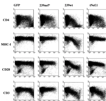
from the progressing animals 29810-24 and 27021-46N, respec-tively (20). The mutanttnef.1andtnef.2alleles were generated by splice overlap extension PCR and cloned into both a bicistronic vector coexpressing GFP (pCGCG.tNef.1 and pCGCG.tNef.2) and a proviral SIVmac239 construct essen-tially as described previously (12, 19). Flow cytometry analysis of Jurkat T cells transiently transfected with the bicistronic vectors (9, 11, 12) revealed that, in contrast to 239wt Nef, the tNef.1 and tNef.2 proteins did not decrease the cell surface expression of CD4, CD28, and MHC-I molecules (Fig. 2) (data not shown). However, both tNef forms down-regulated the cell surface expression of the T-cell receptor (TCR):CD3 * Corresponding author. Mailing address for Jacek Skowronski:
Cold Spring Harbor Laboratory, 1 Bungtown Rd., Cold Spring Harbor, NY 11724. Phone: (516) 367-8407. Fax: (516) 367-8454. E-mail: [email protected]. Mailing address for Frank Kirchhoff: Abteilung Virologie-Universita¨tsklinikum, Albert-Einstein-Allee 11, 89081 Ulm, Germany. Phone: 49 (731) 50023344. Fax: 49 (731) 50023389. E-mail: [email protected].
12360
on November 8, 2019 by guest
http://jvi.asm.org/
complex as efficiently as 239wt Nef (Fig. 2) (data not shown). Down-modulation of TCR:CD3 is a conserved function of SIVmac239 and HIV-2 Nef proteins (2, 10, 21).
To test the ability of the tNefs to stimulate SIV replication and particle infectivity, virus stocks were generated in 239T cells transiently transfected with the respective wild-type and mutant proviral genomes (8). As expected (16), no significant
differences in the replication kinetics of SIVmac239 viruses containing the 239wt nef, tnef.1, and tnef.2 alleles were ob-served in CEMx174 cells (Fig. 3A). Western blot analysis re-vealed that CEMx174 cells infected with the SIVmac239 vari-ants expressed tNef proteins of the expected size of about 25 kDa (data not shown). Infection of rhesus macaque peripheral blood mononuclear cells (rhPBMC) (Fig. 3B) and the herpes-FIG. 1. Amino acid sequence alignment of SIVmac239 wild-type and variant Nef proteins. Amino acid sequences surrounding the deleted regions are shown for Nef⌬152and Nef⌬182; the truncated Nef proteins that emerged in vivo in macaque 29810-24 (tNef24) or in macaque 27071-46N (tNef46); the tNef proteins modeled on tNef24 and tNef46 used in this work (tNef.1 and tNef.2, respectively); and Nef⌬153and Nef⌬183,
resulting from repair of frameshift mutations innef⌬152andnef⌬182. Dots represent identical amino acids, dashes represent missing amino acids,
[image:2.603.119.468.345.676.2]and asterisks represent premature in-frame termination codons introduced by frameshift mutations.
FIG. 2. The SIVmac239 tNef.1 protein retains the ability to down-regulate TCR:CD3 cell surface expression. Jurkat T cells were transiently transfected with a control plasmid expressing green fluorescent protein (GFP) alone (left panels) or with plasmids coexpressing GFP and 239nef*, 239wt Nef (middle panels), or tNef.1 (right panels). 239nef* contains a premature in-frame TAA stop signal at the 93rd codon ofnef(13). Expression of CD4, MHC-I, CD28, CD3, and GFP was analyzed by two-color flow cytometry as described previously (9, 11, 12). Similar results were obtained in two independent experiments.
on November 8, 2019 by guest
http://jvi.asm.org/
virus saimiri-transformed macaque T-cell line 221 (Fig. 3C) (1) demonstrated that, in contrast to the 239wtnefallele, thetnef.1
and tnef.2alleles were unable to stimulate SIVmac239
repli-cation. Additionally, the tNef proteins did not increase virion infectivity (Fig. 3D).
Of the six in vitro Nef activities investigated, the only one retained by the tNef proteins was the ability to down-regulate TCR:CD3. Therefore, the previous suggestion that the se-verely truncated tNef proteins were capable of significantly increasing SIV virulence in rhesus macaques was surprising (20). However, in this study, virus was recovered from the progressing animal, 26939-105N, near necropsy, leaving the possibility that mutations elsewhere in the viral genome might have contributed to the virulent phenotype.
To address this possibility, six juvenile rhesus macaques were inoculated intravenously with SIVmac239 tnef.1 virus stock
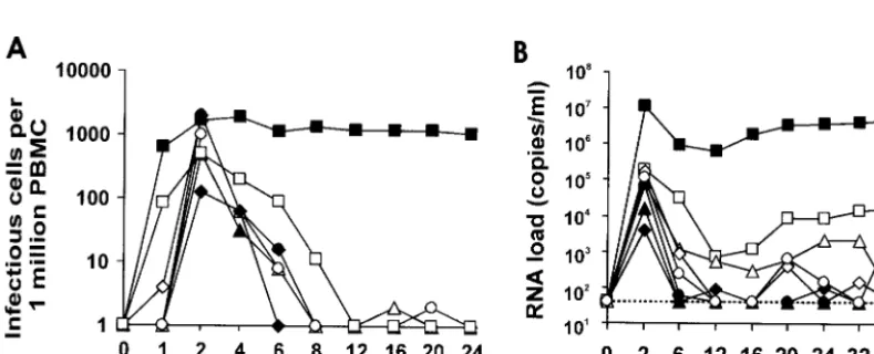
containing 5 ng of p27 produced from transiently transfected 293T cells as described previously (8). The animals were healthy and seronegative for SIV, D-type retroviruses, and STLV-1 at the time of infection. Sera and PBMCs were col-lected from the infected rhesus monkeys at regular intervals. Serological, virological, and immunological analyses of the clinical samples were performed as described before (22, 23, 26). As shown in Fig. 4, following initial spikes in the acute phase, the viral RNA copy numbers and cell associated viral loads were low in all six animals infected with SIVmac239
tnef.1compared to those infected with wild-type SIVmac239.
In agreement with the observed attenuated in vivo replication of SIVmac239tnef.1, the total CD4⫹T cells and CD4⫹CD29⫹
[image:3.603.60.530.67.220.2]memory T cells were essentially unchanged in the infected animals (Fig. 5), all of whom remained healthy throughout the 40-week observation period. Thus, the SIVmac239tnef.1allele FIG. 3. Infectivity and replication of the SIVmac239tnef.1 andtnef.2variants. Stocks of SIVmac239 containing wild type (wt), prematurely terminated (239nef*), ortnef.1andtnef.2variants were generated in 293T cells. Replication of the wild-type and variant viruses in CEMx174 cells (A), rhPBMCs (B), and 221 cells in the absence of IL-2 (C) is shown. Cells were infected with aliquots of virus stocks containing 5 ng of p27. PBMCs were infected immediately after isolation and stimulated 3 days later as described previously (16, 19). The amount of p27 antigen was determined by an SIV/HIV-2 enzyme-linked immunosorbent assay (ELISA) obtained through the AIDS Research and Reference Reagent Program, Division of AIDS, National Institute of Allergy and Infectious Diseases, National Institutes of Health. Reverse transcriptase (RT) activity was determined with a phosphorimager by photon-stimulated light emission (P.S.L.). The infectivity of wild-type and variant SIV to P4-CCR5 cells (5) infected with virus stocks containing 50 ng of p27 was determined as described previously (19), and is shown in panel D. All results are representative of three to five experiments performed with several independently produced virus stocks.
FIG. 4. Replication of the SIVmac239tnef.1variant is attenuated in vivo. The cell-associated viral load (A) and the viral RNA load (B) in Mm10287 (Œ), Mm10295 (‚), Mm10302 (}), Mm10663 (〫), Mm10672 (F), and Mm10677 (E) infected with the SIVmac239tnef.1variant are
shown. For comparison, average values obtained from four rhesus macaques infected with SIVmac239 NU (䊐) and eight who received wild-type SIVmac239 (■) are also shown. The detection limit for viral RNA of approximately 40 copies per ml (26) is indicated by a dotted line.
on November 8, 2019 by guest
http://jvi.asm.org/
[image:3.603.95.490.527.687.2]was severely attenuated, and the infections were well con-trolled in all animals.
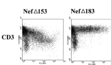
Intact but truncatedtnefORFs were independently restored in several animals infected with SIVmac239 variants containing the 152-bp deletion (20), yet this was never observed in ma-caques infected with an SIVmac239 variant containing a 182-bp deletion (13, 14). Both deletions start at nucleotide position 172 of thenefORF (13, 20). However, in contrast to the 152-bp deletion, the 182-bp deletion removes nucleotides encoding amino acids 111 to 120 of the Nef protein (see Fig. 1). This suggested that amino acids 111 to 120 might be required to down-regulate TCR:CD3 cell surface expression. To ad-dress this possibility, the frameshift mutations introduced by the 152- and 182-bp deletions were repaired by removing an additional single nucleotide in the respectivenef ORFs. The resulting nef variants (nef⌬153 and nef⌬183, see Fig. 1) were
expressed transiently in Jurkat T cells. As shown in Fig. 6, Nef⌬153down-modulated the cell surface expression of TCR:
CD3. In contrast, Nef⌬183 was not able to down-modulate
TCR:CD3 (Fig. 6), and was also defective in all other in vitro assays of Nef functions tested (data not shown). Thus, a selec-tive pressure for TCR:CD3 down-regulation may explain why the truncated Nef proteins emerged only in animals infected with SIVmac239 containing the 152-bp deletion in the nef -unique region. This event was not possible in animals infected with SIVmac239 containing the 182-bp deletion because it removed amino acids required for TCR:CD3 downregulation. In summary, there are two novel implications of this work. First, it is evident that TCR:CD3 down-regulation by SIVmac239 Nef is associated with a selective advantage for the virus in vivo. However, this function alone is insufficient for high viral loads and rapid disease induction by SIVmac239 in infected rhesus macaques. This is consistent with recent evi-dence suggesting that a combination of several independent Nef functions allows SIVmac239 and HIV-1 to replicate effi-ciently in the infected host (3, 5, 12, 19). Second, our data imply that changes elsewhere in the viral genome are required for a pathogenic phenotype of SIVmac239 expressing the tNef proteins. It will be important to determine the nature of such alterations in the SIVmac genome that can compensate for the loss of other Nef functions.
We thank Mandy Krumbiegel and Nathaly Finze for excellent tech-nical assistance and Ingrid Bennett for critical reading of the manu-script. We also thank Thomas Mertens and Bernhard Fleckenstein for constant support and encouragement.
This work was supported by grants from the Wilhelm-Sander Foun-dation and the Deutsche Forschungsgemeinschaft (to F.K.), as well as the Public Health Service (AI-42561 to J.S.).
REFERENCES
1. Alexander, L., Z. Du, M. Rosenzweig, J. U. Jung, and R. C. Desrosiers.1997. A role for natural simian immunodeficiency virus and human immunodefi-ciency virus type 1 Nef alleles in lymphocyte activation. J. Virol.71:6094– 6099.
2. Bell, I., C. Ashman, J. Maughan, E. Hooker, F. Cook, and T. A. Reinhart. 1998. Association of simian immunodeficiency virus Nef with the T-cell receptor (TCR) zeta chain leads to TCR down-modulation. J. Gen. Virol. 79:2717–2727.
3. Carl, S., A. J. Iafrate, S. M. Lang, C. Stahl-Hennig, E. M. Kuhn, D. Fuchs, K. Ma¨tz-Rensing, P. ten Haaft, J. L. Heeney, J. Skowronski, and F. Kirch-FIG. 5. CD4⫹T-cell counts in macaques infected with SIVmac239tnef.1variant. The total number of CD4⫹T cells (A) and CD4⫹CD29⫹
[image:4.603.114.480.69.259.2]memory T cells (B) in the peripheral blood of six animals inoculated with SIVmac239tnef.1over time are shown. Symbols and animal codes are as indicated in the legend to Fig. 4.
FIG. 6. Differential abilities of Nef⌬153and Nef⌬183to down-regu-late cell surface TCR:CD3 expression. The effects of Nef⌬153and Nef⌬183on TCR:CD3 cell surface levels upon transient expression in Jurkat T cells, determined by two-color flow cytometry as described in the legend to Fig. 1, are shown.
on November 8, 2019 by guest
http://jvi.asm.org/
[image:4.603.44.279.540.677.2]7. Deacon, N. J., A. Tsykin, A. Solomon, K. Smith, M. Ludford-Menting, D. J. Hooker, D. A. McPhee, A. L. Greenway, A. Ellett, and C. Chatfield.1995. Genomic structure of an attenuated quasi species of HIV-1 from a blood transfusion donor and recipients. Science270:988–991.
8. Deng, H., R. Liu, W. Ellmeier, S. Choe, D. Unutmaz, M. Burkhart, P. Di Marzio, S. Marmon, R. E. Sutton, C. M. Hill, C. B. Davis, S. C. Peiper, T. J. Schall, D. R. Littman, and N. R. Landau.1996. Identification of a major co-receptor for primary isolates of HIV-1. Nature381:661–666.
9. Greenberg, M. E., A. J. Iafrate, and J. Skowronski.1998. The SH3 domain-binding surface and an acidic motif in HIV-1 Nef regulate trafficking of class I MHC complexes. EMBO J.17:2777–2789.
10. Howe, A. Y. M., J. U. Jung, and R. C. Desrosiers.1998. Zeta chain of the T-cell receptor interacts with nef of simian immunodeficiency virus and human immunodeficiency virus type 2. J. Virol.72:9827–9834.
11. Iafrate, A. J., S. Bronson, and J. Skowronski.1997. Separable functions of Nef disrupt two aspects of T cell receptor machinery: CD4 expression and CD3 signaling. EMBO J.16:673–684.
12. Iafrate, A. J., S. Carl, S. Bronson, C. Stahl-Hennig, T. Swigut, J. Skowronski, and F. Kirchhoff.2000. Disrupting surfaces of Nef required for down-regu-lation of CD4 and for enhancement of virion infectivity attenuates simian immunodeficiency virus replication in vivo. J. Virol.74:9836–9844. 13. Kestler, H. W., III, D. J. Ringler, K. Mori, D. L. Panicali, P. K. Sehgal, M. D.
Daniel, and R. C. Desrosiers.1991. Importance of thenefgene for mainte-nance of high virus loads and for development of AIDS. Cell65:651–662. 14. Kirchhoff, F., H. W. Kestler III, and R. C. Desrosiers.1994. Upstream U3
sequences in simian immunodeficiency virus are selectively deleted in vivo in the absence of an intactnefgene. J. Virol.68:2031–2037.
15. Kirchhoff, F., T. C. Greenough, D. B. Brettler, J. L. Sullivan, and R. C. Desrosiers.1995. Absence of intact nef sequences in a long-term survivor with nonprogressive HIV-1 infection. N. Engl. J. Med.332:228–232.
19. Mu¨nch, J., N. Stolte, D. Fuchs, C. Stahl-Hennig, and F. Kirchhoff.2001. Efficient class I major histocompatibility complex down-regulation by simian immunodeficiency virus Nef is associated with a strong selective advantage in infected rhesus macaques. J. Virol.75:10532–10536.
20. Sawai, E. T., M. S. Hamza, M. Ye, K. E. Shaw, and P. A. Luciw.2000. Pathogenic conversion of live attenuated simian immunodeficiency virus vaccines is associated with expression of truncated Nef. J. Virol.74:2038– 2045.
21. Schaefer, T. M., I. Bell, B. A. Fallert, and T. A. Reinhart.2000. The T-cell receptorchain contains two homologous domains with which simian im-munodeficiency virus Nef interacts and mediates down-modulation. J Vi-rol.74:3273–3283.
22. Stahl-Hennig, C., O. Herchenro¨der, S. Nick, M. Evers, M. Stille-Siegener, K.-D. Jentsch, F. Kirchhoff, T. Tolle, T. J. Gatesmann, W. Lu¨ke, and G. Hunsmann.1990. Experimental infection of macaques with HIV-2BEN a novel HIV-2 isolate. AIDS4:611–617.
23. Stahl-Hennig, C., G. Voss, S. Nick, H. Petry, D. Fuchs, H. Wachter, C. Coulibaly, W. Lu¨ke, and G. Hunsmann.1992. Immunization with Tween-ether-treated SIV adsorbed onto aluminium hydoxide protects monkeys against experimental SIV infection. Virology186:588–596.
24. Swigut, T., A. J. Iafrate, J. Mu¨nch, F. Kirchhoff, and J. Skowronski.2000. Simian and human immunodeficiency virus Nef proteins use different sur-faces to down-regulate class I major histocompatibility antigen expression. J. Virol.74:5691–5701.
25. Swigut, T., N. Shody, and J. Skowronski.2001. Mechanism for down-regu-lation of CD28 by Nef. EMBO J.20:1593–1604.
26. Ten Haaft, P., B. Verstrepen, K. U¨ berla, B. Rosenwirth, and J. Heeney.1998. A pathogenic threshold of virus load defined in simian immunodeficiency virus- or simian-human immunodeficiency virus-infected macaques. J. Virol. 72:10281–10285.