A Dissertation in General Surgery
A COMPARATIVE STUDYBETWEEN APACHE II AND
RANSON SCORING SYSTEMS IN PREDICTING THE
SEVERITY OF ACUTE PANCREATITIS
Dissertation Submitted to the
TAMILNADU DR.M.G.R.MEDICAL UNIVERSITY, CHENNAI, TAMILNADU,
in partial fulfillment of the requirements for the degree of
M.S.GENERAL SURGERY
Branch I
GOVERNMENT
KILPAUK MEDICAL COLLEGE, CHENNAI - 10
DECLARATION BY THE CANDIDATE
I hereby declare that this dissertation entitled “A COMPARATIVE
STUDY BETWEEN APACHE II AND RANSON SCORING SYSTEMS
IN PREDICTING THE SEVERITY OF ACUTE PANCREATITIS” is a
bonafide and genuine research work carried out by me, Under the guidance of
Dr. R.KANNAN, PROFESSOR, Department of General Surgery in
GOVERNMENT ROYAPETTAH HOSPITAL, KILPAUK MEDICAL
COLLEGE,CHENNAI.
Place: CHENNAI Signature of the Candidate Date:
CERTIFICATE BY THE GUIDE
This to certify that this dissertation titled “A COMPARITIVE
STUDY BETWEEN APACHE II AND RANSON SCORING SYSTEM
IN PREDICTING THE SEVERITY OF ACUTE PANCREATITIS” is a
bonafide research work done by Dr. R.SUNDARA PANDIYAN in partial
fulfillment of the requirements for the degree of M.S. General Surgery,
KILPAUK Medical College , CHENNAI.
Place : CHENNAI Signature of the Guide Date :
Name: Dr. R.KANNAN
Professor of Surgery
KILPAUK Medical College
ENDORSEMENT BY THE HOD, PRINCIPAL/
HEAD OF THE INSTITUTION
This is to certify that this dissertation titled “A COMPARITIVE
STUDY BETWEEN APACHE II AND RANSON SCORING SYSTEM
IN PREDICTING THE SEVERITY OF ACUTE PANCREATITIS” is a
bonafide research work done by DR.R.SUNDARA PANDIYAN in partial
fulfillment of the requirements for the degree of M.S.General Surgery,
KILPAUK Medical College, CHENNAI.
Dr. P.N. SHANMUGA SUNDARAM Dr. N. GUNASEKARAN
Professor and HOD DEAN
Department of General Surgery, KILPAUK Medical College,
KILPAUK Medical College, CHENNAI
CHENNAI
Date: Date:
COPYRIGHT
DECLARATION BY THE CANDIDATE
I here by declare that the TAMILNADU DR.M.G.R.MEDICAL
UNIVERSITY, CHENNAI shall have the rights to preserve, use and
disseminate thesis in print or electronic format for academic / research
purpose.
Place : CHENNAI Dr. R. SUNDARA PANDIYAN
Date :
ACKNOWLEDGEMENT
Firstly, I thank almighty GOD for all the grace he has bestowed
upon me. This dissertation is the culmination of the help, encouragement and
guidance from a number of people. I would like to thank them all.
I would like to acknowledge my deep sense of gratitude to
Dr. R. KANNAN, Professor of Surgery, under whose eminent guidance this work was carried out and whose constant encouragement and unstinted
co-operation helped me at every stage of this dissertation.
I express my sincere gratitude to Dr. SHANMUGASUNDARAM, Professor and Head of Department of General Surgery, for his constant
encouragement.
I express my sincere thanks to All my beloved teachers for their
kind Suggestions, support and co-operation. I am indeed grateful to my
respected ASSISTANT PROFESSORS, DR.PRINCESS BEULAH AND
DR.A.RAJESWARAN, who guided me throughout this work.
I am deeply indebeted to all my teachers of the faculty of surgery
for contributing to my knowledge of surgery.
I whole heartedly thank all my collegues, my seniors, my juniors,
Medical Record Department, Library Staff, Nursing staffs of Department of
Surgery, without whose direct or indirect help this work would not have been
possible.
I acknowledge my gratitude to the patients for their co-operation.
LIST OF ABBREVATIONS USED
ACCR - Amylase Creatinine Clearance Ratio
APACHE II - Acute physiology and chronic health evaluation
AUC - Area Under Curve
CAPAP - Carboxy Peptidase Activation Peptide
CFTR - Cystic Fibrosis Transmembrane Regulator
CRAI - Continous Regional Arterial Infusion
CRP - C-reactive Protein
CTSI - Computed Tomography Severity Scoring
IL - Interleukin
NFKB - Nuclear Factor Kappa B
NPV - Negative Predictive Value
PLA2 - Phospolipase A2
PPV - Positive Predictive Value
PSTI - Pancreatic Secretory Trypsin Inhibitor
SAPS - Simplified Acute Physiology Scoring
TAP - Trypsinogen Activated Peptide
TABLE OF CONTENTS
SI. NO Contents Page No
01 INTRODUCTION 1
02 OBJECTIVES 2
03 REVIEW OF LITERATURE 3
04 METHODOLOGY 73
05 OBSERVATION AND RESULTS 75
06 DISCUSSION 83
07 CONCLUSION 88
08 SUMMARY 89
09 BIBLIOGRAPHY 91
10 ANNEXURES
FIGURES 100
PROFORMA 102
LIST OF TABLES
SI. NO. PARTICULARS PAGE NO
01 Atlanta criteria for severe pancreatitis 6
02 Conditions predisposing to acute pancreatitis 15
03 Drugs causing pancreatitis 20
04 Balthazar and CT severity index scoring system 36
05 Differential diagnosis of acute pancreatitis 38
06 Ranson scoring system 41
07 Apache II scoring system 47
08 Modified Glasgow scoring 48
09
Prospective, randomized prophylactic antibiotic trials in
acute pancreatitis 58
10 Complications of acute pancreatitis 66
11 Results of Ranson scoring 76
12 Results of Apache II score 76
13 Analysed data of Ranson and Apache II scores 78
14 Ranson frequency table 79
15 Apache II frequency table 79
16 Percentage table of Ranson and Apache II score 80
17 Apache II predictive performance table 81
18 Ranson predictive performance table 82
19
Comparision of AGE in present study with standard
literature 84
20
Comparision of the SEX in present study with standard
literature 84
Comparision of the causative factors 84
LIST OF FIGURES
SI. NO. PARTICULARS PAGE NO
01 Cullen sign 100
02 Greyturner sign 100
03 CT picture of pancreatic pseudocyst 101
04 CT picture of pancreatic oedema and necrosis 101
INTRODUCTION
Acute pancreatitis is a condition which involves a wide variety of clinical
signs and symptom. The course of which ranges from a mild self limiting
inflammatory process to a more fulminant course which could involve multiorgan
dysfunction and lead to mortality.
The crux of the treatment lies in early diagnosis and appropriate
management. Acute pancreatitis should be differentiated from other diagnoses and
patients should be stratified accordingly and managed appropriately.
There are several scoring systems in predicting the severity of acute
pancreatitis of which the commonly used are APACHE II and RANSONS scoring
systems. But the usage of these two are still under debate and hence the essential for
OBJECTIVES
To compare the RANSON scoring system with APACHE II in predicting the
REVIEW OF LITERATURE
HISTORY
AMBROSE PARE in 1579 gave an early description about acute
pancreatitis. But the importance of the pancreas and its implications on the human
body systems were not felt at that time. Only it was in the middle of the seventeenth
century NICHOLAS SENN indicated that surgical management could be done for
pancreatic gangrene and abscess formation.
In 1889, Reginald Fitz suggested early operative intervention was harmful
and dangerous, after presenting its classic clinical and pathologic presentation.
In 1901, during the autopsy OPIE found impacted gallstone at ampulla of
vater and suggested it could be the cause of acute pancreatitis and death.
The acceptable explanations of acute pancreatitis were given by
OPIE,HALSTEAD and OSLER while working at john Hopkins hospital.
SIR BERKELY MOYNIHAN in 1925 quoted that “acute pancreatitis is the
most horrible of all the abdominal calamities”.
Acute pancreatitis as a disease or disorder ranges from mild localized
inflammation to life threatening multisystem organ failure such as sepsis, renal
The treatment of acute pancreatitis for decades remained the same in the
form of supportive measures instead of treating the root cause that is the culprit organ
pancreas. Outcomes are improving now a days due to the efficient scoring systems
and advent of the advanced imaging modalities and minimally invasive procedures.
The new imaging modalities revolutionized the management of acute pancreatitis and
make the correct staging of the disease process such as localized inflammation,
DEFINITIONS
Acute Pancreatitis is an inflammatory process which involves local tissues
and more far distant remote tissues which thereby becomes a systemic illness.
COMPUTERISED TOMOGRAPHY AND ENDOSCOPIC RETROGRADE
CHOLANGIO PANCREATOGRAPHY (ERCP) differentiate the disease process
from acute and chronic.
ACUTE PANCREATITIS could be mild or severe
MILD disease does not involve any organ dysfunction where as the severe
disease involves multiple organ failure
The approved markers of acute severe pancreatitis are 3 or more of
RANSON criteria for non gallstone pancreatitis and 8 or more of the ACUTE
PHYSIOLOGY ANC CHRONIC HAELTH EVALUATION .
Contrast enhanced CT scan differentiates the interstitial from necrotizing
pancreatitis.
PSEUDOCYST is defined as fluid collection encapsulated by granulation
tissue or fibrous wall for 4 to 6 weeks.
PANCREATIC NECROSIS is a diffuse or focal area of inflammation with
AN ACUTE FLUID COLLECTION in contrast to the pseudocyst is
localized collection of fluid which is not walled by granulation tissue or fibrous wall.
NATURAL HISTORY
The natural course of the disease in 80% of the patients are usually mild or
self limiting confined to the pancreas and remaining 20% of the patients suffer from
extreme severe disease course with the mortality ranging from 2 to 11%.
The disease process is very rapid with bimodal peaking time. Most deaths in
united states, about one half in the first week or two.
In latin American nations most deaths occur within 24 hours (25%). One
third of the deaths occur within 48 hours.
Young patients with no comorbid illness have better clinical outcome than
those with other comorbidities who suffer much from the disease .inflammation and
scarring of the pancreatic tissues make the life miserable in patients who survive ths
acute episode. Most patients due to the resulting stricture of pancreatic duct develop
malabsorption, diabetes mellitus and obstructive pancreatitis.
Patients who are obese develop local complications. Severe acute
pancreatitis and acute respiratory distress syndrome more frequently than non obese
PATHOLOGY
Acinar cell injury particularly peripheral acinar cells which are most
vulnerable to the ischemia remains the mainstay in the pathology of acute pancreatitis
. other injuries are due to fat necrosis and autodigestion. Infectious agents, toxins are
directly noxious to the acinar cells .in contrast to the above ductal necrosis is the
earliest lesion produced due to hypotension in pancreatitis.
Pathologically pancreatitis can be divided into necrotizing and interstitial
Interstitial pancreatitis involves edema of the pancreatic acinar cells
associated with the inflammatory cells infiltration. Usually interstitial pancreatitis runs
a milder course.
Acute necrotising pancreatitis involves pancreatic fat necrosis of larger area
involvement and necrosis of larger area which usually has macroscopic and
microscopic involvement .severe necrosis usually seen in the periphery of the
pancreatic cells but in due course may also involve the major part of the gland.
Macrophages and granulocytes demarcate the areas of inflammation from normal
areas. Clinically acute necrotizing pancreatitis involves a more severe clinical course
and the outcome is usually poor and associated with large scale morbidity.
PATHOGENESIS OF ACUTE PANCREATITIS
Trypsin is the major culprit in the pathogenesis of acute pancreatitis.
Normally tryps in is the enzyme which catalyses the conversion of trypsinogen tn
conversion of proenzymes including elastase, phospholipase and carboxypeptidases.
The trypsin in excess amounts are usually inactivated and excess trysin if any are
inactivated. The inactivation factors such as PANCREATIC SECRETORY
TRYPSIN INHIBITOR(PTSI) which inactivates the 20%of the enzyme activity.
Mesotrypsin and enzme Y are the other other enzmes trypsin itself acts to inactivate
excess trypsin. antiproteases such as ANTITRYPSIN and uz-macroglobulin. other
methods of protection of pancreatic cells from injury are intracellular sequestration of
trypsin during synthesis and transport and the separation of cathepsin B as they travel
through the golgi apparatus .cathepsinB activates the trypsinogen to trypsin.
Autoactivation of trypsin are also prevented by low intracellular, intra acinar
concentrations of calcium.
Various etiological hypothesis has been postulated for the acute pancreatitis
which involves the activation of trypsin and inactivation of the enzyme.
COLOCALIZATION of pancreatic enzymes ,which is followed by injury to the
acinar cells is a widely accepted hypothesis.
Inhibition of cathepsin B prevents activation of trypsinogen. Colocalization
of cathepsin B to the lysosomal enzymes which forms the unstable vacuoles which are
easily destabilized leads to cascade of pancreatic inflammatory reaction.
Cholecystokinin analogue cerulean prevents the trypsinogen activation by inhibiting
the pancreatic cathepsin B which strongly supports the colocalization hypothesis.
Various experimental model suggests that with in 10 minutes the trypsin gets
activated and trypsin activation peptide getting accumulated with in the pancreas and
leading to pancreatic cell damage and acinar cell necrosis. The cleavage of TAP when
trypsinogen gets activated and its concentrations gets constantly elevated in
plasma,urine and gets sequestered as ascitic fluid.
MUTATION THEORY
CFTR (cystic fibrosis transmembrane conductance regulator) gene mutations
are common in the etiopathogenesis of acute pancreatitis .more than 1200 mutations
are there for cftr gene so far in world literature ,the severity of these various mutations
ranges from mild to severe and fulminant producing a highly viscus pancreatic juice
,concentrated acid, and pancreatic insufficiency in infancy. These are usually seen In
homozygotes, whereas heterozygote mutation cause recurrent pancreatitis by ductal
cell dysfunction and alteration of acinar cell function. Bicarbonate conductance gets
altered. The normal function of the CFTR channel is to secrete the chloride and
bicarbonate anions into the ducts flushing away of the liberated enzymes and
proenzymes in to the duodenum.
SPINK 1 is a gene which is protective in function for pancreatic acinar cells.
mutations of which causes the damage to the acinar cells. The accurate mechanism is
PATHOGENESIS OF GALLSTONE PANCREATITIS
Recent passage of gallstone could injure the sphincter of oddi and causes the
reflux of bile into the pancreatic duct and cause the gallstone related pancreatitis. Bile
reflux secondary to edema or stone at the ampulla of vater when it passes through the
common bile duct gets impacted at the ampulla at sphincter of oddi. Inherent
incompetence of the sphincter of oddi in addition to the gallstone disease could lead
to more severe disease .but the exact mechanism remains unclear .
Mixture of bile juice with pancreatic enymes could lead to the pancreatic
acinar cell inflammation due to the excess permeability of the main pancreatic duct.
The theory of common channel remains a debate. Normally the intra-pancreatic duct
pressure is higher than the pressure in the common bile duct, thereby making the
reflux of the bile unlikely. Likewise bile reflux from the duodenum is also unlikely
due to the fact that after sphincterotomy or sphincteroplasty by open or endoscopic
methods the resultant pancreatitis is unlikely, thereby making the theory of common
passage or pathway theory unlikely.
Impacted gallstone at the distal common bile duct which causes the
obstruction of the confluence of the ducts causes the acute pancreatitis and this theory
is popular and supported by the fact that ligation of the main pancreatic duct causes
severe necrotizing pancreatitis and lead to acinar cell damage and this within 3 days if
not decompressed lead to progression of acinar cell necrosis and thereby resulting
PATHOPHYSIOLOGY
The pathophysiology of acute pancreatitis involves the usual inflammatory
cascade and lead to initial acinar cell injury which has to tamed initially, failing which
leads to propagated, unchecked responses ranging from local inflammation, later
leading to systemic response. Which later lead to release of inflammatory cytokines,
super radical induced injury to the ductal and acinar cell epithelium, causing leakage
of excessive pancreatic fluid in the loco regional tissues and bacterial translocation to
the pancreas and later it enter into the systemic circulation.
Endothelial cell damage lead to the initial injury to the pancreatic ductal and
acinar cell which is usually mediated by the VCAM 1(vascular cell adhesion
molecule)
The ongoing inflammation and ischemia in the pancreatic tissues sometimes
eventually lead to the pseudocyst formation due to the disruption of the pancreatic
duct leading to local fluid accumulation within the pancreatic tissues and surrounding
it.
Acute pancreatitis due to the microvascular damage not only affects pancreas
but also it elicits as the systemic response affecting the lungs, kidney, heart, and
numerous metabolic derangements. Pleural effusion, myocardial depression, acute
respiratory distress syndrome, systemic inflammatory response syndrome all
mediated by the pancreatic enzymes which are activated such as phospholipases,
.mediators released from the pancreas to the portal circulation. Once reaching the
portal circulation mediators reach the kupffer cells of the liver induce hepatic
expression and cytokine expression into the systemic circulation. CRP and IL-6 are
the acute phase reactants which are the major culprits which evokes the cell damage
in the lungs, heart, kidney and terminates in the multiorgan dysfunction and failure.
Metabolic complications such as hypocalcemia, hyperlipidemia,
hyperglycemia sometimes hypoglycemia occurs in acute pancreatitis. ARDS due to
the inefficient surfactant production and destruction by the active phopholipase A
(lecithinase) which digests it .acute renal failure is attributed to the hypovolemia and
hypotension, myocardial depressant factor, a major factor induces myocardial
depression and death. The pathogenesis of hypocalcemia is multifactorial and it is
explained on the basis of soap formation. Hypomagnesimia, hypoalbuminemia are
also attributed to the cause as hypocalcemia.
Hormonal imbalances such as parathormone, calcitonin, and glucagon ,the
effects of which binds free calcium by forming fatty acid –albumin complexes,
intracellular translocation of calcium and exposure to endotoxin.
Pancreatic infection (pancreatic necrosis / abscess) occur from the
translocation of colonic bacteria and from the lymphatics due to the breakage of the
protective barrier. Normally there is a protective barrier, so that bacterias cannot
translocate to the inflamed areas. Due to the break in the protective mechanisms in
pancreatitis the impermeability is insulted. Gut ischemia due to the hypovolemia and
CAUSES OF ACUTE PANCREATITIS
Conditions predisposing to acute pancreatitis are listed in the in following
table. Hope the list is not exhaustive and will continue to grow. In India alcoholism
accounts for 70% of the causes. Gall stone diseases also has the major share in the
etiology.
OBSTRUCTIVE CAUSES
Gall Stones
Gallstones are the most common obstructive cause sof acute pancreatitis
accounting for about 40% of the cases of it. The incidence of gallstones causing
pancreatitis is higher in men than in women . cholecystectomy with CBD exploration
reduce the chance of one getting the recurrence of the symptoms and signs of acute
pancreatitis. Smaller stones of less than 5mm in diameter are more prone to cause the
disease than the stones which are greater than 5mm in diameter as the smaller stones
pass easily from the cystic duct to the ampulla.
GALL BLADDER SLUDGE /MICROLITHIASIS
Association of GB sludge and acute pancreatitis is unproved. Sludge
contains cholesterol monohydrate crystals and calcium bilirubinate granules. on
ultrasonography it appears as the mobile, hypodense particles and it gets layered in
the most dependant part of the gall bladder. Usually the patients are asymptomatic.
been implicated in the GB sludge formation due the precipitation reaction with bile
salts.
Prolonged fasting, distal duct obstruction and total parenteral nutrition are
causes implicated.
When the solubility of a substance exceeds in bile, these start to precipitate
and result in sludge. The association between the sludge and pancreatitis remain
unclear. Cholecystectomy has not been proved yet in curing acute pancreatitis in case
of GB sludge.
TUMOURS
Intraductal mucinous tumours of pancreas by obstructing the ductal opening
can elicit acute pancreatitis and it is commoner in patients older than 40 years and
they are usually recurrent. Large adenomas ,metastatic tumours and adenocarcinomas
in a minority of cases also can cause acute pancreatitis.
Duodenal diverticula, choledochoceles, space occupying parasites such as
CLONORCHIS and ASCARIAS and annular pancreas also cause the pancreatitis by
obstructive means.
Drug induced pancreatits involves several pathogenetic mechanisms, of
which hypersensitivity reactions appear to a major factor. The pancreatitis features
in the rechallenge phase it ensues suddenly after taking the drug. Amiinosalicylates
,azathioprine, tetracyclines are the drugs which acts by this mechanism.
Other mechanism by which it causes acute pancreatitis is the accumulation
of toxic metabolites due to prolonged exposure to the drug for the months altogether.
Examples are didanosine and valproate.
Tamoxifen, thiazides and isotretinoin are the drugs that produce
hypertriglyceridemia.
Normally the course of drug induced pancreatitis is mild and self limited.
TOXINS
Smoking causes the risk of acute pancreatitis in alcohol induced and not in
gall stone pancreatitis. Hyperstimulation of pancreas by the organophosphorus
compounds and the venom of the scorpion of Trinidad also lead to acute pancreatitis.
Methyl alcohol is also implicated in the aetiology of acute pancreatitis.
Acute inflammation which has its classical manifestations such as
vasodilatation initially followed by vasoconstriction which leads to stasis and due to
which causing the increased permeability of the capillary endothelium leading to
acinar cell damage and cellular death, due to the progressive ischemia and propagated
inflammatory reaction .
Microcirculation gets affected due to the above mentioned responses and
induced by the free radicals. Though the certainty of the ISCHEMIA-REPERFUSION
theory of cell damage is not clear in pancreas, it is widely accepted .
Recruitment of macrophages and granulocytes into the area of inflammation
is mediated by complement mediated activation of proinflammatory cytokines.
Macrophages and granulocytes release the inflammatory mediators such as
INTERLEUKINS, TUMOUR NECROSIS FACTOR, NUCLEAR FACTOR KAPPA,
leucocyte adhesion molecule and ICAM 1, VCAM 1, TRANSFORMNG GROWTH
FACTOR BETA, IL-6, IL-8, IL-1.
Other mediators of inflammation include arachidonic acid metabolites such
as leukotrienes, prostaglandins, platelet activating factor, nitric oxide, reactive oxygen
species that overwhelm the scavengers of the antioxidant systems that act
endogenously.
Usually these substances act on the pancreatic microcirculation which causes
endothelial cell damage and thrombus formation and vascular leakage which leads to
haemorrhage and lead to acute haemorrhagic pancreatitis leading to pancreatic
necrosis. Acinar cell glutathione concentrations leading to oxidative stress and
permanent cell damage.
ALCOHOL DRUGS AND TOXINS
The machanism of alcohol induced pancreatitis is unclear. There are several
hypothesi s related to this issue. One such is that the relaxation if sphincter of oddi
inflammatory process.it also increases the synthesis of pancreatic enzymes and
lysosomal enzymes of pancreatic acinar cells. Some other hypothesis include
increased protein concentrations in accumulated pancreatic juice in long term alcohol
consuming persons, which obstructs the small ductules of the pancreatic acinar cells
and other theory suggests that alcohol particularly ethanol and one of its metabolite
induces a direct injury to the pancreatic acinar cells.
Genetic background is suspected by the fact that all chronic alcohol
consumers are not developing chronic pancreatitis. But till date no strong genetic
hypothesis has been postulated in this regard.
DRUGS
These are undoubtedly the important cause for acute pancreatitis. But most
cases are unconvincing . Drug induced pancreatitis as a diagnoses can be made after
ruling out the other possible causes and there should convincingly an appropriate time
interval between the initiation of drug consumption and its effect.
Drugs causing hypertriglyceridemia causes acute pancreatitis and noticingly
it is produced with rechallenge with the drug. The table in the ensuing page shows the
individual specific drug causing acute pancreatitis . most cases are idiosyncratic and a
HEREDITARY AND GENETIC CAUSES
Pancreatitis with hereditary etiology is an autosomal dominant disorder
with penetrance as variable and there are various gene mutations in the etiology of
acute pancreatitis. Various gene mutations have been implicated in this disease.
Mutations of various protein products and enzymes such as trypsinogen and CFTR
are the established etiologies in this.
CFTR mutations accounts for 3 to 40% in atleast one allele causes idiopathic
pancreatitis or recurrent or acute pancreatitis or acute on chronic pancreatitis and in a
similar proportion of patients presents with PANCREAS DIVISUM .most patients
with CFTR mutations have sweat chloride values as normal and mucosal potential
difference values as normal, the significance of which is unknown. Recently genomic
testing of the entire CFTR gene is also available commercially.
Another mutation of very mild importance is the SPINKI mutation. The
association is very weak with the acute pancreatitis because of the fact that this
mutation is very common in the common population to the extent of about 2%. Also
only 0.5% of the carriers experience the disease in their lifetime and the severity of
the disease is also very similar to the patients without mutations. Hence the
significance is very weak. Mutations ofN34S is ery common.
Commercial kits of SPINKI analysis and CFTR genome and PRSSI are
Trypsinogen mutations associated with PRSSI are the most common cause
of acute pancreatitis and more than 30 such mutations are described so far and the
commonly implicated are R122H and N291. Randomized trials so far shoes that out
of these 25%of these are N291 and 52% of these mutations are associated with
R122H.
TRAUMA
Blunt trauma of the abdomen in the epigastrium and penetrating injury of
the abdomen can damage the pancreas and lead to the liberation of enzymes which
lead to activation of enzymatic cascade of reactions leading to acute pancreatitis and
thereby elevation of serum amylase and injury could also expand to adjacent organs
also.
The plan of treatment is usually laparotomy and proceed and before surgery
complete evaluation of pancreas is essential as to include pancreatectomy or not in the
surgical procedure.
The injury associated could be to the duct also and include duct rupture and
VASCULAR CAUSES
Pancreatitis could also be caused due to the ischemia, which is usually mild
but also lead to necrotizing pancreatitis. Ischemia of the pancreatic vessels may be due
to atheroembolism, systemic lupus erythematosus or also due to the sudden
hypotension and introperative injuries or embolization of fat plaques, usually from the
aorta due to the invisibe procedures such as transabdominal angiography.
Other invasive vascular procedures such as arterial embolization through
catheter for hepatocellular carcinoma. MARATHON runners who run a longer tracks
also develop acute pancreatitis which is explained on the ischaemic basis. Patients
undergoing CABG and pericardial tamponade and cardiogenic shock also develop
acute pancreatits due to ischemia .
SURGICAL CAUSES
Surgical causes of acute pancreatitis are the most mortal of all the disease as
it ranges from 30% to 50%. Most common surgeries that may lead to acute
pancreatitis include cardiopulmonary bypass and various intraabdominal and
intrathoracic causes and more than 10%follows liver transplantations. More than 30?
Of the patients undergoing cardiac surgeries will have hyperamylaesmia and
subsequently develop acute necrotizing pancreatitis. Delay in the diagnosis and the
mismanagement of hypotension, medicines such as calciumchloride and azathioprine
ENDOSCOPIC RETROGRADE CHOLANGIOPANCRETOGRAPHY
SPHINCTER OF ODDI DYSFUNCTION, difficult cannulation, pancreatic
sphincterotomy , dilatation of the biliary balloon , multiple contrast injection into the
pancreas , and various patient characteristics such as diabetes mellitus ,female gender,
abnormal serum hyperbilirubinemia, prior existing acute pancreatitis, are the various
factors implicated in the causative as the post ERCP pancreatitis .
Technical difficulties and various patient factors are the variables associated
with post ERCP. About 10%of the patients with diagnostic ERCP and 15% of the
patients with therapeutic ERCP. Patients with prior history of post ERCP pancreatitis
definitely develop recurrent attack of recurrent pancreatitis.
PANCREAS DIVISUM
PANCREAS DIVISUM, the congenital malformation which is the most
common cause of acute as well as recurrent pancreatitis is due to the various
hypothetical issues and facts. Pancreatic divisum with normal main or accessory
pancreatic ductular systems are the the cause as acute or recurrent pancreatitis is
established, based on the various facts.
1) there is a higher frequency of acute or recurrent pancreatitis in patients with
pancreatic divisum.
2) placement of the stent or endoscopic sphincterotomy reduces the risk of the
3) patients without stents or prior sphincterotomy are prone for developing
recurrent pancreatitis.
SPHINCTER OF ODDI DYSFUNCTION
40 mm of hg or higher as the intra sphincter pressure is the cause of acute
severe pancreatitis. Open or endoscopic sphincterotomy as a cause can relive the
sphincter of oddi pressure and relieve the acute pancreatitis, which supports the fact
that sphincter of oddi dysfunction as the cause.
MISCELLANEOUS CAUSES
1) sulfasalaziline
2) crohn’s disease
3) celiac disease.
4) burns
5) smoking.
CLINICAL FEATURES OF ACUTE PANCREATITIS
The clinical signs and other physical findings are relatively not going to
guide you for the exact diagnosis as pancreatitis as most of them are similar to that of
HISTORY
Biliary colic is the common presentation of acute pancreatitis. Pain is usually
found in the epigastrium or in the left subcostal ragion with radiation to the back .
Pain starts to appear very rapidly and the pain is typically constant and does
not vary with the position and radiation to the back is pathognomonic of acute
pancreatitis.
Pain relieved on changing the position and getting relieved by simple
medications suggests some other diagnosis such as biliary colic or costochondritis or
gastritis.
VOMITING AND NAUSEA
Inflammation of the posterior gastric wall causes retching and vomiting.
vomiting does not alleviate the pain. Nearly 100 % of the patients with acute
pancreatitis develop acute bilious vomiting and nausea.
PHYSICAL EXAMINATION
1) abdominal tenderness
2) abdominal distension due to colonic ileus
3) epigastris pain
4) gastric ileus or small bowel ileus
6) tenderness and guarding
7) abdominal rigidity is due to diffuse peritonitis difficult to differentiate it
from the perforated viscus
8) bowel sounds are reduced or absent
9) flank ecchymoses (turner’s sign)
10) flank ecchymoses (cullen’s) sign, due to the extravasation of
haemorrhagic pancreatic exudates
11) brawny erythema of the flanks
12) palpable epigastric mass (pseudocyst or large inflammatory mass)
13) tachycardia with HR>100 to 150 Bpm
14) blood pressure may be normal or low due to the third space losses and
hypovolemia
15) temperature may be normal or increase to 101 to 103 degree Fahrenheit
owing to the inflammatory mediators
16) tachypnea and shallow respirations due to the sub diaphragmatic
inflammatory exudates
17) congestive cardiac failure ,pleural effusions
18) atelactasis
19) acute respiratory distress syndrome due to the lecithinase which
20) aletred sensorium, hallucinations, disorientation, coma
21) electrolyte imbalances, fever, hypoxia. All due to the action of
the pancreatic enzymes on the central nervous system
22) icterus in patients with choledocholithiasis
23) edema of the pancreas causing obstructive jaundice.
24) coexisting liver disease also can cause icterus
25) nodular fat necrosis appearing over the buttocks, scalp,trunk
26) alcoholic pancreatitis manifest as xanthomas of tendons,spider
angiomas and hepatomegaly
27) hyperlipidaemic pancreatitis lipemia retinalis
28) band keratopathy seen in hypercalcemia
LABARATORY DIAGNOSIS
Non specific markers of inflammation, specific enzymes of pancreas and
urine levels of non enzymatic secretions of the pancreas, and various other
miscellaneous tests. Serum lipase increased to about 2 to 3 fold in cases of acute
URINE AND SERUM AMYLASE
Serum amylase as an marker of inflammation and normal secretion by
various organs in the human body. Salivary amylase accounts for about 55 % to 60%
and the remaining of the amount secreted by the pancreas . this shows that the amylase
level elevation is not only confined to pancreas but also the salivary glands. Moreover
the enzyme secreted by the pancreas is a ligand termed as the P – isoamylase.
However in practice the measurement of this enzyme is rarely employed when
compared to serum amylase which is a non specific marker.
Because of its wider availability and economicity serum amylase level is
routinely done when compared to the P-ISOAMYLASE. Serum amylase is rapidly
increased on acute pancreatitis in the first 24 hours of inflammation and its level
normally decreased by 4 to 6 days after inflammation. Clearance of amylase is
usually by plasma clearance and only 23% is cleared by renal route. The earliest
marker of inflammation in pancreas is by the measurement of serum amylase when
compared to the other biochemical markers or radiological evidences or surgery
,which usually takes more time to present with the signs of acute pancreatitis.
The major disadvantage of serum amylase as a marker of acute pancreatitis is
its lack of sensitivity and specificity.
In extreme cases of very severe and fulminant pancreatitis where majority of
In very milder cases of acute pancreatitis also the serum amylase level does
not serves as a marker of the disease .during the cases of acute on chronic pancreatitis
also the amylase level is not increased.
Serum amylase level is also not increased or normal in cases of triglyceride
induced pancreatitis. In these cases serial measurements of serum amylase levels are
very important to make the diagnose.
HYPERAMYLASEMIA
Hyperamylasemia is not specific for acute pancreatitis and more than 50%
of the cases of hyperamylasemias are not due to the pancreatic inflammation. Various
conditions associated with this are
1) parotitis
2) salpingitis
3) papillary cystadenoma of the ovary
4) benign cyst of the ovary
5) carcinoma of the lung
6) perforated viscus
7) intestinal infarction
9) renal failure also implicated in the cause of acute pancreatitis
though the amylase as such is not cleared by kidney per se.
Creatinine clearance does not match or correlate with the elevations of
amylase levels.
Linear cause and the effect relationship doesnot exists between the creatinine
clearance and the elevation of the serum amylase, because of the fact that majority of
the patients with profound renal failure have normal levels of serum amylase.
There is a condition termed MACROAMYLASEMIA. It is a condition in
which the serum amylase is bound to a very large protein or an abnormal protein or
abnormal immunoglobulin ,the size of which is too large to be filtered by the
glomerulus. Macroamylasemia as a condition can cause the complication in the
diagnosis of acute pancreatitis.
If the serum amylase level is normal and its urinary value is elevated, the
condition called MUNCHAUSEN’S syndrome should be ruled out . it is a syndrome
in which the patient mixes the saliva in urine deliberately ,thereby confusing the
diagnoses.
ACCR in urine amylase to creatinine clearance ratio is usually increased in
cases of acute pancreatitis in about 3% to 7% of the patients. The importance of which
The importance of urinary amylase also lies only in the diagnosis of
macroamylasemia and nothing else.
Apart from the indirect measurements of macroamylasemia through the
urinary amylase and ACCR, macroamylasemia could also be measured directly.
SERUM LIPASE
Serum lipase as a marker of inflammation in acute pancreatitis is claimed to
be far more specific than the serum amylase .the specificity of which is 85% to 100
%.the claim of the serum lipase to be the marker of acute pancreatitis is due to the
fact LIPASE originates from the pancreas only. Minimal amount of lipase ,though
meagre is due to the elevation of gastric lipase.
This specificity is due to the fact that serum amylase is elevated in variety of
gynaecologic malignancies, tumours,salivary gland dysfunction and macroamylasemia
.serum lipase gets elevated in the first day of the illness and it gets elevated
persistently than the serum amylase . variety of the authors have difference of opinion
regarding the usage of serum amylase and serum lipase in combination as the
diagnostic tool for acute pancreatitis .
Likewise of the serum amylase which gets increased two to three fold
increase in pancreatitis, the lipase value also increase 3 fold in cases of pancreatitis
and also in chronic renal insufficiency, thereby adding confusion in the usage as a
Opinion differs among researchers regarding the usage of lipase alone or
combination with serum amylase in the diagnostic kit.
OTHER ENZYMES
1) pancreatic colipase
2) carboxyester lipase
3) elastase
4) carboxypeptidase A
5) trypsin
6) PLAz
7) ribonuclease
These are non specific enzymes and none of them has diagnostic
implications in the acute pancreatitis.
ROUTINE BLOOD INVESTIGATIONS
1) alanine aminotransferases
2) aspartate aminotransferases
3) hematocrit
4) white blood cell count
6) glucagon
7) serum bilirubin
8) alkaline phophatase
9) serum triglycerde levels
OTHER BLOOD TESTS
1) pancreas associated protein (PAP)
2) pancreas specific protein (PSP)
Magnetic resonance cholangiopancreatography (MRCP) is another noninvasive
modality hat is highly accurate in determing whether common duct stones are present.
If a common duct stone is found at surgery, it is removed either at operation or
endoscopically after surgery. Laparoscopic exploration of the common bile duct is as
PREDICTORS OF SEVERITY:
Predicting severity of pancreatitis early in the course of disease is critical to
maximize therapy and to prevent and minimize organ dysfunction and complications.
Clinical assessment, multiple prognostic scoring lists (Ranson’s, Glasgow/Imrie Coma
scales, APACHE II), peritoneal fluid analysis, organ failure scores, individual
laboratory tests, and CT scanning have all been touted as helpful for this purpose.
SCORING SYSTEMS:
Clinical signs:
Clinical evidence of severe pancreatitis includes signs of peritonitis, shock, and
respiratory distress. At 48 hours after admission (the height of their accuracy),
sensitivity of these signs is less than 40%, but specificity exceeds 95%. The positive
predictive value ranges from 65% to 100%, and the negative predictive value from
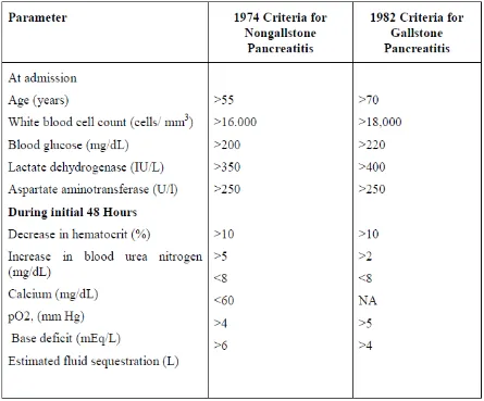
RANSON’S CRITERIA (TABLE 6)
Table 6 Ranson’s 11 Prognostic Criteria for Pancreatitis
Data from Ranson JHC, Rifkind KM, Roses DF, et al: Prognostic signs and the
role of operative management in acute pancreatitis. Surg Gynecol Obstet 139:69,
1974; and Ranson JHC Etiological and prognosis factors in human acute pancreatitis:
A review.Am J Gastroenterol 77: 633, 1982. NA, not applicable.
Ranson and colleagues identified 11 criteria that had prognostic significance
during the first 48 hours of pancreatitis. The original list was analyzed in patients who
primarily suffered from alcoholic pancreatitis and was modified 8 years later for those
severedisease. In mild pancreatitis (score ≤2) the mortality is 2.5%, and in severe
pancreatitis (score 2:3) the mortality is 62%. Also the higher the Ranson’s score, the
higher the incidence of systemic complications, necrosis, and infected necrosis. These
criteriacontinue to remain in wide use in both the United states and Europe.
The Ranson criteria have several drawbacks. First, the two lists are
cumbersome Second, an accurate Ranson’s score takes 48 hours to compute, and the
criteria have not been validated beyond the 48 – hours time limit. Third, not all
laboratories measure all the parameters in routine blood tests (e.g., lactic
dehydrogenase). Fourth, the overall sensitivity of the Ranson criteria (using 3 signs,
as, the cutoff) for diagnosing severe disease is only 40%to 88% and the specificity
43% to 90%. The positive predictive alue is approximately 50% and the negative
predictive value around 90%. Therefore, the best use of Ranson’s criteria is to exclude
severe disease.78, 79, 82
THE APACHE SYSTEM:
Acute physiology score and chronic health evaluation.
The first major attempt at a system to quantify severity of illness in ICU
patients was the APACHE system, by Knaus et al in 1981.
APACHE I
In the original form, APACHE contained 34 physiologic measurements and
included many continuous variables. A value of 0 to 4 was assigned to each variable,
according to its degree of abnormality. Shortly after its introduction apache 1 system
was disfavoured, because of practical problems like collection of large number of
assumed to be normal and weighted as zero. This gave rise to questions about the
models general applicability. Another major criticism of original APACHE system
was that the variables were chosen by a group of physicians and hence there was a
potential of bias. These inaccuracies in the original APACHE system prevented its
widespread use. However, it did serve as a prototype for the development of two
subsequent systems.
SAPS
The simplified acute physiology score was developed from APACHE I system
and incorporated 13 variables that had the most discriminate power and were the most,
frequently measured variables to cover all major organ systems. SAPS score is – still
used but has essentially been replaced by APACHE II in many centres.
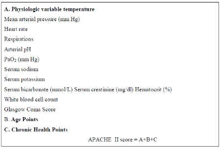
APACHE II
Published in 1985 by the same author this is the second version of the
APACHE system and it contains refinements based on experience with the original
APACHE system. APACHE II has been extensively used and has received far more
attention in the literature then any of the other methodologies for ICU outcome
prediction. It contains 12 continuous variables from the original APACHE system and
also takes into account age of the patient, pre- morbid conditions and Glasgow coma
scale.
DEVELOPMENT OF APACHE II
Using clinical judgement and documented physiologic relationships to choose
variable and assign weights remains the essence of APACHE II. The number of
osmolality, lactic acid level, skin testing for anergy were deleted. Serum BUN was
replaced by more specific serum creatinine and serum pH was retained in preference
to bicarbonate. Many variables crucial in patient care, such as serum glucose, albumin,
CVP and urinary output were found to have less explanatory power. Most of these
variables were sensitive to variations in therapeutic decisions than severity of disease.
Some of the thresholds and weights for the physiologic variables have been
changed ego Glasgow coma score, serum creatinine. Also since Alveolar – arterial O2
gradient (p [A-a] O2 is heavily dependent on inspired O2 (F1O2) a direct weighting
was given to all paO2 values when F1O2 is less than 0.5
To eliminate the problem of missing values and concerns about the assumption
that an unmeasured variable was normal, measurement of all 12 variables was made
mandatory for usage of APACHE II. The recorded values of the variables are based
on the most deranged values during the past 24 hours.
Because age and severe chronic health problems reflect diminished physiologic
reserve, they have been directly incorporated into APACHE II. Also, emergency
surgery and non operative patients with severe chronic organ system dysfunction
were given additional five additional five points in comparison to elective surgical
patients who were given only two points because patients with severe chronic
conditions are not considered to be candidates for elective surgery.
The maximum possible APACHE II score is 71. In the experience of the author
of APACHE II no patient had exceeded 55.The strengths of APACHE II system are:
i) It has a well defined outcome (hospital death)
Short comings of APACHE II system:
Because of extensive usage, important sources of error and bias in the
APACHE II system were revealed. First, APACHE II performs well overall in several
ICU population but it is inaccurate when looking at specific disease categories
because the data base from which it was derived, through large, did not contain many
patients in major disease subsets such as cardiac surgery, oncology etc. Second
APACHE II does not account for prior treatment or clinical course before the patient
enters ICU, this has been labeled as lead time bias. Third, APACHE II requires
determination of a single admission diagnosis, a subjective process prone to bias.
Finally, despite the reduction in number of variables, measurement error from bedside
data collection are still on issue.
APACHE II has been recently refined into APACHE 0, where 0 represents
obesity, and this is a better predictor of prognosis than APACHE II. Another
modification of APACHE II is the APACHE III system which is now being applied
widely to acute pancreatitis clinical trials.
APACHE II has the advantage of being able to be used on a daily basis and its
positive and negative predictive values are similar to those of the Ranson score at 48
hours after admission. The Apache II system assigns points for 12 physiologic
variables,for age, and for chronic health status, in generating a total point score. The
12 physiologic variables are temperature, heart rate, respiration rate, mean arterial
Table 7: Acute Physiology and Chronic Health Evaluation (APACHE)- II
Scoring
System of Disease Severity
Glasgow Score
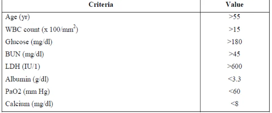
The Glasgow score is a slightly simplified list (8criteria) that is used commonly
in the United Kingdom. Its drawbacks are similar to those of the Ranson score. Other
investigators evaluated organ dysfunction risk factors in a qualitative way and found
that the presence of one risk factor predicted serious complications and more than 50
Table 8: Modified Glasgow Criteria: Within 48 Hours of Admission
failure might be more predictive of a fatal outcome than other parameters. The Goris
score assigns an organ failure value of 0, 1 or 2 or each of seven main Organ system
(respiratory, renal, cardiovascular, hepatic, central nervous, hematopoietic, and GI). A
score of 14 is the maximum and indicates severe disease in all systems. A study from
Scotland demonstrated that the Goris score was more predictive of death than the
Glasgow/Imrie score. In this study of 279 patients with acute pancreatitis, there were
a Goris score of 1 to 4, and 10 deaths (67%) in the 15 patients with a score higher than
5. Greater use of organ failure scores are likely to improve prognostication in acute
pancreatitis.
acute pancreatitis. Levels of catalytic type II PLAz have been reported to differentiate
between mild and severe disease within 24 hours of admission.
Urinary Trypsinogen activation peptide
TAP is the aminoterminal peptide cleaved from trypsinogen during activation of
trypsin, providing a rationale for its use as a marker of acute pancreatitis. It can be
measured in plasma, ascites fluid, and urine. The urinary TAP level appears to be the
most useful and, if measured within 24 hours of onset of symptoms, distinguishes
mild from severe pancreatitis. The sensitivity, specificity, and positive and negative
predictive values of TAP measurement for distinguishing sever from mild acute
pancreatitis at 214 hours compare favorably with those for CRP value and APACHE
A serum and urinary carboxypeptidase activation peptide (CAPAP) assay has
also been shown to predict early severe acute pancreatitis.
Serum Amyloid A: Serum amyloid A is another early acute- phase reactant that is
synthesized in the liver and is associated with the extent of tissue inflammation. Two
studies have demonstrated that the level of this serum protein can differentiate mild
from severe disease.
Procalcitonin The pro peptide procalcitonin is another acute- phase reactant that has
been shown to differentiate mild from severe acute pancreatitis within the first 24,
hours after symptom onset. A serum strip test has been developed for this
measurement that has a sensitivity of 86% and a specificity of 95% in detecting organ
TREATMENT
GENERAL CONSIDERATIONS
The patient with acute pancreatitis requires aggressive intravenous hydration
and adequate analgesia to eliminate or markedly reduce pain. An order for no oral
intake (NPO) is usually in force until nausea and vomiting have subsided. Abdominal
pain is treated with analgesics, given parenterally every 3 hours. Morphine can also be
used. Dosing is monitored carefully and adjusted daily according to ongoing needs.
Although morphine has been reported to increase sphincter of Oddi tone and to raise
serum amylase levels, its use to treat the pain of pancreatitis has not been shown to
adversely affect outcome. Nasogastric intubation is not used routinely because it is not
beneficial in mild pancreatitis. This modality is used only to treat gastric or intestinal
ileus or intractable nausea and vomiting. Similarly, proton pump inhibitors and
histamine Hz receptor blocking agents are not beneficial and are not used.
Each patient should be carefully monitored for any signs of early organ failure
such as hypotension and pulmonary or renal insufficiency via close following of vital
sings and urinary output. Rapid respiratory rate should not be assumed to be due to
abdominal pain, and blood gas measurements and oxygen supplementation are
mandatory in this situation. In cannot be overly emphasized that any patient who
exhibits signs of early organ dysfunction should be immediately transferred to
intensive care monitoring because deterioration can be rapid and fatal. This may be
the use of prophylactic antibiotics in patients with servere necotizing pancreatitis.
However, given this latest information, another reasonable approach would be to
[image:68.595.69.528.220.494.2]withhold antibiotics pending signs of infection.
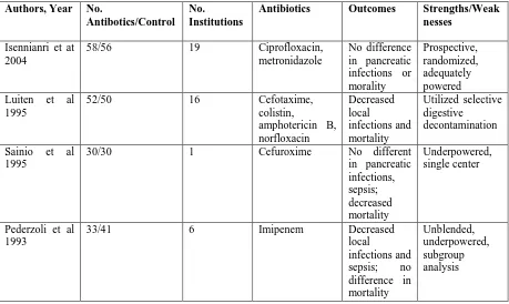
Table 9: Prospective, Randomized Prophylactic Antibiotic Trials in Acute Pancreatitis
Authors, Year No.
Antibotics/Control No.
Institutions
Antibiotics Outcomes Strengths/Weak
nesses
Isennianri et at 2004
58/56 19 Ciprofloxacin,
metronidazole No difference in pancreatic infections or morality Prospective, randomized, adequately powered Luiten et al
1995
52/50 16 Cefotaxime,
colistin, amphotericin B, norfloxacin Decreased local infections and mortality Utilized selective digestive decontamination
Sainio et al 1995
30/30 1 Cefuroxime No different
in pancreatic infections, sepsis; decreased mortality Underpowered, single center
Pederzoli et al 1993
33/41 6 Imipenem Decreased
local
Table 10: complications of acute pancreatitis Local Sterile necrosis Infected necrosis Abscess Pseudocyst Gastrointestinal bleeding: Pancreatitis- related:
Splenic artery rupture or splenic artery pseudoaneurysm rupture
Splenic vein rupture
Portal vein rupture
Splenic vein thrombosis leading to gastroesophageal varies with rupture
Pseudocyst or abscess hemorrhage
Postnecrosectomy bleeding
Non- pancreatitis – related:
Mallory- Weiss tear
Alcoholic gastropathy
Stress- related mucosal gastropathy
Splenic injury:
Infarction
Rupture
Hematoma
Fistulization or obstruction of small or large bowerl
Right- sided hydronephrosis
Systemic
Respiratory failure
Renal failure
Shock (circulatory failure)
Hyperglycemia
Hypocalcemia
Disseminated intravascular coagulation
Subcutaneous nodules due to fat necrosis
Retinopathy
And prologns hospitalization. There are several complications of endoscopic drainage
METHODOLOGY
The present study is a prospective study of 50 cases of Acute
pancreatitis admitted in Royapettah Hospital, Kilpauk medical college Chennai,
during the study period from May 2014 to august 2014.fifty cases for the
purpose of the study were selected on the basis of the nonprobability
(purposive) sampling method.
Source of study:
All patients diagnosed with acute pancreatitis admitted in Govt.
Royapettah Hospital, Kilpauk Medical College, Chennai 10.
Inclusion criteria:
All patients diagnosed with acute pancreatitis based on the clinical
suspicion and elevated serum amylase.
Exclusioncriteria:
• Hyperamylasaemia due to other causes
• Chronic pancreatitis
• Acute on chronic pancreatitis
Method of collection of data:
All patients diagnosed with acute pancreatitis based on the clinical
suspicion and increased serum amylase levels admitted in govt .royapetteh
hospital are assessed with multiple clinical and laboratory variables of both
Ranson and Apache II scoring system and the final score of the patient from
both the scoring systems are assessed to know their efficacy in predicting the
severity of the disease (higher the score more severe the disease).
STATISTICAL METHODS APPLIED:
Data was analysed statistically using WILCOXON SIGN RANK TEST
and FISHERS EXACT TEST by SPSS version 17.
OBSERVATION AND RESULTS
A total of 50 patients were included in the study .All had an admitting
diagnosis of acute pancreatitis .All 50 patients fulfilled the inclusion criteria:
o Clinical suspicion of pancreatitis
o Increased amylase
o Features of Pancreatitis on USG ABDOMEN
Of the 50 patients ,age range was 24-60years (mean-42years), 29
(58%)were men and 21(42%) women. The causes of acute pancreatitis included
biliarystone 22(44%),alcoholism 16(32%),idiopathic12(24%).10(20%)patients were
chronic smokers and 13(26%) had atleast one co-morbid disease. The common
concomitant diseases were hypertension (37.5%), diabetes mellitus(25%) ,ischaemic
heart disease(5%).
Overall,12(24%)patients suffered from severe pancreatitis and 38(76%)
had mild acute pancreatitis of which all 12 had severe attack as per APACHE
II score(>8) and only 4 of these were considered severe by RANSON
score(>3) .The systemic complications were multiorgan failure in 3(6%)
,respiratory2(4%) and renal 2(4%) all seen in patients with severe score as per
APACHEII .No death occurred and mortality was nil .Local complications
occurred in 3 patients (6%) and both had acute fluid collection .All the
complications were seen in patients with severe score as per APACHE II and
RANSON Scores:
Table11: Ranson scoring system results
Score Frequency Percentage
<3 46 92%
3– 4 4 8%
5-6 Nil -
>6 Nil -
TOTAL 50 100%
(Score>3 suggests severe pancreatitis)
In our study only 4 patients had score more than 3, suggesting that
only 8% of them were considered to be having severe pancreatitis as per
Ransons criteria.
APACHEII Scores:
Table12: Apache II scoring system results :
Score Frequency Percentage
0-5 36 72%
6– 10 7 14%
11–15 4 8%
>15 3 6%
TOTAL 50 100%
(Score>8 suggest severe pancreatitis)
In our study 12 patients were diagnosed to have score more than 8 of
the 50 cases, suggesting that 24% had severe pancreatitis as per Apache II
Data was analysed using Wilcoxon sign rank test & Fishers exact test
.The value at cutoff point was expressed as sensitivity, specificity, ppv, npv &
area under the ROC curve. P<0.05 was considered to be significant.
Table 13: Analysed data of Ranson and ApacheII scores:
Severityofacute PancreatitisScore
Median Interquartil eRange
Z P
ApacheII 2 7 4.491 <0.0005
Ranson 0 1
Table 14: Ranson – patient frequency
Frequency Percent
Valid Mild
Severe
Total
46
4
50
92
8
100
Table 15: Apache II – patient frequency
Frequency Percent
Valid Mild
Severe
Total
38
12
50
76
24
[image:89.595.127.472.289.405.2]Table 16: Percentage table (both scoring system together) Ranson
Total Mild Severge
Apache Mild Count
% of Total
38 76% 0 .0% 38 76%
Severe Count 8
16%
4
8%
12
24%
Total Count
% of Total
46 92% 4 8% 50% 100.0% Graph:2
(Percentage of mild and severe pancreatitis)
ApacheII Ranson 10 0 9 0 8 0 7 0 6 0 Mil Sever
APACHE II
Table17: Predictive performance: Area under curve =0.717 a Apache b
Total
Mild Severe
Serum Positive Count
amylase % within Apache
8 20.0% 12 100.0% 20 40% Negative Count
% within Apache
30.4% 80.0% 0 .0% 30 60%
Total Count
% within Apache
38 100.0% 12 100.0% 50 100.0% P<0.0005
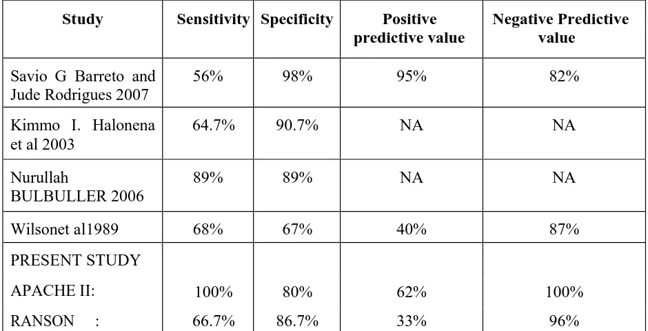
Sensitivity = 100%
Specificity = 80%
ppv = 62%
Ranson
Table18:Predictive performance:- Area under curve = 0.667
Ranson
Total Mild Severe
Serum Positive Count
amylase % within Ranson
1 25% 3 75% 4 8% Negative Count
% within Ranson
45 86.7% 1 25% 46 92% Total Count
% within Ranson