Product development specifications for a follicular sampling
device for use in a human in-vitro fertilisation clinic
A thesis presented in partial fulfilment of the requirements for the degree of Master of Engineering
Ill
Bioprocess Engineering
at Massey University, Palmerston North, New Zealand.
Hamish Alexander Harding
Abstract:
The likelihood of pregnancy in human in-vitro fertilisation is heavily dependent on the condition of the embryos that are re-implanted into the patient's uterus. The condition of embryos is in turn dependent on the quality of the oocytes, from which they grew. It has been suggested previously that oocyte quality could be related to the level of dissolved oxygen in the ovarian follicle. The first objective of this work was to develop a set of product development specifications for a device that would be used routinely in a fertility clinic for sampling follicular fluid for dissolved oxygen determination. The second objective was to design and construct a prototype so that the relationship between dissolved oxygen and oocyte quality could be established.
A length of time was spent at two fertility clinics, one in Hamilton, New Zealand and one in Auckland, New Zealand. The experiences at these clinics, as well as technical constraints, were translated into a set of product development specifications. These specifications canvassed issues relating to cleanliness, potential damage to the oocyte and preservation of the dissolved gas equilibrium in the sample. A prototype device was designed and developed and found to be wanting in the clinical environment. Further clinical constraints were identified from this experience, allowing a second prototype device to be developed. This second device was found to be suitable for clinical use and it is anticipate that in the future the sampling device will re-emerge in a new, more suitable form, based on the specifications developed in this thesis.
Acknowledgments
Thanks are due firstly to my supervisors Dr John Bronlund, Dr Alan Hart and Mr John Gawith, without whose wisdom and guidance very little would have been achieved. Though your collective efforts, I have begun this research, worked though the rocky parts and come out relatively unscathed at the end. Thanks also to Gabe Redding for being an outstanding colleague, brimming with useful information, advice and opinion. Thanks are due to the staff at Fertility Associates Isis Clinic, in Hamilton and Fertility Associates Auckland, especially John Peek, Andrea Coxhead and the embryologists at Fertility Associates Hamilton, who tolerated my ignorance of fertility treatment and answered all of my idiot questions.
I would like to thank the patients who consented to be a part of my study who, for obvious ethical reasons must remain nameless. You know who you are.
Finally, I would like to thank everybody else who has supported me through my studies; my family, my friends, my flatmates, my ex-colleagues from Medlab Central Ltd. and my colleagues at Youthline Palmerston North. I appreciate what you have so freely given me, allowing me to focus on my studies. I wish I could list everybody by name but I'm only allowed one page for my acknowledgments.
I would like to thank the Beatles for the White Album.
Table of contents
Chapter 1. Introduction ... 1
1.1 Objectives ... 2
1.2 Product specifications ... 2
Chapter 2. Review of the literature ... 3
2.1 Ovarian physiology and the follicular cycle ... 3
2.1.1 Follicular structure and growth ... 4
2.1.2 Structure of the human ovaries ... .4
2.1.3 Structure of the oocyte and cumulus oophorus ... 6
2.1.4 Follicular fluid ... 8
2.1.4.1 Physical properties of follicular fluid ... 9
2.1.4.2 Composition of follicular fluid ... 9
2.1.4.3 Follicular fluid pO2, pCO2 and pH ... 11
2.1.5 Post-ovulatory events ... 13
2.1. 5 .1 Post fertilisation development... ... 14
2.2 In-vitro fertilisation ... 14
2.2.1 The IVF process ... 15
2.2.1.1 Hormone treatment ... 15
2.2.1.2 Oocyte harvest. ... 16
2.2.1.3 Culture and fertilisation ... 17
2.2.2.1 Factors affecting success rates ... 18
2.3 Prediction of oocyte developmental competence ... 18
2.3 .1 Measures of oocyte/embryo quality ... 19
2.3 .2 Maternal age ... 20
2.3.3 Blood flow characteristics ... 20
2.3.4 Follicular gas composition ... 21 2.4 The product development process ... 22
2.4.1 The philosophy of product development... ... 22
2.4.2 Stages of the product development process ... 22
2.4.2.1 Project planning ... 23
2.4.2.2 Concept development.. ... 23
2.4.2.3 System-level design ... 23
2.4.2.4 Detailed design, testing and refinement.. ... 24
2.4.3 Identification of customer needs and initial specifications ... 24
2.5 Sampling liquids for dissolved oxygen determination (and reduction of pre-analytical errors) ... 25
2.5.1 Sampler material. ... 25
2.5.2 Gas bubbles ... 25
2.6 Measurement of dissolved oxygen ... 26
2.6.1 Henry's law ... 26
2.6.1.l Temperature dependence of gas - liquid equilibria ... 26
2.6.3 The Clark electrode ... 27
2. 7 Conclusions ... 31
Chapter 3. Clinical aspects of follicular sampling ... 32
3.1 The clinics ... 33
3.2 Personnel at the fertility clinic and their roles ... 33
3.2.1 Clinicians ... 34
3 .2.2 Embryologists/laboratory staff ... 34
3.2.3 Nurses ... 34
3.2.4 Support staff ... 35
3.3 What happens during IVF treatment? ... 35
3 .4 Equipment used in the fertility clinic ... 36
3 .4.1 Laboratory and laboratory equipment ... 37
3.4.2 Clinical/theatre equipment ... 38
3.4.3 Other miscellany ... 39
3.5 Oocyte pick up ... 40
3.5.l Preparation ... 40
3 .5 .2 The aspiration procedure ... 41
3.5.3 Physical dimensions ... 42
3.5.4 Usage ... 42
3.5.5 Sampler retention volume ... .43
3.5.7 Post procedure activity ... .43
3.5.8 Time delay before measurement ... .44
3.5.9 Sample storage temperature and cellular consumption of oxygen ... .44
3.5.10 Tracing the source of a sample ... .46
3.6 Summary of specifications ... 46
3.7 Conclusions ... 47
Chapter 4. Cytological constraints on sampling devices ... .48
4.1 Minimising effects to the oocyte - cumulus complex (OCC) ... 48
4.1.l Minimise effect of shear forces on OCC ... .48
4.1.2 Minimise temperature effects ... 49
4.1.3 Loss of oocyte ... 50
4.2 Minimise embryotoxicity of materials ... 50
4.2. l Materials selection ... 50
4.2.2 Materials preparation/cleaning ... 51
4.2.3 Embryotoxicity testing ... 51
4.3 Summary of specifications ... 51
4.4 Conclusions ... 52
Chapter 5. Physical aspects of measuring dissolved intrafollicular oxygen ... 53
5.1 Minimisation ofpreanalytical errors in follicular samples ... 53
5 .1.1 Blood contamination ... 53
5.1.1.2 Formulation ... 55
5.1.2 Sampler materials ... 58
5 .1.2. l System description ... 58
5.1.2.2 Formulation ... 59
5.1.3 Bubble contamination ... 61
5 .1.3. l System description ... 61
5 .1.3 .2 Formulation ... 61
5.2 Downstream compatibility ... 63
5.3 Specification summary ... 64
5.4 Conclusion ... 64
Chapter 6. Design details of a working follicular sampling device and protocols for dissolved oxygen determination in a fertility clinic ... 65
6.1 Description of the sampling device ... 65
6.2 Minimisation of pre-analytical errors ... 67
6.2.1 Evaluation of K-ATS-1000 syringe for sampling for dissolved oxygen ... 67
6.2.1.1 Materials and methods ... 68
6.2.1.2 Results and discussion ... 68
6.2.2 Effect of bubbles ... 69
6.2.3 Time delay before measurement ... 70
6.2.4 Sample temperature control. ... 70
6.2.6.1 Materials and methods ... 72
6.2.6.2 Results and discussion ... 72
6.3 Tracking samples and oocytes after sampling ... 74
6.4 Minimise effect of shear forces on the oocyte-cumulus complex ... 75
6.5 Minimise temperature effects on OCC ... 77
6.6 Loss of oocyte ... 77
6.7 Minimise embryo-toxic effects ... 77
6.7.1 Materials selection ... 78
6.7.1.1 Syringes ... 78
6. 7 .1.2 Silicone tubing ... 79
6. 7 .1.3 Other materials ... 79
6.7.2 Materials preparation/cleaning ... 79
6.7.3 Embryo-toxicity testing with mouse embryos ... 79
6.8 Sterilisation ... 80
6.8.1 Material selection ... 81
6.8.2 Sterilisation regime ... 82
6.9 Sampler retention volume ... 83
6.10 Sample volume ... 83
6.11 Use of the sampling device ... 84
6.12 Measurement of dissolved oxygen ... 84
6.12. 1.1 Oxygen electrode method description ... 85
6.12.1.2 Oxygen cell construction materials ... 86
6.12.3 Ensuring linearity of probe response ... 87
6.12.2 iStat method ... 88
6.12.2.1 Description of the iStat analyser ... 88
6.12.2.2 Validation of iStat pO2 measurements with non-blood liquids ... 90
6.13 Conclusion ... 91
Chapter 7. Performance of the sampling device in the clinical environment ... 92
7 .1 Ethical considerations ... 93
7 .2 Clinical protocol ... 93
7.3 Performance results ... 95
7.4 iterative improvement.. ... 96
7.4.1 Improvements on airtightness ... 96
7.4.2 Improved aseptic performance ... 97
7.4.3 Improvements regarding ergonomic factors ... 97
7 .5 Performance of the improved sampling device ... 97
7 .6 What went wrong? ... 98
7. 7 Summary of specifications ... 99
7 .8 Conclusions ... 100
Chapter 8. Conclusions ... 101
Appendix 1. Calculation of the Effect of Addition of Blood to non-Blood Liquids on Dissolved Oxygen ... l 09
Chapter 1. Introduction
Earth's increase, foison plenty, Barns and gamers never empty, Vines with clustering bunches growing,
Plants with goodly burden bowing; Spring come to you at the farthest
In the very end of harvest! Scarcity and want shall shun you;
Ceres· blessing so is on you.
The Tempest IV, i. William Shakespeare
While Ferdinand and Miranda had the benefit of Ceres' blessing, it is often medical intervention, rather than divine intervention that is sought in the 21st century for issues of fertility. Since the birth of a child in 1978 (Steptoe & Edwards, 1978) as a result of in vitro fertilisation (IVF), an entire industry has grown to supply treatment to those affected by infertility.
This thesis describes the work carried out over a 12 month period directed towards the design of a product for use in the modern fertility clinic that would allow clinicians
some insight into the conditions in which an egg had developed in a patient's ovary.
In vitro fertilisation is a treatment that is used to aid couples that are experiencing
difficulties conceiving. The woman is usually given hormones that encourage her
ovaries to produce more oocytes (eggs) than would occur during a normal ovarian cycle.
These oocytes grow inside small (up to approximately 10ml) blisters, filled with fluid on the surface of the ovaries. These blisters are called follicles. At the appropriate time a clinician will extract the follicular fluid and oocytes and the oocytes will be cultured in a laboratory. The oocytes are fertilised in vitro and the resulting embryos are grown further. After a few days the embryos can be implanted into the woman's uterus, frozen or discarded.
grade embryos is a reduced likelihood of pregnancy and live birth. Thus, if the clinician has information regarding the oocytes, then the patient's treatment can be managed to
improve the chances of a successful treatment.
1.1 Objectives
The primary objective of this project is to develop a set of product specifications that will help to guide the design of a device for sampling follicular fluid. The samples will
be used to determine the dissolved oxygen content of the follicular fluid.
The secondary objective of this project is to develop a prototype device and use it in a fertility clinic. This will help to establish a correlation between dissolved intra-follicular
oxygen and oocyte developmental competence, and will help to validate and refine the
specifications from the primary objective.
1.2 Product specifications
Product specifications are a set of constraints that describe what a product is going to achieve. It is not a description of what the product is or how it works, it is a description
of what it does. A specification is a measurable parameter that meets a need that has been expressed by a customer or identified through research.
The sampling system is to be composed of several modules (for technical reasons that
are disused later in section 2.6.3). The first is the module that will interface with the
surgical equipment that is used to extract the oocytes and will divert part of follicular
fluid away from the bulk flow as a sample. The second part is a container that will hold the sample after it has been removed from the bulk of the follicular fluid. The third part
of the system is the instrument that will be used to determine the dissolved oxygen
levels in the fluid and the final part is the set of operating procedures that determines how the device will be prepared to clinical standards, used and treated after use.
A detailed understanding of the physiology surrounding IVF treatment is needed in
order to describe an appropriate system.
Chapter 2. Review of the literature
And so she said to Abram, "The Lord has kept me from having children. Why don't you sleep with my slave-girl? Perhaps she can have a child for me." Abram agreed with what Sarai said.
Genesis 16:2
It wasn't until the ripe age of 90 that Sarai bore a child (Isaac) to Abram herself, as the
result of a divine act. Clearly recognition of infertility, and some of the anxieties
associated with this condition, can be found in some of the earliest writings. This
literature review deals with more contemporary publications.
In order to achieve the objectives stated in section 1. 1, there are several fields of
learning that need to be reviewed. These topics are the nature and physiology of ovaries,
follicular growth and oocyte maturation (section 2.1 ), the IVF process (section 2.2), an
overview of previous efforts to predict developmental capacity of human oocytes based
on follicular properties (section 2.3),the product development process and how it can be
applied to biomedical devices (section 2.4) and finally, a description of the field of
dissolved oxygen determination in human bodily fluids (sections 2.5 and 2.6).
ra A
"'••--=--
-1--.--:-1--•• - - . J &.I-- .£-11:-••·-- -•• ... I -L. I VVCll lelll p11y::,1UIU!:fY Cll lU LIIC IUlll\,UIQI \,Y\,IICTo give this work some background, an overview of the nature of the ovaries, their
function and their behaviour must be considered. Even though their presence was likely
to have been noticed by Hippocrates (460-370BC) during his extensive examination of
the uterus, the ovaries were largely overlooked as being the source of the elements
required to form a foetus for several centuries. Galen (130-200AD) proposed that the
ovaries contributed some sort of blood filtrate, via the fallopian tubes, to the uterus,
which would mix with male semen and coagulate, forming a foetus. Regnier de Graaf
(1641-1673) regarded the vesicular structures of the ovaries (ie. the tertiary follicles) to
be the eggs (analogous to bird eggs) which, after being fertilised, would undergo
embryonic and foetal development. Although he was not the discoverer of mammalian
eggs (that honour belongs to William Cruikshank), von Baer found the source of
mammalian eggs (oocytes) in 1827. More detailed accounts of ovarian history are in ready supply, such those of Short (1977), O'Dowd & Philipp (1994) and of Hunter
2.1.1 Follicular structure and growth
Each germ cell (oocyte) is contained inside a structure called a follicle in the ovarian
cortex (in the outer layer of the ovary). At birth each ovary contains about 500,000
follicles, about 83000 at puberty, 30,000 at age 35 and <1000 at age 50. 400-500 are used in ovulation. Most of them degenerate by a process called atresia before the follicle
has fully matured. The peak in germ cell numbers occurs in the prenatal stage,
approximately 6 months after conception. The significance of this is that each female
has a number of oocytes at birth and, unlike spermatozoa in males, no more will be
produced.
Most of the follicles in an ovary are primordial follicles. They are about 50µm in
diameter, lie in the periphery of the cortex and consist of an oocyte and a single layer of granulosa cells, known as the membrana granulosa. Granulosa cells are gland cells,
which means that they are capable of producing hormones and secreting these hormones
into the surrounding space or into blood stream for distribution around the body. At this
stage the granulosa cells are pressed flat against the oocyte.
2.1.2 Structure of the human ovaries
The ovaries are the gonads in the female, that is, a gland that produces germ cells, or
gametes. Toe ovaries are found in the human female lying on each side of the upper
pelvic cavity (figure 2.1), against the back of the pelvic wall, near the uterus. Each
ovary "resembles an almond in size and shape" (Anderson, 2002). Toe ovary is covered
in a thin layer called the ovarian epithelium, below which is dense tissue called the
ovarian cortex and less dense tissue, called the ovarian medulla. The medulla contains
blood vessels, lymphatic vessels and nerves. The medulla and cortex form the ovarian
stroma ( connective tissue framework). The female germ cells are in the cortex. The ovaries are connected to the uterus by the mesovarium and broad ligament, and are in
contact with the fallopian tubes via the infundibula, formed by the fimbriae tubae at the
distal ends of the fallopian salpinges. The ovaries are heavily vascularised and are
C.
a.
1. Ovary 2. Broad hgament
3. Fallopian Tube 4. Fimbriae tubae 5. Uterine cavity 6. Uterine Endometrium
7. Cervix
8. Vaginal Fomix
9. Vagina 10. Primordial Follicles 11. Primary Follides 12. Secondary Follicles 13. Tertiary (antral) follicles
14. Ovulated oocyte and corpus haemorragicum 15. Corpora lutea
Figure 2.1. Anatomy of the human female reproductive tract". (a) Position of human female reproductive tract in the abdomen. (b) Structure of the human female reproductive organs and genital tract, with blood supply (note that the ovarian vascularity have not been included to preserve clarity the of the diagram). (c) Structure of the human ovary, showing the
phases of follicular development
0
Adaptedfrom Cull (1989)
Some of the primordial follicles grow to become primary follicles. They are about
1 00µm in diameter. The granulosa membrane is still a single layer but the cells change
shape from being flat to more cuboid. The connective tissue covering the granulosa
[image:17.573.85.500.71.550.2]Some of the pnmary follicles grow to become secondary follicles. In these, the granulosa cells have divided by mitosis to become 2-6 cells across the membrane. The theca is still a single layer of cells.
Some of the secondary follicles grow to become tertiary follicles. These are much larger
at about 200µm in diameter. The granulosa cells have begun secreting fluid that fills the extracellular spaces and these pockets of fluid pool to form the antral space (filled with the antral, or follicular, fluid). Additional fluid passes through the theca from the blood vessels and this serous fluid augments the secretions from the granulosa cells. About 80% of the proteins found in blood are present in antral fluid (Lipner, 1973). This fluid contains proteins, steroid and protein hormones, anticoagulants (as some of the protein present is fibrin and thus there is a potential for the fluid to clot), enzymes (also some of the protein present) and electrolytes.
The theca has by now grown to form an inside (theca intema) which contains glandular cells and small blood vessels and an outside (theca extema) with connective tissue and
larger blood vessels. This phase of development is heavily influenced by secretion of follicle stimulating hormone (FSH) by the pituitary gland, at the base of the brain.
Tertiary follicles are graded by their size. In humans, l-9mm qualifies as a resting tertiary follicle, l 0- l 4mm qualifies a ripe tertiary follicle and l 5-25mm is called a
Graafian follicle, after Regnier de Graaf ( 1641-1673). The fluid from the tertiary follicles is aspirated during IVF treatment (Hunter, 2003).
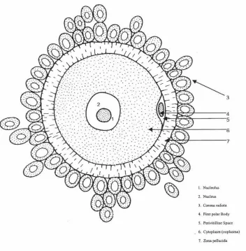
2.1.3 Structure of the oocyte and cumulus oophorus
This section provides a brief overview of the main features of the human oocyte and its associated structures. Detailed reviews of this topic are given by Crosby and Moor
(1984), Crisp ( 1992) and Hunter (2003).
The primordial human oocyte exists in a state of arrested development, at the prophase of meiosis. At this stage it has a nucleus (known as a germinal vesicle), which contains the genetic material. The oocyte will remain in this diploid state until almost
immediately before ovulation, although a number of other changes and interactions
Growth of the early follicular (granulosa) cells promotes activity in the oocyte. Most notably, the growth of an acellular layer called the zona pellucida (ZP). The zona pellucida can first be seen in the secondary follicle and a complete, uniform zona pellucida can be seen before the follicle reaches the graafian stage. The zona pellucida originates mainly from secretions of the growing oocyte. Areas of uniform, fine, fibrillar substances begin to appear close to the oocyte surface and develop into a mesh of interconnected filaments. As the oocyte grows, the plasma membrane (or oolemma), which surrounds the cytoplasm of the oocyte, becomes increasingly undulated. Eventually the shape of the oolemma becomes such that it forms microvilli, which project into the ZP and allow junctional contact with the extra-zonal cells, whose plasma membranes are undergoing similar changes. These gap junctions allow the transfer of material across the zona. Thus, the oocyte is not isolated from the follicular cells by the zona pellucida (Hunter, 2003).
Within the oocyte during pre-antral follicular growth there is a considerable amount of protein synthesis and accumulation of vitilline (cytoplasmic) reserves (analogous to the yolk of bird eggs). There is also an increase in cell diameter to approximately 120µm. Much of this development is directed by the action of honnones (gonadotrophin) that affect the gonads; at this stage follicular development is controlled by the action of follicle stimulating hormone (FSH), which is secreted by the pituitary gland at the base of the brain.
As the follicle develops an antral space, the oocyte is suspended in the antral fluid, held in place by a stalk of granulosa cells called the cumulus oophorus. There is a layer of cells packed closely to the oocyte within the cumulus called the corona radiata. The rest of the cumulus consists of granulosa cells dispersed in a gel-like matrix of hyaluronic acid. It is the coronal cells that have projected microvilli invested in the zona and are in junctional contact with the ooplasm. The cumulus oophorus contains up to 11,000 cells (Oritz & Croxatto, 1979 cited in Mohr, 1984) and measures 3-5mm in diameter (Crosby & Moor, 1984).
a gonadotrophinal hormone, leutinising hormone (LH), which causes a large amount of
fluid to accumulate in one of the follicles (the dominant follicle) and approximately 36
hours after the LH surge, ovulation occurs.
l. Nucleolus
2. NuclCUJ 3. Corona ndiata
4. First polar Body
S. Pcrivitillinc Space
. 6. Cytoplasm (ooplasma)
7. 1.ona pcllucida
Figure 2.2 Structure of the human oocyte showing the zona pel/ucida and corona radiata •.
• Adapted from Cull ( 1989)
2.1.4 Follicular fluid
What follows is a description of the liquid that fills the antral space because it is this
fluid that is to be collected and analysed for dissolved oxygen levels. It is important to
understand its functionality and physical properties since these will have an impact on
the collection and sensing operations. Fischer, et al. (1992) reports the average diameter
of ripe human follicles to be 16.6mm, meaning that each follicle yields but a few
millilitres. It is an increasingly difficult subject of study, since work with human
[image:20.575.74.426.165.524.2]difficult to work with small sample volumes, and to sample reliably to show variation
within a follicle over time. Consequently, there is much still to be understood regarding
the nature of the liquor folliculi.
The literature relating to the properties of follicular fluid has been described in excellent
reviews by Edwards (1974), by McNatty (1978) and by Gosden, et al. (1988).
2.1.4.1 Physical properties of follicular fluid
The liquid that inhabits the interior of the follicles is straw coloured. The yellow hue has
been shown to be due to the presence of bilirubin and carotenoids (Bayer et al., 1992).
Spectrophotometric examination shows that follicular fluid that is free from blood has
an absorbance peak at 458nm and gains several more absorbance peaks at 418nm,
540nm and 575nm as blood is introduced (Huyser et al., 1993).
Rheological studies have shown that the viscosity of human follicular fluid decreases
with increasing shear rate in the range 75-750s-1 and when exposed to a constant shear
rate the viscosity falls linearly with time at a rate of l 3µP min-\Luck et al., 2000).
Studies on the intrafollicular fluid pressure in the rabbit have shown that it is similar to
the pressure exerted on capillaries, and that it increases with increasing antral volume
(Blandau & Rumery, 1963; Espey & Lipner, 1963). The significance of these findings is
that the ovulatory follicle is unlikely to burst as a result of the internal pressure and is
more likely to collapse as a result of structural changes in the follicular wall. The
pressure is sufficient to push the liquid from the follicle slowly, as the follicle collapses.
Suction is applied during aspiration of this fluid for IVF treatment because the flow of
fluid is not sufficiently fast for aspiration purposes.
Observations have been made on the temperature of follicles preceding ovulation, and
have shown, somewhat counter-intuitively, that the graafian follicle is slightly (but
significantly) cooler than the surrounding ovarian tissue in rabbits (Grinsted et al.,
1980), pigs (Hunter et al., 1995) and in humans (Grinsted et al., 1985).
2.1.4.2 Composition of follicular fluid
fluid has been measured and compared to that found in blood serum and/or plasma for various species (Gosden et al., 1988). Table 2.1 shows the concentrations of electrolytes
in human follicular fluid and blood plasma/serum and the normal reference blood levels.
Table 2.1. Electrolyte concentrations (mmol.L"1
) of human follicular fluid (FF) and blood plasma (P) or serum (S)".
Na· I\
Normal Blood Range
136-145 3.5-5.0 (Anderson, 2002)
FF P/S FF P/S
Shalgi el al. (1972) 124 145 4.4 4.6
Edwards (1974) 147.8 150.5 5.2 5.7 Chong, et al. (1977) 143 154 5.4 5.4
·Adapted from Gosden, et al. (/988)
The protein content of follicular fluid has been investigated extensively showing that the
total protein concentration is lower than in blood serum and that follicular fluid is lacking the blood proteins with higher molecular weights, due to the filtering effect of the thecal and granulosa layers. The protein composition of human follicular fluid is
summarised in Table 2.2.
Table 2.2. Summary of studies into the protein content of human follicular fluid with respect to human blood serum (mg/mL).
Normal Blood Serum Range (Anderson, 2002)
Perloff, et al. (1955)
Manarang-Pangan, et al. (1971)
Shalgi, el al. (1973)
values are for blood plasma.
Total Protein
55-80
FF Serum
61.2 71.7
35.8 75.7
58 58
Albumin Fibrinogen •
33-55 2-4
FF Serum FF Plasma
27.4 28.8
40 30 1.1 3.7
Shalgi, et al. (1973) show that the mural layers that enclose the antral fluid have a filtering effect on plasma proteins, allowing passage of about 50% of proteins with a
molecular weight of 250,000 and blocking high molecular weight proteins (greater than MW=850,000) to the point where they are undetectable. They also observed that
fibrinogen is blocked to a greater extent than its MW could explain, and suggested that there are further mechanisms operating, possibly similar to those that block fibrinogen
2.1.4.3 Follicular fluid p02, pC02 and pH
Research into the dissolved gas compositions of human follicular fluid is in short
supply. The work to date is summarised in Table 2.3. For cross-species comparison,
values have also been given for porcine follicles.
Table 2.3. Summary of studies of partial pressures (mmHg) of oxygen and carbon dioxide and pH in human follicular fluid
and arterial blood.
Normal Arterial Blood· Range(Anderson, 2002
Shalgi et al.(1972)
Fraser et al.(1973)
Fischer et al. ( 1992)
lmoedemhe et al.(1993)
Huey et al.(1999)
Knudsen et al. (1978)
Gosden, R. G. & Hunter, R. H. F Unpublished, cited in Gosden et al. (1988)
Basini et al.(2004)
02
83-108
FF Blood
54.3
103.5 109.8
59.8 102
138.33
100.5
51 41 ...
-96.0
CO2 H
32-45 7.35-7.45
FF Blood FF Blood
35.1 7.267
41.7 44.1 7.344 7.306
46.9 38.3 7.33 7.41
40.17 7.36
34.8 7.35
-
...45 53 7.41
52.1
"Values are normal reference ranges for adult females. ··values are for porcine follicular fluid .... Values are for porcine venous blood.
Two of these studies show that as the size of ovarian follicles increase, the partial
pressure of oxygen falls dramatically in humans (Fischer et al., 1992) and in pigs
(Basini et al., 2004). This has been mathematically modelled, showing that the
reduction in intrafollicular oxygen is due in part to an increased distance for oxygen diffusion from the follicular perimeter through the bulk of the liquid and also due to
increased mural layers consuming most of the available oxygen (Gosden &
Byatt-Srnith, 1986).
The research of Shalgi, et a/.(1972) describes the determination of oxygen tension in
intrafollicular fluid using excised ovarian tissue. The fluid was drawn from the follicles
into syringes, avoiding contamination by air and blood and the gas concentrations were
determined immediately. While disruption to the gas equilibria was likely avoided after
[image:23.576.97.508.203.431.2]acidosis can be induced after ischaemic periods relative to measurements on in vivo ovarian tissue (Knudsen et al., 1978). Secondly, the construction material of the
syringes is not mentioned. It is well accepted that plastics are capable of disrupting the
gas equilibria in liquids as a response to gases dissolved in the polymer matrix (Scott et
al., 1971; Restall et al., 1975; Beaulieu et al., 1999; Anderson, 2002). However, the
measurements of oxygen tension in follicular fluid are consistent with the studies of Fischer, et al. (1992) and of Knudsen, et al. (1978).
Fraser, et al. (1973) reported the oxygen tension in follicular fluid to be 103.5mm Hg and the pCO2 to be 41.7mm Hg. It is worth noting that the investigators took measures to prevent changes to the oxygen concentration in their samples by using glass syringes
with the head space filled with heparinised saline. However, they note that their measurement of follicular dissolved gases may not be valid.
The researchers who undertook the next study of follicular oxygen (Fischer et al., 1992) preserved the dissolved gas concentration in the sample in a clever way. They inserted a
heparinised glass capillary between the needle used to puncture the follicles and the collection tube, which was removed, inspected for the presence of an oocyte and was analysed for pO2, pCO2 and pH. This technique avoided any contact with air before the measurements were made and also avoided materials that may have disrupted the dissolved gas equilibrium of the follicular fluid, thus reducing errors in their measurements. Their results are comparable to those of Shagi, et al. ( 1972).
Further research was carried out, examining the effects of carbon dioxide pneumoperitoneum (a technique where the peritoneal cavity is inflated with carbon dioxide gas) for laparoscopic collection of oocytes, and the changes in follicular fluid
gas concentrations (Imoedemhe et al., 1993). They demonstrated that the oxygen
tension of follicular fluid was 138.3mm Hg, considerably higher than previous studies,
and indeed, higher than is normally available to most tissues in the body (Anderson (2002) puts normal arterial blood oxygen partial pressure in the range 83-108mm Hg). The methods used in their study may help to explain this anomaly.
presumably was in equilibrium with air, in order to prevent contact with the air. The authors do not state that the paraffin oil had been pre-equilibrated with any other gas mixture and so the partial pressure of oxygen in the paraffin oil is likely to be much higher than that of the follicular samples.
The authors stated that each sample was examined for the presence of an oocyte and transported to the laboratory for dissolved oxygen determination. They show that the time lag has no significant effect on the oxygen concentration for up to l 0 minutes. It is possible that in the preceding time, during examination for the absence of an oocyte, that the liquids were extensively contacted (thus increasing the rate of mass transfer between the two liquid phases) and that the two liquids were largely equilibrated before the zero time measurements were made.
Huey, et al. (1999) reported the partial pressure of oxygen in human follicular fluid to be 100.5 mmHg. The fluid was sampled from the collection tube at the end of the follicle aspirating needle. It has been shown that aspiration into the collection tube causes a significant rise in the oxygen concentration of the follicular fluid (Redding,
unpublished data), and consequently, the quality of these measurements is questionable.
There are two observations that can be made from reviewing the literature published on the topic of dissolved gases in human follicular fluid:
• The volume of literature is extremely limited on this topic
• What has been published shows that there is still much to be understood about the dissolved gases in human follicular fluid since the few papers that have been published do not show consistent measurements.
2.1.5 Post-ovulatory events
its functionality. In the event of fertilisation the corpus luteum produces increasing
amounts of progesterone for the first weeks of pregnancy.
The oocyte, after leaving the follicle is entrained by the infundibula and guided into the fallopian tube. Contraction of the cillia inside the tube push fluid through the tubal isthmus and towards the uterus (Bellve & McDonald, 1970). At this point the oocyte is
either fertilised, in which case, it will adhere to the uterine lining and develop or it will
be lost amidst the menstrual material as the uterine lining breaks up. A review of
implantation is given by Findlay (1984).
2.1.5.1 Post fertilisation development
This section will very briefly describe the changes that occur within the oocyte after fertilisation. This process of embryonic development is a very important part of IVF treatment, since the embryo is cultured for some time before being re-implanted. Entire volumes have been written on the subject, and such detail is not provided here. For more information see McLaren ( 1972) or Paulson ( 1997).
The ingress of a sperm cell triggers the resumption of meiosis from the metaphase II
stage, ending with the extrusion of the second polar body into the perivitilline space. At
this stage, there are two pronuclei, each having half the normal number of chromosomes. These two pronuclei join in a process called syngamy and form the nucleus of a cell so that the cell now has a full, diploid, set of chromosomes. Over the
course of a few days the cell will cleave, still inside the zona pellucida. The cells,
therefore, maintain about the same volume as the initial oocyte and sperm cells had. When the internal mass of cells has grown sufficiently, the zona ruptures and the
embryo "hatches". The cells continue to divide (although any synchrony in the divisions
has long been lost) and form an "outside" section of cells and a fluid filled internal cavity, which also contains a few cells. The outer layer is destined to interact with the endometrial layers and become the placenta and associated structures and the cells in the cavity develop to form the foetus.
2.2 In-vitro fertilisation
mother did not have functional fallopian tubes and the fertilisation occurred in vitro (for
further reading in the history of IVF, see Biggers (1984) or O'Dowd and Philipp (1994)
both of whom give fairly detailed overviews of the development ofIVF).
2.2.1 The IVF process
The course of in-vitro fertilisation and zygote/embryo/blastocyst transfer is tailored to
the patient. This includes selecting the course of hormone treatment, the number of oocytes harvested and fertilised, the number of embryos implanted and the fate of any
spare embryos.
However, all IVF cycles involve some aspects of ovarian stimulation, oocyte harvest,
oocyte culture and fertilisation and reimplantation. Each of these phases is briefly
described below.
2.2.1.1 Hormone treatment
In the early days of IVF the patients' follicular cycle was allowed to run naturally, meaning that only a few oocytes could be recovered, since normally only one follicle ovulates, and there are only a few antral follicles. In order to increase the likelihood of
pregnancy, procedures were developed to stimulate the ovaries to super-ovulate by
administration of hormones and hormone analogues.
Most cycles now involve a pituitary down-regulation stage with a gonadotrophin
releasing hormone agonist (GnRHa) such as Leuprolide or Buserelin, starting on day 21
of the menstrual cycle (Paulson & Thornton, 1997). After a period of 10-18 days the ovaries are in a resting state, since the normal follicular cycle has been halted (Paulson
& Thornton, 1997). The effect of this is that there is less chance of the development of a single dominant follicle (as there is normally) and the clinician is able to time the
growth of follicles for greater convenience. At this stage gonadotropins are
administered, such as human menopausal gonadotrophin (hMG) or follicle stimulating
hormone (FSH). Gonal F and Puregon are brands of these drugs. They are continued
along with the GnRHa to prevent a premature endogenous leutinising hormone surge.
When the clinician is satisfied with the status of the follicles (usually by oestradiol 1713
induces the final stages of follicle development and ovulation. The subject of induction
of super-ovulation has been described extensively elsewhere in the literature. For a
more detailed description of hormone treatment the reader is referred to the work of
Paulson and Thornton ( 1997).
2.2.1.2 Oocyte harvest
Approximately 36 hours after administration of hCG, ovulation will occur. This is
undesirable during an IVF cycle because the oocytes will be liberated from the follicles
and lost to the clinician. In order to prevent ovulation, the clinician aspirates the fluid
from the follicles 34-36 hours after the hCG dose (Paulson & Thornton, 1997). It is this
harvesting process that forms the focus of this work and the fluid that is collected is to
have the oxygen levels characterised.
The most common method of retrieving the oocytes is by ultrasound-guided
trans-vaginal aspiration. During this procedure an aspirating needle is attached to an
ultrasound probe that is placed against the vaginal fomix. The ultrasound scan shows
the ovary and follicles and allows the needle to be guided through the vaginal wall and
into each follicle. When the needle has punctured the follicular wall, a vacuum is
applied and the antral fluid (along with the oocyte and some of the mural granulosa
cells) flows through the needle and into a collection tube. The aspirated fluid is
examined by an embryologist for the presence of an oocyte. If the oocyte is absent from
the fluid, the follicle is flushed with a liquid which is re-aspirated and frequently
contains the oocyte. Oocytes are recovered from about 60-70% of aspirated follicles
(Awonuga et al., 1996).
The size, shape and materials of the needle and strength of vacuum have been correlated
with damage to the oocyte, ZP cumulus and corona and other developmental parameters
in IVF (Awonuga et al., 1996; Home et al., 1996; Miller et al., 2004). Generally, higher
vacuums, finer needles ·and rougher needle interiors lead to more visible damage to the
oocytes and related structures and reduced
Typically the needle is 16 gauge and a vacuum of 100mm Hg is applied, although some
clinics apply vacuum using a syringe, which is unlikely to be constant or well
Prior to the use of the transvaginal technique, aspiration under laparoscopic guidance
was the norm. Since this technique has been superseded, it is not described here. The
reader is referred to the work of Kovacs, et al. (1984) for more information on this
technology.
2.2.1.3 Culture and fertilisation
After the oocyte has been located in the aspirated fluid, it is allowed to further develop
in an in-vitro environment. This environment attempts to simulate that of the interior of
the human fallopian tubes in terms of temperature, dissolved gases and other dissolved
substances (such as proteins, hormones, electrolytes, osmolality, pH, etc.). Optimisation
of this culture environment is still largely empirical and so there are several different
types of media (such as Human Tubal Fluid (HTF) and Ham's F-10). The atmospheric
composition is usually 5% oxygen, 5% carbon dioxide in nitrogen and the temperature
is always 3 7°C. Details of the materials and conditions required for one method of
oocyte and embryo culture are given by Wang and Gill (2004).
The two most common methods of fertilisation are insemination and intracytoplasmic
sperm injection (ICSI). Insemination, the simpler of the two methods, involves the
oocyte being exposed to a number of pre-treated, viable spermatozoa, and events are
allowed to take their "natural" course. ICSI is used in cases where there is a significant
male factor in the infertility, and a single sperm cell is injected through the zona
pellucida and oolemma into the ooplasm. ICSI is much more labour intensive. A
detailed description of fertilisation techniques is given by Paulson and Francis ( 1997).
After fertilisation the embryos are cultured for several more days until they are
discarded, frozen or implanted. This usually happens at the embryo (6-8 cell) stage
anytime up to the blastocyst stage.
2.2.2 Success rates of IVF
Despite the efforts of the assisted reproduction community, the success rates are rather
low (bear in mind, however, that the community deals with people for whom natural
conception has failed). Table 2.4 shows some of the statistics published by the U.S.
Two features to notice are the fact that success rates decline sharply with increasing age
and that the success rates overall averaged less than 30%.
Table 2.4 American ART success rates for 2001 by maternal age·.
Maternal Age
<35 35-37 38-40 41-43
Number of cycles started 35984 17791 16283 7044
%Cycles resulting in pregnancy 40.6 34.4 26.2 17.3
%Cycles resulting in live birth 35.2 28.4 19.6 10.4
%Retrievals resulting in pregnancy 38.9 33.1 23.8 13.2
% Transfers resulting in live birth 41.1 35.1 25.4 14.5
Average number of embryos transferred 2.8 3.1 3.4 3.7
·Adapted from Assisted Reproductive Technology Success Rates - National Summary and Fertility Clinic Reports (2003)(Assisted Reproductive Technology Success Rates - National Summary and Fertility Clinic Reports).
2.2.2.1 Factors affecting success rates
Clearly maternal age has a very large impact on the likelihood of birth following ART. One of the reasons for this is believed to be a reduced response to ovarian hyperstimulation. The success of ART cycles is heavily dependent on the number of embryos replaced, which in turn is dependent on the number of embryos produced and the number of oocytes obtained at aspiration. If a patient does not respond well to the gonadotropins, then fewer follicles will develop, resulting in fewer oocytes and fewer embryos and fewer high quality embryos for implantation. One method employed to counter this effect is to implant more embryos (see Table 2.4) to increase the chance of transferring a high quality embryo, which leads to pregnancy and eventually live birth. This carries an increased risk of multiple pregnancy, which can have serious implications for both the mother and the children. In New Zealand the number of embryos transfered is limited to one or two.
As noted by Shuttleworth (1909), chromosomal abnormalities become more frequent with increasing maternal age.
2.3 Prediction of oocyte developmental competence
[image:30.576.56.511.60.328.2]reasoning has led researchers to believe that certain parameters of this environment can
give an indication of the developmental capacity of an oocyte resulting in myriad
correlations being calculated, some stronger than others. The appeal of having a
measurable parameter that correlates strongly with oocyte quality arises from the
difficulty in testing an oocyte for its capacity to develop nonnally without impairing ( or
completely devastating) that capacity (bearing in mind that the oocyte is only a single
cell and that its haploid state leaves it particularly susceptible to chromosomal damage).
2.3.1 Measures of oocyte/embryo quality
The first opportunity to measure oocyte quality comes after it is liberated from the
follicle by microscopic examination. The presence of a germinal vesicle (and absence of
the first polar body) can indicate poor quality, since the first meiotic division has not
occurred, and consequently, the cell has the wrong number of chromosomes. Light
microscopy can also show the state of the zona and the cumulus, which have roles to
play during fertilisation, and may not function properly if damaged (Hunter, 2003).
Immediately post-fertilisation, light microscopy should show the presence of two
pronuclei ( one from the sperm and one from the oocyte) and not a third pronucleus,
which is indicative of polyspermy (ie. penetration of more than one spermatozoan into
the oocyte ). Mohr ( 1984), provides a six point list of characteristics that can be used to
grade human embryos:
• The shape and relative size ofblastomers within an embryo
• The number, location, size and general appearance of nuclei within the blastomers
• The number of small anucleate cytoplasmic fragments (blebs) in the perivitilline
space or between the blastomers (ie. the degree of fragmentation)
• The general appearance and distribution of cytoplasmic organelles
• Formation of junctions between the blastomers and compaction of the blastomers
An emerging option for determination of embryo quality is the use of pre-implantation
genetic diagnosis (PGD). This technique involves taking a cell from an embryo (about 8 cells) or blastocyst (note that several cells can be taken from the trophoderm of the blastocyst with no detrimental effects) and analysing the cell(s) for genetic aberrations. An excellent review of PGD is given by Findlay (2000). This technique falls outside of the scope of this thesis and will not be discussed further.
2.3.2 Maternal age
It was first noted in literature that with advanced maternal age came a higher incidence of "mongolism" (Down's syndrome) by Shuttleworth (1909) (cited by O'Dowd & Phillipp, 1994). Subsequently, maternal age has been shown to affect not only the
"quality" of the oocytes, with respect to the number of chromosomes (trisomy 21, for example), but also the number of oocytes produced (the implications of this are
described in section 2.3.3.1).
Paulson and Thornton (1997) state that the likelihood of miscarriage increases with
increasing maternal age. The rate in women undergoing assisted reproductive treatment
(ART) less than 40 years old is 18.5%, whereas in women over 40 years, the rate increases to 35.1 %. The reasons for this trend are unclear.
2.3.3 Blood flow characteristics
It has become well accepted that the degree of vascularisation and blood flow around a follicle strongly influences the oocyte that resides inside (remembering that blood does not flow through the follicle and that the blood vessels are in the thecal layers, and do not permeate the antral space). The use of colour doppler ultrasonography has allowed relatively non-invasive imaging of bloodflow characteristics about the ovaries and the follicles therein. There is a host of literature that supports the view that follicles with better vascularisation produce oocytes that are more likely to fertilise and grow normally (Van Blerkom et al., 1997; Van Blerkom, 1998; Bhat et al., 1999; Coulam et
measurements of blood velocity in a clinical setting. Bhal, et al. (1999) show that the
amount of follicle that is surrounded by blood vessels(graded as <25%, <50%, <75%
and >75%) can be correlated strongly with the likelihood of a pregnancy.
2.3.4 Follicular gas composition
It was first postulated by Gaulden ( 1992) that reduced dissolved follicular oxygen could
impair the quality of the developing oocyte. She proposed that an hypoxic environment
would induce production of excess lactate, reducing the pH of the oocyte surroundings. Such an acid environment would be capable of disrupting the microtubules that form the
meiotic spindle and after a disrupted chromosomal abstriction, aneuploidy would be
more likely, bringing an increased risk of abnormal embryonic development, spontaneous abortion and post natal conditions (such as trisomy 21 - Down's syndrome).
Subsequently, there have been two studies in the human attempting to relate follicular oxygen tension to oocyte developmental competence. The first was by Van Blerkom, et al. ( 1997), in which they claim that oocytes from severely hypoxic follicles were more
likely to have chromosomal abnormalities than oocytes from follicles with a greater
dissolved oxygen content. They found also that the ability of an oocyte to develop
in-vitro to the metaphase II stage and to fertilise was not related to the oxygen content but
that the ability to develop to the 6-8 cell stage did correlate to dissolved oxygen significantly. The measurements of oxygen in this study involved immersing an oxygen probe into the aspirated fluid after it had been collected. It has been shown that the
aspiration of fluid into a vacuum-sealed collection tube significantly alters the concentration of dissolved oxygen (Redding, unpublished data). Surprisingly they do
not report the actual oxygen tension in the fluids, instead, oxygen content is reported to
be ~1.5%, 1.5-2.5% or 3.0-5.5%.
The second study that attempted to correlate dissolved oxygen with developmental
competence in the human oocyte was by Huey, et al. (1999). They show that ability to
fertilise and embryo morphology does not correlate significantly with the concentration
2.4 The product development process
The previous sections of this review have demonstrated the need for improved success rates in IVF treatments, and outlined the potential value in ovarian follicular fluid oxygen levels for providing information on oocyte quality. It is also clear that successful design of an apparatus to allow this to be done in a modern fertility clinic must consider a vast array of issues, from logistics, to clinical use, to technical feasibility.
A product development process has been adopted for this project so that there was a well defined, structured, process by which these issues were considered in order to achieve the objectives from Section l. l:
• To develop a set of product specifications that will help to guide the design of a device for sampling follicular fluid for dissolved oxygen determination, and
• To develop a prototype device and use it in a fertility clinic
This section will describe the philosophy and purpose of the product development concept, the anatomy of a product development project and the specific stages that were anticipated to be carried out through this project.
2.4.1 The philosophy of product development
The reason for the product development process is to help new products to be successful on the market. There are many variations on the theme but all of them have this goal in mind. Cooper (2001) thoroughly describes the purpose of a structured product development process, pointing out that careful planning in the early development stages is more efficient (and cheaper) than making substantial changes later in the process, as result of poor forethought.
2.4.2 Stages of the product development process
Figure 2.4 The product development process consists of several distinct phases, each with its own objective. Adapted from Ulrich and Eppinger (2000).
Project
Planning
Concept Development
System-level design
2.4.2.1 Project planning
Detail Design
Testing and Refinement
Production ramp up
Ulrich and Eppinger {, 2000 #49} explain that this stage identifies opportunities and
sorts the wheat from the chaff The projects that appear to have merit are allocated
resources and are passed on to the next phase. The planning takes into account the
situation of the organisation and ( especially for commercial businesses) the portfolio of
developments that the organisation sees as being ideal.
2.4.2.2 Concept development
This phase is concerned with identifying what the customers want, what the product
should do, how these things can be achieved and what the best way of achieving them
is. This best way of achieving an end (the product concept) is passed on to the
system-level design phase.
This project is concerned with the identification of the customers' needs and what the
product should do. Thus, these points are expanded in more detail in section 2.4.3.
2.4.2.3 System-level design
The system-level design is concerned with sorting out the nuts and bolts of how a
product will work. It may involve computer modelling/CAD designs, mathematical
models of performance and working prototypes of parts of the product or even a whole
working product. At this stage the designers begin to take the manufacture of the
product into account and how the customer will interact with/operate the product.
When the working product has been designed and tested it is passed to the detailed
2.4.2.4 Detailed design, testing and refinement
These phases are concerned with designing the final product, how it will look, operate
and perform. As the title indicates, this is an iterative process, where a product is tested,
tweaked (only small changes should be required at this stage) and retested.
After the testing and refinement have been completed, then the fine details of
production can be resolved and the product is released to the market.
2.4.3 Identification of customer needs and initial specifications
This phase of development begins by understanding the needs of the customers. Ulrich
and Eppinger (2000)identify six distinct objectives for this exercise:
• Ensure that the product is focused on customer needs
• Identify latent or hidden needs as well as explicit needs
• Provide a fact base for justifying the product specifications
• Create an archival record of the needs activity of the development process
• Ensure that no critical customer need is missed or forgotten
• Develop a common understanding of customer needs among members of the
development team
They suggest that these needs can be identified through interviews with individuals
from the customer group, facilitated focus groups and observing the product in action.
Griffin and Hauser (1993) discuss the various strengths and weaknesses of these
methods at length.
The next stage is to take the needs that have been identified and sort them into a
hierarchy of importance to the customer. The final part is to assign each need with a
parameter which can be measured. The target performance of the product regarding
each parameter is assigned and recorded. This is the set of initial specifications.
As stated in section 1.1 one of the objectives of this project is to form a list of initial
analysis. The rest of this chapter is concerned with the nature of gases dissolved in follicular fluid, which influence the product design and will require appropriate product specifications.
2.5 Sampling liquids for dissolved oxygen determination (and
reduction of pre-analytical errors)
Preservation of the dissolved gas equilibrium before analysis has been identified as one of the most important factors in ensuring high quality information from dissolved gas determination (Muller-Plathe, 1998). The two most important influences on dissolved
gas equilibrium are the materials with which the liquid is in contact and the presence of an unequilibrated gas phase in the sample container.
2.5.1 Sampler material
It has been shown that polymeric materials can disrupt gas equilibria in liquids (Scott et al., 1971; Stevens, 1992; Carignan & Gachter, 1994; Beaulieu et al., 1999). Thus it is
important to consider the nature of the materials used in the construction of both the
sampling device and the equipment that will be used to measure the concentrations of the dissolved gases. Generally, glass is the material of choice ( d'Ortho et al., 2001 ), followed by metals (although these have the disadvantage of not being transparent) and plastics. Carignan ( 1994) shows that the extent to which a plastic affects the amount of
dissolved gas is polymer specific, as summarised in table 2.5. This is backed up by the
extensive material data available in Pauly ( 1999).
Table 2.5 Specific oxygen release from several different polymers into water. Adapted from Carignan & Gachter (1994).
2.5.2 Gas bubbles Polymer
Teflon
Polycarbonate Acrylic
Polyvinyl chloride Polyformaldehyde
High Density Polyethylene Polyvinylidene
02 released (µrnol cm· )
0.18
0.23 0.10
0.07 0.04
0.04 0.05
Gas bubbles in liquid samples have been identified as a major problem in dissolved
oxygen measurement (Mueller et al., 1976; Biswas et al., 1982; Pruden et al., 1986; C.
2.6 Measurement of dissolved oxygen
A key aspect of this project is the measurement of dissolved oxygen. What follows is a
description of the nature of gases dissolved in aqueous solutions and common issues
associated with the measuring of dissolved oxygen.
2.6.1 Henry's law
The simplest description of the solubility of a gas is Henry's Law.
Heruy's Law of dissolved gases.
f =k.x
fugacity of gas (Pa)
k Henry's law constant (Pa)
x mole fraction of gas in solution
Henry's law states that the fugacity of a gas in solution is proportional to its mole
fraction in solution. Fugacity can be interpreted as partial pressure above the solution
provided that the gas behaves in a manner not too dissimilar from the ideal gas ( and thus
the ideal gas law). The mole fraction of gas in solution can also be interpreted as being a
concentration, if the proportionality constant is adjusted appropriately. Restating
Henry's law: The concentration of a gas in solution is proportional to the partial
pressure of that gas above the solution. Henry's law applies to solutions where the
solute particles are sufficiently dilute that there is no solute-solute interacton. It also
applies only to isothermal and isobaric situations where there is no other solute
dissolved in the solvent (Hitchman, 1978).
The relationship in Henry's law can be adjusted to apply to different units of
concentration, other than mole fractions, by using different values (and units) of the
Henry's law constant.
2.6.1.1 Temperature dependence of gas - liquid equilibria
An equation can be derived from basic thermodynamics to describe the variation of gas
solubility in a solvent. The relationship is given below (Hitchman, 1978):
The relationship between solubility of a gas in a solvent and the temperature of the system.
T Absolute temperature (K)
R Ideal Gas Constant (8.314 J/mol.K)
t>.H Heat of solution (J/mol)
P Isobaric system pressure (Pa)
This relationship holds for ideal solutions (very dilute solutions approximate ideality).
2.6.2 Applications of dissolved oxygen measurement
Once samples have been collected they must have their dissolved oxygen levels quantified. The oxygen measurement method could place constraints on the amount of
sample required, or possibly other factors. To identify these potential impacts a brief overview of dissolved oxygen measurement was undertaken.
Measurement of dissolved oxygen finds applications over a variety of fields, most commonly in aqueous environmental monitoring (dissolved oxygen in rivers, streams,
etc.), in wastewater treatment, in industrial fermentation operations and in clinical monitoring. Each of these applications have developed their own techniques and, with care, solutions to problems relating to dissolved oxygen monitoring in one situation can
be transferred to another.
2.6.3 The Clark electrode
The current in a galvanic cell is characterised by its magnitude and direction. Thus, if
the reduction of molecular oxygen was to occur at one of the electrodes (ie. the
cathode), then there will be a current to that electrode.
The amount of oxygen reduced at the electrode (w, grams) is proportional to the amount
of charge (q, coulombs) that is passed from the electrode (ie. q=it) and is related by
M
o2
Mit
Mo2 Mass of oxygen reduced at thenF
electrodeM molecular weight of oxygen
current (A)
time (s)
n number of electrons per reaction
F Faraday's constant (96,486.7C/mol)
Assuming that the rate of the reaction at the electrode obeys first order kinetics, then the rate of reaction can be expressed as:
N number of moles of reduced species produced
kRed rate constant for the reduction reaction
k0 , rate constant for the oxidation reaction
Co, Concentration of the oxidised species at the
electrode
CR"" Concentration of reduced species at the
electrode
Thus, the current generated by the reaction per unit area of electrode can be expressed:
That is, there is a measurable current, which is proportional to the concentration of molecular oxygen at the surface of the electrode (Hitchman, 1978).
There are several major disadvantages to having an electrode in contact with a test solution. The first is that the surface of the electrode can be degraded by impurities in the environment to be tested, reducing the available area for the reaction to take place and thus reducing the measured current ( causing false measurements or requiring frequent calibration). The second disadvantage is that the electrolyte concentration of the test solution may not be known or may be changing, which will affect the electrode potential, the current and thus the integrity of the measurements (which maybe be erroneous or require frequent recalibration). Thirdly, it is possible that alternative reactions could be occurring at the electrode surface and generating additional current. This would serve to inflate the measured values of oxygen in solution.