Limbs joint position sense in patients with
chronic non-specific back pain
Natalija Pahomova1and Daina Smite2 1Jelgava polyclinic, Latvia
2R¯ıga Stradin¸š University, Latvia
Abstract. Purpose: To analyze limbs joint positions’ sense and its relationship with pain intensity and duration in patients with chronic non-specific back pain. Methods: Study design: cross-sectional study. For assessment of patients, the following elements were used: general data collection protocol; evaluation of pain intensity using the Visual Analogue Scale; assessment of limb kinesthesia: determination of reposition precision of a 90-degree flexion angle in shoulder, elbow, hip and knee joints. Subjects. The study included 100 patients (88 women, 12 men) with chronic non-specific back pain, who met the selection criteria for the study. The average age of patients was 45.9±11.6 years, and it ranged from 19 to 64 years. Results: All participants of the study were found to have diminished upper and lower limbs kinesthesia. The study results showed plausible (p <0.05) relation between limbs kinesthesia and pain intensity and duration, and these correlations were significantly determined by pain localization. Conclusion: Limb kinesthesia is plausibly related to the manifestation of pain in patients with chronic non-specific back pain.
Key words: kinesthesia, proprioception, joint position sense, chronic back pain.
1 Introduction
Within one year, prevalence of lower back pain in Europe is 25–63%, while the upper back pain prevalence is 20–40% [1,2]. More than 90% of back pain is non-specific [1].
Joint position and movement sense is a part of proprioceptive sense (sometimes it is also called “kinesthesia”). Proprioception means the awareness of the relative position of body segments and the position and movement of the body in space [3]. Patients with chronic back pain have reduced proprioceptive sense of the respective spine segments, which correlates with pain intensity and function restrictions [4–6]. It has been proved that people with chronic, idiopathic neck pain feel worse than others at health control at head-to-neutral repositioning tests [7]. The result was confirmed in a recent systematic review that higher cervical joint reposition sense error was found in people with traumatic, as well as with non-traumatic neck pain than in healthy controls [8]. Paulus and Brumagne (2008) found that for people with recurrent neck pain not only head position control, but also shoulder joint position sense is disturbed [8]. Huysmans et al. (2010) discovered impaired position sense acuity of the upper extremities in patients with chronic neck pain [9,10]. It was suggested that spinal proprioception reduction in body position control (mainly spine global stability) is provided by lower extremity joints kinesthesia [11–13]. Zazulak et al. (2007) and others have shown that impaired lumbar proprioception increases the risk of knee injury [14]. Shakoor et al. (2008) found impaired joint kinesthesia in both lower and upper limbs in patients with
hip osteoarthritis. This finding may be explained in two ways – either one hip degenerative changes and proprioceptive disorders in the long term lead to another joint proprioceptive changes, or generated changes in a proprioceptive deficit is the risk factor for the development of osteoarthritis [15].
Proprioception disorders and back pain cause-and-effect relationship remains debatable. Transmission of sensory information due to tissue damage or pain is altered, in its turn, proprioceptive deficit leads to increased range of intervertebral motion that is a precursor to further paraspinal tissues injury [16, 17]. Neurophysiological research determined the role of central sensitization in various musculoskeletal disorders, including low back pain, osteoarthritis [18]. It is known that pain inhibits afferent proprioceptive information transmission to the spinal cord and higher centers of the central nervous system, which allows us to explain postural control disorders and pathological movement patterns in patients with chronic pain syndrome [4,19–21]. On the other hand, there is still lack of evidence for central sensitization in idiopathic, non-traumatic neck pain [22].
Moseley et al. (2012) gave a hypothesis that in a patient with chronic back pain body orientation representation in the cerebral cortex could be altered [23]. Brugmane et al. (2004) demonstrated proprioceptive reweighting from the trunk to the ankle in patients with low back pain [11]. In recent studies, it was supposed that a reduced proprioception from the lower limbs may contribute to an altered postural control in patients with chronic back pain and that a reduced proprioceptive feedback from segments of the body other than trunk may affect postural control in back pain patients [24].
2 Methods
2.1 Participants
This study examined 100 patients (88 female, 12 male) who were admitted to Jelgava outpatient rehabilitation department from 01.07.14 to 31.10.14 with chronic non-specific back pain (pain duration more than 12 weeks, localized in neck (upper back) and/or low back). The age group of the participants was 45.9±11.6 years. The exclusion criteria included the following: any specific cause of back pain, traumatic injury of limbs or joint arthritis, muscle strength in extremity muscles less than 3 grades (after Kendall); peripheral or central paresis, previous spine and/or limb surgery, pregnancy and less than two years after childbirth, additional disease that causes functional limitation and/or contraindication for functional tests.
2.2 Design and procedure
The study was performed as a cross-sectional analysis. Each participant was evaluated before the conduction of rehabilitation sessions by the study assessment methods. The approval for this study was obtained from the Ethics Committee of Riga Stradins University, and each subject was required to sign a consent form prior to the participation in the study.
2.3 Assessment methods
Table 1. Pain intensity (VAS) and duration (current episode, time from the first onset) in the sub-groups (mean (SD)).
Pain characteristics Group 1 n=33 Group 2 n=19 Group 3 n=48 Pain intensity (VAS; mean (SD)) 5.9 (1.9) 4.6 (1.8) 5.2 (2.1) Current pain episode duration (weeks; mean (SD)) 15.6 (6.3) 15.2 (6.7) 14.1 (0.9) Time from pain onset (years; mean (SD)) 9.8 (7.1) 6.9 (4.9) 12.7 (9.3)
were performed in supine position, but knee testing – in prone position. Movements where performed without visual control.
Visual analogue scale (0 “no pain”, 10 “worst possible pain”) – was used to evaluate the pain intensity. Duration of current pain episode (weeks), time from first pain episode (years) and pain localization (“neck and upper back” and/or “low back”) were detected.
2.4 Statistical analysis
The results were analyzed using SPSS.V20 software. We used the mean and standard deviation to describe the results for each joint reposition error score and pain characteristics. ANOVA analysis of variances was used to calculate the difference between mean values of joints reposition error scores in three independent groups based on pain localization. Spearman correlation analysis was used to reveal the correlation between joints reposition error scores and pain characteristics. The statistically significant level wasp <0.05.
3 Results
3.1 Pain characteristics
Almost half of participants (n=48) marked pain both in upper and lower back, 33 patients complained about lower back pain, but others (n=19) about upper back (neck) pain. Presuming that back-pain localization is an important factor to predict manifestation of chronic pain syndrome in analysis, three subgroups were formed: Group 1: patients with lower back pain (n=33); Group 2: patients with upper back pain (n=19); Group 3: patients with both upper and lower back pain (n=48). Based on ANOVA test results, division of patients into subgroups based on gender, age, education, employment, occupational risk factors and physical activity had no statistically significant difference (p >0.05).
The pain intensity and duration in participants is outlined in Table1. The pain intensity was higher (p <0.05) in Group 1 and Group 3, than in Group 2 (F =1.296,p=0.028). There was no difference between the subgroups based on the current pain episode duration (ANOVA,F =0.423,p=0.656), but the time from first pain episode onset was longer in Group 1 (p <0.05) than in Group 2, but in Group 3 it was longer (p <0.05) than in both Group 1 and Group 2 (F =3.805,p=0.026;F =5.714,p=0.009).
3.2 Joint reposition error tests results
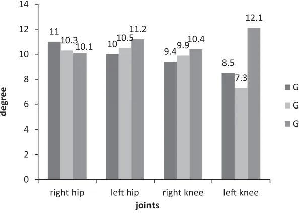
Joint position sense impairment was found for all the participants in at least three out of eight joints examined. Mean joint reposition error in upper and lower limb joints is illustrated in Fig.1and Fig.2.
11.9
13.9
8.8
11.2 11.3
10.7 11.3
9.5
11.2 11.8
9.7 9.5
0 2 4 6 8 10 12 14 16
right shoulder le shoulder right elbow le elbow
deg
ree
joints
Group 1
Group 2
[image:4.482.76.405.72.285.2]Group 3
Fig. 1. Mean reposition error (degrees) of upper limb joints in the three subgroups.
11
10
9.4
8.5
10.3 10.5
9.9
7.3 10.1
11.2
10.4
12.1
0 2 4 6 8 10 12 14
right hip le hip right knee le knee
deg
ree
joints
Group 1
Group2
Group 3
Fig. 2. Mean reposition error (degrees) of lower limb joints in the three subgroups.
[image:4.482.79.376.332.547.2]Table 2. A schematic description of significant (p <0.05) correlation (based on Spearman’s correlation analysis) between joint reposition error and pain characteristics in the study subgroups. The table shows only joints whose kinesthesia’s disorders correlated with pain syndrome.
Group Pain intensity (VAS) Duration of current Time from the first pain episode (weeks) pain episode (years) Group 1 – left hip – right elbow – left elbow
– both knees – right hip Group 2 – both elbows – left elbow
Group 3 – right shoulder – right shoulder – right hip – left knee – left knee
3.3 Correlation analysis
The results confirmed some reliable (p <0.05) moderate to weak correlations between the test results of pain intensity (VAS) and joint reposition error: in Group 1 with knee and hip (R =0.478,p=0.005;R=0.410,p=0.018); in Group 2 with elbow (R=0.519,p= 0.023); in Group 3 with shoulder and knee (R =0.299,p=0.039;R=0.290,p=0.044). In Group 2 and Group 3 joint reposition error test results in the same joints showed significant correlation with the duration of current pain episode: Group 2 with elbow (R=0.47, p=0.042), Group 3 with shoulder and knee (R=0.298,p=0.04;R=0.331,p=0.022). In Group 1 duration of current pain episode correlates with reposition error in elbow and hip (R=0.371,p=0.034; R=0.356,p=0.042). Time from the first pain episode revealed significant correlation (p <0.05) with reposition error in elbow joint for Group 1 (R=0.435,p=0.011) and hip joint for Group 3 (R=0.303,p=0.036). The significant (p <0.05) correlations between joint reposition error test results and pain characteristics in the three subgroups are schematically shown in Table2.
4 Discussion
The main findings of our study were the following: (1) all studied patients with chronic back pain had both upper and lower limb joint position sense impairment regardless of back pain localization; (2) back pain intensity had reliable relationship between joint position sense in limb adjacent to pain localization; (3) limb joint position sense impairment showed the tendency to be more expressed in patients with an extended current pain episode.
Based on the study methodology, the studied patients formed a sufficiently homogeneous group by a structural damage of the spine and pain type (so-called, chronic non-specific back pain). Although the previous studies revealed that a chronic pain syndrome is a disease and is characterized by the dysfunction of the nervous system and pain localization has only a secondary role [25], our assumption on the back pain localization as an important factor to predict manifestation of chronic pain syndrome proved to be useful. It could also be proved by recent evidence from the systematic review that central sensitization is not a characteristic feature for all chronic neck pain patients [21].
cortex in major chronic back pain cases [17,30,31]. Shakoor et al. (2008) found impaired joint kinesthesia in both lower and upper limbs in patients with hip osteoarthritis [15], but in recent systematic review, Luch et al. (2014) concluded that both peripheral mechanisms and hypersensitivity of CNS are involved in osteoarthritis pain syndrome [32].
Taking into account the bio-psycho-social aspects of the chronic pain syndrome [33] there is a reason to suggest that limb joints reposition error tests results were influenced by such emotional characteristic of chronic pain patients as anxiety. It has been stated in different studies that there is a connection between anxiety and disturbed motor task performance, also, anxiety diminished body representation in CNS [34,35].
Considering the correlation between more expressed joint position sense impairment with extended length of current pain episode and in patients with lower back and both lower and upper back pain localization with longer time from the first pain episode, increased risk of limb joints injuries or motor control disorders and subsequent pain can be presumed as long – term consequences. It is very important, regarding the weight bearing joints (hip, knee) that in clinical setting lower limb motor control disorders in patients with chronic lower back pain are usually explained with biomechanical adaptive changes, but our findings point out the necessity to pay the same amount of attention to neuromuscular component (as general proprioception disorder). As our aim of the research was to analyze the relationships, the cause-effect relation remains unclear. It can be presumed that poor limb joint proprioception (which could be due to some peripheral mechanism (e.g., soft tissue injury)) has an effect on prolonging the back-pain episode. These circumstances emphasize the need for future research in longitudinal studies.
Our research results confirmed that more intense back pain is related to higher joint reposition error in limb adjacent to pain localization. There were only few studies that confirm concurrent action of pain and proprioception stimulus in spinal cord level (“gate control mechanism”) [18,31], that could partly explain our findings. In addition, previous studies have found close relationship between pain intensity and anxiety in chronic back pain patients [33], thus it can be presumed that anxiety and fear were the main mediators between higher pain intensity and greater joint position sense impairment. For patients with chronic back pain the component of somatization and fear (especially fear of movements) has been observed already in previous studies [33]. Our finding in the relationship between pain intensity and joint reposition error in limb adjacent to pain localization could be mainly explained by fear and excessive awareness of the painful site (back part), which could result in poor motor task performance (e.g. joint reposition). The motor control of spine (that was more diminished in patients with higher pain intensity) leads to less effective motor performance in distal segments (limb joints).
As the methodological strength of our research, the homogenous study group could be pointed out. The established sub-groups were useful and important initially, the assessment methods and study design was appropriate and allowed to reach the goal of the study. In further research, we could advise to use longitudinal design and assessment supplement with methods to detect muscle synergy patterns in limbs and psycho-emotional state. As well as investigations of interventions directed towards the improvement of limb joints proprioception in patients with chronic back pain could be conducted.
5 Conclusions
between the joint position sense in limb adjacent to pain localization, that should be taking into account when choosing multimodal therapeutic approaches.
References
[1] A. Farioli, S. Mattioli, A. Quaglieri, S. Curti, F. Violante, D. Coggon, Scand. J. Work Environ. Health 40(1), 36–46 (2014)
[2] Systematic Literature Report. European Federation of IASP Chapters (2012),http:// www.sip-platform.eu/tl_files/redakteurbereich/Home/Reflection Process_screen.pdf
[3] U. Proske, S. Gandevia, Physiol. Rev. 92, 1651–1697 (2012)
[4] A. Lee, J. Cholewicki, N.P. Reeves, B.T. Zazulak, L.W. Mysliwiec, Arch. Phys. Med. Rehabil. 91, 1327–31 (2010)
[5] R.B. Grahama, L.Y. Oikawa, G.B. Ross, J. Biomechanics 47, 1459–1464 (2014) [6] I. Paulus, S. Brumagne, J. Rehabil. Med. 40, 426–432 (2008)
[7] T.R. Stanton, H.B. Leake, K.J. Chalmers, G.L. Moseley, Somatosens Mot. Res. 33(2), 93–8 (2016)
[8] J. de Vries, B. Ischebeck, L. Voogt, J. van der Geest, M. Janssen, M. Frens, G. Kleinrensink. Phys. Ther. 96(6), 876–87 (2016)
[9] M. Huysmans, M. Hoozemans, A. van der Beek, M. de Looze, J. van Dieën, J. Rehabil. Med. 42, 876–883 (2010)
[10] J. Sandlund, U. Röijezon, M. Björklund, M. Djupsjöbacka, J. Rehabil. Med. 40, 366–374 (2008)
[11] S. Brumagne, P. Cordo, S. Verschueren, Neurosci. Lett. 366, 63–66 (2004)
[12] K. Claeys, S. Brumagne, W. Dankaerts, H. Kiers, L. Janssens, Eur. J. Appl. Physiol.
111, 115–123 (2011)
[13] T. Popa, M. Bonifazi, R. Della Volpe, A. Rossi, R. Mazzocchio, Exp. Brain. Res. 177, 411–418 (2007)
[14] B.T. Zazulak, T.E. Hawett, P. Reeves, B. Goldberg, J. Cholewicki, Am. J. Sports Med.
35(7), 1123–1130 (2007)
[15] N. Shakoor, K.J. Lee, L.F. Fogg, J. Block, Arthritis & Rheumatism 59(9–15), 1237–1240 (2008)
[16] G. Ebenbichler, L. Oddsson, J. Kollmitzer, Z. Erim, Med. Sci. Sports Exerc. 33(11), 1889–1898 (2001)
[17] S. Brumagne, P. Cordo, R. Lysens, S. Verschueren, S. Swinnen, Spine 25(8), 989–994 (2000)
[18] J. Nijs, L. Daenen, P. Cras, F. Struyf, N. Roussel, R.A. Oostendorp, Clin. J. Pain. 28(2), 175–181 (2012)
[19] M.J. Comerford, S.L. Mottram, Manual Therapy 6(1), 15–26 (2001)
[20] C. Demoulin, V. Distrée, M. Tomasella, J.M. Crielaard, M. Vanderthommen, Annales de réadaptation et de médecine physique 50, 677–684 (2007)
[21] G.L. Moseley, Manual Therapy (Elsevier Ltd, 2003)
[22] A. Malfliet, J. Kregel, B. Cagnie, M. Kuipers, M. Dolphens, N. Roussel, M. Meeus, L. Danneels, W. M. Bramer, J. Nijs, Pain Physician 18(3), 223–236 (2015)
[23] G.L. Moseley, H. Flor, Neurorehabil. Neural. Repair 26(6), 646–652 (2012)
[24] R. Della Volpe, T. Popa, F. Ginanneschi, R. Spidalieri, R. Mazzocchio, A. Rossi, Gait and Posture 24, 349–355 (2006)
[25] S.M. Rubinstein, Best Pract. Res. Clin. Rheumatology 22(3), 471–482 (2008) [26] H.J. Jo, A.Y. Song, K.J. Lee, D.C. Lee, Y.H. Kim, P.S. Sung, Eur. Spine J. 20,
[27] H.Y. Lee, J.D. Vang, G. Yao, S.F. Wang, Manual Therapy 13, 419–425 (2008) [28] C. Teng, H. Chai, D.M. Lai, S.F. Wang, Manual Therapy 12, 22–28 (2007)
[29] R. Astfalck, P. O’Sullivan, A. Smith, L. Straker, A. Burnett, Manual Therapy 18, 410–417 (2013)
[30] B.M. Wand, L. Parkitny, N. O’Connel, H. Luomajoki, J. McAuley, M. Thacker, G.L. Moseley, Manual Therapy 16, 15–20 (2011)
[31] C.J. Woolf, M.W. Salter, Science 288(9), 1765–1768 (2000)
[32] E. Lluch, R. Torres, J. Nijs, J. Van Oosterwijck, Int. J. Clin. Pract. 70(1), 31–44 (2016) [33] D. Šmite, G. Anc¯ane, Nat. Exact. Appl. Sci. 64(5/6), 202–208 (2010)
[34] J.R Davis, B.C. Horslen, K. Nishikawa, K. Fukushima, R. Chua, J.T. Inglis, M.G. Karpenter. J. Neurophysiol. 106, 3082–3090 (2011)