1-1-1980
Changes in pulmonary function and hepatic flow
associated with a surgically induced porto-caval
shunt
Paul Joseph Antol
Iowa State University
Follow this and additional works at:https://lib.dr.iastate.edu/rtd Part of theEngineering Commons
This Thesis is brought to you for free and open access by the Iowa State University Capstones, Theses and Dissertations at Iowa State University Digital Repository. It has been accepted for inclusion in Retrospective Theses and Dissertations by an authorized administrator of Iowa State University Digital Repository. For more information, please [email protected].
Recommended Citation
Antol, Paul Joseph, "Changes in pulmonary function and hepatic flow associated with a surgically induced porto-caval shunt" (1980).
Retrospective Theses and Dissertations. 18071.
'
/
with a surgically induced porto-caval shunt
_r
.s
IA I 9 f/OA
,..,
gg> byc..3
Paul Joseph Antol, Jr.
A Thesis Submitted to the
Graduate Faculty in Partial Fulfillment of the Requirements for the Degree of
MASTER OF SCIENCE
Major:. Biomedical Engineering
Signatures have been redacted for privacy
Iowa State University Ames, Iowa
1980
TABLE OF CONTENTS
Page
FOREW0RD iv
INTRODUCTION 1
REVIEW OF LITERATURE 3
Alcoholism and the Lung 3
Effects of Portal-Systemic Anastomoses 5
Porto-Caval Shunts and Hepatic Flow 7
Porto-Caval Shunts and Pulmonary Function 7
MATERIALS AND METHODS 11
Surgical Preparation 11
Pulmonary Function Tests 11
Liver Function Tests 13
Clinical Tests 14
Post-Mortem 15
CALCULATIONS 16
Lung Compliance, Resistance, and Functional Residual Capacity 16
Liver Function 18
RESULTS 22
Pulmonary Function 22
Liver Function Tests 25
Clinical Tests 29
Post-Mortem Observations 29
DISCUSSION 31
Pulmonary Studies Liver Function Studies Mathematical Model
CONCLUSIONS AND RECOMMENDATIONS SUMMARY
LITERATURE CITED ACKNOWLEDGMENTS
APPENJ;>IX A: SURGICAL PROCEDURE
APPENDIX B: CALCULATION PROGRAMS FOR TI-SS CALCULATOR Compliance-Resistance Program
Functional Residual Capacity Program Percentage Disappearance .Rate Program APPENDIX C: CLINICAL TEST RESULTS
APPENDIX D: POST-MORTEM OBSERVATIONS Gross Observations
Histopathological Observations
Page
32
3S
37
39
41
42
4S
46
Sl Sl
S2
FOREWORD
The world (parenthetically speaking) has a mistaken notion about the nature of science and scientists • . . scientists often seem to be matter of fact people, unromantic and even hard-headed. ·That is because the scientific attitude is realistic, as opposed
INTRODUCTION
The many effects of alcoholism have been.investigated over the years and yet there are marty questions· that remain to be answered. As it will
be described later, chronic alcoholism usually leads. to cirrhosis of the liver (or Laennec's cirrhosis), portal hypertension, and other
physiologi-cal disorders. Such disorders as esophageal varices, ascites, and the
establishment of collateral vessels that divert portal blood directly into the system~c circulation are directly correlated with portal hypertension.
It is these collateral vessels, either porto-caval or porto-pulmonary shunts, that are of direct interest in this research.
The main purpose of this research is to investigate the .effects on pulmonary function of a porto-caval shunt which changes portal flow.
Because of the porto-caval shunt, blood is diverted from the
mesen-teric and splanchnic vasculature into the vena cava. As a result of this shunt, the lung capillary system receives the metabolites and other
ab-sorbed compounds that originate in the gastrointestinal outflow. There-fore, digestive compounds· normally removed or altered by the liver may
alter pulmonary function.
In the alcoholic human, the study of these effects is complicated by
the prese~ce of the liver cirrhosis itself, poor diet, smoking, and the
It has been reported in the literature that humans who have cirrhosis
of.the liver and a surgical porto-caval anastomosis appear to demonstrate some cardio-pulmonary dysfunction. The cardio-pulmonary dysfunction in-cludes pulmonary hypertension, cardiomegaly, and a hypertrophy of the i11terstit.ium of the· lung.
Therefore, the question arises as to whether the disorders observed in
lung function arise as a result of the cirrhotic liver, the porto-caval
shunts, or are due to some other possibility, such as smoking.
In order to demonstrate an altered portal flow and the exposure of
the lung to portal blood (i.e., hepatic cirrhosis), porto-caval
anasto-moses were performed on five healthy dogs. The surgical procedure is
known as the Eck fistula (Markowitz et al., 1964, pp. 542-546). Using
each dog ~sits-own control, pulmonary and liver function studies were
conducted at various times after surgery in an attempt to determine the
REVIEW OF LITERATURE
·-'
During the last twenty years, a great quantity of information has been compiled on the effects of alcohol on living organisms. These effects are numero.us and occur in many organ systems including heart, liver and the central nervous system, as described by Lieber (1976}. More
recently, data have been obtained which suggest an interaction of alcohol
with the lung. Since alcoholism is such a complex condition with many dy.sfunctions occurring simultaneously, it remains difficult to ascertain
the exact cause or causes for the impairment of lung function (Burch and
Depasquale, 1967). This review will attempt to develop a foundation of
literature to support the function of porto-caval shunts in the normal and
alcoholic individual.
Alcoholism and the Lung
Several studies have looked at pulmonary function in alcoholics. Banner (1973) studied thirty patients described to be long-term, heavy drinkers. Of the eleven pq.tients selected for pulmonary mechanics tests,
four had normal pulmonary mechanics., four had signs of airway restriction,
one had signs of emphysema, and two were not consistent with any known pulmonary dysfunction. From these data, he suggested that the ultra-structure of the lung may be damaged by alcohol. Emirgil et al. (1974)
reported a decrease in total lung capacity, vital capacity, and residual
volume in twenty-three chronic alcoholics. They suggested alcohol as a
causative agent in producing lung disease independent of the effects of
con-firmed the results of Banner (1973) and stated that little information
exists with regard to the effects of alcohol or the effects of alcohol metabolites on the lungs. Furthermore, Emirgil and Sobol observed im-paired lung function could be related to hepatic dysfunction, not to the
alcohol itself.
Ruff et al. (1971) reported that in 80% of the hepatic cirrhosis cases studied, the closing volume (the lung volume at which the dependent
lung zones begin to trap gas as a result of airway closure) was increased and exceeded the functional residual capacity (FRC). This indicates some
of the airways are closed during normal breathing. Two suggestions for premature closure were made: 1) the loss of elastic recoil in the de-pendent. zones of the lungs, and 2) a decrease in resistance during
me-chani~al compression of the small airways from distended vessels and from
interstitial pulmonary .edema.
It was suggested by Ruff et al. (1971) that the compression of small airways and presence of interstitial pulmonary edema was responsible for
the impaired diffusion and lowered arterial oxygen levels in cases of liver cirrhosis. Although no direct evidence of pulmonary· edema was found by Ruff et al., separate necropsy studies by Berthelot et ·al. (1966) and
Cameron· (1948) not only demonstrate dilatation of precapillary sphincters,
but also pulmonary edema in cases of cirrhotic patients.
Heinemann (1960) proposed the lowered arterial oxygen levels were due to venous-to-arterial shunts. He observed that administration of 100% oxygen to patients with liver disease experiencing hypoxemia did not abolish the problem of hypoxemia.
Effects of Portal-Systemic Anastomoses
Cirrhosis of the liver is accompanied by m~rked changes in the hepatic an4 splanchnic vasculature. This· leads to reduced hepatic blood flow, increased portal venous pressure, and the development of collateral
vessels. The formation of collateral vessels allows portal blood to
by-pass the diseased liver. The·portal blood enters either the inferior vena cava through port9-caval shunts or the superior vena cava through
porto-pulmonary shunts (Bradley et al., 1952; Taylor and Myers, 1956; and
Heinemann, 1960, 1977).
Calabresi and Abelmann (1957), indicated that lowered arterial oxygen
tensions may be due to either uneven ventilation-perfusion. relationships or to .anatomical shunts bypassing the lungs from the portal vein. They
demonstrated anatomical shunts created by anastomoses of the portal bed with the superior vena cava and pulmonary vein. Their data defined the
shunts ·as follows: 1) a porto-caval shunt would exist when blood passes from the portal vein through the peri-esophageal and the mediastinal or azygos veins .to the superior vena cava, and 2) a porto-pulmonary shunt would exist when blood passes from the portal vein through the
peri-esophageal and mediastinal veins to the bronchial veins and finally to the
pulmonary vein. A porto-pulmonary shunt would therefore bypass the lungs
and result in the lowering of arterial blood oxygen levels (Calabresi and Abelmann, 1957).
Heinemann (1960) supported the findings of Calabrasi and Abelmann
(1957) by suggesting the volume of porto-pulmonary shunted blood depends
metabolites from the gastrointestinal tract would have direct access to the pulmonary arteries via cirrhotic shunted or surgically induced
anastomoses of the portal vein to the vena cava. He points out that the development of cardiac hypertrophy, particularly predominant right
ven-tricular .hypertrophy,. occurs in patients with liver cirrhosis. Since cardiac output is elevated in cirrhotic patients, it would account for the
occurrence of pulmonary hypertension as well as cardiac hypertrophy.
How-ever, Heinemann alludes to the idea that vasoactive substances drained by the sp,lanchnic vessels and normally inactivated by the liver could reach
the pulmonary and systemic circulation via the collateral vessels.
Shaldon et al. (1961) concluded there was no relationship between
arterial oxygen unsaturation and porto-pulmonary anastomoses, which is contrary to Calabresi and Abelmann (1957) and Heinemann (1960).
The difference in opinion between Shaldon et al. (1961), and
Cala-bresi and Abelmann (1957) and Heinemann (1960) was further complicated by
Williams and Abelmann (1963), who measured porto-pulmonary shunt flow in
one patient as 27% of total portal flow, and also by Nakamura et al.
(1965), who measured shunt flows in two patients at 11% and 14%. However,
both Williams and Abelmann and Nakamura et al. agreed on two points:
1) Arterial oxygen unsaturation is not due to the shunts, which supports Shaldon et al. (1961).
Porto-Caval Shunts and Hepatic Flow
The discrepancy· in this review concerning anatomical porto-caval and porto-pulmonary shunts and their effects is complicated by the surgical treatment for portal hypertension in advanced cases of cirrhosis. In cirrhotic patients who are afflicted with either bleeding esophageal varices or the buildup of ascitic fluid due to increased pressure in the
portal vasculature, the treatment is to create a porto-caval shunt. This
is usually done with either a side-to-side or an end-to-side surgical
anastomosis of the portal vein to the vena cava. This· results in part or
all of the portal blood being diverted directly into the systemic vascula-ture and the portal system undergoes decompression (Shackelford, 1955,
pp. 677-688).
Bra<lley et al. (1952, 1953) observed hepatic circulation in cirrhosis
and saw.that hepatic blood flow decreased during any cirrhotic process.
They stated that hepatic ischemia and relative tissue hypoxia appear to be characteristic stigmas and speculated that changes in hepato-cellular
oxygen metabolism may reflect some serious derangement of cellular physi-ology. These changes may be followed by necrobiosis and alteration in the
tissue and vascular structure of the liver.
Porto-Caval Shunts and Pulmonary Function
Cotes et al. (1968) evaluated lung function on three patients who had
porto-caval anastomoses performed five to six years previously. In
ob-serving dilatation of the precapillary vessels of the lung, however, they
should be no blood flow through any of the porto-pulmonary shunts which may have been present. Cotes et al. proposed precapillary pulmonary
vasodilatation to be one of the consequences of reduced portal blood flow. There was evidence in the three patients for the syndrome of defective gas
transfer, which is usually accompanied by features suggestive of fibrosing
alveolitis. They concluded that the surgical technique which prolongs the
lives of these cirrhotic patients may increase the incidenc.e of further
pulmonary complications.
Sallam and Watson (1970) discussed complications arising in the lung as a result of micro-thromboemboli originating in the splenic and portal
veins. They described a porto-caval anastomosis performed on a patient diagnosed with Banti's syndrome six years previously. Although pulmonary
function tests and biopsy of the liver were shown to be normal, there was
considerable evidence of pulmonary hypertension as seen in X-ray films. Sallam and Watson came to no conclusion in this study other than the
problem was.probably due to a thrombus in the splenoportal system
re-leasing microemboli.
Senior et al. (1968) conducted pulmonary function studies on four
patients with hepatic cirrhosis and surgical porto-caval shunts. These
patients had clinical signs of pulmonary hypertension with cor puimonale
and normal chests as .evidenced by ECG. and chest X-rays prior to porto-caval anastomosis. On later examinations, there were marked changes in
outputs were normal while minute alveolar ventilation and pulmonary arterial pressure were abnormally elevated. Arterial hemoglobin satura-tion was reduced (hypoxemia). Necropsy studies on two of these patients showed microscopic evidence of thromboemboli in small pulmonary vessels. The evidence included intimal thickening, medial hypertrophy, and partial occlusion and recanalization of vessel lumens. However, no sources of the
thromboemboli were found at autopsy.
Lebrec et al. (1979) observed the same symptoms as Senior et al.
(1968) in nine patients with similar histories of hepatic cirrhosis and
surgical porto-caval anastomosis. They concluded that diversion of portal
venous blood into the inferior or superior vena cava plays a major role in
the development of-pulmonary hypertension. Lebrec et al. suggested a
vasoconstrictive-agent produced in the splanchnic territory could bypass the liver and gain access to the pulmonary arteries, thus inducing
pulmonary hypertension.
Johnson and Lambert (1967) studied changes in cardiac output in normal dogs that were given end-to-side porto-caval anastomosis.
Mor-tality rate was 50% due to factors outside of the surgery and its conse-quences. Of the survivors, mean cardiac output was shown to increase 35%.
They proposed that the increase in cardiac output was the result of a
cyclic pattern in which portal blood flows into a low resistance system
(i.e., the inferior vena cava) through the shunt. This results in an
in-creased arterial inflow to the portal system, increasing total portal flow
Johnson and Lambert observed a decrease in blood pressure and no change in pulse rate or right atrial pressure.
In summary, this literature review has suggested that alcoholism and the resulting cirrhosis causes numerous complications in lung, heart and
liver. Pulmonary hypertension, increased cardiac output, and decreased hepat:Lc blood flow are the major effec.ts of cirrhosis of the liver.
MATERIALS AND METHODS
Surgical Preparation
Six normal, healthy adult mongrel dogs ranging in weight from
13.6-22. 7 kg were obtained and housed at Laboratory Animal Resources, Iowa State University. Porto-caval anastomoses were created surgically using
the ,technique for the straight Eck fistula previously described by
Markowitz, Archibald, and Downie (1964, pp. 542-546). The procedure for the Eck fistula is detailed in Appendix A. This surgery created a 1-2 cm
fistula between the portal vein and the vena cava just above the splenic vein in each dog. This allowed portal blood to bypass the liver and gain
access to the lung vasculature.
No major problems were associated with each approximately six hour
surgery and each dog was up and walking twelve to eighteen hours after surgery. Only one dog, #1656, experienced a post-operative localized skin infection from suture. Antibiotic treatment effectively controlled the infection.
Dog #1582 was used only to test the surgical procedure and was not subjected to pulmonary function testing but was included in
histopatholog-ical examinations at termination.
Pulmonary Function Tests
D?gs #1588, 1598, 1656, 1704, and 1740 were treated similarly in all
placed in dorsal recumbency. A SO cm 7-French catheter imbedded into a rubber stomach tube was filled with normal sali~e and was then inserted
into the esophagous to about mid-thorax. The animal was then connected via the endotracheal tube to a FleischR #0 pneumotach (Instrumentation
R
Associates, Inc., New York, New York) which was attached to a Statham
PMS differential pressure transducer (Statham Laboratories, Inc., Hato Rey, Puerto Rico) for measurement of airflow. Volumes were determined by
electronic integration and all measurements of flow, volume, and
esopha-R
geal pressure were measured on a four-channel Beckman R611 Dynagraph (Beckman Instruments, Inc., Schiller Park, Illinois). All channels were
balanced and calibrated before measurements. The esophageal cannula was connected to a Statham PR23 low-pressure transducer and after flushing
with normal saline, the position of the esophageal cannula was adjusted to
give a slight positive peak at the onset of expiration. After the animal and physiological recordings were stable,' a continuous recording (2S mm/
sec) for one minute was obtained. Compliance (C) and resistance (R) values were then determined.
Functional residual capacity (FRC) measurements were conducted using a multiple breath nitrogen washout method. The dogs were attached to a
R
digital nitrogen analyzer (Hewlett-Packard ll47302A, Hewlett-Packard, Inc., Palo Alto, California) and a digital pneumotach (Hewlett-PackardR
ll47303A). Values for nitrogen content and inspired tidal volume were taken from the analog outputs of both devices and recorded on a two-channel BrushR 220 recorder (Gould, Inc., Cleveland, Ohio). When the
breathing room air to 100% oxygen. At the same time, expired air was collected in an evacuated meterological balloon. When the expired nitro-gen content fell below 10% in each breath, the animal was removed from the pneumbtach and the nitrogen content of the expired air in the balloon was determined with the nitrogen analyzer. Two separate FRC measurements were performed and the results were averaged.
Samples for blood gas determination were drawn when the nitrogen con-tent. returned to normal, allowing.the blood to re-equilibrate after FRC
measurement. Blood gases were measured on an Instrumentation Labora-toriesR 513 blood gas analyzer (Instrumentation Laboratories, Lexington, Maryland) and values for pH, partial pressure of carbon dioxide (Pco2), and partial pressure of oxygen (P02) were obtained. The arterial blood was sampled from either the right or left femoral artery and the venous blood was sampled from either the right or left cephalic vein.
Liver Function Tests
The function of the liver was determined 'by injection of a calculated amount of sterile indocyanine green (Cardio GreenR, Hynson, Westcott, and Dunning, Baltimore, Maryland) and observing the clearance of the dye from the blood over a time period of twenty minutes.
and blood samples were withdrawn through the intravenous (IV) catheter at times of 1, 5, 10, 15, and 20 minutes post-injection. Blood samples were then added immediately to test tubes containing 0.1 ml of 1% (1 g/100 ml
saline) heparin, and were centrifuged at maximum speed in a clinical centrifuge for ten minutes. After the plasma was removed from the packed cells, the amount of indocyanine green in the plasma was determined
spectrophotometrically_at 805 nm and calculated from a standard curve. If the absorbance was greater than 0.8, dilutions were made to increase the
accuracy of the determination. In most cases, no more than a 1:3 dilution
was required. The clearance curves for t)le dye were then produced by
plotting on a semi-logarithmic scale concentration (mg/100 ml) vs. time for each dog. The clearance curves were subjected to a linear regression program and a correlation coefficient was determined to verify the
linearity of the clearance relationship. F~ve experimental and two normal
dogs were tested on two separate occasions.
Clinical Tests
Blood samples were drawn four days prior to euthanasia and analyzed
by the Clinical Laboratory, Department of Veterinary Pathology, Iowa State University. The levels of blood urea nitrogen (BUN), glucose,
bilirubin, albumin, alanine amino transferase (~SGPT)., and alkaline
phos-phatase were assayed with the assistance of Dr. A. E. Ledet, Department of
Ameasurement of activated clotting time (ACT) was also made using Vaccu-TainerR #3865 tubes (Becton-Dickinson and Company, Rutherford, New
Jersey). Elevated clotting time would give an indication of abnormal liver function at the cellular level since the majority' of the clotting
proteins are synthesized by the liver (White et al., 1973, Chapter 30).
Post-Mortem
Animals were euthanized at an average of 32 weeks from the time of pre-surgical measurements. Samples of brain, lung, heart, liver,
jejeunum, kidney, spleen, thyroid, and parathyroid weresectionedand ' '
examined by Dr. Robert Glock, Department of Veterinary Pathology, and Dr. John Andrews, Diagnostic Laboratory, College of Veterinary Medicine, Iowa
CALCULATIONS
Lung Compliance, Resistance, and Functional Residual Capacity
Lung compliance and resistance are calculated from values for
air-flow, tidal volume, and esophageal pressure (Figure 1). A perpendicular line is drawo through the recordings at zero airflow and at maximum air~
flow. When the airflow is zero, there is no effort required by the lung to overcome airway resistance and, therefore, the tidal volume and
esophageal pressure are due only to the elastic resistance of the lung at that point. Consequently, compliance can then be calculated using the
tidal volume (VT) and the esophageal pressure (PT) at zero airflow: Compliance (C ) L = V /P T T
From the perpendicular line drawo at maximum airflow, a tidal volume
at ·a flow (VTF), a pressure at a flow (PTF) and the value for the flow (F) are determined. Assuming lung compliance is constant over the range of
tidal volumes, the esophageal pressure required to overcome elasti.c
re-sistance (compliance pressure) at a given lung volume may be calculated:
This pressure is subtracted from the total esophageal pressure
meas-ured at an airflow to yield the pressure generated to overcome airway
resistance to airflow:
Resistance pressure (P ) = P - P
R F CL
The resistance of the airways to airflow is calculated from the re-sistance pressure and the measured flow (F):
Resistance (R) = P /F
F
.... 500
...
3.4
..,
Figure 1. Recording used to calculate lung compliance (CL) and airway resistance (R) · (Engwall, 1980)
F = maximum airflow
VT = maximum tidal volume
VTF = tidal volume at maximum airflow
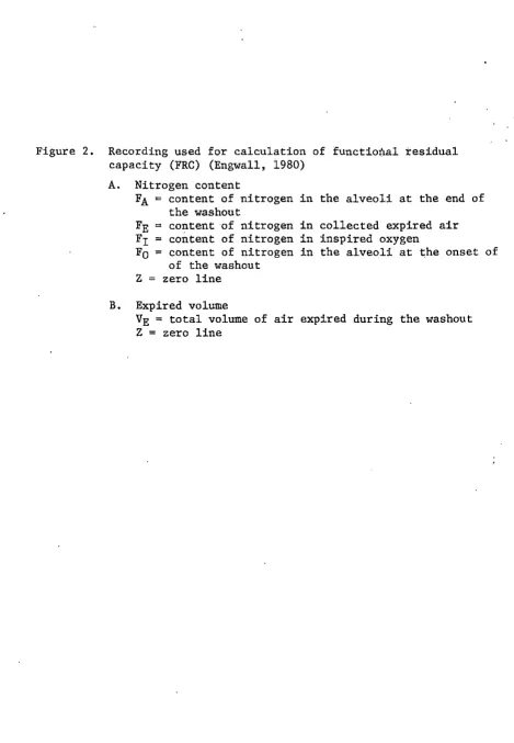
[image:22.588.39.490.45.713.2]FRC was calculated from the following.formula (Figure Z):
FRC (ml)
where VE = volume of expired air during washout (ml) VDS = dead space of the apparatus (about 45 ml)
FE = content of nitrogen in the expired air FI = content of nitrogen in the inspired oxygen F = 0 content of nitrogen in the alveoli pre-washout
FA= content of nitrogen in the alveoli post-washout
Since lung compliance and resistance can be affected by the frequency of respiration, all values have been normalized by dividing by the
fre-quency. Since FRC is affected by body weight, those values were corrected
by d;J.viding by the individual weights.
Liver Function
From the clearance curves for indocyanine green, the percentage
disappearance rate was calculated using the following formula: Percentage Disappearance Rate (PDR) = (1
-D = (Ln CZ - Ln
c
1)/(Tz - T1)where
c
1 = concentration at time T1 (mg/100 ml)CZ concentration at time T z (mg/100 ml)
D
e ) x 100
R
All calculations were performed on a programmable TI-55 calculator (Texas. Instruments, .Incorporated, Dallas, Texas). Programs for the
A. Nitrogen content
FA = content of nitrogen in the alveoli at the end of the washout
FE = content of nitrogen in collected expired air Fr = content of nitrogen in inspired oxygen
Fo = content of nitrogen in the alveoli at the onset of of the washout
Z = zero line
B. Expired volume
[image:24.569.52.522.51.729.2]n
8
·-Z--z-
A3
:I:
-·00~--Statistical data were provided by the SAS computer system, Iowa State University, with the assistance of D. J. Meerdink, Department of
RESULTS
Pulmonary Function
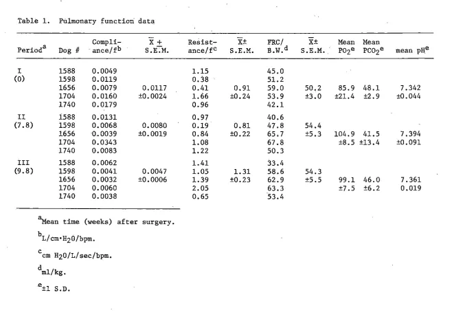
Results of the pulmonary function tests performed on the five dogs
are listed in Table 1. Measurements ·of compliance (C), resistance (R),
and functional residual capacity (FRC) were performed on all dogs at
selected times during the experimental period.
Control values are designated in period 1 for all measurements. They
are within the limits determined in our laboratory for normal dogs and are
also within the range reported by Engwall (1980).
Four of the five dogs studied showed 66.6% decreases in compliance in the first 10-12 weeks after surgery. Three of those four then returned to
control in subsequent weeks. The fifth dog showed a 26.5% increase in compliance which then decreased to approach the control value by the 18th
week.
Three of the five dogs exhibited a 46.3% increase in resistance during the first 10-12 weeks but subsequently returned to control levels.
The ,other two. dogs showed 42.5% decreases in resistance in the first eight
weeks, followed by sharp increases averaging 100% in the next two weeks, and then subsequent return toward normal by the 18th week.
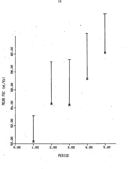
Functional residual capacity (FRC) increased 16 .. 3% in four dogs and
decreased 25.8% in one dog during the initial 10-12 weeks. However, no
Compli- X+ Resist- X± FRC/ X± Mean Mean
Period a Dog II ance/fb S. E;M. ance/fC S .E .M. ll.w.d S.E.M .. Po2e PCOze mean pH.e
I 1588 0.0049 1.15 45.0
(0) 1598 0.0119 0.38 51.2
1656 0.0079 0.0117 0.41 0.91 59.0 50.2 85.9 48.1 7.342 1704 0.0160 ±0.0024 1.66 ±0.24 53.9 ±3.0 ±21.4 ±2. 9 ±0.044
1740 0.0179 0.96 42.1
II 1588 0.0131 0.97 40.6
(7. 8) 1598 0.0068 0.0080 0.19 0.81 47.8 54.4
1656 0.0039 ±0.0019 0.84 ±0.22 65.7 ±5.3 104.9 41.5 7.394
1704 0.0343 1.08 67.8 ±8.5 ±13.4 ±0.091
1740 0.0083 1.22 50.3
III 1588 0.0062 1.41 33.4
...,
N(9. 8) 1598 0.0041 0.0047 1.05 1.31 58.6 54.3
1656 0.0032 ±0.0006 1.39 ±0.23 62.9 ±5.5 99.1 46.0 7.361
1704 0.0060 2.05 63.3 ±7.5 ±6.2 0.019
1740 0.0038 0.65 53.4
~ean time (weeks) after surgery.
b L/cm•HzO/bpm. c cm H20/L/sec/bpm. d ml/kg.
[image:28.779.62.721.42.519.2]Compli- Resist- X± FRC/ X± Mean Mean
Period Dog II ance/f X±S.E.M. ance/f S. E .M. B,W. S.E.M. P02 PC02 mean pH
IV 1588 0.0051 0.36 31.8
(17.8) 1598 0.0104 0.0098
o.
36 0.83 46.0 57.291.6f 43.2f 7.410f 1656 0.0029 ±0.0026 0.39 ±0.28 64.0 ±8.5
1704 0.0163 1.54 81.3 ±12.7 ±5.4 ±0.016
1740 0.0145 1.51 62.9
v
1588 0.0050 1.09 28.2(31. 8) 1598 0.0106 0.0096 0.35 0.93 75.5 60.1
1656 0.0031 ±0.0026 0.60 ±0.21 51.9 ±9.6 90.5 41.6 7.370
1704 . 0.0176 1. 56 82.8 ±8.3 ±3.4 ±0 .. 048
1740 0.0118 1.04 62.1
N
[image:29.781.64.728.70.282.2]Since the surgical procedure on all five dogs was not performed on the.same day, the time of pulmonary function measurements after surgery was variable for each dog. Therefore, the times of measurement from the
initial were averaged along with the individual measurements of C, R, and FRC to aid in the statistical evaluation. By averaging these data, the
time after surgery was· condensed into five periods and the means and
standard error were calculated.
There was a significant decrease (P < 0.05) in compliance (Figure 3)
from the control for all five dogs. This was accompanied by a concurrent significant increase (P < 0.05) in resistance (Figure 4). The mean FRC
showed an increasing trend overall, but the increase was not significant
(Figure 5).
The changes in mean arterial oxygen content (P02), carbon dioxide (PC02), and pH were not significantly different from the control.
Liver Function Tests
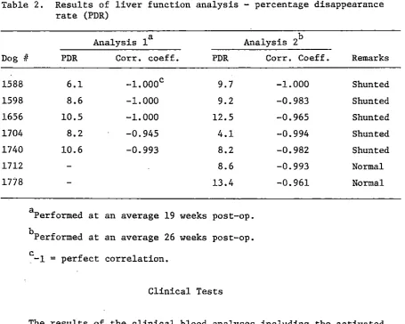
The res.ults from the liver function tests including percentage
dis-appearance rate and correlation coefficients from the clearance curves are given ~n Table 2. The data for analysis 1 were obtained at an average of
19 weeks after surgery for all animals. The percentage disappearance rate
for each animal during both liver function analyses shows no significant difference between the first and second analysis,. which was made seven
.
.,....,
N I a r-l x ~ ~ ::;;::: 0.. i:o... a
:£!
:.:: u ... -' ~ LU w z c:x: --' 0..::;;::: a
w z c:x: LU :;:: Cl
::P
.
~
Cl
t\J
.
~ Cl Cl ~ Cl ID Cl Cl
ID
.
0
0 ::P
o-+~~~~~..-~~~~~~~~~~..-~~~~~~~~~~..-~~-0. 00 1.00 2.00 3.00 l!.00 5.00
PERIOD
,....
:E
CL
~ u
UJ
[image:32.576.40.513.59.640.2]"'
...
__J...
D N
=
>E:u ... LW u z
""'
t;:;
·-
(/} •LW. 0:: z er UJ :E 0"'
-0C\J
.
-0
-.
-0 0
.
-0"'
0 0 <D ci-+~~~~~-r-~~~~-..~~~~~-r-~~~~-..~~~~~..-~~0.00 l,OO 2.00 3.00 ~.00
s.oo
PERIOD
~ CJ :,..:::
...
--' ::;: '--' . u ~ u.. z:i'5
:;::: 0 0 0 IO 0 0..
ID
lfl
0 0
.
IOlfl
0 0
.
::J' lfl 0 0
VI
0 0 0-+~~~~-r~~~~-.-~~~~-r-~~~~-r-~~~~.--~~ lflo.oo
1.00;::.oo
3.00. ILOOs.oo
PERIOD
Table 2. Results of liver function analysis - percentage disappearance rate (PDR)
Analysis la Analysis 2 b
Dog II PDR Corr. coeff.. PDR Corr. Coeff. Remarks
1588 6.1 -1. oooc 9.7 -1.000 Shunted
1598 8.6 -1. 000 9.2 -0.983 Shunted
1656 10.5 -1. 000 12.5 -0.965 Shunted
1704 8.2 -0. 945 4.1 -0.994 Shunted
1740 10.6 -0.993 8.2 -0.982 Shunted
1712 8.6 -0.993 Normal
1778 13.4 -0.961 Normal
a Performed at an average 19 weeks post-op.
b Performed at an average 26 weeks post-op. c . -1 = perfect correlation.
Clinical Tests
The results of the clinical blood analyses including the activated
coagulation times (ACT) for all five dogs are displayed in Appendix C. All values were normal at termination.
Post-Mortem Observations
Post-mortem examination results for all· six surgical an~mals indicate
that all animals demonstrated mild to moderate right heart dilatation.
Two of the dogs showed mild congestion in the lung, and four dogs showed congestion in the liver (Appendix D). Although three dogs showed signs of
[image:34.570.59.515.88.452.2]Gross examination of the portal vein and vena cava in the vicii;iity of the shunt revealed a nonpatent shunt in all dogs. Judging by the condition of the vessels, the shunt had been closed for some time.
Histop_athological examination revealed all ~nimals to have varying degrees of fibrosis and congestion in the liver, and also varying degrees of interstitial thickening, fibrosis, and congestion in the lungs. Most
DISCUSSION
The Fate of the Shunt
At termination of the experiment that the shunts were closed and apparently had been nonfunctional for some time. This finding immediately ruled out the major assumption made at the beginning of this project. The
assumption the shunts would remain patent indefinitely was based on two reasons: 1) the pressure gradient between the portal vein and the
vena cava would increase blood flow through shunt; and 2) since the vena cava is a lower resist~nce system than the liver, there would be a
tenden-cy for blood flow to pass through shunt. From these experimental data,
the two reasons appear valid for only the first ten weeks after surgery.
In preliminary research by the author, the portal pressure at the level of the splenic vein in one normal dog was 8 mmHg. Taylor and Myers
(1956) found portal pressures in normal cats to range from 5-13 mmHg with a mean pressure of 8 mmHg. The pressure in the.vena cava at the level of the liver is 2 mmHg (Swenson, 1977, p. 135). Therefore, the pressure dif-ference between the two vessels could be predicted to be 6 mmHg.
If the portal vein had been ligated as outlined in the original
pro-cedure (Mar~owitz et al., 1964), there is no doubt patency wo.uld have been
maintained. However, the complications from a complete shunt may have been a.greater problem. Markowitz et al. described in detail the, symptoms
of meat intoxication in dogs with complete Eck fistulas. They noted
re-curri?g instances of coma and lethargy in shunted dogs. Johnson and
losing 50% of their animals due to the same complications described by
Markowitz et al.
Because the portal vein was not ligated, the problems reported by
,
Markowitz et al. (1964) and Johnson and Lambert (1967) were notexperi-enced in this project. The liver was able to receive some portal blood.
The apparent reason for shunt failure can be attributed to not
ligating' the portal vein and possibly the method for producing the shunt.
The cut edges of the vessel walls would have provided enough connective tissue surface that platelets and fibrin strands could have been laid' down
immediately following surgery. Since the predicted pressure difference was small, it r.emains doubtful the shunt flow 'Would have been enough to
prevent further deposition of material in the shunt. Therefore, the shunt
would gradually become sealed over time.
Pulmonary Studies
Although the fistula failed, there was still a marked change observed
in both pulmonary structure and function. In all dogs subjected to
pulmonary studies, the maximum change in compliance and resistance values
occurred at an average of ten, weeks after surgery (period 3). After ten
weeks post-surgery, complianc~ and' resistance values approached,
psurgery levels. Although a positive correlation cannot be made, the re-turn of pulmonary measurements to normal may indicate the closure of the
shunt. Functional residual capacity (FRC) increased slightly. These
lung. There were marked signs of interstitial thickening and pulmonary edema accompanied by varying degrees of fibrosis in the dog lungs.
In cases of pulmonary edema, compliance decreases due to reduction in lung volume and interference with the elastic properties in the lung. 'concurrently, lung resistance increases due to the narrowing of the·small
airways as a result of the thickening of the peribronchial cuff. FRC usually increases when there is an increase in airway resistance. The
presence of fibrotic areas would also tend to decrease compliance, but
resistance and FRC would tend to either remain normal or decrease sli.ghtly
(West, 1977).
·The changes observ,ed in compliance, resistance, and FRC may be due to
not only interstitial edema.but also to the presence of microemboli, which were most prominant in ·dog 111582. Microemboli could play a P'.irt· in ·:·t~e.···
development of pulmonary hypertension (Sallam and Watson, 1970). '.!;here ·
was considerable right heart dilatation observed in all dogs which may have been produced by pulmonary hypertension. The various factors that
lead to pulmonary hypertension include obliteration of the capillary bed
by destruction of alveolar walls or interstitial fibrosis, and hypertrophy
of smooth muscle in the walls of small arteri~s (West, 1977). All dogs
demonstrated areas of tissue degeneration and fibrosis in the lung. Dr.
Andrews of the Diagnostic Laboratory, Iowa State University, observed that the
severity of tissue abnormality in the dogs of this project was correlated
to the amount of time passed since surgery was performed.
et al. (1968) demonstrated marked changes in anatomical structure of heart
and lungs after surgery for porto-caval anastomosis. Prior to surgery, these patients had liver cirrhosis and portal hypertension. However, after surgery, pulmonary hypertension and cor pulmonale was noted.
In patients with liver cirrhosis, Lebrec et al. (1979) agreed with
Senior et al. (1968) that the existence of pulmonary emboli was the cause
of the pulmonary hypertension. However, neither Lebrec et al. or Senior et al. could find the source of the emboli at autopsy.
Since no gross thrombi were found in this project, the pulmonary and
cardiac abnormalities that were observed are more related to the
porto-caval anastomosis than to the liver damage. Unlike the studies by Lebrec et al. (1979) and Senior et al. (1968), all dogs used in this project were
normal, healthy dogs. There were no symptoms or direct evidence of liver
cirrhosis, portal hypertension, excessive alcohol intake, smoking, or
other problems that were experienced by Lebrec et al. or Senior et al.
The.gross and microscopic abnormalities produced by the shunt in the dogs in this project were still apparent at termination 22 weeks later.
Because the shunt may be responsible for the changes in pulmonary function instead of the liver, the following question arises, "What
com-pound is contained in portal blood that would produce these changes?" In normal systemic circulation, compounds that would be normally detoxified by the liver could cause either the destruction of lung tissue or the
for-mation of microemboli. Both Lebrec et al. (1979) and Heinemann (1960,
1977) have proposed the existence of agents from the splanchnic or
There are several possibilities for the nature of this agent:
1) Microemboli that would form normally in the walls of the intestine could reach the lung parenchyma directly through the shunt.
2) Bacterial cells (particularly anaerobes) that gain access to the
portal vein from the intestine could exhibit a thrombogenic or
vasoconstrictive response when trapped by the lung capillaries.
The cell-mediated immune system could cause an edematous response in removing these bacterial cells.
3) Endotoxins from these bacterial cells could exhibit a hypertensive
response.
4) The circulating levels of digestion products would become
ab-normally elevated in the lung capillaries. Particularly, the concentrations of amino acids and fatty acids after a meal would
require an increased time to be cleared from the general
circula-tion. This may be enough to cause trauma to the lung vasculature.
Any of the four possibilities mentioned could be responsible for causing the pulmonary hypertension. These suggestions remove the major
emphasis from the liver as being the causitive agent responsible for changes in the lung.
Liver Function Studies
All the values for percentage disappearance rate (PDR) are within the
laboratory, Iowa State University. It should be noted that in some of the measurements, .the values are approaching the limits of normality. The activated coagulation time (ACT) results were within the normal range re-ported by Byars et al. (1976).
The function of the liver as determined by dye clearance methods
allowed the cellular function of the liver to be measured. It would also
have allowed information relating to the patency of the shunt to be
ob-tained. However, the dye clearance determinations were performed after
the shunt had probably closed. Stekiel et al. (1960) and Banaszak et al. (1960) reported a method to calculate mean hepatic transit time using
indo-cyanine green in dogs. The method required hepatic cannulation with the
assistance of a fluoroscope. and only total hepatic flow, not portal flow, could be established. Therefore, no simple noninvasive technique is
avail-able to measure portal flow or shunt flow without performing lapai:otomy. Although liver function was normal at termination, the liver was
apparently affected by the presence of the shunt. Histological examination revealed a marked disorganization in the liver parenchyma and an overall
fibrosis of the central veins in all dogs. Bradley et al. (1952, 1953) ' observed similar chan!les in cirrhotic livers in humans. Hepatic ischemia
and tissue hypoxia are common in the cirrhotic process, and these factors
were observed ·in this research. The cellular abnormalities observed in
the dogs in this project suggests serious results when portal blood flow
is reduced. The liver of the dog is highly dependent on portal blood for oxygen (Swenson, 1977, p. 135).
established by the clinical laboratory, Iowa State University. It should
be noted that in some of the measurements, the values are approaching the limits of normality. The activated coagulation time (ACT) results were within the normal range reported by Byars et al·. (1976).
Mathematical Model
A mathematical model of the liver and the shunt was developed to
characterize the flow patterns around the liver. The liver was treated as a single pool with two inputs and one output. Output from the lymphatic vessels of the liver is very small and was omitted. Two first-order
dif-ferential equations. were determined for both cases (normal and shunted)
and their derivations are given in Appendix E. The resulting equations
were programmed on a PDP-SA computer (Digital Equipment Corporation, Maynard, Massachusetts) and the results were displayed on a storage
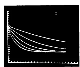
os-cilloscope. Figure 6 demonstrates both the normal and shunted hepatic ·clearance of a liver-specific dye (e.g., indocyanine green). As the
shunt diverts increasing amounts of blood away from the portal vein, the
ability of the liver to remove the dye from the general circulation becomes lessened and .the clearance time increases. When the ratio of normal
half-time (T~) to shunted half-time (T'~) was calculated, a relationship was established relating this ratio to the product of the shunt fraction (o)
and the ratio of total hepatic venous flow (QL) to the hepatic arterial
flow (QH):
T~ QL
From this relationship, the shunt fraction may be calculated if the half-time ratio and the liver flow ratio is known. Therefore, a
nonin-vasive technique may be available to determine the amount of shunt in an
animal.
[image:43.574.119.430.247.506.2]CONCLUSIONS AND RECOMMENDATIONS
In reviewing this research project, there is no doubt regarding the importance of maintaining a patent shunt. The results (or lack of
re-sults) obtained depended on a patent shunt at termination. More knowledge
concerning the function of the shunt has been obtained and future
experi-ments will be based on this knowledge.
An incomplete shunt (portal vein intact) appears to be patent for
only ten weeks. Therefore, a more concentrated study must be devoted to
the early period of.patency.
A complete shunt (portal vein ligated) or implanted static shunts may be necessary to maintain the patency of the shunt and produce the maximum
change in pulmonary and liver function. However, the complications
asso-ciated with portal ligation would cause adverse conditions that would affect collection of reliable data. Furthermore, there are no cases
re-ported of a complete anatomical shunt. Therefore, the complete shunt would not be an accurate model for the cirrhotic condition.
More data must be collected if changes in pulmonary and liver func~
tion are to be more accurately described. This will require more animals
and more frequent pulmonary and liver function, and clinical tests. This will hopefully eliminate any possible questions regarding events occurring
between long periods of time, which was a problem in this research.
and tested. The model would enable the researcher to obtain information regarding the dynamics of a porto-caval shunt without resorting to
SUMMARY
Five normal dogs were given porto-caval shunts using a modified tech-nique of the Eck fistula. Measurements of pulmonary function including compliance, resistance, and functional residual capacity, and arterial
oxygen· levels demonstrated significant decreases in compliance and in-creases in resistance. Functional residual capacity and arterial oxygen levels did not change significantly. Liver function determined by
clearance of indocyanine green and clinical blood tests including blood
urea nitrogen, glucose, bilirubin, albumin, SGPT, and alkaline phosphatase
were normal at termination. There was marked right heart dilatation in all dogs. Histologically, the lung demonstrated considerable edema and
interstitial thickening in all dogs. The liver showed mild to moderate
ischemia and fibrosis. A mathematical model simulating dye clearance in
LITERATURE CITED
Banaszak, E. F., W. J. Stekiel, R. A. Grace, and J. J. Smith. 1960. Estimation of hepatic blood flow using a single injection dye clear-ance method. Am. J. Physiol. 198(4):877-880.
Banner, A. S. 1973. Pulmonary function in chronic alcoholism. Am. Rev. Resp. Dis. 108:851-857.
Berthelot, P., J. G. Walker, S. Sherlock, and L. Reid. 1966. Arterial changes in the lungs in cirrhosis of the liver-lung spider nevi. N. Engl. J. Med. 274:291-298.
Bradley, S. E., F. J. Ingelfinger, and G. P. Bradley. 1952. Hepatic circulation in cirrhosis of the liver. Circulation 5:419-429. Bradley, S. E., C. M. Smythe, H.F. Fitzpatrick, and A.H. Blakemore.
1953. The effect of a portalcaval shunt on estimated hepatic blood flow and oxygen uptake in cirrhosis. J. Clin. Invest. 32:526-537. Burch, G. E., and N. P. Depasquale. 1967. Alcoholic lung disease - an
hypothesis. Am. Heart J. 73(2):147-148.
Byars, T. D., G. V. Lung, N. A. Ferris, and K. S. Keeton. 1976. vated coagulation time (ACT) of whole blood in normal dogs. Vet. Res. 37(11):1359-1361.
Acti-Am. J.
Calabresi, P., and W. H. Abelmann. 1957. Porto-caval and porto-pulmonary anastomoses in Laennec's cirrhosis and in heart failure. J. Clin. Invest. 36:1257-1265.
Cameron, G. R. 1948. Pulmonary oedema. Br. Med. J. 1:965-972.
Cotes, J. E., G. B. Field, G. J. A. Brown, and A. E. Read. 1968. Impair-ment of lung function after portacaval anastomosis. Lancet
1:952-955.
Emirgil, C., and B. J. Sobol. 1977. Pulmonary function in former alcoholics. Chest ?2(1) :45-51.
Emirgil, C., B. J. Sobol, B. Heymann, and K. Shibutani. 1974. Pulmonary function in alcoholics.
Am.
J. Med. 57:69-77.Engwall, M. J. associated
Iowa State
1980. Changes in pulmonary hemodynamics and lung function with an acute intravenous dose of ethanol. M. S. thesis. University, Ames.
Heinemann, H. O. 1977. Alcohol and the lung - a brief review.
Am.
J. Med. 63:81-85.Johnson, G., and J. Lambert. 1967. Cardiac output after porto-caval shunt. Ann. Surg. 166:207-212.
Lebrec, D., J.P. Capron, D. Dhumeaux, and J.P. Benhamou; 1979.
Pulmonary hypertention complicating portal hypertention.
Am.
Rev. Resp. Dis. 120:849-856.Lieber,
c.
S. 1976. The metabolism of alcohol. Sci.Am.
234:25-33. Markowitz, J., J. Archibald, and H. G. Downie. 1964. Experimentalsurgery. 5th ed. The Williams and Wilkins Company, Baltimore, Md. 659 pp •
. Nakamura, T., S. Nakamura, T. Tazawa, S. Abe, T. Aikawa, and K. Tokita. 1965. Measurement of blood flow through porto-pulmonary anastomosis in portal hypertension·. J. Lab. Clin. Med. 65:114-121.
Ruff, F., J.M. B. Hughes, N. Stanley, D. McCarthy, R. Green, A. Aronoff, L. Claton, and J. Milic-Emili. 1971. Regional lung function in patients with hepatic cirrhosis. J. Clin. Invest. 50:2403-2413. Sallam, M., and W. C. Watson. 1970. Pulmonary hypertension due to
micro-thromboembolism from splenic and portal veins after portacaval anastomosis. Br. Heart J. 32:269-271.
Senior, R. M., R. C. Britton, G. M. Turino, J. A. Wood, G. A. Langer, and A. P. Fishman. 1968. Pulmonary hypertension associated with cirrho-sis of the liver and with portacaval shunts. Circulation 37:88-96. Shackelford, R. T. 1955. Bickham-Callendar surgery of the alimentary
tract. Vol. 1. W. B. Sanders Co., Philadelphia. 862 pp.
Shaldon, S., J. Caesar, L. Chiandussi, H. S. Williams, E. Sheville, and S. Sherlock. 1961. The demonstration of porta-pulmonary anastomoses in portal cirrhosis with the use of radioactive krypton. N. Engl. J. Med. 265:410-414.
Stekiel, W. J., J.P. Kampine, E. F. Banaszak, and J. J. Smith. 1960. Hepatic clearance of indocyanine in the dog.
Am.
J. Physiol. 198(4): 881-885.Taylor, W. J., and J. D. Myers. 1956. ization in the study of the normal noncirrhotic portal hypertension.
Occlusive hepatic venous catheter-liver, cirrhosis of the liver and Circulation 13:368-380.
West, J. B. 1977. Pulmonary pathophysiology - the essentials. The Williams and Wilkins Company, Baltimore, Md. 227 pp.
Wheeler, .H. O. , W. L Cranston, and J. I, Meltzer. and biliary excretion of indocyanine green in Exptl. Biol. Med. 99:11-14.
1958. Hepatic uptake the dog. Proc. Soc.
White, A., P. Handler, and E. L. Smith. 1973. Principles of bio-chemistry. 5th ed. McGraw-Hill Inc., New York.
Williams, J, H., and W. H. Abelmann. 1963. Portopulmonary shunts in
ACKNOWLEDGMENTS
I would like to express my most sincere gratitude to my committee members Dr. Richard Engen, Dr. Richard Seagrave, and Dr. Bernard White for tolerating my lack of knowledge during the course of this research .•
I would also like to extend my.heart-felt thanks to Mrs. Joyce Feavel for
her expertise in surgical procedure, Dr. Robert Glock and Dr. John Andrews
for their assistance with the pathological aspects of this research, to Mr. Denis Meerdink for the time he spent playing with the computer on my
behalf, and to Charm Nickey and Barb Steele for their expert skills used
in the preparation of this thesis. Finally, I wish to extend my thanks and gratitude to Mr. Mark Darrah for helping me find the courage and the
APPENDIX A: SURGICAL PROCEDURE
The following is a detailed description of the methods used to create a porto-caval shunt from the procedure for performing an Eck fistula, as
described by Markowitz et al. (1964, pp. 542-546).
After fasting 24 hours, each dog was anesthetized with sodium pento-barbital (28.6 mg/kg). The animal was then intubated and prepared for
surgery by shaving hair from dorsal to ventral midline on the right side extending from mid-thorax to the hind limb. Following preparation of the
surgical site asceptically, the animal was placed on the table in dorsal
recumbency with exposure favoring the lateral right side. The table was
equipped with a circulating heating water pad to control body temperature during the procedure.
The incision was made starting from the xiphoid process down the
midline for approximately 4 cm, then down at an angle paralleling the
costal margin to just beyond the nipple line, extending to about the level
of the umbilicus. Stay sutures were placed on the muscle groups to be in-cised to aid in alignment on closure and care was taken to control
bleed-ing as the Rectus abdominus, internal abdominal oblique, a section of the external abdominal oblique, and the Transversus abdominus were cut. Upon
entering the peritoneum, the incision was retracted and after wrapping the viscera in the mesenteric membrane, the viscera were removed from the
cavity and placed in a moist, sterile turkish towel and placed to the left side of the animal.
After stripping fat and overlying fascia away from both portal vein
cardio-\
Figure 7.. The Eck fistula; the portal vein (right) has been .att:a.ched tp the vena cava (left) by stay sutures (arrows) a!ld the posterior · suture line is laid with the bottom needle. (Markowitz eLal.·,
[image:52.576.57.534.52.569.2]vascular si~k to attach the two vessels together. Two needles were used for the two stay sutures and the needles were left intact for the next
step.
A posterior suture line was then placed continuously between the two vessels using a length of 5-0 silk distal to the liver. Care was taken to
control bleeding as the needle was passed through the portal wall, as the
wall is very thin and tears quite easily.
Upon completion of the posterior suture line, a cutting thread of
1-0 gastrointestinal silk was then placed in both vessels near and
paral-lel to the suture line (Figure 8a).
After the cutting· thread was placed, the 5-0 silk proximal to the liver was used to place the anterior suture line in the same manner as before (Figure 8B). When completed, the two vessels are aligned (Figure
8C) with the cutting thread buried between them.
The cutting thread was carefully pulled free in a back-and-forth
motion, cutting the walls of both vessels and creating the fistula in the process. In some cases, there was some bleeding from the exit point of the cutting thread which was controlled by placing a single interrupted
suture of 5-0 silk to further close the exit point.
Once it was certain hemostasis had been achieved, the viscera were replaced, the retractor removed, and the incision was closed using 3-0
silk in a continuous pattern, taking care to align the layers of muscle
and fascia. The skin was closed using VetafilR (S. Jackson, Inc.,
c.
A.
B.
Figure 8. The Eck fistula; the portal vein is on the right and the vena cava is on the left. A) The cutting thread is introduced
parallel to the posterior suture line. This thread will create the fistula when pulled free. B) The anterior suture line draws both vessels together and buries the cutting thread be-tween the two vessels. C) Principle of action of the cutting thread. The vessels are aligned as shown in cross-section. Pulling the cutting thread free will complete the fistula.
[image:54.571.61.475.72.616.2]The technique described above was slightly modified from the original description (Markowitz et al., 1964) in that the portal vein was not
APPENDIX B: CALCULATION PROGRAMS FOR TI-55 CALCULATOR
Compliance-Resistance Program
Key stroke Step Key code Key stroke Step Key code
RCL 00 61 = 16 85
2 01 02 + 17 45
+ 02 45 RCL 18 61
RCL 03 61 0 19 00
4 04 04 = 20 85
05 85 R/S 21 86
R/S 06 86 RST 22 87
l/X 07 34 23 00
x
08 55 24 00RCL 09 61 25 00
1 10 01 26 00
= 11 85 27 00
+/-
12 84 28 00+
13 75 29 00RCL 14 61 30 00
3 15 03 31 00
Memory locations Value for
STO 0 Flow (F)
STO 1 Tidal volume at a flow (VTF)
STO 2 Tidal volume (VT)
STD 3 Pressure at a flow (PTF) STO 4 Esophageal pressure (PT)
Procedure
1) Store values from Dynagraph recordings into memory.
Functional Residual Capacity Program
Key stroke Step Key code Key stroke Step
STO 00 51 R/S 16
0 01 00 STO 17
~y 02 31 1 18
SUM 03 71 ~y 19
0 04 00
+/-
20·R/S 05 86 SUM 21
STO 06 51 1 22
1 07 01 EXC 23
X:tY
08 31 1 24+/-
09 84 l/X 25SUM 10 71 PROD 26
.l 11 01 0 27
EXC 12 66 RCL 28
1 13 01 0 29
PROD 14 76 R/S 30
0 15 00 RST 31
Memory locations 0 and 1 are used for temporary purposes.
Procedure
1) Enter deadspace volume (VDs> = 45.0. 2) Push interchange key (~Y).
3) Enter expired air volume (VE).
4) Push run (R/S) key - disregard display.
5) Enter nitrogen content of inspired oxygen (F.1).
6) Push interchange key.
7) Enter nitrogen content of expired air (FE). 8) Push run key - disregard display.
9) Enter post-was~out alveolar nitrogen content (FA).
10) Push interchange ~ey ..
11) Enter pre-washout alveolar nitrogen content (F0). 12) Push run key - display shows calculated FRC.
Key code
Percentage Disappearance Ra~e Program
Key stroke Step Key code Key stroke Step Key code
STO 00 51 RCL 16 61
1 01 01 3 17 03
X*Y 02 31 = 18 85
• 03 45 INV 19 21
RCL 04 61 PROD 20 76
1 05 01 2 21 02
= 06 85 RCL 22 61
lnX 07 23 2 23 02
STO 08 51 eX 24 24
2 09 02
+/-
25 84R/S 10 86
+
26 75STO 11 51 1 27 01
3 12 03 = 28 85
Y.;toY 13 31 R/S 29 86
+/-
14 84 RST 30 87+
15 75 31 00Memory locations 1, 2, and 3 are temporary only. Procedure
1) Enter value for
c
2.2) Push interchange key (X.,,Y) • 3) Enter value for
c
1•4) Push run (~/S) key.
5) Enter value for
r
1 .6) Push interchange key (Y.;toY). 7) Enter value for
r
2 .8) Push run (R/S) key.
9) Multiply display value by 100.
10) Display shows calculated percentage disappearance rate.
APPENDIX C: CLINICAL TEST RESULTS
Dog II
Test 1588 1598 1656 1704 1740
Urea nitrogen (rng/dl) 12 18 18 21 16
Glucose (mg/dl) 102 75 93 81 89
To.tal bilirubin (mg/dl) <1.0 <1.0 <1.0 <1.0 <1.0 Direct bilirubin (mg/dl) <1.0 <1.0 <1.0 <1.0 <1.0
Albumin (gm/ dl) 3.6 3.3 4.1 3.2 3.0
SGPT (IU/l) 19.2 15.4 17.5 15.7 62.4
\
APPENDIX D: POST-MORTEM OBSERVATIONS
The following observations were made at termination for the six dogs used in this project. Included are gross as well as histopathological
observations, as determined by Dr. Robert Glock, Department of Veterinary Pathotogy, Iowa State University, and Dr. John Andrews, Diagnostic
Laboratory, College of Veterinary Medicine, Iowa State University.
Gross Observations
Observations were made only if organ or tissue had visible signs of
abnormality. All other normal tissues were not listed. The animal's
breed, sex, weight, and time since surgery are given.
Dog #1582: Irish Setter, male, 22.7 kg, 43 weeks. The lung had
raised grey plaques 1-2 cm on both diaphragmatic lobes and what appeared to be pleural thickening. There was moderate right heart dilatation.
Dog #1588: Husky, female, 25.4 kg, 33 weeks. Hair on right side never grew back after surgery and numerous skin lesions were noted. The brain had moderate internal hydrocephalus. The liver was 740 gm and
appeared slightly congested with rounded edges. The heart showed very mild right heart dilatation with a calcified plaque on the endocardial
surface of the right ventricular wall. The kidneys were normal except
for a surgical sponge adhered to the anterior pole of the right kidney. There was a mass of adhesions in the mesentery.