R E S E A R C H
Open Access
Fitness components and natural selection: why
are there different patterns on the emergence of
drug resistance in
Plasmodium falciparum
and
Plasmodium vivax
?
Kristan A Schneider
1,2*and Ananias A Escalante
3,4*Abstract
Background:Considering the distinct biological characteristics ofPlasmodiumspecies is crucial for control and elimination efforts, in particular when facing the spread of drug resistance. Whereas the evolutionary fitness of all malarial species could be approximated by the probability of being taken by a mosquito and then infecting a new host, the actual steps in the malaria life cycle leading to a successful transmission event show differences among Plasmodiumspecies. These“steps”are called fitness components. Differences in terms of fitness components may affect how selection imposed by interventions, e.g. drug treatments, differentially acts on eachPlasmodiumspecies. Thus, a successful malaria control or elimination programme should understand how differences in fitness components among different malaria species could affect adaptive evolution (e.g. the emergence of drug resistance). In this investigation, the interactions between some fitness components and natural selection are explored.
Methods:A population-genetic model is formulated that qualitatively explains how different fitness components (in particular gametocytogenesis and longevity of gametocytes) affect selection acting on merozoites during the erythrocytic cycle. By comparingPlasmodium falciparumandPlasmodium vivax,the interplay of parasitaemia and gametocytaemia dynamics in determining fitness is modelled under circumstances that allow contrasting solely the differences between these two parasites in terms of their fitness components.
Results:By simulating fitness components, it is shown that selection acting on merozoites (e.g., on drug resistant mutations or malaria antigens) is more efficient inP. falciparumthan inP. vivax. These results could explain, at least in part, why resistance against drugs, such as chloroquine (CQ) is highly prevalent inP. falciparumworldwide, while CQ is still a successful treatment forP. vivaxdespite its massive use. Furthermore, these analyses are used to explore the importance of understanding the dynamic of gametocytaemia to ascertain the spreading of drug resistance. Conclusions:The strength of natural selection on mutations that express their advantage at the merozoite stage is different inP. vivaxandP. falciparum. Species-specific differences in gametocytogenesis and longevity of gametocytes need to be accounted for when designing effective malaria control and elimination programmes. There is a need for reliable data on gametocytogenesis from field studies.
Keywords:Fitness components, Natural Selection, Primaquine, Artemisinin based combination therapy, Malaria elimination, Gametocytogenesis
* Correspondence:[email protected];[email protected]
1
Department of MNI, University of Applied Sciences Mittweida, Mittweida, Germany
3
Center for Evolutionary Medicine and Informatics, Arizona State University, Tempe, USA
Full list of author information is available at the end of the article
Background
Malaria burden has made this disease a significant bar-rier for reaching global development. Malaria, however, is not one disease. Whereas Plasmodium falciparum is responsible for most cases in Africa, endemic regions outside of Africa are characterized by the presence of Plasmodium vivax, the second most important malaria parasite in terms of its morbidity. Although they are found in sympatry across endemic regions worldwide, there are well-known differences between P. falciparum and P. vivax. Indeed, they differ in key aspects of their basic biology, geographic range, and evolutionary origins [1-4]. Such distinctive characteristics affect control and elimination managements of these two malarias; specif-ically in terms of (i) treatment of their associated clinical diseases and (ii) the design of new control tools, such as choosing candidates for anti-malarial vaccines.
Regardless of these noticeable biological differences, it has not been properly discussed yet how interventions acting as selective forces (e.g. anti-malarial drugs) differ-entially affect each species. Such assessment is important considering that malaria elimination will face areas where these two parasites are prevalent. Even in Africa, where P. vivax is rightfully neglected when compared with the impact of P. falciparum, there is increasing evidence indicating that this parasite is present, but at a lower prevalence [5,6].
An example of differences between these two species, in terms of emerging adaptations hampering the efficacy of interventions, can be found in the onset of chloro-quine (CQ) resistance. Whereas in P. falciparum CQ resistance originated and spread worldwide as result of several independent events [7], it remains the drug of choice for treating P. vivax in most areas worldwide. CQ-resistantP. vivax strains are typically present–if at all – at low prevalence, and resistance-levels are low compared with P. falciparum [8]. Why did drug resist-ance emerge slower in P. vivax? There aread hoc expla-nations for this observation, e.g., resistance may have a greater cost inP. vivaxor that CQ targets a different path-way in this parasite. Although these explanations are un-doubtedly intuitive, they might confuse other mechanisms. Likewise, there is extensive evidence of balancing selection acting on major malaria antigens expressed in the mero-zoite stage inP. falciparum,whereas inP. vivaxsuch pat-terns are harder to detect [9-12]. Again, it could be argued that the immune system does not necessarily recognize the same antigens equally efficiently in both parasites. More-over, there are some species-specific proteins involved in the invasion of the red blood cells that could alter the over-all immune response ([13] and references within there).
Regardless of the intellectual merit of thesead hoc expla-nations, it is worth exploring alternative arguments based on formal foundations; this allows proposing hypotheses
susceptible to be tested with empirical data. Consequently, operational research will be enriched by providing new perspectives that will support sustainable evidence-based malaria-elimination programmes. Following this logic, this article discusses how the distinct biological characteristics of different Plasmodium species may explain, at least in part, the differences observed in terms of patterns asso-ciated with some forms of positive selection.
selection has on the parasite population in terms of spreading mutations that may compromise the lifespan of tools used for control and elimination.
Methods
Basic terminology and assumptions
For the present purposes, parasitaemia and gameto-cytaemia are considered as population averages. In a single malaria episode, parasitaemia increases in discrete cycles and will be under strong stochastic fluctuations. However, averaged over all malaria episodes in the population, parasitaemia will approximately follow a deterministic, continuous growth function (see Figures 1 and 2). The same holds for the gametocytaemia dynamics. In particular, not every malaria infection has gameto-cytaemia, but the population average will again follow a deterministic growth function.
In what follows‘resistant’,‘sensitive’, and‘drug pressure’ can be substituted (with the necessary modifications) by ‘beneficial’,‘deleterious’, and ‘selective force’, respectively, to obtain a more general interpretation. Resistant para-sites might be associated with metabolic costs [17,18]. Hence, the population-average dynamics of resistant and sensitive parasites will be different, even in the absence of drug pressure. Notably, the emphasis of this population genetic model is on the ensemble of the various fitness components for each parasite species, not on the specifics of the parasitaemia and gametocytaemia dynamics. Overall, this population genetic model is used to compare P. vivax and P. falciparum in terms of how two specific fitness components, time of gametocyte production and their lifespan, affect the dynamics of an advantageous mutation expressed in the merozoite stage. Thus, the figures properly summarize the results. In order to contrast the importance that differ-ences in gametocytaemia dynamics may have, a specific example on the effect of patient isolation may have on the spread of mutations associated with resistance in P. vivax and P. falciparumis discussed. The mathem-atical detail and the parameters chosen are described below. Readers shall feel free to skip the mathematical details as the figures guide the intuitive argument without mathematical formalism.
Model
Evolutionary dynamics
Schneider and Kim [19,20] derived a model for the spread of resistant associated mutations tailored to the transmission cycle ofP. falciparum. The model incorpo-rated the possibility of co-infections, different numbers of sporozoites or sporozoite-strains infecting hosts, and heterogeneity among hosts (e.g., due to treated and untreated infections). Denoting the relative frequency of resistant parasites in generationtby p(t), it was shown
that the evolutionary dynamics follow the recursion equation
p tð Þ ¼ pð Þ0 λ t
pð Þ0 λtþð1pð Þ0 Þμt¼
1
1þ1pð Þp0ð Þ0 λμ t ; ð1Þ
where timetis measured in discrete units of transmission cycles (i.e., one generations corresponds to one full trans-mission cycle - from sporozoite to sporozoite). Moreover,λ and μ are the fitnesses of resistant and sensitive parasites averaged over heterogeneous clusters of hosts with different responses to the infection, e.g., over treated and untreated hosts. The average fitnessesλandμare the likelihoods that, respectively, resistant or sensitive sporozoites causing an infection lead to gametocyte-offspring that are taken by a mosquito during its blood meal. Therefore, fitness is the results of various events that take place during the life cycle, the fitness components. Particularly,λandμdepend on the average parasitaemia and gametocytaemia dynamics within infections. In the following, it is explained how the various fitness components assemble. From (1) it is clear that only the ratio λμ (the relation between the fitnesses of resistant and sensitive parasites) is considered in the evolu-tionary dynamics. If a mutation occurs that renders a para-site to be resistant–either in the human host or mosquito vector–initially only few sporozoites in one or a few mos-quitoes will be resistant. Hence, ifNdenotes the number of sporozoites counted over all mosquitoes participating in a given transmission cycle, the initial frequency of resistant parasitesp(0) is on the order of 1
N. Notably, the formulation of the model incorporated co-infections by several mosqui-toes. However, the dynamics and spread of resistant-associated mutations (at a single locus) are independent from the likelihood of co-infections (for more details see [19]). The situation would change with mutations interact-ing at two linked loci; however, such a more complex scenario is not considered at this point.
The transmission cycle forP. vivaxis similar to that of P. falciparum, an important difference is that P. vivax infections involve liver-stage hypnozoites that can result in relapses. In a model that considers different merozoite variants [19,20], this can be approximated by assuming a higher likelihood of co-infections, since relapses will generate overlapping parasite generations. Ideally, the fitnesses of resistant and sensitive parasites inP. vivaxneed to be adjusted accordingly by considering the evolutionary dynamics of hypnozoites. This is especially important if hypnozoites are never treated in the population. However, such models involve different sets of assumptions that should be explored somewhere else.
Parasitaemia dynamics
respectively, resistant and sensitive parasites in the absence of drugs is modelled according to the Holling Type III functions
PSð Þ ¼t 1þPmaxξρτ
S andPRð Þ ¼τ Pmax
1þξρτ
R
where Pmax is the threshold parasitaemia, i.e., the
max-imum number of parasites that can be sustained within an infection. Here, PS and PR correspond to growth
para-meters of resistant and sensitive parasites, andξis a fitting parameter. The above dynamic imply exponential growth 0 2 4 6 8 10 12 14 16 18 20 22 24 26 28
0. 0.1 0.2 0.3 0.4 0.5
0. 0.2 0.4 0.6 0.8 1.
time (days after blood stage starts)
Parasites
(x10 )/
µ
L in
P. falc.
6
Parasites
(x10 )/
µ
L in
P. vivax
4
symptoms
start
gametocytes
in
P
.
vivax
treatment
gametocytes
in
P
.falc
.
Sensitive Resistant
0 2 4 6 8 10 12 14 16 18 20 22 24 26 28 0.
40. 80. 120. 160. 200.
0. 100. 200. 300. 400. 500.
time (days after blood stage starts)
gametocytes/
µ
L in
P. falc.
gametocytes/
µ
L in
P. vivax
symptoms
start
gametocytes
in
P
.
vivax
treatment
gametocytes
in
P
.falc
.
Sensitive P. vivax
Resistant P. vivax
Sensitive P. falciparum
Resistant P. falciparum
0 500 1000 1500 2000 2500 3000
0. 0.2 0.4 0.6 0.8 1.
0. 0.5 1. 1.5 2.
generations
prevalence of resistanc
e
fitness res./sens
.
P. vivax P. falciparum
B
A
[image:4.595.59.538.89.556.2]C
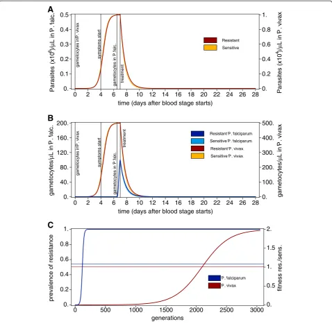
Figure 1Intra-host and population-level dynamics resulting from fitness components. A) Population-average dynamics of asexual
0 2 4 6 8 10 12 14 16 18 20 22 24 26 28 0.
0.1 0.2 0.3 0.4 0.5
0. 0.2 0.4 0.6 0.8 1.
time (days after blood stage starts)
time (days after blood stage starts)
Parasites (x10 )/
µ
L in
P. falc.
6
symptoms
start
gametocyte
s
in
P
.
vivax
treatment
gametocyte
s
in
P
.falc
.
Sensitive Resistant
0 2 4 6 8 10 12 14 16 18 20 22 24 26 28 0.
40. 80. 120. 160. 200.
0. 40. 80. 120. 160. 200.
gametocytes/
µ
L in
P. falc.
gametocytes/
µ
L in
P. vivax
symptoms
start
gametocytes
in
P
.
vivax
treatment
gametocytes
in
P
.falc
.
Sensitive P. vivax
Resistant P. vivax
Sensitive P. falciparum
Resistant P. falciparum
0 500 1000 1500 2000 2500 3000 3500 4000 0.
0.2 0.4 0.6 0.8 1.
0. 0.5 1. 1.5 2.
generations
prevalence of resistanc
e
fitness res./sens
.
P. vivax P. falciparum
0 50 100 150 200 250 300
0. 0.2 0.4 0.6 0.8 1.
0. 0.5 1. 1.5 2.
generations
prevalence of resistanc
e
fitness res./sens
.
P. vivax P. falciparum
A
B
C
[image:5.595.59.537.84.713.2]D
of sensitive and resistant parasites at low density after a slow initial establishment. However, once the parasitaemia approachesPmax,it will level off due to a carrying capacity. The Greek letterτrefers to the time scale of the duration of an average infection. Moreover,τ= 0 denotes the start of blood stage parasitaemia (erythrocytic cycle), and τis scaled in days. The relationρS>ρRimplies metabolic costs
for resistance, i.e., a slower growth of resistant parasites [17,18].
After, the onset of drug treatment, parasitaemia will decrease exponentially. In particular, the dynamics are modelled by PSð Þ ¼τ 1þPmaxξρτ
S e
δsðττ0Þ and P
Rð Þ ¼τ 1þPmaxξρτ R eδRðττ0Þ for τ > τ
0, where τ0denotes the time that drugs become effective, andδS, andδRdenote the death rates of sensitive and resistant parasites, respectively. Naturally,δS>δRholds.
Moreover, Pmax is replaced byPmaxfalc and Pmaxvivax to spe-cifically modelP. falciparum and P. vivax. Hence, apart from a scaling constant both parasite species are assumed to follow the same parasitaemia dynamics. In a more compact form these are given by
Pð Þ ¼τ P
1þξρτfð Þτ ð2Þ
where fð Þ ¼τ eδ∗ðττ0Þ for τ> τ
0 and f(τ)≡1 for τ>
τ0 (the asterisk in the subscript is a placeholder for R
andS, and that in the superscript forfalcandvivax). The above parasitaemia dynamics are population averages including all treated and untreated infections.
Gametocytaemia dynamics
While parasitaemia dynamics determine the performance of parasites within an average malaria episode, parasites’ fitnesses on a population scale are determined by their ability to transmit to other hosts. The fitness component determining transmissibility is summarized by the gameto-cytes’dynamics. The population-average gametocytaemia, after its appearance, is assumed to be proportional to the population-average parasitaemia.
InP. falciparumepisodes gametocytes appear at a later stage of the infection [1,14-16]. The average dynamics amongP. falciparuminfections are assumed to follow
GfalcS ð Þ ¼τ φPSfalcð Þτ gfalcð Þτ and
GRfalcð Þ ¼τ φPRfalcð Þτ
ð3Þ
whereφis the percentage of blood-stage parasites develop-ing into gametocytes. Moreover, gfalc(τ) = 0 for τ1falc>τ> 0
gfalc(τ) = 64(τ−τ1falc)3gfalc(τ) = 0 forτ1falc>τ> 0,gfalc(τ) = 64(τ
−τ1falc)3(1−τ+τ1falc)3for τfalc1 þ12>τ>τ
falc
1 andg
falc
(τ) = 1 for τ>τfalc1 þ1
2. The function g
falc
(τ) ensures that gametocytaemia has a smooth onset over 12 hours and does not immediately jump from zero toφPmaxfalc. This is
mainly for illustrative purposes. The outcome of the model is independent of the particular choice of this function.
Gametocytaemia appears readily inP. vivaxinfections, i.e., at timeτ1vivax= 0 [1,14-16]. Hence, the gametocytae-mia dynamics follow
GvivaxS ð Þ ¼τ vPvivaxS ð Þτ fSð Þτgvivaxð Þτ and
GvivaxR ð Þ ¼τ vPRvivaxð Þτ fRð Þτ gvivaxð Þτ
ð4Þ
wherevis the percentage of blood-stage parasites devel-oping into gametocytes in P. vivax and gvivax(τ) is defined asgfalc(τ)withτ1falcreplaced byτ1vivax.
Note, that the functions g(τ) account for the different
onsets of gametocytogenesis inP. falciparumandP. vivax. Namely, gametocytogenesis starts with the erythrocytic cycle in P. vivax, (about a week before drug treatment starts). However, in P. falciparum gametocytogenesis starts shortly before drug treatment starts [1,14,16].
Life-span of gametocytes
Alternatively to the above, more realistic gametocytaemia dynamics can be assumed by including an additional fitness component. Namely, gametocytes are non-reproducing; they derive from merozoites and die after a while if they are not transmitted by a mosquito vector.
Hence, the number of gametocytes at time τ in, respectively,P. falciparumandP. vivaxare
Gfalcð Þ ¼τ
Zτ
Max½0;τβfalc
φPfalcð Þz fSð Þz gfalcð Þz dz ð5Þ
and
(See figure on previous page.)
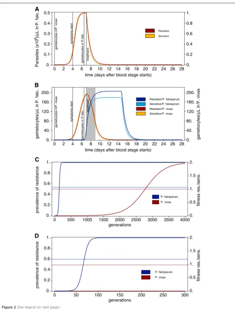
Figure 2Intra-host dynamics with refined gametocytaemia. A) As Figure 1A with parameters described in Methods.B) As compared to
Gvivaxð Þ ¼τ
Zτ
Max½0;τβvivax
νPvivaxð Þz fð Þz gvivaxð Þz dz
ð6Þ
whereβvivaxandβfalcare the life spans of gametocytes in
P. vivaxandP. falciparum, respectively.
Fitness
The various fitness components can be combined to determine fitness. The fitness of a parasite is the prob-ability that a gametocyte offspring is transmitted to a different host, and hence can further participate into the transmission cycle. Any gametocyte can be picked up by a mosquito whenever gametocytaemia exceeds a threshold Gcritvivax, or Gcritfalc forP. vivaxor P. falciparum,
respectively. Hence, the fitnesses of parasites are pro-portional to the respective mean value of the average gametocytaemia, which is taken over the time window when gametocytaemia exceeds its threshold. Hence fitness
is proportional to
Zb
a
Gð Þτ dτ.
Here, (aRvivax,bRvivax), (avivaxS ,bvivaxS ), (aRfalc,bRfalc), (aSfalc,bSfalc),
denote the time intervals in which resistant and sensitive gametocytes exceed the threshold gametocytaemia to be transmittable in P. vivax, and P. falciparum, respectively. This implies a density-dependence in fitness.
Thus, the fitness advantage of resistant over sensitive parasites inP. vivaxandP. falciparumare
λ μ¼
ZbR
aR
GRð Þτ dτ
ZbS
aS
GSð Þτ dτ
: ð7Þ
Note, that in Schneider and Kim [19]λandμare aver-aged over treated and untreated hosts (or heterogeneous clusters of hosts). Here this is already accounted for by regarding the population-average parasitaemia and game-tocytaemia dynamics.
Isolation of patients
As a standard healthcare procedure, malaria patients might be isolated by using bed nets. During isolation, mal-aria cannot be transmitted, which results in an additional fitness component. Hence, this needs to be accounted for when deriving fitnesses of resistant and sensitive parasites on a population level. In particular, if the patient isolation
is sustained ForτQ days, the fitnesses are proportional to
Zb
a
Gð Þτ I½τ0;τ0þτQð Þτ dτ;and the fitness ratios become
λ μ¼
ZbR
aR
GRð Þτ I½τ0;τ0þτQð Þτ dτ
ZbS
aS
GSð Þτ I½τ0;τ0þτQð Þτ dτ
: ð8Þ
Here, Iτ0;τ0þτ Q
½ ð Þ ¼τ 1 for τ0<τ<τ0+τQand 0
other-wise. In other words, whereas gametocytaemia during the patient isolation period is assumed irrelevant in terms of transmission, isolation of patients changes the relative fatnesses of resistant versus sensitive parasites. Of note, the changes in fitness are qualitatively and quantitatively different in P. falciparum and P. vivax, since dynamics of gametocytaemia are different between these species.
Parameter choice
To lay out the formal arguments, parameters are chosen that are intuitive and lead to within-host and evolution-ary dynamics that biologically meaningful.
First, note that the ratio of resistant and sensitive fatnesses or λμ is independent of the choice of Pmaxvivax,
Pmaxfalc, v, and φ, if Gcritvivax and Gcritfalc are properly scaled.
Hence, the merit of keeping these parameters is to provide examples with realistic values for gametocytaemia and parasitaemia when P. falciparum and P. vivax are con-trasted. Moreover, note that the parameters were chosen to achieve illustrative and representative examples. Hence, their concrete values are of secondary importance.
The following parameters determining parasitaemia dynamics are assumed: Pmaxvivax= 104/μL, Pmaxfalc = 5 * 105/μL
[21], and ξ=105 For the basic model determined by Equations (1) – (4) the parameters ρS¼100
7 ; ρR¼ 100
7:25; δS¼1:5 and δR¼1:35 are used, which implies slight metabolic cost for resistance. For the model account-ing for gametocytes’ life-span, ρS¼100
7 ; ρR¼1007:5; δS¼ 1:5 and δR¼1:25 are used. These choices guarantee that the maximum parasitaemia will be nearly reached after five to seven days after the onset of the erythrocytic cycle. Fur-ther, treatment is assumed to start atτ0=7.
Regarding gametocytaemia specific parameters, the onset of gametogenesis in P. vivax is immediately after the start of the erythrocytic cycle, i.e.,τ1vivax= 0, whereas
it is 6.5 days later in P. falciparum, i.e., τ1falc= 6.5. The
In the basic modelv= 0.05, andφ= 0.0004 were used.
In the model accounting for gametocytes’life-spanv= 0.01,
andφ= 0.0004 were used. Moreover, longevity of
gameto-cytes is set to 8 days forP. falciparum(βfalc= 8, [22]) and
2 days forP. vivax(βvivax= 2; [23]).
When isolation of treated patients is incorporated, 2 days of isolation are assumed, i.e.,τQ= 2 in equation (8).
Results
Dynamics for the simple model
The argument that is laid out here assumes quantita-tively identical parasitaemia dynamics for P. falciparum and P. vivax episodes. Even further, the dynamics are identical up to a scaling constant to ensure meaningful merozoite counts for both species. The merit of this ap-proach is to keep some fitness components constant allowing contrasting the impact of the remaining com-ponents. Figure 1A shows the population-average para-sitaemia dynamics of resistant and sensitive parasites in P. falciparum andP. vivax. Note, that resistant parasites are just slightly less efficiently removed than sensitive parasites. (In fact such differences might not even mani-fest in clinical outcomes).
Gametocytes are produced at earlier stages in P. vivax infections when exposure to antimalarial drugs is un-likely. Hence, during an infection, the time window in which sensitive parasites are selectively advantageous and produce gametocytes at the same time is prolonged inP. vivax. This is depicted in Figure 1B. Therefore, over the course of the infections, disproportionally more sensi-tive gametocytes contribute toP. vivaxtransmission when compared to P. falciparum. Consequently, this translates into smaller fitness differences between resistant and sen-sitive parasites in P. vivax, since fitness is determined as the overall prevalence of gametocytes during the popula-tion average course of infecpopula-tions. The dynamics for the spread of resistance prevalence is depicted in Figure 1C. While under these circumstances resistance spreads within 200 generations in P. falciparum it takes 3,000 generations in P. vivax. This corresponds to 20–40 years inP. falciparumand 300–600 years forP. vivax, assuming 5 to 10 malaria generations per year. This notorious differ-ence is observed by simply considering that gametocytes in P. vivax are produced earlier than in P. falciparum during the course of an infection.
Incorporation the life span of gametocytes
The argument as laid out above, still applies if the di-fferences in the life-span of gametocytes is incorporated (Equations 5 and 6), as shown in Figure 2. From similar parasitaemia dynamics (Figure 2A), the gametocytaemia dynamics are derived (Figure 2B). Again, the prevalence of resistant compared with sensitive gametocytes is higher in P. falciparum episodes, resulting in stronger selection for
drug-resistance, and faster spread of resistance prevalence inP. falciparumthan inP. vivax(cf. Figure 2C).
Isolation of patients
Surprisingly, the strength of selection for drug resistance and hence its spread, will increase by short isolation measures (Equations 7 and 8) in clinical episodes of P. falciparum, whereas it can be an effective mechanism to prevent the spread of drug-resistance in P. vivax. Figure 2D shows the spread of resistance derived from the gametocytaemia dynamics for Figure 2B when two-day isolation is assumed in clinical cases (see Methods). Hence, the fitnesses are calculated as for Figure 2C, except that the dynamics two days after the start of treatment do not contribute to determine fitness - grey shaded area in Figure 2B. Isolation prevents the spread of drug resistance in P. vivax, since gametocytes have a short lifespan and they will likely die after the onset of drug-treatment.
InP. falciparum episodes the situation differs because gametocytes are longer lasting. Hence, during isolation, proportionally the transmission of sensitive gametocytes is more severely reduced than that of resistant gameto-cytes. Shortly after the onset of drug treatment, more sensitive than resistant gametocytes could be transmit-ted, which however, is precluded precisely by the isola-tion. By comparing Figure 2C with Figure 2D it becomes clear that the spread of drug resistance is facilitated by short-term isolation measures. Namely, it spreads within 100 generations instead of 200 generations, correspond-ing to 10–20 years compared with 20–40 years when as-suming 5–10 generations (transmissions cycles) per year. Notably, these results are very sensitive on the duration of isolation. In principle, as inP.vivax, resistance evolu-tion can be prevented in P. falciparum if the isolation period exceeds the longevity of gametocytes, i.e., if isola-tion would be sustained for several weeks. However, such possibility has no basis in reality.
Discussion
Importantly, the assumptions on the parasitaemia and gametocytaemia dynamics made here are by no means crucial, since they affect the results only quantitatively but not qualitatively. Indeed, in the illustrative examples presented here, reasonable values for the parameters were used from the literature, which might not exactly agree with every other study. However, for the argu-ments laid out here, only the qualitative picture that gametocytes occur earlier in P. vivax but persist longer in P. falciparum matters. Indeed this was confirmed in many studies (despite ambiguous values among studies) [1,14-16]. Overall, what matters is that late gametocyto-genesis during the infection and longer lifespan of game-tocytes will promote the spread of drug resistance. Thus, when contrasting these two malarial parasites, resistance will spread easier in P. falciparum than in P. vivax. It shall be pointed out that the role of hypnozoites was roughly approximated in terms of a higher prevalence of co-infections in P. vivaxthan inP. falciparum. The role of hypnozoites, especially if they are not properly trea-ted, should be explored elsewhere.
For simplicity, the methods and results were tailored to-wards resistance against drugs that target solely the mero-zoite stage; however, an almost identical reasoning implies that any traces of selection occurring acting on blood stage merozoites will be more visible forP. falciparum (gametocytogenesis; longevity of gametocytes).
Based on the results presented here, the use of drugs that can clear gametocytes, e.g., primaquine (PQ), could prevent the spread of drug-resistant solely on the basis of its effect on critical fitness components. Whereas one could make a solid argument about its benefits based on its blocking transmission effect, the proposed decompos-ition of fitness into basic components allows for under-standing the potential benefits of using this drug in terms of how selection acts on mutations associated with resistance at the population level. By clearing game-tocytes from parasites with mutations that confer resist-ance against the primary drug, their likelihood of transmitting will be reduced reducing their fitness ad-vantage compared to sensitive parasites. Hence, PQ use can potentially impede or delay the onset and spread of resistant mutations by having an effect on the evolution-ary dynamics of such mutations.
Transmission is the ultimate measure of the parasites’ fitness so, at this level, epidemiologic and evolutionary arguments are expected to converge to the same con-clusions as shown in this investigation. However, the results here highlight the importance of gametocyte dy-namics with a different perspective. Investigating gam-etocyte dynamics is not only important in the usual context of understanding and blocking transmission in an epidemiologic sense, but it is also critical in the context of sustaining successful malaria control strategies. Indeed,
gametocyte dynamics determines how those control tools (e.g. antimalarial drugs) act as selective forces on the para-site population compromising their long-term efficacy. Namely, they affect the spread of mutations that are selected by interventions, in this case, drug resistance. Therefore, more detailed studies on the gametocytaemia dynamics are needed. Unfortunately, most studies reported only on the prevalence of gametocytes in clinical episodes on certain census points after the beginning of treatment. However, a description of population-average gametocytae-mia as employed here would be highly desirable. Since such studies do not exist, we were unable to tailor our model towards more realism. Furthermore, existing studies [15] are often based on data from neurosyphilis patients, and hence may not be representative for an average en-demic area. However, as argued above, the qualitative pic-ture will remain unchanged as it is independent of the particular choice of gametocytaemia parameters.
Whereas this model focused on comparing two para-sites with clear differences in their gametocytaemia dy-namics, its findings regarding the interplay between the efficacy of selection acting on mutations associated with drug resistance and gametocytaemia should be consid-ered in a broader context. E.g. it is important to consider that the prevalence and density of gametocytaemia are affected by particular drug treatments. For instance, Robert et al. [25] showed that gametocytaemia preva-lence and density is substantially higher in SP treated patients than in CQ treated patients. Possibly, SP even triggers gametocytogenesis or CQ prevents gametocyto-genesis. On the other hand, artesunate seems to reduce gametocytaemia prevalence substantially in the course of ACT [26-30]. On the contrary, it seems that amodia-quine (AQ) - administered as AQ or AQ+SP - leads to high gametocytaemia prevalence compared with other treatments [26,31-34]. Thus, it seems that there is evi-dence indicating that some combination drug therapies could be prone to spread resistant mutations by affecting gametocytaemia under circumstances that need to be properly explored. Detailed studies on the population average gametocytaemia under different drug treat-ments, and whenever sensitive and resistant/tolerant parasites are present, would provide desirable informa-tion to refine our understanding of resistance evoluinforma-tion.
Conclusions
explain, at least in part, the observed differences in terms of the spreading of drug resistance between these two major malarial parasites.
The proposed model allowed exploring the differential effect of isolating patients on these two species in terms of the spread of mutations associated with drug resist-ance. Even further, if transmission can be affected by other suitable measures (other than isolation), then the rise in frequency of advantageous mutations associated with drug resistance at the merozoite stage will be delayed or reduced drastically depending on the relative role of the different fitness components. Like in previous epidemiologic or population-biology models, this ap-proach suggests that by employing drugs that success-fully reduce gametocytes, the onset of drug resistance could be delayed (e.g. using primaquine to contain the spread of mutations associated with drug resistance or tolerance in P. falciparum). Noteworthy, this argument is more refined than the usual transmission-blocking argument, namely it consists of two parts: (i) by clearing parasites while they are advantageous against the pri-mary drug, the fitness of resistant mutations is effect-ively reduced; (ii) reduced gametocytaemia reduces the likelihood of transmitting malaria, hampering the onset and spread of resistance.
Whereas this investigation focused in compared very dif-ferent gametocyte dynamics (P. vivaxagainstP. falciparum), it emphasizes the importance of gametocytaemia in terms of the strength of selection acting on the parasite. Overall, there is a need of monitoring gametocytaemia more care-fully over long periods at a population level (note that gametocytes might be carried up to 55 days [25]). More-over, more studies on the mechanisms that may trigger gametocytogenesis (e.g. specific drug combinations) and the importance of submicroscopic gametocytaemia seem highly desirable.
Competing interest
The authors declare that they have no competing interests.
Authors' contributions
KS and AE designed the investigation. KS adapted the mathematical model and performed the simulations. KS and AE discussed the analyses and drafted the manuscript. Both authors read and approved the final manuscript.
Acknowledgements
The authors gratefully acknowledge Yuseob Kim’s and Maria Pacheco’s comments on the manuscript and the fruitful discussions with them. The valuable comments of two anonymous reviewers that helped to improve the manuscript were highly appreciated. This research is supported by National Institute of General Medical Sciences of the National Institutes of Health under award number R01GM084320 to AE. The content is solely the responsibility of the authors and does not represent the official views of the NIH.
Author details
1
Department of MNI, University of Applied Sciences Mittweida, Mittweida, Germany.2Department of Mathematics, University of Vienna, Vienna, Austria. 3
Center for Evolutionary Medicine and Informatics, Arizona State University, Tempe, USA.4School of Life Sciences, Arizona State University, Tempe, USA.
Received: 2 July 2012 Accepted: 4 January 2013 Published: 11 January 2013
References
1. Coatney G, Collins W, Warren M, Contacos P:The primate malarias. Bethesda: U.S. National Institute of Allergy and Infectious Diseases; 1971. 2. Escalante AA, Cornejo OE, Freeland DE, Poe AC, Durrego E, Collins WE, Lal
AA:A monkey's tale: the origin ofPlasmodium vivaxas a human malaria parasite.Proc Natl Acad Sci USA2005,102:1980–1985.
3. Liu W, Li Y, Learn GH, Rudicell RS, Robertson JD, Keele BF, Ndjango JB, Sanz CM, Morgan DB, Locatelli S, Gonder MK, Kranzusch PJ, Walsh PD, Delaporte E, Mpoudi-Ngole E, Georgiev AV, Muller MN, Shaw GM, Peeters M, Sharp PM, Rayner JC, Hahn BH:Origin of the human malaria parasite
Plasmodium falciparumin gorillas.Nature2010,467:420–425. 4. Pacheco MA, Battistuzzi FU, Junge RE, Cornejo OE, Williams CV, Landau I,
Rabetafika L, Snounou G, Jones-Engel L, Escalante AA:Timing the origin of human malarias: the lemur puzzle.BMC Evol Biol2011,11:299. 5. Dhorda M, Nyehangane D, Rénia L, Piola P, Guerin PJ, Snounou G:
Transmission ofPlasmodium vivaxin south-western Uganda: report of three cases in pregnant women.PLoS One2011,6:19801.
6. Wurtz N, Mint Lekweiry K, Bogreau H, Pradines B, Rogier C, Ould Mohamed Salem Boukhary A, Hafid JE, Ould Ahmedou Salem MS, Trape JF, Basco LK, Briolant S:Vivax malaria in Mauritania includes infection of a Duffy-negative individual.Malar J2011,10:336.
7. Wootton JC, Feng X, Ferdig MT, Cooper RA, Mu J, Baruch DI, Magill AJ, Su XZ:Genetic diversity and chloroquine selective sweeps inPlasmodium falciparum.Nature2002,418:320–323.
8. Douglas NM, Anstey NM, Angus BJ, Nosten F, Price RN:Artemisinin combination therapy for vivax malaria.Lancet Infect Dis2010,10:405–416. 9. Escalante AA, Cornejo OE, Rojas A, Udhayakumar V, Lal AA:Assessing the
effect of natural selection in malaria parasites.Trends Parasitol2004, 20:388–395.
10. Sawai H, Otani H, Arisue N, Palacpac N, de Oliveira Martins L, Pathirana S, Handunnetti S, Kawai S, Kishino H, Horii T, Tanabe K:Lineage-specific positive selection at the merozoite surface protein 1 (msp1) locus of
Plasmodium vivaxand related simian malaria parasites.BMC Evol Biol 2010,10:52.
11. Pacheco MA, Elango AP, Rahman AA, Fisher D, Collins WE, Barnwell JW, Escalante AA:Evidence of purifying selection on merozoite surface protein 8 (MSP8) and 10 (MSP10) inPlasmodiumspp.Infect Genet Evol 2012,12:978–986.
12. Pacheco MA, Poe AC, Collins WE, Lal AA, Tanabe K, Kariuki SK, Udhayakumar V, Escalante AA:A comparative study of the genetic diversity of the 42 kDa fragment of the merozoite surface protein 1 in Plasmodium falciparum and P. vivax.Infect Genet Evol2007,7:180–187.
13. Grimberg BT, Udomsangpetch R, Xainli J, McHenry A, Panichakul T, Sattabongkot J, Cui L, Bockarie M, Chitnis C, Adams J, Zimmerman PA, King CL:Plasmodium vivaxinvasion of human erythrocytes inhibited by antibodies directed against the Duffy binding protein.PLoS Med2007, 4:337.
14. Boyd MF, Kitchen SF:On the infectiousness of patients infected with
Plasmodium vivaxandPlasmodium falciparum.Am J Trop Med Hyg1937, 17:253–262.
15. McKenzie FE, Jeffery GM, Collins WE:Gametocytemia and fever in human malaria infections.J Parasitol2007,93:627–633.
16. Shute PG, Maryon M:A study of gametocytes in a west African strain of
Plasmodium falciparum.Trans R Soc Trop Med Hyg1951,44:421–438. 17. Felger I, Beck HP:Fitness costs of resistance to antimalarial drugs.
Trends Parasitol2008,24:331–333.
18. Babiker HA, Hastings IM, Swedberg G:Impaired fitness of drug-resistant malaria parasites: evidence andimplication on drug-deployment policies. Expert Rev Anti Infect Ther2009,7:581–593.
19. Schneider KA, Kim Y:An analytical model for genetic hitchhiking in the evolution of antimalarial drug resistance.Theor Popul Biol2010,78:93–108. 20. Schneider KA, Kim Y:Approximations for the hitchhiking effect caused by
the evolution of antimalarial-drug resistance.J Math Biol2011, 62:789–832.
21. Muhamad P, Ruengweerayut R, Chacharoenkul W, Rungsihirunrat K, Na-Bangchang K:Monitoring of clinical efficacy and in vitro sensitivity of
22. Bousema T, Drakeley C:Epidemiology and infectivity ofPlasmodium falciparumandPlasmodium vivaxgametocytes in relation to malaria control and elimination.Clin Microb Rev2011,24:377–410.
23. Carte R, Graves PM:Gametocytes. InMalaria: principles and practice of malariology. Volume 1. Edited by Wernsdorfer WH, McGregor I. Edinburgh: Churchill Livingstone; 1988:253–306.
24. Méndez F, Muñoz A, Plowe CV:Use of area under the curve to characterize transmission potential after antimalarial treatment.Am J Trop Med Hyg2006,75:640–644.
25. Robert V, Awono-Ambene HP, Le Hesran JY, Trape JF:Gametocytemia and infectivity to mosquitoes of patients with uncomplicatedPlasmodium falciparummalaria attacks treated with chloroquine or sulfadoxine plus pyrimethamine.Am J Trop Med Hyg2000,62:210–216.
26. Arango EM, Upegui YA, Carmona-Fonseca J:Efficacy of different primaquine-based antimalarial regimens againstPlasmodium falciparum
gametocytemia.Acta Trop2012,122:177–182.
27. Bousema T, Okell L, Shekalaghe S, Griffin JT, Omar S, Sawa P, Sutherland C, Sauerwein R, Ghani AC, Drakeley C:Revisiting the circulation time of
Plasmodium falciparumgametocytes: molecular detection methods to estimate the duration of gametocyte carriage and the effect of gametocytocidal drugs.Malar J2010,9:136.
28. Oesterholt MJ, Alifrangis M, Sutherland CJ, Omar SA, Sawa P, Howitt C, Gouagna LC, Sauerwein RW, Bousema T:Submicroscopic gametocytes and the transmission of antifolate-resistantPlasmodium falciparumin Western Kenya.PLoS One2009,4:e4364.
29. Tangpukdee N, Krudsood S, Srivilairit S, Phophak N, Chonsawat P, Yanpanich W, Kano S, Wilairatana P:Gametocyte clearance in uncomplicated and severePlasmodium falciparummalaria after artesunate-mefloquine treatment in Thailand.Korean J Parasitol2008,46:65–70.
30. Shekalaghe SA, Bousema JT, Kunei KK, Lushino P, Masokoto A, Wolters LR, Mwakalinga S, Mosha FW, Sauerwein RW, Drakeley CJ:Submicroscopic
Plasmodium falciparumgametocyte carriage is common in an area of low and seasonal transmission in Tanzania.Trop Med Int Health2007, 12:547–553.
31. Sowunmi A, Balogun T, Gbotosho GO, Happi CT, Adedeji AA, Bolaji OM, Fehintola FA, Folarin OA:Activities of artemether-lumefantrine and amodiaquine-sulfalene-pyrimethamine against sexual-stage parasites in falciparum malaria in children.Chemotherapy2008,54:201–208. 32. Sowunmi A, Balogun ST, Gbotosho GO, Happi CT:Plasmodium falciparum
gametocyte sex ratios in symptomatic children treated with antimalarial drugs.Acta Trop2009,109:108–117.
33. Sowunmi A, Gbotosho GO, Adedeji AA, Tambo E, Fateye BA, Happi CT, Amoo AO:The effects of artemether-lumefantrine vs amodiaquine-sulfalene-pyrimethamine on the hepatomegaly associated with
Plasmodium falciparummalaria in children.Parasitol Res2007, 100:511–517.
34. Sowunmi A, Balogun T, Gbotosho GO, Happi CT, Adedeji AA, Fehintola FA: Activities of amodiaquine, artesunate, and artesunate-amodiaquine against asexual- and sexual-stage parasites in falciparum malaria in children.Antimicrob Agents Chemother2007,51:1694–1699.
doi:10.1186/1475-2875-12-15
Cite this article as:Schneider and Escalante:Fitness components and natural selection: why are there different patterns on the emergence of drug resistance inPlasmodium falciparumandPlasmodium vivax?.
Malaria Journal201312:15.
Submit your next manuscript to BioMed Central and take full advantage of:
• Convenient online submission
• Thorough peer review
• No space constraints or color figure charges
• Immediate publication on acceptance
• Inclusion in PubMed, CAS, Scopus and Google Scholar
• Research which is freely available for redistribution