R E S E A R C H
Open Access
Transcriptome sequencing and analysis of
Plasmodium gallinaceum
reveals polymorphisms
and selection on the apical membrane antigen-1
Elvin J Lauron
1*, Khouanchy S Oakgrove
1, Lisa A Tell
2, Kevin Biskar
1, Scott W Roy
1and Ravinder NM Sehgal
1Abstract
Background:Plasmodiumerythrocyte invasion genes play a key role in malaria parasite transmission, host-specificity and immuno-evasion. However, the evolution of the genes responsible remains understudied. Investigating these genes in avian malaria parasites, where diversity is particularly high, offers new insights into the processes that confer malaria pathogenesis. These parasites can pose a significant threat to birds and since birds play crucial ecological roles they serve as important models for disease dynamics. Comprehensive knowledge of the genetic factors involved in avian malaria parasite invasion is lacking and has been hampered by difficulties in obtaining nuclear data from avian malaria parasites. Thus the first Illumina-basedde novotranscriptome sequencing and analysis of the chicken parasite Plasmodium gallinaceumwas performed to assess the evolution of essentialPlasmodiumgenes.
Methods:White leghorn chickens were inoculated intravenously with erythrocytes containingP. gallinaceum. cDNA libraries were prepared from RNA extracts collected from infected chick blood and sequencing was run on the HiSeq2000 platform. Orthologues identified by transcriptome sequencing were characterized using phylogenetic,ab initioprotein modelling and comparative and population-based methods.
Results:Analysis of the transcriptome identified several orthologues required for intra-erythrocytic survival and erythrocyte invasion, including the rhoptry neck protein 2 (RON2) and the apical membrane antigen-1 (AMA-1). Ama-1of avian malaria parasites exhibits high levels of genetic diversity and evolves under positive diversifying selection, ostensibly due to protective host immune responses.
Conclusion:Erythrocyte invasion byPlasmodiumparasites require AMA-1 and RON2 interactions. AMA-1 and RON2 ofP. gallinaceumare evolutionarily and structurally conserved, suggesting that these proteins may play essential roles for avian malaria parasites to invade host erythrocytes. In addition, host-driven selection presumably results in the high levels of genetic variation found inama-1 of avianPlasmodiumspecies. These findings have implications for investigating avian malaria epidemiology and population dynamics. Moreover, this work highlights theP. gallinaceum transcriptome as an important public resource for investigating the diversity and evolution of essentialPlasmodium genes.
Keywords:Avian malaria, Transcriptome, Apical membrane antigen I, Synonymous, Non-synonymous, Polymorphism
* Correspondence:[email protected] 1
Department of Biology, San Francisco State University, San Francisco, CA 94132, USA
Full list of author information is available at the end of the article
Background
Immuno-evasion is likely a major factor that influences the evolution of Plasmodium parasites, the causative agent of malaria [1]. Parasites with antigenic diversity may have frequency-dependent advantages and as a re-sult evolve under strong positive/diversifying selection [2]. The genetic diversity maintained by positive selec-tion in target malaria antigens poses a major problem in the development of effective malaria vaccines [3,4]. In general, most genes that encode antigens ofPlasmodium parasites are highly polymorphic and encode proteins that are important targets for host protective antibody responses [5]. Indeed, polymorphisms in the circumspor-ozoite protein, a cell surface protein required for sporo-zoites to attach and invade target cells, appear to be maintained by selective pressures exerted via host pro-tective immune responses [6,7]. Evidence for positive se-lection has also been reported for the Plasmodium surface proteins DBP (Duffy-binding protein), EBA-175 (erythrocyte-binding antigen 175) and a large number of other antigens [8].
Polymorphisms maintained by selective pressures within erythrocyte-binding ligands may alter the host receptor-specificity of Plasmodium parasites [3,9,10]. In Plasmo-dium parasites that infect multiple host species through host-switching events, e.g., avian malaria species, such polymorphisms may contribute to a broad host-specificity range [11]. The host-specificity of avian malaria parasites is diverse: some parasites can infect hosts from multiple families or even multiple orders; others are restricted to a single avian family or even species [12-14]. Therefore, avian Plasmodium parasites provide an exceptional model for studying host specificity and host-parasite co-evolutionary dynamics in natural populations [15,16].
Host-parasite co-evolutionary relationships are thought to maintain genetic diversity in both host and parasite populations [17]. Indeed, there is evidence for parasite-driven diversifying selection in avian hosts [18-20]. How-ever, little is known regarding host-driven selection in avianPlasmodiumparasites. This is largely due to the dif-ficulties of identifying and obtaining data on nuclear genes: since erythrocytes are nucleated in bird hosts, it is hard to isolate parasite DNA/RNA from the much more abundant host material [21]. Therefore, Plasmo-dium gallinaceum, a parasite of the domestic chicken (Gallus gallus), was chosen to perform transcriptome sequencing and analysis in this study since it is rela-tively easy to propagate in chickens and generating high parasitemia is readily achieved with this strain.
Plasmodium gallinaceumhas been an important model for understanding cellular biological mechanisms involved in malaria parasite transmission [22-24], and can yield insight applicable to Plasmodium falciparum [25], as P. falciparum shares high similarity with the genome
of P. gallinaceum. This is supported by phylogenetic evidence [26-31] and biochemical data that function-ally confirm the evolutionary relationships [25]. Here, the goal was to identify orthologues of essential and well-characterizedP. falciparumgenes from theP. gal-linaceum transcriptome; some of these include the long chain fatty acid elongation enzyme (ELO3), LCCL domain-containing protein (CCp2), and serine hydro-xymethyltransferase (SHMT).
SHMT is highly upregulated throughout the intra-erythrocytic development stages [32] and plays an indis-pensable role in the de novo pyrimidine biosynthesis pathway in Plasmodium parasites; the essentiality of SHMT has been confirmed through SHMT-knockout parasites [33]. ELO3 and CCp2 also play essential roles during intra-erythrocytic development and are specific-ally required for Plasmodium gametocytogenesis, as demonstrated by transposon-mediated insertional muta-genesis [34]. In addition, two orthologues that are essen-tial for erythrocytic invasion, AMA-1 (a major malaria vaccine candidate) and RON2 (the AMA-1 receptor) were characterized in this study.
Plasmodium invasion of erythrocytes can be blocked by antibody-mediated inhibition of AMA-1-RON2 inter-actions [35-37], and the vaccine potential of AMA-1 has been well demonstrated in various animal models [38-40]. In spite of these promising results, different iso-lates of the same species exhibit polymorphisms inama -1 that may allow parasites to avoid inhibitory effects of natural anti-AMA1 antibodies produced by host protect-ive immune responses [41]. Moreover, natural immune responses to AMA-1 have also revealed polymorphic B and T cell epitopes within AMA-1 that are maintained by positive selection [42,43]. Given that AMA-1 is a highly polymorphic antigen that is unique to apicom-plexan parasites [44,45], the level of diversity and selec-tion on avian Plasmodium ama-1 was evaluated. The results of this study have implications for studying erythrocyte invasion, host immune responses and the population genetics and epidemiology of avian malaria parasites.
Methods
Infection of chickens
immediately flash frozen in an ethanol-dry ice bath, or used for blood smear examination. Blood smears were stained with Giemsa, and the infection status was veri-fied by microscopy and PCR amplification of the cyto-chrome bgene [46].
Generation of cDNA libraries, sequencing and data analysis of thePlasmodium gallinaceumblood stages
Total RNA was prepared directly from the frozen sam-ples of parasitized erythrocytes. RNA was extracted using Phase Lock Gel and ethanol precipitation methods [47]. The RNA quality was checked on the Bioanalyzer 2100 (Agilent Technologies Inc., USA). cDNA libraries were prepared from RNA extracts and sequenced at the qb3 Genomics Sequencing Laboratory at the University of California, Berkeley, USA as follows: rRNA was de-pleted from RNA extracts using Ribo-Zero™ (Epicentre, USA) prior to generating cDNA libraries using TruSeq™ (Illumina Inc, USA). Sequencing was run in one lane as paired-end reads of 100 base pairs (bp) on the HiSeq2000 platform. The quality of all Illumina reads was assessed with FastQC [48]. Overall, the sequence reads were of good quality (average quality score of 38 per read). Seventy-five per cent of the sequence reads had a quality score≥30. Thus, no quality trimming was required nor performed, so as to minimize loss of the dataset. Blat/ Bowtie [49,50] query of the Illumina reads against the G. gallus (chicken) genome was run to remove chicken sequences. Adapters were removed and the remaining paired-end reads were used for thede novo reconstruc-tion of the P. gallinaceum transcriptome using Trinity [51]. To identifyP. gallinaceumprotein-coding transcripts involved in erythrocyte invasion, intra-erythrocytic sur-vival and gametocytogenesis, Tblastn of theP. gallinaceum transcriptome against the P. falciparum transcriptome was performed using Geneious 7.0.4. An E-value cut-off of 1e-10 was chosen for identifying putative orthologues. The P. falciparum transcriptome was downloaded from PlasmoDB [52].
Sample collection
Plasmodium lucensisolates used in this study came from blood samples collected from a single species, the Olive Sunbird (Cyanomitra olivacea), in Cameroon during the period 2005 to 2007 [46]. Plasmodium globularis was isolated from blood samples collected from the Yellow-whiskered Greenbul (Andropadus latirostris) in Ghana, 2007 [53]. Plasmodium megaglobularis isolates came from blood samples collected from the Olive-bellied Sunbird (Cinnyris chloropygius) in Cameroon during 1990 [53]. Plasmodium lineage spp. PV16 isolates were from blood samples collected from the Olive Sunbird in Cameroon during 2005 [14]. Plasmodium homopolare isolates were collected from various birds in China Creek
County Park, California, USA (Additional file 1) during 2011 to 2013 [54]. All birds were caught with mist-nets and banded. Blood samples were collected from the bra-chial vein and samples were stored in lysis buffer (10 mM Tris-HCL pH 8.0, 100 mM EDTA, 2% SDS).
PCR amplification and DNA sequencing
DNA was extracted from whole blood following a DNeasy kit protocol (Qiagen, USA). Identification of avianPlasmodiumspecies was based on PCR assays and sequences of thecytochrome bgene [55]. TheP. gallina-ceum ama-1coding sequence was identified using theP. gallinaceum RNA-seq data, and ama-1 domain I primers were designed based on conserved regions among P. gallinaceum and other mammalian Plasmo-diumspecies. A nested PCR was used to amplify the hy-pervariable domain I region of ama-1 corresponding to 444-906 bp or 271-732 bp according to P. falciparum ama-1 or P. gallinaceum ama-1, respectively. The fol-lowing primers were used for the first round of amplifi-cation:Pg_AMA1F1 (GATTTAGGTGAAGATGCAGAA GT) and Pg_AMA1R1 (TTAATTAAACATGTTGGTTT TACAT). The amplification conditions were as follows, first, 4 min at 94°C, followed by 20 cycles with 0.5 min of denaturation at 94°C, annealing at 50°C for 1 min, and elongation at 72°C for 1.2 min. After 20 cycles, a final elongation step at 72°C for 5 min was carried out. The amplified products of 785 bp were used for the sec-ond round of amplification with the following primers: Pg_AMA1F2 (ATGTCCAGTTTTTGGAAAAGGTAT) and Pg_AMA1R2 (CCATCAACCCATAAT CCAAATTT). The second round amplification conditions were as follows first, 1 min at 94°C, followed by 40 cycles with 0.5 min of de-naturation at 94°C, annealing at 53°C for 1 min, and elong-ation at 72°C for 0.7 min. After 40 cycles, a final elongelong-ation step at 72°C for 5 min was carried out. The amplified prod-ucts of 500 bp were run out on a 1.8% agarose gel using 1 × TBE, and visualized by ethidium bromide staining under ultraviolet light. Resulting amplicons were purified using ExoSap (following manufacture’s instructions, USB Corp, USA) and sequenced by ElimBio (Hayward, USA), see Additional file 2 and Additional file 3 for accession num-bers. Several attempts to amplify domain II and III were unsuccessful, which may have been due to the low GC content in these regions and the difficulty in designing highly specific primers.
Phylogenetic analyses
ML, Modeltest v3.7 [59] was used to determine the most appropriate nucleotide substitution model based on the Akaike Information Criterion (AIC) [60]. ML methods for theama-1andSHMTgenes were implemented using the GTR + I + G model that permits rate variations in all six base substitution types for unequal base composition, invariable sites and among site rate variation. ML methods for the RON2, ELO3, CCp2 genes were imple-mented using the GTR + G model. A thorough ML search was performed along with 10,000 bootstrap inferences.
Protein structure modelling
Three-dimensional (3D) models representing tertiary protein structures of AMA-1, domain I of AMA-1 alone, and RON2 was generated using an ab initio approach with the iterative implementation of the threading as-sembly refinement (I-TASSER) method [61,62]. The ac-curacy of the 3D models was assessed based on the confidence (C) score and the template modelling (TM) score. The quality of the top-ranked 3D models, mea-sured by LGscore and MaxSub values, was further assessed using protein quality predictor ProQ [63]. 3D models with an LGscore greater than 2.5 are considered very good models, where as values above 4 indicate ex-tremely good models. 3D models with MaxSub values above 0.1 are considered fairly good models, whereas values above 0.5 indicate very good models. Secondary structures of AMA-1 or domain I of AMA-1 alone and RON2 were determined by PSIPRED v3.3 [64].
Statistical analyses of genetic diversity
A total of 51P. lucens ama-1sequences consisting of 12 different haplotypes were compared with 49 P. falcip-arum ama-1 sequences. The McDonald-Kreitman test [65] was performed on domain I ofama-1to determine whether this region is evolving under selection. ThedN/
dS(non-synonymous substitutions per non-synonymous
sites divided by synonymous substitutions per synonym-ous sites) ratio was evaluated using a sliding window method to investigate selection across the region, as im-plemented in DNAsp v5.10 [66]. Significant differences betweendN and dSwere evaluated for the entire region
of domain I and for regions with highdN/dSratios using
the Nei and Gojobori method with the Jukes and Cantor correction, and a one-tailed Z-test with 1,000 bootstrap pseudosamples, as implemented in MEGA v5.2.2 [67]. Between-species divergence (K) using Jukes and Cantor correction was calculated with MEGA (see Additional file 4 and Additional file 5). The nucleotide diversity (π) across domain I of P. lucens ama-1 was also evaluated using a sliding window method.
Tajima’s test was performed to determine if sequences departed significantly from neutral variation patterns. With Tajima’s test, departure from neutrality is measured
by differences betweenπand the nucleotide diversity ex-pected under neutrality (θ). π is expected to increase above that ofθ as a result of a rare allele being selected and maintained at intermediate frequencies under posi-tive selection. Thus, a posiposi-tive test statistic (D) value under positive diversifying selection is expected [68,69]. Fu and Li’s test was also performed usingP. gallinaceum as outgroup to determine whether mutations are select-ively neutral. Similarly, a positive value of D* and F* under positive diversifying selection is expected. When comparing estimates ofθbased on singleton sites to that derived from theD* orF* index, an excess of intermediate frequency polymorphisms and lower number of singleton sites makes the statistics values positive [69,70]. To check for clustering, a metric multidimensional scaling analysis was performed in R using the bios2mds package.
Results
Transcriptome sequencing
A total of 100 Gb comprising of 220 M 100 nucleotide (nt) paired-end sequencing read was obtained. Sixty-three percent of the total sequence reads obtained were removed after running a Bowtie query against the G. gallusgenome. The remaining 82 M sequence-read pairs were assembled de novo using Trinity. Long open read-ing frames (ORFs) within the assembled transcriptome were identified. These putative coding sequences (CDS) were compared to the P. falciparum (isolate 3D7) tran-scriptome. Eighty-one per cent of P. falciparum CDS sequences had a significant BLAST hit within theP. gal-linaceum transcriptome, suggesting that the P. gallina-ceum transcriptome obtained was fairly complete. The size distribution for the CDS that showed homology to P. falciparumCDS is shown in Additional file 6.
Identification and phylogenetic analysis of genes essential for intra-erythrocytic stage survival, and gametocytogenesis
A full-length cDNA sequence encoding an orthologue of SHMT in P. gallinaceum, with an 84% amino acid se-quence identity in a pairwise comparison toP. falciparum SHMT, was identified.The translatedSHMTsequences of all analysedPlasmodium species resulted in proteins with identical lengths of 442 amino acids. Twelve cysteines were present in P. gallinaceum SHMT, eight of which were conserved in position.
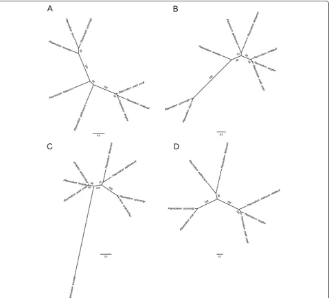
79 and 74% identity in pairwise comparisons to P. falcip-arum ELO3 and CCp2, respectively. The P. gallinaceum CCp2 amino acid sequence (1,348 amino acids) that was analysed contains 17 cysteines, all of which were conserved between mammalian Plasmodiumspecies. The translated P. gallinaceum ELO3 full-length transcript sequence re-sulted in a protein of 521 amino acids and was approxi-mately 121 amino acids shorter than the P. falciparum ELO3 protein. Phylogenetic analyses suggested that these essential orthologues inP. gallinaceumare most similar to P. falciparum (as compared to other mammalian Plasmodiumspecies) (Figure 1A-C). The high degree of
conservation suggests that these orthologues may also play important roles during the intra-erythrocytic stages ofP. gallinaceum.
Identification and phylogenetic analysis ofama-1and the
ama-1receptorRON2inPlasmodium gallinaceum
[image:5.595.59.539.246.681.2]The coding region ofP. gallinaceum ama-1was 1,692 bp with an A + T content (72%) greater than P. falciparum (around 70%). The translated protein sequence was 556 amino acids long, shared 55% amino acid identity toP. fal-ciparum ama-1, and contains 17 cysteines. All 16 cyste-ines within the three cysteine-rich domains of AMA-1
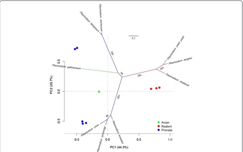
were conserved in number and position when the aligned protein sequences of individual domains were analysed, with the exception of domain III. Domain III contains six cysteine residues [71,72], which were present in both P. gallinaceum and P. falciparum ama-1. However, domain III of P. gallinaceum AMA-1 contained five amino acid deletions between positions 465-471 (relative toP. falcip-arum AMA-1). Therefore, the position of cysteines and the number of amino acids in domain III varied slightly between P. gallinaceum and P. falciparum. Phylogenetic analysis of ama-1 revealedP. gallinaceum ama-1 as sig-nificantly divergent from all mammalianPlasmodium spe-cies analysed (Figure 2). A metric multidimensional scaling (MDS) analysis was performed to complement the phylogeny and to visualize the evolutionary trajectories of ama-1 on a low dimensional space. Principal component analysis (PCA) plots showed clustering consistent with that of theama-1phylogeny (Figure 2). Phylogenetic and metric multidimensional scaling analysis of allama-1 se-quences compared in this study placed avianPlasmodium parasites into a strongly supported monophyletic clade and cluster (Additional file 7).
In addition to identifying AMA-1 in P. gallinaceum, a full-length cDNA sequence encoding a version of P.
gallinaceumRON2 was identified.P. gallinaceumRON2 was truncated at the N terminus by approximately 620 amino acids, in comparison to the translated full-length cDNA sequence ofP. falciparumRON2. The coding se-quence of P. gallinaceum RON2 was 4,641 bp and en-codes a protein of 1545 amino acids with 71% identity to P. falciparum RON2. Two conserved cysteines that are required for RON2 to bind the AMA-1 pocket [73] were conserved in P. gallinaceum RON2 (Figure 3). A phylo-genetic analyses of RON2 groupsP. gallinaceumwithP. falciparum(Figure 1D).
ama-1polymorphisms in avianPlasmodiumfield isolates
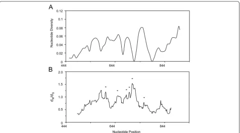
[image:6.595.57.540.391.693.2]The entire ama-1 domain I region consisting of 468 bp was sequenced and analysed from a total of 51P. lucens isolates collected from African rainforest birds. A sliding window analysis ofπ using a window of 30 bp moved in steps of nine sites reveals polymorphisms across the en-tire region of domain I (Figure 4A). The region 787-816 bp appears to be the most polymorphic region in domain I ofP. lucens ama-1. This region corresponds to the naturally immunogenic T cell epitope located within residues 259-271 of P. falciparum AMA-1, which was also reported to be polymorphic [42]. Three-hundred
and eighty-seven monomorphic sites and 81 poly-morphic sites were detected in domain I of P. lucens ama-1. Three polymorphic sites at positions 116, 138 and 205 exhibited three different nucleotides, whereas the remaining sites had only two. A total of 77 muta-tions were detected, 27 of which were synonymous and 50 of which were non-synonymous. π for all 51 se-quences analysed was 0.043237 ± 0.005210 SD, which is
more polymorphic than previously reportedπ (0.01361-0.01764) forP. falciparumisolates [42,74].
[image:7.595.58.540.89.230.2]An alignment of the translated sequences revealed three additional amino acid residues (Glu-Phe-X) positioned near hydrophobic amino acids that line the hydrophobic trough of domain I (Additional file 8). The additional amino acids are located between residues 184-185 relative toP. falciparumAMA-1. Considering that it is unknown
Figure 3Three-dimensional models of the avianPlasmodiumRON2 protein. A)Stereo views of the C-terminus ofP. gallinaceumRON2 (left) andP. falciparumRON2 (right).B)Sequence alignment ofP. gallinaceumandP. falciparumRON2 are shown with the secondary structure of the corresponding amino acid regions above the alignment. Helices are colored red. The line with connecting arrows indicates disulfide bonds. Conserved amino acids are highlighted in red.
Figure 4Sliding window plot of A)πand B)dN/dSfor domain I ofPlasmodium lucens ama-1.Nucleotide positions are relative to the
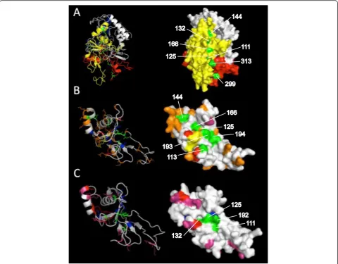
[image:7.595.58.540.427.694.2]whether the tertiary structure of AMA-1 in avian Plas-modium species contains a hydrophobic trough, a 3D model of the full-lengthP. gallinaceum AMA-1 was gen-erated. Tertiary structure-based analysis of the resulting 3D model was found to be satisfactory (LGscore = 3, Max-Sub = 0.3). Conserved hydrophobic amino acid residues that line the hydrophobic trough, according to P. falcip-arum AMA-1 positions found by Bai et al. [75], were highlighted in green. All highlighted residues (111, 125, 132, 144, 299, 313 relative toP. gallinaceumAMA-1) also appeared to reside in a small yet extended pocket of P. gallinaceum AMA-1 (Figure 5A), with the exception of residues 193 and 194. 3D models for the AMA-1 domain I of P. lucens and P. falciparum were also generated to visually compare intraspecific polymorphisms (Figure 5B).
[image:8.595.58.539.280.655.2]To determine whether these polymorphisms and additional amino acids are present among other avian Plasmodium species, domain I of ama-1 from 28 P. homopolare (a newly identified host-generalist), three P. megaglobularis (host-generalist), three Plasmodium lineage spp. PV16, and two P. globularis(host-specialist) isolates [14,54] were sequenced. Between-species diver-gence (K) ranged 0.062-0.349 (Additional file 4), approxi-mately two to three-fold greater than K calculated for cytochrome b(0.030-0.103). Attempts to sequence domain I from other avian species, includingPlasmodium relictum (GRW11 and SGS1) isolates were unsuccessful, likely due to low parasitaemia or parasite genomic DNA concentra-tions. Interestingly, no intraspecific polymorphisms were observed. However, additional amino acids in domain I
Figure 5Three-dimensional models of the avianPlasmodiumAMA-1 protein.Theab initio-generated models are based on theA)556 aa sequence ofP. gallinaceumAMA-1,B)155 aa residues in AMA-1 domain I ofP. lucens, andC)152 aa residues in AMA-1 domain I ofP. falciparum. Both stereo (left) and surface (right) views are shown. Domain I, II and III ofP. gallinaceumAMA-1 are coloured yellow, red and blue, respectively. Conserved hydrophobic residues that line the putative hydrophobic trough are labelled and highlighted in green.Plasmodium lucensandP. falciparum
were present in all field-caught avian Plasmodium spe-cies included in this study. Similar toP. lucens, domain I of P. megaglobularis, lineage PV16, and P. globularis AMA-1 sequences contain three additional amino acids (Glu-Phe-X) between residues 184-185, whereas the do-main I of P. homopolare AMA-1 contains two amino acid (Arg-Asp) insertions between residues 187-188 (Additional file 8). All additional amino acids observed were located within or near hydrophobic amino acids that line the hydrophobic pocket.
Comparison ofPlasmodium lucens ama-1andPlasmodium falciparum ama-1sequences
Forty-nine P. falciparum ama-1 sequences were com-pared with 51 P. lucens ama-1 sequences in an align-ment covering the entire domain I region. There were a total of 120 fixed nucleotide differences between the spe-cies. Of these differences, 33% (39) were synonymous and 67% were (81) non-synonymous. There were a total of 121 polymorphic sites within-species, of which 22% (27) sites were synonymous and 78% (94) were non-synonymous. A McDonald-Kreitman test with domain I sequences detected significant departure from neutrality in theP. lucens ama-1domain I region (Neutrality Index (NI) = 1.676, P = 0.07; NI with Jukes and Cantor correc-tion = 2.035, P = 0.009), suggesting that polymorphisms at the domain I are maintained under positive diversify-ing selection.
A dN/dS analysis of the entire domain did not detect
significant differences between non-synonymous and synonymous changes. To determine if regions with high dN/dSratios are under selection, a sliding-window
ana-lysis (90 bp with a step size of three bases) ofdN/dSwas
conducted. Significant difference betweendN anddSwas
detected throughout domain I (at the midpoint nt position of region 543-650, dN/dS= 3.51, P < 0.0005; mid point nt
position of region 609-698, dN/dS= 1.90, P < 0.05; mid
point nt position of region 645-735, dN/dS= 1.97, P <
0.05; mid point nt position of region 657-746,dN/dS=
1.66, P = 0.05; mid point nt position of region 669-758, dN/dS= 2.01, P < 0.05) (Figure 4B), suggesting that these
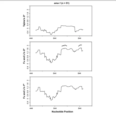
regions are under positive diversifying selection. No significant departure from the neutral expectation was detected using Tajima’s D (D= -0.12236, P > 0.10). Similar results were obtained for the Fu and Li D* (1.32144, P > 0.10), whereas the F* value of 1.79767 was significant with P < 0.02. Sliding-window analysis with a window of 30 bp and a step size of nine bases did not detect significantly low or high statistics values for all neutrality tests along the domain I region ofama-1. To ensure that the true level of variation is not obscured from choosing too small of a window, the sliding-window analysis with a sliding-window of 90 bp and a step size of three bases was performed. Significantly high F*
values were observed between 658-786 bp and 718-831 bp (Figure 6). These results of the population-based methods also support the findings from comparing dN
todS, and provide additional evidence that the domain I
region of P. lucens ama-1 is under positive diversifying selection.
Discussion
Assessing the genetic diversity and selection in erythro-cyte invasion genes of avian malaria parasites may pro-vide insight on the population and the transmission dynamics of malaria. Higher rates of malaria transmis-sion are expected to occur in areas with high parasite di-versity [76]. This prediction is supported by the high levels of diversity in nuclear genes from malaria parasites found in avian hosts of Hawaii, including the thrombospondin-related anonymous protein (trap) gene, which encodes a protein involved in immuno-evasion and erythrocyte inva-sion [77]. Although trap evolves under positive selection in human malaria parasites, no evidence for positive selec-tion was found in trap of avian malaria parasites [78]. Here, for the first time, evidence of positive selection as a driving force in the evolution and diversification of an erythrocyte invasion gene (ama-1) in avian malaria para-sites is provided. These results show thatama-1is a useful nuclear marker for investigating the adaptive evolution of avian malaria parasite populations. In addition, these re-sults suggest that the roleama-1plays in erythrocyte inva-sion is evolutionarily conserved across avian Plasmodium species.
Avian Plasmodium ama-1 is relatively conserved in comparison with orthologous genes of mammalian Plas-modium species. The conservation of the hydrophobic trough in P. gallinaceum AMA1 underscores the func-tional importance of AMA-1 in avianPlasmodium para-sites. The hydrophobic trough of AMA-1 binds to the RON complex via AMA-1-RON2 interactions [73,79]. These interactions form the invasion machinery required to mediate erythrocyte invasion [80,81]. Moreover, the identification ofP. gallinaceumRON2 and the conserva-tion of the RON2 helices involved in AMA-1 binding further supports the notion that this junction-dependent invasion process is evolutionarily conserved in avian Plasmodium parasites. Future studies will be important to investigate the signatures of selection within regions ofP. gallinaceumRON2, and to further the understand-ing of junction-dependent invasion processes in avian Plasmodiumparasites.
domain are unique to Plasmodium. More importantly, domain III of P. falciparum AMA-1 is antigenic and elicits growth-inhibiting antibodies [83]. Domain III con-tains two conserved immunodominant epitopes. The first conserved epitope is located at position 459-464, whereas the second is located at position 467-475. Do-main III ofP. gallinaceumAMA-1 lacks four amino acid residues within the second immunodominant epitope (between position 468-473 with respect toP. falciparum AMA-1). These differences may also be present in other avian Plasmodium species and possibly result in a lack
of or escape from inhibitory antibodies directed against the second immunodominant epitope. It is thus tempting to speculate that the observed differences in the domain III region ofP. gallinaceumAMA-1 may contribute to the diverse host range of avianPlasmodium parasites. Unfor-tunately, efforts to PCR amplify and assess the domain III regions in ama-1 of other avian Plasmodium parasites were unsuccessful.
[image:10.595.61.537.88.551.2]In spite of this, several avian Plasmodium species ex-hibited genetic diversity and contained additional amino acids present within the domain I region of AMA-1.
Figure 6Sliding window plot of Tajima’sD, Fu and Li’sD* andF* tests for domain I ofPlasmodium lucens ama-1.The window length is 90 bp with a step size of 3 bp. Asterisks indicate regions where significant departure from neutrality was observed. n represents the number of
Plasmodiumisolates from African birds contained three additional amino acids in domain I, whereasP. homopo-lare isolates from California contained two additional amino acids. Surprisingly, domain I of P. homopolare ama-1 was highly conserved with no genetic diversity found between 28 different isolates. One possibility for this finding is that domain I of P. homopolare ama-1 is subject to negative selection, as negative selection has been shown on the rhoptry-associated protein 1 (RAP-1), which is also involved in erythrocyte invasion [84]. How-ever, this possibility is difficult to investigate considering the lack of diversity in P. homopolare ama-1. Alterna-tively, the lack of diversity may result from a recent demo-graphic sweep (bottle-neck) or host switching events that lead to recent population expansions [85,86]. The latter is favoured, as P. homopolare was found in five different families representing nine bird species [54]. Therefore, any reciprocal selection acting on P. homopolare ama-1 by host protective immune responses may be weak rela-tive to the selection imposed on a host specialist by a sin-gle host (e.g., the Olive Sunbird and P. lucens). A lack of diversity was also found in the host generalistP. megaglo-bularis. However, more robust sampling and sequencing of parasites is required as the true diversity may be underestimated due to low sample sizes. These results are in stark contrast to the level of genetic diversity found inP. lucens ama-1.
These findings provide significant evidence that poly-morphisms in P. lucens ama-1 are maintained by posi-tive selection. The patterns of genetic diversity across domain I ofP. lucens ama-1 are consistent with studies on mammalian Plasmodium species [72,74,87,88]. Like-wise, sliding window plots of πand Fu and Li’sF*value indicate that the region corresponding to a natural T cell epitope in domain I is highly polymorphic and is under positive selection in P. lucens ama-1, which is also in agreement with earlier studies [42,89]. This observation suggests that avian Plasmodium AMA-1 may induce T cell responses in infected bird hosts and has implications for studying immune responses in bird populations that are naturally exposed to malaria parasites. In addition, humoral immune responses against AMA-1 in bird pop-ulations may also provide valuable information for immuno-epidemiologic studies. Such studies can be par-ticularly important for terrestrial ecosystems that are sen-sitive to losses in native bird populations, especially since native birds play essential roles in many terrestrial ecosys-tems [90,91]. Therefore, there is a great need to under-stand host-parasite interaction and its relationship to host immune responses in avianPlasmodiumparasites.
Conclusions
Ultimately, these findings provide insight into the erythrocyte invasion process of avian Plasmodium, are
the first evidence of host-driven selection in an avian Plasmodium species, and demonstrate the substantial applications of theP. gallinaceumtranscriptome. TheP. gallinaceum transcriptome dataset represents a major public genomic resource that will serve to progress re-search on the functional genomics and evolution of Plas-modium. Further analyses of invasion and immuno-evasion-related genes could reveal additional nuclear markers for phylogenetic applications, as these genes may exhibit high levels of diversity [21]. Identifying add-itional markers has been difficult as the majority of se-quenced Plasmodium genomes are from mammalian Plasmodium species; therefore, primer development is often facilitated using mammalian Plasmodium species sequence data and the resulting primers may not be suit-able for PCR amplification of non-mammalian Plasmo-diumspecies DNA. However, with the recent sequencing and availability of the P. relictumtranscriptome [21] and the addition of the P. gallinaceumtranscriptome, signifi-cant progress towards adding reliable markers for more thorough phylogenetic and evolutionary studies of Plas-modiumor closely related genera is expected.
Additional files
Additional file 1:Host and parasite species locality.The table shows the source of isolates used to analyse sequence diversity.
Additional file 2:GenBank Accession numbers for the parasite taxa.
GenBank Accession numbers for the parasite taxa used in this study are provided.
Additional file 3:Genbank accession numbers for theama-1
sequences.The table shows Genbank accession numbers for theama-1
sequences obtained fromPlasmodiumfield isolates used in this study. Additional file 4:Between-species divergence.The table shows between-species divergence (K) calculated using Jukes and Cantor correction.
Additional file 5:Genetic distances and amino acid similarity.The table shows genetic distances computed from DNA sequences and amino acid similarity betweenPlasmodium gallinaceumandPlasmodium falciparum. Additional file 6:The size distribution forPlasmodium gallinaceum
transcripts.The figure shows the size distribution forPlasmodium gallinaceumtranscripts that show homology toPlasmodium falciparum
transcripts.
Additional file 7:Phylogenetic and MDS analysis ofPlasmodium
parasites.Anama-1phylogeny and MDS analysis shows clustering of avian malaria parasites according to host species.
Additional file 8:AMA1- domain I amino acid sequence alignment.
The figure shows aligned amino acid sequences with the AMA-1 domain I ofPlasmodium falciparum,Plasmodium lucens,Plasmodium megaglobularis,
Plasmodium globularis,Plasmodiumlineage spp. PV16, andPlasmodium homopolare.
Competing interests
The authors declare that they have no competing interests.
Authors’contributions
collection and RNA preparations. LAT performed the chicken infections and assisted with the blood sample collections. KB performed the transcriptome assembly. SWR performed the characterization of the transcriptome. RNMS conceived and participated in the design of the study, and helped draft the manuscript. All authors read and approved the final version of the manuscript.
Acknowledgements
This work was supported by a National Institute of Health grant SC2AI089120-01A1, and used the Vincent J. Coates Genomics Sequencing Laboratory at UC Berkeley, supported by NIH S10 Instrumentation Grants S10RR029668 and S10RR027303. We would like to thank Dr. Robert Gwadz for kindly providing us withP. gallinaceumsamples. We thank Eric Routman (San Francisco State University) for his assistance with statistical analysis, and thank Erika Walther and other scientists involved in sampling efforts.
Author details
1Department of Biology, San Francisco State University, San Francisco, CA
94132, USA.2Department of Medicine and Epidemiology, School of
Veterinary Medicine, University of California, Davis, CA 95616, USA.
Received: 12 June 2014 Accepted: 17 September 2014 Published: 26 September 2014
References
1. Ferreira MU, da Silva Nunes M, Wunderlich G:Antigenic diversity and immune evasion by malaria parasites.Clin Diagn Lab Immunol2004, 11:987–995.
2. Escalante AA, Lal AA, Ayala FJ:Genetic polymorphism and natural selection in the malaria parasitePlasmodium falciparum.Genetics1998, 149:189–202.
3. Takala SL, Plowe CV:Genetic diversity and malaria vaccine design, testing, and efficacy: Preventing and overcoming“vaccine resistant malaria”.
Parasite Immunol2009,31:560–573.
4. Roy SW, Irimia M:Origins of human malaria: Rare genomic changes and full mitochondrial genomes confirm the relationship ofPlasmodium falciparumto other mammalian parasites but complicate the origins of Plasmodium vivax.Mol Biol Evol2008,25:2511–2511.
5. Deitsch KW, Moxon ER, Wellems TE:Shared themes of antigenic variation and virulence in bacterial, protozoal, and fungal infections.Microbiol Mol Biol Rev1997,61:281–293.
6. Hughes AL:Circumsporozoite protein genes of malaria parasites (Plasmodiumspp.): evidence for positive selection on immunogenic regions.Genetics1991,127:345–353.
7. Escalante AA, Grebert HM, Isea R, Goldman IF, Basco L, Magris M, Biswas S, Kariuki S, Lal AA:A study of genetic diversity in the gene encoding the circumsporozoite protein (CSP) ofPlasmodium falciparumfrom different transmission areas—XVI. Asembo Bay Cohort Project.Mol Biochem Parasitol2002,125:83–90.
8. Baum J, Thomas AW, Conway DJ:Evidence for diversifying selection on erythrocyte-binding antigens of Plasmodium falciparum and P. vivax.
Genetics2003,163:1327–1336.
9. Mayer DCG, Mu J-B, Feng X, Su X, Miller LH:Polymorphism in aPlasmodium falciparumerythrocyte-binding ligand changes its receptor specificity.J Exp Med2002,196:1523–1528.
10. Mayer DCG, Mu J-B, Kaneko O, Duan J, Su X, Miller LH:Polymorphism in thePlasmodium falciparumerythrocyte-binding ligand JESEBL/EBA-181 alters its receptor specificity.Proc Natl Acad Sci U S A2004,101:2518–2523. 11. Iyer J, Grüner AC, Rénia L, Snounou G, Preiser PR:Invasion of host cells by
malaria parasites: a tale of two protein families.Mol Microbiol2007, 65:231–249.
12. Ricklefs RE, Fallon SM:Diversification and host switching in avian malaria parasites.Proc R Soc Lond B2002,269:885–892.
13. Fallon SM, Bermingham E, Ricklefs RE:Host specialization and geographic localization of avian malaria parasites: a regional analysis in the Lesser Antilles.Am Nat2005,165:466–480.
14. Loiseau C, Harrigan RJ, Robert A, Bowie RCK, Thomassen HA, Smith TB, Sehgal RNM:Host and habitat specialization of avian malaria in Africa.
Mol Ecol2012,21:431–441.
15. Bensch S, Stjernman M, Hasselquist D, Ostman O, Hansson B, Westerdahl H, Pinheiro RT:Host specificity in avian blood parasites: a study of
PlasmodiumandHaemoproteusmitochondrial DNA amplified from birds.
Proc Biol Sci2000,267:1583–1589.
16. Ricklefs RE, Fallon SM, Bermingham E:Evolutionary relationships, cospeciation, and host switching in avian malaria parasites.Syst Biol
2004,53:111–119.
17. Hellgren O, Pérez-Tris J, Bensch S:A jack-of-all-trades and still a master of some: prevalence and host range in avian malaria and related blood parasites.Ecology2009,90:2840–2849.
18. Loiseau C, Zoorob R, Robert A, Chastel O, Julliard R, Sorci G:Plasmodium relictuminfection and MHC diversity in the house sparrow (Passer domesticus).Proc Biol Sci2011,278:1264–1272.
19. Sepil I, Lachish S, Hinks AE, Sheldon BC:Mhc supertypes confer both qualitative and quantitative resistance to avian malaria infections in a wild bird population.Proc Biol Sci2013,280:20130134.
20. Westerdahl H, Stjernman M, Råberg L, Lannefors M, Nilsson J-Å:MHC-I affects infection intensity but not infection status with a frequent avian malaria parasite in blue tits.PLoS One2013,8:e72647.
21. Hellgren O, Kutzer M, Bensch S, Valkiūnas G, Palinauskas V:Identification and characterization of the merozoite surface protein 1 (msp1) gene in a host generalist avian malaria parasite,Plasmodium relictum(lineages SGS1 and GRW4) with the use of blood transcriptome.Malar J2013,12:381. 22. Carter R, Chen DH:Malaria transmission blocked by immunisation with
gametes of the malaria parasite.Nature1976,263:57–60. 23. Gwadz RW:Successful immunization against the sexual stages of
Plasmodium gallinaceum.Science1976,193:1150–1151.
24. Vinetz JM, Valenzuela JG, Specht CA, Aravind L, Langer RC, Ribeiro JMC, Kaslow DC:Chitinases of the avian malaria parasitePlasmodium gallinaceum, a class of enzymes necessary for parasite invasion of the mosquito midgut.J Biol Chem2000,275:10331–10341.
25. Li F, Patra KP, Vinetz JM:An anti-Chitinase malaria transmission-blocking single-chain antibody as an effector molecule for creating aPlasmodium falciparum-refractory mosquito.J Infect Dis2005,192:878–887.
26. Waters AP, Higgins DG, McCutchan TF:Plasmodium falciparumappears to have arisen as a result of lateral transfer between avian and human hosts.Proc Natl Acad Sci U S A1991,88:3140–3144.
27. Escalante AA, Ayala FJ:Phylogeny of the malarial genusPlasmodium, derived from rRNA gene sequences.Proc Natl Acad Sci U S A1994, 91:11373–11377.
28. Escalante AA, Ayala FJ:Evolutionary origin ofPlasmodiumand other Apicomplexa based on rRNA genes.Proc Natl Acad Sci U S A1995, 92:5793–5797.
29. McCutchan TF, Kissinger JC, Touray MG, Rogers MJ, Li J, Sullivan M, Braga EM, Krettli AU, Miller LH:Comparison of circumsporozoite proteins from avian and mammalian malarias: biological and phylogenetic implications.Proc Natl Acad Sci U S A1996,93:11889–11894. 30. Kissinger JC, Souza PCA, Soarest CO, Paul R, Wahl AM, Rathore D,
McCutchan TF, Krettli AU:Molecular phylogenetic analysis of the avian malarial parasitePlasmodium(Novyella)juxtanucleare.J Parasitol2002, 88:769–773.
31. Pick C, Ebersberger I, Spielmann T, Bruchhaus I, Burmester T:Phylogenomic analyses of malaria parasites and evolution of their exported proteins.
BMC Evol Biol2011,11:167.
32. Nirmalan N, Wang P, Sims PFG, Hyde JE:Transcriptional analysis of genes encoding enzymes of the folate pathway in the human malaria parasite Plasmodium falciparum.Mol Microbiol2002,46:179–190.
33. Pornthanakasem W, Kongkasuriyachai D, Uthaipibull C, Yuthavong Y, Leartsakulpanich U:Plasmodiumserine hydroxymethyltransferase: indispensability and display of distinct localization.Malar J2012,11:387. 34. Ikadai H, Shaw Saliba K, Kanzok SM, McLean KJ, Tanaka TQ, Cao J,
Williamson KC, Jacobs Lorena M:Transposon mutagenesis identifies genes essential forPlasmodium falciparumgametocytogenesis.Proc Natl Acad Sci U S A2013,110:E1676–E1684.
35. Deans JA, Alderson T, Thomas AW, Mitchell GH, Lennox ES, Cohen S:Rat monoclonal antibodies which inhibit the in vitro multiplication of Plasmodium knowlesi.Clin Exp Immunol1982,49:297–309. 36. Thomas AW, Deans JA, Mitchell GH, Alderson T, Cohen S:The Fab
fragments of monoclonal IgG to a merozoite surface antigen inhibit Plasmodium knowlesiinvasion of erythrocytes.Mol Biochem Parasitol1984, 13:187–199.
ofPlasmodium falciparummerozoites.Proc Natl Acad Sci U S A2003, 100:12295–12300.
38. Deans JA, Knight AM, Jean WC, Waters AP, Cohen S, Mitchell GH: Vaccination trials in rhesus monkeys with a minor, invariant,Plasmodium knowlesi66 kD merozoite antigen.Parasite Immunol1988,10:535–552. 39. Collins WE, Pye D, Crewther PE, Vandenberg KL, Galland GG, Sulzer AJ,
Kemp DJ, Edwards SJ, Coppel RL, Sullivan JS:Protective immunity induced in squirrel monkeys with recombinant apical membrane antigen-1 of Plasmodiumfragile.Am J Trop Med Hyg1994,51:711–719.
40. Xu H, Hodder AN, Yan H, Crewther PE, Anders RF, Good MF:CD4+ T cells acting independently of antibody contribute to protective immunity to Plasmodium chabaudiinfection after apical membrane antigen 1 immunization.J Immunol2000,165:389–396.
41. Coley AM, Parisi K, Masciantonio R, Hoeck J, Casey JL, Murphy VJ, Harris KS, Batchelor AH, Anders RF, Foley M:The most polymorphic residue on Plasmodium falciparumapical membrane antigen 1 determines binding of an invasion-Inhibitory antibody.Infect Immun2006,74:2628–2636. 42. Escalante AA, Grebert HM, Chaiyaroj SC, Magris M, Biswas S, Nahlen BL, Lal
AA:Polymorphism in the gene encoding the apical membrane antigen-1 (AMA-1) ofPlasmodium falciparum.X. Asembo Bay Cohort Project.Mol Biochem Parasitol2001,113:279–287.
43. Hodder AN, Crewther PE, Anders RF:Specificity of the protective antibody response to apical membrane antigen 1.Infect Immun2001,69:3286–3294. 44. Michon P, Stevens JR, Kaneko O, Adams JH:Evolutionary relationships of
conserved cysteine-rich motifs in adhesive molecules of malaria parasites.Mol Biol Evol2002,19:1128–1142.
45. Remarque EJ, Faber BW, Kocken CHM, Thomas AW:Apical membrane antigen 1: a malaria vaccine candidate in review.Trends Parasitol2008, 24:74–84.
46. Valkiūnas G, Iezhova TA, Loiseau C, Smith TB, Sehgal RNM:New malaria parasites of the subgenusNovyellain African rainforest birds, with remarks on their high prevalence, classification and diagnostics.Parasitol Res2009,104:1061–1077.
47. Martinez C, Marzec T, Smith CD, Tell LA, Sehgal RNM:Identification and expression of maebl, an erythrocyte-binding gene, inPlasmodium gallinaceum.Parasitol Res2013,112:945–954.
48. Babraham Bioinformatics.[http://www.bioinformatics.babraham.ac.uk/] 49. Kent WJ:BLAT—The BLAST-Like Alignment Tool.Genome Res2002,
12:656–664.
50. Langmead B, Trapnell C, Pop M, Salzberg SL:Ultrafast and memory-efficient alignment of short DNA sequences to the human genome.
Genome Biol2009,10:R25.
51. Grabherr MG, Haas BJ, Yassour M, Levin JZ, Thompson DA, Amit I, Adiconis X, Fan L, Raychowdhury R, Zeng Q, Chen Z, Mauceli E, Hacohen N, Gnirke A, Rhind N, di Palma F, Birren BW, Nusbaum C, Lindblad-Toh K, Friedman N, Regev A:Full-length transcriptome assembly from RNA-Seq data without a reference genome.Nat Biotechnol2011,29:644–652.
52. PlasmoDB: [http://plasmodb.org]
53. Valkiūnas G, Iezhova TA, Loiseau C, Chasar A, Smith TB, Sehgal RNM:New species of haemosporidian parasites (Haemosporida) from African rainforest birds, with remarks on their classification.Parasitol Res2008, 103:1213–1228.
54. Walther EL, Valkiūnas G, González AD, Matta NE, Ricklefs RE, Cornel A, Sehgal RNM:Description, molecular characterization, and patterns of
distribution of a widespread New World avian malaria parasite (Haemosporida: Plasmodiidae), Plasmodium (Novyella) homopolare sp. nov.Parasitol Res2014,113:3319–3332.
55. Waldenström J, Bensch S, Hasselquist D, Östman Ö:A new nested polymerase chain reaction method very efficient in detecting PlasmodiumandHaemoproteusinfections from avian blood.J Parasitol
2004,90:191–194.
56. Galtier N, Gouy M, Gautier C:SEAVIEW and PHYLO_WIN: two graphic tools for sequence alignment and molecular phylogeny.Comput Appl Biosci
1996,12:543–548.
57. Maddison WP, Maddison DD:Mesquite: a modular system for evolutionary analysis.Volume Version 2.75. 2011. http://mesquiteproject.org.
58. Stamatakis A:RAxML-VI-HPC: maximum likelihood-based phylogenetic analyses with thousands of taxa and mixed models.Bioinformatics2006, 22:2688–2690.
59. Posada D, Crandall KA:MODELTEST: testing the model of DNA substitution.Bioinformatics1998,14:817–818.
60. Huelsenbeck JP, Crandall KA:Phylogeny estimation and hypothesis testing using maximum likelihood.Annu Rev Ecol Syst1997,28:437–466. 61. Zhang Y:Template-based modeling and free modeling by I-TASSER in
CASP7.Proteins2007,69(Suppl 8):108–117.
62. Wu S, Skolnick J, Zhang Y:Ab initiomodeling of small proteins by iterative TASSER simulations.BMC Biol2007,5:17.
63. Wallner B, Elofsson A:Can correct protein models be identified?Protein Sci
2003,12:1073–1086.
64. Buchan DWA, Minneci F, Nugent TCO, Bryson K, Jones DT:Scalable web services for the PSIPRED Protein Analysis Workbench.Nucleic Acids Res
2013,41:W349–357.
65. Egea R, Casillas S, Barbadilla A:Standard and generalized McDonald-Kreitman test: a website to detect selection by comparing different classes of DNA sites.Nucleic Acids Res2008,36:W157–162. 66. Rozas J, Sánchez-DelBarrio JC, Messeguer X, Rozas R:DnaSP, DNA
polymorphism analyses by the coalescent and other methods.
Bioinformatics2003,19:2496–2497.
67. Tamura K, Peterson D, Peterson N, Stecher G, Nei M, Kumar S:MEGA5: molecular evolutionary genetics analysis using maximum likelihood, evolutionary distance, and maximum parsimony methods.Mol Biol Evol
2011,28:2731–2739.
68. Tajima F:Simple methods for testing the molecular evolutionary clock hypothesis.Genetics1993,135:599–607.
69. Alexandre JS, Kaewthamasorn M, Yahata K, Nakazawa S, Kaneko O:Positive selection on thePlasmodium falciparum clag2gene encoding a component of the erythrocyte binding rhoptry protein complex.Trop Med Int Health2011,39:77–82.
70. Fu YX, Li WH:Statistical tests of neutrality of mutations.Genetics1993, 133:693–709.
71. Hodder AN, Crewther PE, Matthew ML, Reid GE, Moritz RL, Simpson RJ, Anders RF:The disulfide bond structure ofPlasmodiumapical membrane antigen-1.J Biol Chem1996,271:29446–29452.
72. Kocken CH, Narum DL, Massougbodji A, Ayivi B, Dubbeld MA, van der Wel A, Conway DJ, Sanni A, Thomas AW:Molecular characterisation of Plasmodium reichenowiapical membrane antigen-1 (AMA-1), comparison withP. falciparumAMA-1, and antibody-mediated inhibition of red cell invasion.Mol Biochem Parasitol2000,109:147–156.
73. Srinivasan P, Beatty WL, Diouf A, Herrera R, Ambroggio X, Moch JK, Tyler JS, Narum DL, Pierce SK, Boothroyd JC, Haynes JD, Miller LH:Binding of Plasmodiummerozoite proteins RON2 and AMA1 triggers commitment to invasion.Proc Natl Acad Sci U S A2011,108:13275–13280.
74. Mardani A, Keshavarz H, Heidari A, Hajjaran H, Raeisi A, Khorramizadeh MR: Genetic diversity and natural selection at the domain I of apical membrane antigen-1 (AMA-1) ofPlasmodium falciparumin isolates from Iran.Exp Parasitol2012,130:456–462.
75. Bai T, Becker M, Gupta A, Strike P, Murphy VJ, Anders RF, Batchelor AH: Structure of AMA1 fromPlasmodium falciparumreveals a clustering of polymorphisms that surround a conserved hydrophobic pocket.Proc Natl Acad Sci U S A2005,102:12736–12741.
76. Conway DJ:Molecular epidemiology of malaria.Clin Microbiol Rev2007, 20:188–204.
77. Farias MEM, Atkinson CT, LaPointe DA, Jarvi SI:Analysis of the trap gene provides evidence for the role of elevation and vector abundance in the genetic diversity ofPlasmodium relictumin Hawaii.Malar J2012, 11:305.
78. Jarvi SI, Farias ME, Atkinson CT:Genetic characterization of Hawaiian isolates ofPlasmodium relictumreveals mixed-genotype infections.Biol Direct2008,3:25.
79. Lamarque M, Besteiro S, Papoin J, Roques M, Vulliez-Le Normand B, Morlon-Guyot J, Dubremetz J-F, Fauquenoy S, Tomavo S, Faber BW, Kocken CH, Thomas AW, Boulanger MJ, Bentley GA, Lebrun M:The RON2-AMA1 interaction is a critical step in moving junction-dependent invasion by Apicomplexan parasites.PLoS Pathog2011,7:e1001276.
80. Collins CR, Withers-Martinez C, Hackett F, Blackman MJ:An inhibitory anti-body blocks interactions between components of the malarial invasion machinery.PLoS Pathog2009,5:e1000273.
82. Kato K, Mayer DCG, Singh S, Reid M, Miller LH:Domain III ofPlasmodium falciparumapical membrane antigen 1 binds to the erythrocyte membrane protein Kx.Proc Natl Acad Sci U S A2005,102:5552–5557. 83. Mueller MS, Renard A, Boato F, Vogel D, Naegeli M, Zurbriggen R, Robinson
JA, Pluschke G:Induction of parasite growth-inhibitory antibodies by a virosomal formulation of a peptidomimetic of loop I from domain III of Plasmodium falciparumapical membrane antigen 1.Infect Immun2003, 71:4749–4758.
84. Andreina Pacheco M, Ryan EM, Poe AC, Basco L, Udhayakumar V, Collins WE, Escalante AA:Evidence for negative selection on the gene encoding rhoptry–associated protein 1 (RAP-1) inPlasmodiumspp.Infect Genet Evol2010,10:655–661.
85. Escalante AA, Cornejo OE, Freeland DE, Poe AC, Durrego E, Collins WE, Lal AA:A monkey’s tale: The origin ofPlasmodium vivaxas a human malaria parasite.Proc Natl Acad Sci U S A2005,102:1980–1985.
86. Krief S, Escalante AA, Pacheco MA, Mugisha L, André C, Halbwax M, Fischer A, Krief JM, Kasenene JM, Crandfield M, Cornejo OE, Chavatte J-M, Lin C, Letourneur F, Grüner AC, McCutchan TF, Rénia L, Snounou G:On the diversity of malaria parasites in African Apes and the origin of Plasmodium falciparumfrom Bonobos.PLoS Pathog2010,6:e1000765. 87. Oliveira DA, Udhayakumar V, Bloland P, Shi YP, Nahlen BL, Oloo AJ, Hawley WE, Lal AA:Genetic conservation of thePlasmodium falciparumapical membrane antigen-1 (AMA-1).Mol Biochem Parasitol1996,76:333–336. 88. Polley SD, Conway DJ:Strong diversifying selection on domains of the Plasmodium falciparumapical membrane antigen 1 gene.Genetics2001, 158:1505–1512.
89. Lal AA, Hughes MA, Oliveira DA, Nelson C, Bloland PB, Oloo AJ, Hawley WE, Hightower AW, Nahlen BL, Udhayakumar V:Identification of T-cell determinants in natural immune responses to thePlasmodium falciparumapical membrane antigen (AMA-1) in an adult population exposed to malaria.Infect Immun1996,64:1054–1059.
90. Şekercioğlu ÇH, Daily GC, Ehrlich PR:Ecosystem consequences of bird declines.Proc Natl Acad Sci U S A2004,101:18042–18047.
91. Tompkins DM, Gleeson DM:Relationship between avian malaria distribution and an exotic invasive mosquito in New Zealand.J Roy Soc New Zeal2006,36:51–62.
doi:10.1186/1475-2875-13-382
Cite this article as:Lauronet al.:Transcriptome sequencing and analysis ofPlasmodium gallinaceumreveals polymorphisms and selection on the apical membrane antigen-1.Malaria Journal201413:382.
Submit your next manuscript to BioMed Central and take full advantage of:
• Convenient online submission
• Thorough peer review
• No space constraints or color figure charges
• Immediate publication on acceptance
• Inclusion in PubMed, CAS, Scopus and Google Scholar
• Research which is freely available for redistribution