By
PAULINE GALLAGHER
A Thesis Submitted for the Degree of Doctor of Philosophy (PhD) in the
Statement (ii)
Acknowledgements (iii)
Abstract Civ)
List of abbreviations (Vi)
Chapter 1
General Introduction Chapter 2
Materials and Methods Chapter 3
Effects of skin grafting and host H-2 phenotype on aberrant H-2 antigen expression of spleen cells from semiallogeneic radiation chimeras Chapter 4
Does antibody blocking of H-2 antigens cause the reduced expression of P2 antigens observed on spleen cells of (Pi x P2)Fi -*■ P radiation chimeras ?
Chapter 5
Self tolerance and H-2 antigen expression in semiallogeneic radiation chimeras are affected by the extrathymic host environment
Chapter 6
H-2 restriction and H-2 phenotypes of spleen cells from thymus-grafted radiation chimeras: evidence consistent with intrathymic negative selection and extrathymic suppression
Chapter 7
General discussion: a summary in perspective
Apart from the skin grafting described in Chapter 3, which was performed by Mrs C.E. Woodhams, all the experiments presented in this thesis were my own work.
Pauline Gallagher,
Department of Microbiology,
John Curtin School of Medical Research, Australian National University,
Canberra, A.C.T.
ACKNOWLEDGEMENTS
The work presented in this thesis was performed during tenure of an Australian National University Ph.D. Scholarship in the Department of Microbiology, John Curtin School of Medical Research.
I wish to thank my supervisor Bob Blanden for his pragmatism, encouragement and constructive criticisms of my work throughout my course; I thank Gordon Ada for his advice and support in difficult times. I am also extremely grateful to Helen O'Neill and Bob
Ashman for many useful discussions. To Cathy Woodhams, who taught me many experimental techniques and who assisted repeatedly in mouse operations, I especially extend my thanks for her friendship, practical advice and capable technical assistance.
ABSTRACT
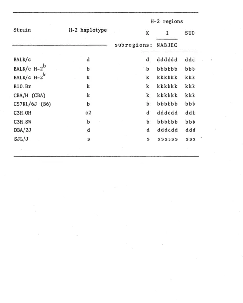
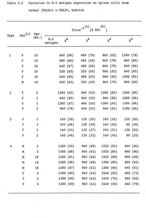
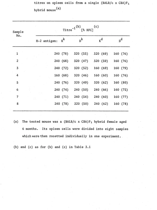
Cytotoxic T cells from (Pi x P2)Fi -> Px semiallogeneic radiation chimeras show a preference towards the H-2 type of the host in H-2 restricted responses to foreign antigens. Some of these chimeras may be incompletely tolerant of the P2 haplotype, and P2 H-2 K and D antigens may be expressed on their spleen cells at much lower levels than is
found on normal (Pi x P2)Fi hybrid cells. Experiments described in this thesis were designed to provide more basic information on the immuno biology of radiation chimeras; semiallogeneic chimeras were analysed firstly by a rosetting technique for changes to their H-2 antigen phenotypes, and secondly for the functional capabilities of their T
cells with respect to both self tolerance and H-2 restriction preferences, by means of (i) rejection of parental skin grafts in vivo3 (ii) spleen cell lysis of parental targets after stimulation in vitro with parental cells, and (iii) secondary in vitro responsiveness against ectromelia virus-infected parental cells.
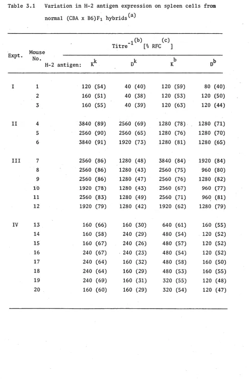
The phenomenon of reduced P2 H-2 antigen expression on spleen
cells from (Pi x P2)Fi -> Pi chimeras was reproduced in Chapter 3. Grafting the chimeras with P2 skin was not necessary for this aberrant H-2
phenotype to occur, and it appeared that once P2 H-2 levels were reduced on the chimeric lymphocytes, they remained at low levels indefinitely in the animal. However, the aberrant phenotype was shown to be
reversible. When bone marrow stem cells from chimeras that expressed low P2 levels were transferred into lethally irradiated (Pi x P2)Fi
hosts, the progeny splenic lymphocytes expressed the same concentrations of P2-type H-2 antigens as normal (Pi x P2)Fi hybrid cells.
determinants after overnight culture, and therefore failed to provide evidence that anti-P2 antibody might be masking and/or capping P2 H-2 molecules off the cell membranes.
Experiments on chimeras that had been grafted with parental thymuses showed that the H-2 type of the thymus did not modulate H-2 antigen expression of spleen cells. Instead, the extrathymic host
environment played a more significant role. All chimeras that displayed reduced levels of P 2 H-2 could mount responses both in vivo and in vitro against the P2 haplotype, thereby demonstrating a strong association in these animals between quantitative alterations to non-host H-2 antigen levels and loss of tolerance to that haplotype.
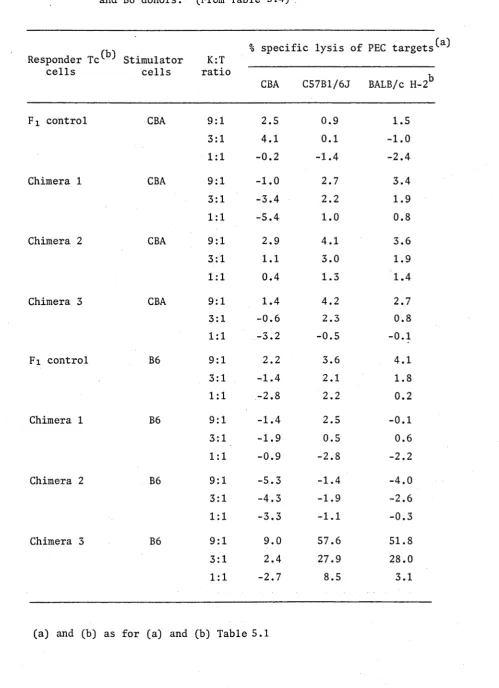
Experiments designed to investigate the H-2 restriction capabilities of cytotoxic T cells in thymus-grafted chimeras demonstrated preference in favour of the H-2 type of the thymus for lysis of virus-infected target cells. The bias varied between undetectable and absolute among individual animals that expressed normal F x levels of H-2 antigens, but absolute
restriction was seen in only 4 out of 21 chimeras. In the other 17 chimeras, T cells restricted to the non-host, non-thymic parental H-2 type were
readily detected. Chimeras with thymus grafts from both parental strains showed reduced T cell activity, suggesting that an extrathymic suppression mechanism was controlling T cell function in these chimeras.
The results were discussed in terms of a model for negative
B cell Bursa of Fabricius (or bone marrow)-derived lymphocyte
BM bone marrow
B6 C57B1/6J strain mouse CBA CBA/HJ strain mouse
CML cell-mediated lympholysis DTH delayed-type hypersensitivity FACS fluorescence activated cell sorter FCS foetal calf serum
FL foetal liver
GvH graft-versus-host reaction
H-2 the major histocompatibility complex of the mouse la I region-associated
Ig immunoglobulin
i.p. intraperitoneal Ir gene immune response gene i.v. intravenous
K:T killer:target ratio
MEM Eagle's minimum essential medium MHC major histocompatibility gene complex ml millilitre (10-3 litre)
yl microlitre (10~6 litre) MLR mixed lymphocyte reaction ND not determined
NT not tested
List of
PEC PFU Pi P 2 P 3 RFC SAMIg SEM SRBC T cell Tc cell Td cell Th cell Ti cell Ts cell Thy-1 X 2-ME
Abbreviations, continued
peritoneal exudate cells plaque-forming units parent 1
parent 2 parent 3
rosette-forming cells
sheep anti-mouse immunoglobulin serum fraction standard error of the mean
sheep erythrocytes
thymus-derived lymphocyte cytotoxic T cell
delayed-type hypersensitivity-mediating T cell helper T cell
1 LYMPHOCYTES AND IMMUNITY 4
1.1 Introduction to the immune system 4
1.2 Haemopoietic stem cells 5
1.3 T lymphocytes 7
1.3.1 Regulatory T cells 9
1.3.2 Effector T cells 11
1.4 B lymphocytes 12
2 THE THYMUS 14
2.1 Historical considerations 14
2.2 Thymus as a primary lymphoid organ 15
2.3 Ultrastructure
16-2.4 Cellular components 16
2.4.1 Thymic lymphocytes 17
2.4.2 Thymic macrophages 18
2.5 Thymic cell surface antigens 18
2.6 Cell traffic through the thymus 20
2.7 Thymic hormones 23
3 THE MAJOR HISTOCOMPATIBILITY COMPLEX OF THE MOUSE 25
3.2 Main regions of the H-2 complex 26
3.2.1 K and D regions 26
3.2.2 I region 29
3.2.3 S region 31
3.2.4 T region 32
3.2.5 U region 33
4 THE H-2 GENE COMPLEX AND IMMUNITY 33
4.1 H-2 restriction of T cell function 33
4.1.1 Involvement in lymphocyte collaboration 34 4.1.2 Involvement in antiviral cytotoxicity 34
4.2 H-2 molecules as self markers 35
4.3 Alloreactivity 39
4.4 Immune response genes 40
4.5 Interaction between virus and H-2 molecule 41
4.6 The T cell antigen receptor 42
5 RADIATION CHIMERAS 45
5.1 Chimeras in immunology 45
5.2 Establishment of chimerism 45
5.2.1 Radiation effects on the immune system 46 5.2.2 Reconstitution by haemopoietic stem cells 48
5.3 Side effects of lethal irradiation 50
1. LYMPHOCYTES AND IMMUNITY
1.1 Introduction to the Immune System
The immune system of higher vertebrates is characterised by certain features: it is specific for individual antigens; it is
diverse in that it can mount a response to a vast number of different antigens; it is flexible in that it may vary its repertoire to permit the response to virtually any antigen that may appear in nature to invade the animal; it is adaptable to antigenic stimulation so that a second encounter with the same antigen provokes a faster and greater immune response, i.e. it demonstrates immunological memory; it is tolerant to self so that under normal conditions an immune system does not react to its self components. All these properties are determined mainly by the lymphocyte, a small round cell with scanty cytoplasm, which was for a long time considered to be an end cell lacking further proliferative or differentiation potential.
Cochrane § Dixon 1962, Gowans § McGregor 1963) of the recipient; they could transfer delayed-type hypersensitivity (DTH) (Chase 1945, Wessldn 1952, Bloom § Chase 1967), initiate the rejection of allo geneic skin grafts (Gowans
et al.
1962, Billingham $ Silvers 1963), confer immunity to transplantable tumours (Mitchison 1955), and provide immunological tolerance (Gowanset al.
1962).Because lymphocytes occur in the blood as well as the lymphoid tissues, they are generally classified as belonging to the system of cell populations that generate blood cells, otherwise known as the haemopoietic cell system, In the mouse embryo, haemopoietic cells arise initially in the yolk sac, and migrate sequentially to the liver, spleen and bone marrow of the foetus; in adults, haemopoiesis occurs largely in the bone marrow and to a lesser extent in the spleen
(Metcalf $ Moore 1971).
1.2 Haemopoietic stem cells
The concept of a line of multipotential stem cells, from which all the various blood cell types were derived, was first proposed by Maximow who postulated the existence of a "haemocytoblast" (Maximow 1924). Such a cell has indeed been defined in haemopoietic tissues: the transfer of normal murine bone marrow cells into irradiated hosts causes the development within the recipient spleens of cell colonies containing differentiating erythrocytes, granulocytes, megakaryocytes
bone marrow cells bearing chromosomal abnormalities randomly generated by irradiation, which were transferred into unirradiated recipients
(Wu
et at.
1968). The fact that each colony originated from a single cell demonstrated the multipotential nature of these stem cells.Stem cells can undergo a series of divisions during which they are capable of differentiating into the precursors of the various functional populations, or renewing themselves as multipotential cells (Siminovitch
et at.
1963). There is controversy about whatdetermines the line of development along which a stem cell will proceed (Till
et at.
1964, Curry § Trentin 1967, Van Zant $ Coldwasser 1977). While a vast literature has accumulated on the humoral factors known to drive differentiation along given lines, it is unclear whether or not these factors determine stem cell commitment.Cells, other than lymphocytes, derived from the haemopoietic system are: erythrocytes, megakaryocytes which produce platelets, granulocytes, eosinophils, basophils, and monocytes which migrate into the tissues to become macrophages. The development of these non-lymphoid lines has been reviewed elsewhere (Metcalf § Moore 1971, Till § McCulloch 1980) and is not of direct relevance to this thesis.
Stem cell progenitors of lymphocytes can differentiate along two alternative pathways: they may become bursa-derived lymphocytes (B cells*), the immunologically competent precursors of immunoglobulin- producing plasma cells, or they may develop into Thy 1-bearing
derived lymphocytes (T cells).
1.3 T lymphocytes
T cell progenitors which migrate to the thymus from the haemo- poietic tissues such as bone marrow lack all identifying T cell
markers before they enter the thymus (Scheid
et at.
1973). However,they are already unipotential, being separable from the progenitors
of B cells (Lepault
et al
. 1985) and distinguishable from multipotentialstem cells on the basis of their cell density (El-Arini § Osoba 1973), and their drug and irradiation sensitivities (Berman § Kaplan 1959). As will be discussed later, these unipotential stem cells differentiate into T cells in the thymus; they acquire Thy 1 antigen (Lepault §
Weissman 1981) and various lymphocyte alloantigens of the Lyt series
(Mathieson
et al.
1979, Scollay £ Weissman 1980, Lepaultet al.
1983),their expression of histocompatibility antigens increases (Scollay 1982) , and they become the resting precursors of immunologically active T cells
(Stutman 1978). It has been accepted that during their development
in the thymus T cells establish tolerance to self and acquire the T cell receptor repertoire needed for their specific responsiveness to
antigen (Immunol. Rev.
A2_,
1978, 1 - ). These issues are highlycontentious today; they provide the theoretical basis for this thesis, and we await the definition of the T cell receptor before these points can become established fact.
Post-thymic T cells migrate via the blood and lymph to the T-dependent areas of the peripheral lymphoid organs, viz, spleen, lymph nodes and Peyer's patches (Parrott § De Sousa 1971, Greaves
et al.
1973). They also recirculate from blood to lymph via theMcGregor 1965). Eighty to ninety percent of mouse thoracic duct lymphocytes are Thy 1 positive; T cell depletion markedly decreases the number of lymphocytes in the thoracic duct (Miller § Mitchell 1969, Tyler et al. 1968), and nude mice, which have no thymus, have very few lymphocytes in their thoracic duct lymph (Sprent § Miller 1972) . There is evidence that not all T cells recirculate (Stobo et at. 1972, Stobo § Paul 1973), but a very large proportion of them do not remain sessile in the lymphoid organs once they have migrated there.
T cells which are not activated by antigen can be very long-lived, varying in their lifespan from 4-5 days up to several months as
measured by the interval between two successive cell divisions (Everett et al. 1964). Attempts to categorise T cells on the basis of their
longevity have been largely arbitrary; however, it would seem logical for immunological memory to be a function of cells which last for long periods of time without division. There is some evidence that long-lived T cells may have a longer average lifespan than long-lived B cells (Sprent § Miller 1972).
molecule, is present on some T cells for no known reason.
The functional and antigenic heterogeneity of T cells has permitted their classification into a series of subsets (Katz 1977, Snell 1978), which can be categorised broadly as regulatory and effector cell subpopulations. Regulatory cell groups are able to modulate the responses of the effector lymphocytes in both positive
and negative directions; effector T cells carry out cell-mediated immunity.
1.3.1. Regulatory T cells
Initiator T cells (Ti), when sensitised to an alloantigen, function to recruit allospecific lymphocytes from the lymph nodes
(Cohen § Livnat 1976). Their sensitisation to alloantigen occurs rapidly, within 4-6 hours, requires protein but not DNA synthesis, and once established is not destroyed by irradiation in vitro of up to 2000 R (Snell 1978). They are found most frequently in the spleen and are probably short-lived. They can also respond to soluble
antigens (Steinman et at. 1977), are involved in the adoptive transfer of protection against the bacterium Listeria monocytogenes to naive recipients (Kaufmann et at. 1979), and may collaborate in vitro with other T cells in the anti-Listeria immune response (Kaufmann et at.
1982). However, attempts to identify them in primary antiviral immune responses have been unsuccessful (R.V. Blanden, personal communication).
It has been proposed that their role in immunity is to patrol
associated (la) antigens, and are Lyt 1+ , 2/3+ , i.e. they express high levels of the antigens Lyt 1 and Lyt 2/3 which are found on
lymphocytes (Feldmann
et dl.
1977).Helper T cells (Th) collaborate in the induction of immune responses, helping B cells to mount specific antibody responses
(Claman
et dl.
1966, Davieset dl.
1967, Miller § Mitchell 1968,Mitchison 1971) or assisting other T cells in their effector capacities (Cantor $ Asofsky 1970, 1972). They function by activating B cells or effector T cells under the appropriate circumstances for an efficient immune response, but their possible role after that in the effector phase of immunity is an unresolved question. They are Lyt 1+ , 2/3 . The Th cell subclass involved in B cell responses has been subdivided further into two groups on the following bases (Marrack § Kappler 1976, Tada
et dl.
1978): Th 1 lacks antigens coded byI-A
andI-J
subregions of the
H-2
gene complex (I-A , I-J ), and acts exclusively on the B cells specific for the stimulating antigen; Th 2 is I-A+ , I-J+ , and once activated secretes antigen non-specific factors to trigger the B cell response. Th 1 develops early, within the first week of the antibody response, whereas Th 2 appears several weekslater (Takatsu
et dl.
1980).Suppressor T cells (Ts) (Gershon 1974) exert the opposite
effect to Th cells in that they depress both T and B immune responses. They are generally recognised by their expression of
I-J
codedantigenic determinants (Murphy
et dl.
1976), although as mentioned above, Th 2 also express I-J antigens. However, Ts generally carry the Lyt 1 , 2/3+*phenotype to distinguish them from Th cells (Beverleyet dl, 1976, Cantor et dl, 1976, Jandinski et dl, 1976). Ts are also a heterogeneous subset of cells, as reflected for instance in the description of some non-specific suppressors displaying the Lyt 1+ , 2/3+ phenotype (Pickel § Hoffmann 1977, Mosier et al, 1977).
1.3.2 Effector T cells
Cytotoxic T cells (Tc) are defined in vitro as having the capacity to lyse infected or chemically modified syngeneic target cells, H-2 incompatible targets, and under some special conditions non-H-2 disparate cells with differences in minor histocompatibility antigens. The H-2 restriction governing their function will be
discussed later. Tc cells are the specific agents in the elimination of viral infections (Blanden 1974a). They have been shown to transfer antimicrobial resistance to Listeria monocytogenes in both rats and mice (Jungi et dl. 1982, Cheers £ Sandrin 1983) and have been implicated
in the recovery of cattle from the protozoan parasite Theileria.parva (Eugui § Emery 1981). Tc cells are involved in immunity to some
transplantable tumours (Cerrotini $ Brunner 1974), and in the rejection of allografts along with DTH-mediating T cells (Ascher et al. 1983). They have also been shown to mediate DTH to certain viral infections
(Leung § Ada, 1980a, Ada et al. 1981, Lin § Askonas 1981). Antigenically, Tc cells lack la antigens (Beverley et dl. 1976, Lonai 1975), and are
generally considered to be Lyt 1 , 2/3+ (Cantor § Boyse 1975a,b, Kisielow 1975), although Tc cells bearing Lyt 1+ , 2/3+ have been reported in
certain strains of mice (Beverley et al. 1976, Pang et al. 1976).
classically mediate DTH (Td) are Lyt 1+ , 2/3 (Huber
et al.
1976, Vadaset al.
1976, Leung § Ada 1980b), and do not express la antigens(Vadas
et al.
1976). However, for the successful transfer of DTH to a naive recipient challenged with antigen, the primed donor cells must bearI-A
subregion genes which match those of the recipient animal (Leunget al.
1980, Milleret al.
1975, Vadaset al.
1977). In the recipient, macrophages appear to process and present theantigen to the I-A matched Td cells in order to activate them (Mottram § Miller 1980). Td cells are phenotypically the same as some Th
cells (Bianchi
et al.
1981).1.4 B lymphocytes
B cells in birds mature in the bursa of Fabricius, which is the sole site of humoral antibody-forming capacity (Glick
et al.
1956, Warneret al.
1969, Cooperet al.
1969). Bursal lymphocytes are derivedfrom migrant blood-borne stem cells (Moore § Owen 1966); since there is no mammalian equivalent of this organ, it is generally considered that B cells in mammals differentiate in foetal liver and spleen before birth (Tyan § Herzenberg 1968, Nossal § Pike 1973) and in the bone marrow thereafter (Mitchell $ Miller 1968). Speculations that the gastrointestinal lymphoid tissues provide bursal function (Cooper
et al.
1966, Percyet al.
1968) have never been validated.Newly formed B cells migrate via blood to the peripheral lymphoid organs. Once they settle in the B dependent areas, they tend to remain there. B cells recirculate much less than T cells (Sprent § Miller 1972, Howard
et al.
1972). Like T cells they can be long-lived, but their lifespan is affected by antigenic or mitogenicB cells are characterised by the expression of immunoglobulin (Ig) on their cell membranes. Pre-B cells can be detected in bone marrow by the presence of Ig chains within their cytoplasm well before
Ig appears on the cell surface (Raff
et al.
1976, Melcherset al.
1976, Burrows
et al.
1979, Levitt § Cooper 1980). All newly formed B cells first synthesise IgM (Kincadeet al.
1970, Lawtonet al.
1972), but subsequently, IgD can be produced at the same time as IgM since the majority of peripheral B cells in adult animals carry both theseclasses of Ig (Abney
et al.
1978).The synthesis of antibody by the B lineage requires firstly the rearrangement of genes on the chromosome during ontogeny, and later the
C M f > h c t \
translation of DNA sequences from unlinked genes in order to produce different classes of Ig with the tremendous diversity of antigenic
binding sites (idiotypes) that make up the antibody repertoire (reviewed by Kincade 1981). There are five different classes of Ig: IgM, IgD, IgG, IgA and IgE (Nisonoff
et al.
1975). They vary in their physical properties, concentrations in serum, functional capabilities, and the conditions for their production in an immune response. B cells may switch from the production of one class of Ig to another, e.g.IgM -* IgG, without altering the specificity of the Ig for antigen (Nossal
et al.
1964, Pierce § Klinman 1975, Gearhartet al.
1975), and there are even examples of this switch occurring without obvious antigenic stimulation (Lawton $ Cooper 1974).The differentiation antigens Qal, Qa2 and Qa4 are displayed on B cell precursors and some B cell populations, but their time of appearance depends upon the chronological age of the animal rather than the differentiation stage of the cells (Kincade 1981).
B cells can be separated into subclasses on the basis of their expression of some of the antigens mentioned above as well as on their functional capability in various assays. Definition of these subclasses will not be attempted here.
2. THYMUS
2.1 Historical overview
From as early as the seventeenth century, descriptive studies on the thymus, such as its ultrastructure and growth properties, were extensively documented. Up until the 1960's, the organ was called the thymus gland, although there was no real evidence for a thymic
hormone; it was known to be full of lymphocytes although these lymphocytes did not produce antibody. The function of the thymus remained an
antibodies to some antigens (Miller 1962 a,b).
Sir Macfarlane Burnet wrote: "... we are taking part in the elucidation of the function of the last major organ of the body to remain a mystery." (Burnet 1962). Miller’s work established the fact that the thymus is the site of maturation of the immunologically competent lymphocytes known as T cells.
2.2. Thymus as a primary lymphoid organ
As was mentioned earlier, the thymus and the bursa of Fabricius are the two known primary lymphoid organs. The thymus is distinguished from the secondary lymphoid organs by the absence of executive
immunological events such as antibody formation, an extremely high
level of lymphoid cell proliferation independent of antigenic stimulation, and a prominent compartment of epithelial cells; within 24 hours of
the onset of any serious illness, it atrophies rapidly in adults at the same time as the secondary lymphoid organs hypertrophy (Boyd 1932).
The thymus also has another puzzling characteristic: it grows rapidly in the foetus to reach its maximum size relative to the
animal at birth, continuing to grow in the young animal until puberty (Boyd 1932). Then growth ceases and the organ slowly involutes, i.e. the active thymic tissue is gradually replaced by fatty connective tissue (Hammar 1921, Metcalf 1960a). This apparent shutdown in activity suggests that the thymus must exercise its most important functions early in life. In fact, removal of the adult thymus has
little immediate deleterious effect (Metcalf 1960b). However, involution is not complete and the thymus continues to function throughout life
(Hammar 1935, Metcalf 1966a); adult thymectomy in the long term causes an increasingly severe defect in immune capability (Metcalf 1965,
2.3 Ultrastructure
Anatomically, in most species, the thymus is a moderately large paired organ overlying the heart in the upper anterior mediastinum of the chest. The two creamy-grey-coloured lobes, bound together by a thin connective capsule, are derived from the third pharyngeal pouch during embryogenesis (Crisan 1935, Rygaard 1973). The thymus looks avascular because its blood vessels are small. The framework of the lobes is composed by reticular fibres closely associated with blood vessels
(Smith § Ireland 1941). These fibrous septae break up the organ into lobules, each containing a cortex which surrounds a central medulla. The cortex and the medulla are separated by a thin corticomedullary zone. Before involution the proportion of cortex to medulla is remarkably constant at about 9:1 (Metcalf 1966a). Blood supply to the cortex is by a fine network of capillaries radiating out from thin arteries, whereas in the medulla there is a preponderance of veins and fewer arteries (Smith
et al.
1952). Lymphatic drainage occurs through sheath-like lymph vessels which accompany the medullary veins and arteries (Smith 1955).2.4 Cellular components
The different types of cells that constitute the thymus are as follows: epithelial and reticular cells (Smith 1964), lymphocytes
(Beard 1900, Weissman 1973, Fathman
et al.
1975a, Cantor and Weissman 1976), interdigitating or dendritic cells (Kaiserlinget al.
1974, Rouseet al.
1979, Steinman 1981), eosinophils, and macrophages (Smith 1964, Humeet al.
1983). Chromolipoid cells and some mast cellsdevelop during ageing and after irradiation (Smith 1964) .
a sponge of epithelial and reticular cells within which are embedded the tightly packed lymphoid cells. Cortical lymphocytes made up 90% of the weight of the thymus in young healthy mice.
2.4.1 Thymic lymphocytes
The lymphocytes present can be categorised as small, medium and large, by far the majority being small. It is established that the large cells divide to form the medium which in turn divide to form the small lymphocytes (Weissman 1967), but in view of the facts that
(a) the large lymphocytes are located in the outer cortex subcapsularly, the small in the more central cortex, and the medium mainly but not exclusively in the medulla (Metcalf 1966a, Scollay
et at,
1978) and,(b) there is intense mitotic activity in the cortex with few mitoses in the medulla (Metcalf 1966a), it seems that not all the medium cells give rise to the small lymphocytes. Rather, these medium cells of the medulla seem to be quite different in a number of respects from cortical lymphocytes. Among other features, they are more stable in both age-related and accidental involution of the thymus, more mobile than those in the cortex (Sainte-Marie § Leblond 1958), much more radioresistant (Trowell 1961) and cortisone-resistant (Schlesinger
^ Golokai 1967) than cortical thymocytes, and they are capable of homing back to the thymus and dividing there after intravenous injection into an animal (Kadish § Basch 1975, 1977). Under normal circumstances they persist indefinitely in the thymus, not being completely identical to extrathymic T cells (Elliott 1973).
are unable to repopulate the lymphocyte-depleted thymus of irradiated animals (Ford § Micklem 1963). Cortical thymocytes are not immuno competent. B cells account for less than 0.1% of the total thymic lymphoid component and these probably occur in the vasculature.
2.4.2 Thymic macrophages
Thymic macrophages are found throughout the thymus. They are extensively spread out on the underside of the thymic capsule and lie along the septae which project into the cortex. Throughout the cortex and the corticomedullary junction they occur as very flattened cells with vast membrane processes extending over the epithelial cells. Thus they form a complete network closely associated with lymphocytes.
In the medulla they are more rounded and look more like classical macrophages (Hume et dl. 1983). Earlier work by Metcalf described
a phagocytic reticular type of cell which stained with Periodic Acid-Schiff reagent (PAS cell) scattered throughout the outer third of the cortex (Metcalf 5 Ishidate 1961, 1962). About one-third of these cells could be shown at any given time to have phagocytosed
pyknotic lymphocytes (Ishidate § Metcalf 1963). Even more interestingly, they were associated with a higher incidence of lymphocyte mitoses
when lymphocytes were in contact with them. Metcalf speculated that they could be processing the debris from dead lymphocytes to provide nutrients for other dividing lymphocytes. The function of thymic macrophages will be discussed further later.
2.5 Thymic cell surface antigens
for separation of cell populations and for the definition of stages of cellular differentiation.
Thy 1 or 0 antigen (Reif § Allen 1964, Campbell
et al.
1981) is a cell surface molecule which, in lymphoid tissue, is expressed only on thymus-dependent cells (Raff § Wortis 1970). It occurs at slightly lower levels on peripheral T cells than on thymocytes which express it maximally, and Thy 1 is the most abundant known surface molecule on mouse thymocytes. However, thymocytes can be separated into two groups on the basis of their level of Thy 1 antigen, the subpopulation with lower Thy 1 levels being more like peripheral T cells in this respect (Shortmanet al.
1975). It has been suggested that Thy 1 might be the primordialimmunoglobulin domain (Williams § Gagnon 1982); its function is unknown. The presence of Thy 1 is used routinely in techniques for the separation of T cells from B cells.
TL antigen is found only on the thymocytes and leukaemia cells of certain inbred strains of mice, and is genetically determined by the
Tla
locus of theH-2
gene complex (Schlesinger 1972). It is detectable on most but not all cortical thymocytes and on 30-40% of medullary thymocytes (Scollayet al.
1980a, Schlesinger $ Golokai1967).
The expression of H-2 K and D histocompatibility molecules in the thymus varies considerably with cell type and location. In general, thymic lymphocytes express markedly lower concentrations of H-2 (Gorer $ Boyse 1959, Winn 1960, 1962, Cerrotini $ Brunner 1967) and I-A
negative by FACS analysis; the medium medullary thymocytes express K, D and I-A antigens almost at the same level as peripheral T cells. In the thymic stroma,
K
andI
region antigens are detectable from about the fourteenth day of gestation which is the point at which lymphocyte differentiation is beginning (Jenkinsonet al.
1980). In the medulla of young adult mice,Ks D
andI
region antigens appear confluent by immunofluorescence, whereas in the cortex K and D are found mainly on the epithelial-reticular cells and I-A antigen ismost readily detected on macrophages and dendritic cells (Rouse
et al.
1980, Van Ewijket al.
1980, Jenkinsonet al.
1981). Histocompatibility antigens will be described more fully in Section 3.Two other thymocyte markers frequently used are peanut agglutinin (PNA), a lectin which binds to immature cortical thymocytes (London
et al.
1978), and terminal deoxynucleotidyl transferase (TdT), anenzyme found only in immature thymocytes (Bollum 1960, Chang 1971). TdT is particularly interesting because it adds deoxyribonucleotides to the ends of DNA primers, i.e. it can synthesise new short DNA sequences without a template. It has been suggested that TdT may possibly act as a mutagen to provide diversity for the T cell receptor
(Baltimore 1974).
2.6 Cell traffic through the thymus
Lymphoid cells do not originate in the thymus, but migrate there from bone marrow. This fact was established in a number of ways: 1. If a thymus was irradiated, the lymphocytes within it were
lymphocyte-depleted recipients, the thymus was regenerated before the lymph nodes. If the reconstituting cells were derived from lymph node or thymus, then the lymph nodes were repopulated but not the thymus
(Ford § Micklem 1963). 3. CBA-T6T6 bone marrow cells were transferred into normal CBA mice, and were later demonstrated to have homed to the thymus as well as the spleen, Peyer's patches and lymph nodes (Micklem
et at.
1968). 4. T cell-depleted bone marrow cells were shown to give rise to splenic T cells in irradiated recipients within 15 days of transplantation (El-Arini $ Osoba 1973). Prothymocytes are defined as being among the "null-cell" population of spleen and bone marrow(Basch £ Kadish 1977, Kadish § Basch 1977).
Once inside the thymus, the lymphocytes begin proliferating rapidly in the cortex. Large lymphocytes divide every 6.8 hours and medium lymphocytes every 8.2 hours (Metcalf § Wiadrowski 1966). The
thymic lymphoid population turns over every 5-7 days (Bryant 1972), yet in young adult mice only 1% of these cells migrate out into the
periphery. More than 95% of thymocytes die
in situ.
Such statistics lend themselves easily to the thesis that the thymus is the site of elimination or inhibition of self-reactive clones of T cells, as well as being the site of T cell differentiation (Burnet 1962), but we await elucidation of the precise reason why there is so much cell death in the thymic cortex.Jerne explained this cell death in terms of thymic selection exerted by self histocompatibility antigens on maturing thymocytes, and suppression of "forbidden clones" capable of reacting against self which must not be released into the periphery (Jerne 1971). His
defined
in vitro
as a group of thymic epithelial cells expressing K and D antigens which supposedly engulf cortical thymocytes and permitthem to undergo high mitotic activity (Wekerle § Ketelson 1980, Wekerle
et at.
1980). However, it was subsequently shown that TNCs do notcompletely envelop the thymocytes and that all the thymocytes within
a given TNC are not from one clone (Kyewski £ Kaplan 1982). Nevertheless, purified TNCs when cultured with these thymocytes in the presence of
IL-2 were shown to give rise to Lyt 1 T cells, i.e. TNC-derived
thymocytes were the precursors of cytotoxic/suppressor T cells. It is not yet clear whether Lyt 1 is lost from the thymocytes, thus defining another stage of differentiation, or whether one subclass of thymocytes preferentially associates with the epithelial cells intrathymically.
Histological studies (Metcalf 1966a, Hume
et al.
1983) have shown the close association of macrophages with proliferating corticalthymocytes. Macrophages can stimulate thymocytes to proliferate by the production of IL-1 (Bettens
et dl.
1982), and in the medulla, macrophages or dendritic cells (Ia+ , Thy 1 , Ig ) can induce the proliferation of the relatively mature PNA thymocytes (Howe
et dl,
1970, von Boehmer § Byrd 1972, Beller £ Unanue 1978, Born § Wekerle 1982)possibly for the production of lymphokines such as IL-2 (Yu
et dl,
Newly formed T lymphocytes pass out of the thymus via blood and lymph to seed the spleen, lymph nodes and Peyer's patches, and to re circulate. It is not known whether the thymic emigrants are from the
cortex or the medulla. When they leave the thymus they are immunologically competent and look like peripheral T cells in terms of all known
phenotypic markers (Scollay
et dl
. 1978, Scollay 1982), yet there has not been a set of lymphocytes demonstrated still within the thymus that the emigres obviously derive from.2.7 Thymic hormones
The endocrine role of the thymus has not been ignored since Miller’s discovery of thymic immunological function. In fact, a large number of hormone-like peptides which are produced by the thymus have been characterised (Goldstein
et dl.
19S1). Many of them have been sequenced or extensively analysed biochemically, and all are quite distinct. Histochemical and cytological studies suggest that it is mainly the epithelial-reticular cells of the thymus medulla which are engaged in the production and secretion of these hormones (Trainin 1974).an effective reconstitution for neonatally thymectomised mice, in that wasting syndrome was prevented and antibody production against sheep
erythrocytes was permitted (Osoba § Miller 1964). The implication was that a humoral factor could endow lymphocytes with immunological competence. This was indicated more directly in neonatally thymectomised rats which gained the ability to mount DTK responses to bovine serum albumin after being treated with repeated injections of thymic
extracts (Jankovic et at. 1965).
Only a small number of thymic factors will be mentioned here. The most studied thymic hormones are the thymosin family of
polypeptides of which there are 40-50 members, some active individually and others with no known function (Goldstein et at. 1981). They are all derived from thymosin fraction 5, a potent soup of immunopotentiating agents with molecular weights between 1000 and 15000 daltons (Low et at. 1979, Low § Goldstein 1979). Some examples of these polypeptides are
*f + +
as follows: Thymosin ai can induce the expression of the Lyt 1 2 3
fir\o>ri*ve
antigen phenotype on -hyman lymphocytes, amplify the capacity of murine K
lymphocytes to respond to T cell mitogens, and induce macrophage
inhibition factor (MIF) in guinea pig peripheral blood leukocytes (Low
et at. 1979). It can also modulate the expression of TdT in that it
decreases TdT expression in thymocytes when used at low concentrations, but increases TdT expression at high concentrations (Hu et at. 1980, Goldschneider et at. 1981). Thymosin a 7 is a potent inducer of
suppressor cells (Goldstein et at. 1981). Thymosin B3 and 34 both induce TdT expression in TdT bone marrow cells both in vivo and in vitro (Low
et at. 1981).
precursors (Komuro $ Boyse 1973, Basch $ Goldstein 1974).
THF is a thymic hormonal factor which confers greater resistance to hydrocortisone on thymocytes (Trainin
et al.
1967, 1974, Trainin 1974). FTS or facteur thymique serique is a 900 dalton peptide originating in the thymus but commonly found in normal serum, which confers sensitivity to anti-6 serum and azathioprine on the precursors of thymocytes (Bach § Dardenne 1972, 1973, Dardenneet al.
1977).The list of factors is long and their claimed capabilities are indeed astonishing, but they cannot completely replace a structurally intact thymus in producing immunological competence. Thymus grafts enclosed in diffusion chambers did not permit the full recovery of lymph node, spleen and Peyer's patches in thymectomised hosts (Osoba § Miller 1964), so intrathymic cell contact is probably an important part of the T cell differentiation process. Also, when strips of cortical tissue or single cell suspensions of thymic tissue are used as grafts, there is no reconstitution (Metcalf 1963). Thus the structural integrity of cortex and medulla together is necessary for proper thymic function.
3 THE MAJOR HISTOCOMPATIBILITY COMPLEX OF THE MOUSE
3.1 Historical considerations
That susceptibility and resistance to transplants had a genetic basis was first demonstrated by Loeb in 1902 and subsequently by Little and coworkers, who used inbred mouse strains to reveal the existence of multiple histocompatibility genes. The first identification of an
or not a transplantable tumour would grow in a given mouse strain. This pioneering work on the genes responsible for the stimulation of
alloantibody production and for graft rejection (Gorer 1936, 1937), initially called antigen II, provided the basis for the definition of a group of genes called H-2 (histocompatibility-2) (Gorer
et al.
1948) or the major histocompatibility complex of the mouse.
3.2 Main regions of the H-2 complex
The
H-2
complex on chromosome 17 in the mouse is usually divided into four regions -Ky I3 S
andD
(Figure 1.1), although evidence now suggests that the T region is also part of the complex (Michaeisonet
at,
1977, Stanton § Hood 1980, Soloskiet al.
1981).3.2.1 K and D regions
The
K
andD
regions control the classical transplantation antigens responsible for the discrimination of self from non-self as defined by the rejection or acceptance of grafts. These antigens are believed to be present on the cells of virtually every tissue: spleen expresses maximal levels; other lymphoid organs expressslightly less, whereas brain, skeletal muscle and testes have barely detectable levels (Klein 1975). But more recent studies have shown that different cell types vary significantly in the amount of H-2
antigens on their membranes (Parr 1979, Rouse
et al.
1979); lymphomyeloid cells in particular have high concentrations. It is possible thatK and D r e g i o n s code f o r a s e r i e s o f c l a s s I m o l e c u le s w hich a r e i n t e g r a l membrane g l y c o p r o t e i n s o f m o l e c u l a r w e ig h t 45000 d a l t o n s , n o n - c o v a l e n t l y a s s o c i a t e d w i t h 32 m i c r o g l o b u l i n ( 3 2M) an 11500 d a l t o n
p o l y p e p t i d e ( V i t e t t a Ej C ap ra 1 9 7 8 ). From se q u e n c e a n a l y s i s , t h e
m o l e c u le c o n s i s t s o f a h eav y c h a i n 346 amino a c i d s lo n g w i t h two i n t r a c h a i n d i s u l p h i d e l i n k a g e s and two c a r b o h y d r a t e s i d e g r o u p s . The
a s s o c i a t e d 32M i s 99 amino a c i d s l o n g , c o n t a i n s one i n t r a c h a i n d i s u l p h i d e bond and i s n o t g l y c o s y l a t e d ( C o li g a n e t d l . 1981) . The m o l e c u le i s i n s e r t e d i n t h e l i p i d b i l a y e r o f t h e c e l l membrane by v i r t u e o f a h y d r o p h o b ic r e g i o n n e a r i t s -C00H t e r m i n u s .
I t i s w e l l e s t a b l i s h e d t h a t H-2 a n t i g e n s a r e s e r o l o g i c a l l y v e r y com plex (Amos e t d l . 1 9 5 5 ) , and h a s b een c o n v i n c i n g l y d e m o n s t r a te d w i t h c o n v e n t i o n a l a n t i s e r a t h a t t h e p r o t e i n p o r t i o n o f H-2 m o l e c u l e s , n o t
t h e c a r b o h y d r a t e s i d e c h a i n s , c o n t a i n t h e a n t i g e n i c s p e c i f i c i t i e s (N ath en so n 6 C u l l e n 1 9 7 4 ). However, by t h e u s e o f m o n o c lo n al a n t i b o d i e s , c a r t e
ls
h y d r a t e d e f i n e d a n t i g e n s have a l s o b een d e f i n e d on a H-2K m o le c u le ( O 'N e i l l $ P a r i s h 1981a, O ' N e i l l e t d l . 1 9 8 1 ).
On t w o - d im e n s io n a l p o l y a c r y l a m i d e g e l s , t h e K and D r e g i o n p r o d u c t s a r e a h e t e r o g e n e o u s p o p u l a t i o n o f m o l e c u le s w i t h r e s p e c t t o t h e i r
m o l e c u l a r w e ig h t s and c h a r g e (K r a k a u e r e t d l . 1 9 8 0 ). P a r t o f t h i s h e t e r o g e n e i t y can be a t t r i b u t e d t o p o s t - t r a n s l a t i o n a l m o d i f i c a t i o n s t o t h e c y t o p l a s m i c p r e c u r s o r m o l e c u le s su ch as v a r i a t i o n i n g l y c o s y l - a t i o n . The r e s t i s p r e s u m a b ly a c o n s e q u e n c e o f m u l t i p l e gene p r o d u c t s . C e r t a i n l y t h e K r e g i o n c o n t r o l s two d i s t i n c t m o l e c u le s ( I v ä n y i and Ddmant 1981, Try p h o n as e t d l . 1 9 8 3 ) , and t h e D r e g i o n i s c u r r e n t l y known t o code f o r f o u r p r o d u c t s : D ( S n e l l e t d l . 1 971a, Shimada £
N a th e n s o n 1969, S ch w artz e t d l . 19 7 3 a, Reyes e t d l . 1 9 8 2 ); L (Lemmonier
et dl.
1981). Recent analysis of a library of cosmid clones from BALB/c mice has placed the number of unique class I genes at more than 35 (Steinmetzet dl.
1982a) although a large portion of these are probably encoded by theT
region.Another distinctive feature of H-2 antigens in their extreme polymorphism, that is, there is no single wild-type gene complex but a large number of prevalent alleles or alternative forms of the histocompatibility genes*. Currently there appear to be over 100
alleles for each of the
K
andD
regions alone (Klein $ Figueroa 1981). If this polymorphism is considered together with the observation that the mutation rate of H-2 loci is relatively high (Klein 1978), then the rapid diversification ofH-2
genes does not appear to disadvantage the host species in natural selection but may even be advantageous. The precise function(s) of H-2 products is not known. Theories abound(e.g. J e m e 1971, Bodmer 1972, Ohno 1977, Parish
et dl.
1981). That these molecules cause the rejection of non-sygeneic grafts seems to be more an accident of modern science than a reason for their existence. It is well established however, that for a foreign antigen on aninfected cell to be recognised by a T effector cell, this antigen must associate in some way with H-2 products on the membrane, and that the T immunocyte must be able to interact with the H-2 molecules on the infected cell in order to mount an immune response (Zinkernagel £
Doherty 1974a,b). Presumably not all antigens interact in the appropriate way with each H-2 molecule. Therefore diversity of H-2 molecules
within a given population might permit a greater diversity of foreign antigens to be recognised and responded to by the immune system.
*Unlike the other class I molecules,
T
region products are not very poly morphic, and may be expressed to a very limited extent on cell surfaces3.2.2 I region
The
I
region is located in the e~2
complex betweenH-2K
and
Ss
(Davidet dl.
1973, Hauptfeldet dl.
1973, Sachs $ Cone 1973). As will be explained later, this region was first defined by genes that control the immune responses to a variety of antigens (Ir genes).The cell surface molecules encoded by the J region are known collectively as
I
region-associated (la) antigens, or class IImolecules, and appear to be equivalent to the Ir gene products (Herzenberg § Herzenberg 1974, Klein
et ail
. 1981, McKenzieet dl,
1981). LikeH-2 molecules they are integral membrane glycoproteins. Unlike H-2 molecules, they are limited in their cellular distribution, being
expressed on B lymphocytes, subpopulations of T lymphocytes particularly when activated, macrophages, dendritic cells, epidermis, sperm and
Langerhans cells in the skin (Hämmerling
et dl,
1975, McKenzie § Potter 1979) . On B cells they are closely associated with the Fc receptor which binds antibody-antigen complexes (Dickler $ Sachs 1974).Biochemically, la molecules have a molecular weight of 58000 daltons and exist on the cell surface as two non-covalently associated polypeptide subunits: an a chain of 30000-35000 daltons, and a 3
chain of 25000-30000 daltons (Cullen
et dl.
1976, Cooket dl.
1978). TheI
region has been divided intoI-N3 I-A3 I-B3 I - J I - E
andI-C
subregions using serological criteria.I-N
was serologically defined as a consequence of strong proliferation in mixed leukocyte cultures which mapped betweenK
aRdI-A
(Hayes $ Bach 1980).B-A
polypeptide chains: and which combine to form the A molecule, and A g (E^) which links with E^ encoded by the
I-E
subregion to produce a functional E molecule (Joneset al.
1978, Cooket oil.
1979). Although E^ is the only isolated molecule encoded inI-E3
there may be up to four molecular products of this subregion (Lafuseet al.
1982, Steinmetzet al.
1982a).I-A/E molecules are necessary for the induction and regulation of immune responses. For successful cellular interaction there must be matching of I-A between T cells and B cells (Sprent § Alpert 1981, Asano
et al.
1981) and between T cells and macrophages (Erb § Feldmann1975). I-A antigens are important in mixed lymphocyte reactions (MLR) (Klein 1975, Lonai § McDevitt 1977), graft rejection (Klein 1975,
McKenzie § Henning 1977), antigen-induced T cell proliferation (Schwartz
et al.
1978, Nepomet al.
1981), T cell-dependent antibody responses(McDevitt
et al.
1976, Sprent 1980), and the transfer of delayed-type hypersensitivity (DTH) to proteins and polypeptides (Milleret al.
1975, 1977) other than those that may integrate into the cell membrane. I-A products have also been detected on soluble T cell factors
(Armerding
et al.
1974, Erbet al.
1976), and la antigens on B cells can act as acceptor sites for some of these factors (Taussiget al.
1976).The
I-J
subregion products are serologically well defined (Kannoet al.
1981, Wattenbaugh 1981), but little is known of their biochemistrythat I-J determinants are not encoded between the
I-A
andI-E
subregions(Steinmetz
et dl.
1982b, Kronenberget dl.
1983), contrary to earliermapping studies in recombinant mouse strains (Murphy
et dl.
1976,Tada
et dl.
1976), yet the possibility has not been excluded that acontrol element encoded in
I-J
regulates the expression ofI-J
structuralgenes located outside the H-2 complex.
No molecules encoded by
I-B
orI-C
have yet been isolated. Thereis evidence that
I-B
as a separate subregion may not exist, since itseffects may be accounted for by I-A/I-E interaction (Baxevanis
et dl.
1981). This is supported by DNA sequencing studies (Steinmetz
et dl.
1982b). Nevertheless, it has been claimed that
I-B
controls the immuneresponse to the male specific H-Y antigen for DTH and graft rejection
(Hurme
et dl.
1978, Liew § Simpson 1980).I-C,
the subregion controllingthe immune response to the random terpolymer GLT (Merryman $ Maurer
1974), codes for stimulating determinants in MLRs (Okuda § David 1978)
and for a necessary structural component of a suppressor factor (Rich
et dl.
1979).3.2.3 S region
Even though the
S
region is located within the H-2 complex, itdoes not contain histocompatibility genes. It controls the serum levels
of four known glycoproteins, or class III molecules, which are part of
the complement system (Atkinson
et dl.
1982): Ss, the fourth componentof complement (C4) (Gorman
et dl.
1975, Ddmantet dl.
1973, Lachmannet dl.
1975, Carrol $ Capra 1978); Sip, a molecule very similar to C4but structurally and antigenically distinct, found generally only
in the males of some inbred mouse strains (Hansen £ Shreffler 1976,
B, a factor of the properdin system in the alternative complement
pathway (Roos § Ddmant 1982, Paolucci $ Shreffler 1983). Ss and Sip are 200 000 molecular weight peptidase precursors which become activated in the complement cascade, and C2 and B are 100 000 molecular weight proenzymes which also develop proteolytic activity upon activation.
3.2.4 T region
As mentioned earlier, cloning studies have indicated that there are at least 36 class I genes per haploid genome in the BALB/c mouse, about 30 of which are in the T region (Steinmetz e t d l , 1982a,
Margulies e t d l , 1982). There are at least two histocompatibility loci which map to T (Snell e t a t . 1971b, Boyse e t a t . 1972, Ddmant § Graft 1973, Klein § Chiang 1978), but little is known of the function of its other products. The molecules identified at present are: TL, thymus leukaemia antigen (Old e t a t . 1963) , which is expressed only on the small dense cortical lymphocytes of the thymus (Vitetta § Capra 1978), Qa-1 (Stanton £ Boyse 1976, Rothenberg § Triglia 1981) which is actively synthesised in T and B cells of normal spleen; Qa-2 (Flaherty 1976, Michaelson e t d l , 1981, Soloski e t d l , 1982) which is detectable on a variety of nucleated blood cells and their precursors, e.g. Thy-1+ cells; Qa-3 (Flaherty e t d l , 1978) expressed only on splenic and lymph node lymphocytes; Qat-4 and Qat-5 (Hämmerling
e t d l , 1979) which are limited in expression to a proportion of mature
cell surface of about two hours.
3.2.5 U region
The only other region of possible relevance to this thesis is a recently reported region,
U3
betweenS
andD3
which controlsmolecules resembling la antigens (O’Neill § Parish 1981b). As yet,
U
has been defined only in serological terms and awaits confirmation, although there is evidence that an Ir gene effect maps to the same part of theH-2
complex (Berzofskyet al.
1982).4. THE H-2 GENE COMPLEX AND IMMUNITY
4.1 H-2 restriction of T cell function
The importance of the MHC in determining the rejection of trans plants has already been discussed. Yet, at the time when Medawar concluded that the lymphocyte population evoked by the graft was instrumental in its rejection (Medawar 1957), few people realised the general significance of the MHC in terms of lymphocyte function and immunity.
In 1970, contact hypersensitivity experiments performed in outbred guinea pigs were published, in which sensitisation to dinitrochloro- benzene was found to be most effective if the guinea pigs were primed with autologous dinitrophenylated lymphocytes rather than coupled
4.1.1 Involvement in lymphocyte collaboration
When H-2 compatible thymus cells were transferred into athymic nude mice along with an antigen (sheep erythrocytes or bacteriophage T4), the mice were able to mount a substantial antibody response, but if H-2 incompatible cells were transferred instead, no specific antibody was elicited (Kindred § Shreffler 1972). Therefore, histocompatibility
antigens were involved in the cooperation between T and B cells for antibody production. This finding was reproduced in congenic strains of mice which had all the same background genes but differed at the H-2 complex (Katz
et dl
. 1973a) and the genes responsible were mapped to theI
region (Katzet dl.
1973b).About the same time, the sharing of
I
region genes between the donors of antigen-primed T cells and antigen-presenting macrophages was found to be necessary for the proliferation of specific T cells(Shevach § Rosenthal 1973, Rosenthal § Shevach 1973).
4.1.2 Involvement in antiviral cytotoxicity
Then, in the study of antiviral T cell cytotoxicity, it was noticed that T cells primed
in vivo
with lymphocytic choriomeningitis virus (LCMV) would lyse only those infected targets that had the same H-2 haplotype as the donors of the Tc cells (Zinkernagel § Doherty 1974a).Doherty 1979), as well as H-2 restricted cytotoxicity against minor histocompatibility antigens (Bevan 1975a,b) and the male H-Y antigen
(Gordon et at. 1975). Even though the susceptibility of mice to virus- induced leukemias was known at that stage to be controlled by the MHC
(Sjögren § Ringertz 1962, Lilly et at. 1964), the discovery of H-2 restriction highlighted this fact and implicated T cell responsiveness in the elimination of such tumours (Lilly § Pincus 1973).
H-2 restricted antiviral responses were also found to occur in vivo. In the transfer of virus-specific T cells to infected recipient mice for protection from ectromelia virus (mousepox), the immune T cell donors and the recipients needed to share the same class I genes if virus clearance was to be effective (Blanden 1974b, Kees § Blanden 1976).
Experimental evidence continued to mount in support of the require ment for the MHC in most aspects of T cell function. I region
restriction, and more recently K/D restriction, was found in the immune response to the facultative intracellular bacterium Listeria monocytogenes 3 whereby specific T cells activate macrophages to clear the bacterial
infection (Zinkernagel 1974, Zinkernagel et at. 1977, Jungi et at. 1982, Cheers $ Sandrin 1983). DTH to soluble proteins, chemically modified cells (Miller et at. 1975, Vadas et at. 1977), and viruses
(Leung et at. 1980) was also I region-restricted, as was contact
sensitivity (Schultz $ Bailey 1975, Fachet $ Ando 1977) in that immune response genes selectively affected the different classes of Ig
produced in this response (Thomas et at. 1979).
4.2 H-2 molecules as self markers
the infecting antigen was first proposed by Lawrence (1959). He considered the mechanism of immunity to infection to be simply a modification of the process by which mutant cells are recognised and
destroyed in the body. When Zinkernagel and Doherty discovered antiviral H-2 restriction, they suggested that the Tc cell precursor recognises both H-2 molecules and viral antigens together on the infected cell membrane, or alternatively that the H-2 molecules were self markers antigenically altered in a specific way by each virus, so that the Tc cell reacted against modified self. It is still not clear which alternative is correct.
Zinkemagel and Doherty also proposed that H-2 restriction was not simply a requirement for identity between an effector T cell and its target. Rather, it was a method of eliminating aberrant or infected cells in the body whilst maintaining tolerance to normal healthy self. They found that in virus-sensitised F x hybrid mice, the immune Fx T
cells, which bear H-2 antigens of both parental strains, are restricted to either one or the other parental H-2 type, but not to both (Zinkernagel § Doherty 1974b). This result has been reproduced with cloned lines
of influenza virus specific Tc cells: none of the clones of Fi origin were cytotoxic on both parental targets (Braciale
et dl.
1981b). Thus the hypothesis of a mutual interaction between H-2 antigens on inter acting cells was eliminated, and the antigen-presenting cell was defined as being a determinant of restriction.Other evidence disproving that H-2 identity is required for H-2 restriction has come from studies on T cell-macrophage interactions
from the T cells. H-2 matching is therefore not required for successful collaboration between T cells and macrophages, but rather, recognition of H-2 markers plus antigen is performed by the T cell only.
All the above experimental results can be grouped in terms of class I or class II restricted T cell effects. Class I-type H-2 restriction occurs in situations where the foreign antigen (X) is presented together with class I antigens on cells throughout the body. The effector T cell recognises the self H-2 molecule and X, but it does not respond to X in association with non-self H-2, or self H-2 alone, or to Xalone on the cell surface (Forman $ Vitetta 1976, Zinkernagel § Oldstone 1976, Kees
et dl.
1978). The class II restricted responses invariably require Ia+ antigen-presentingcells such as cells of the monocyte/macrophage family, dendritic cells, Langethans cells and possibly B lymphocytes. Considered non-specific in their roles in immunity, these antigen-processing cells can exert fine control on immune T lymphocytes, firstly by their ability to present particulate antigens on their cell membranes appropriately
for T cell recognition, and secondly by regulating the divisions and activation states of lymphocytes through their secretory products
(Unanue 1981). Their capabilities as antigen-presenting cells for class II restricted responses, as well as their accessory function in
producing lymphokines, are determined by whether or not they express la antigens (Yamashita § Shevach 1977, Cowing