0022-538X/11/$12.00 doi:10.1128/JVI.02177-10
Copyright © 2011, American Society for Microbiology. All Rights Reserved.
A Comprehensive Structure-Function Comparison of Hepatitis C Virus
Strain JFH1 and J6 Polymerases Reveals a Key Residue Stimulating
Replication in Cell Culture across Genotypes
䌤
†
Melanie Schmitt,
1Nathalie Scrima,
2Danijela Radujkovic,
1Ce
´lia Caillet-Saguy,
2Philip C. Simister,
2‡
Peter Friebe,
1§ Oliver Wicht,
1¶ Rahel Klein,
1Ralf Bartenschlager,
1Volker Lohmann,
1* and Ste
´phane Bressanelli
2*
University of Heidelberg, Department of Infectious Diseases, Molecular Virology, Heidelberg, Germany,1and
Laboratoire de Virologie Mole´culaire et Structurale, CNRS UPR3296, Centre de Recherche de Gif, 1 Avenue de la Terrasse, 91198 Gif-sur-Yvette Cedex, France2
Received 15 October 2010/Accepted 23 December 2010
The hepatitis C virus (HCV) genotype 2a isolate JFH1 represents the only cloned HCV wild-type sequence capable of efficient replication in cell culture as well asin vivo. Previous reports have pointed to NS5B, the viral RNA-dependent RNA polymerase (RdRp), as a major determinant for efficient replication of this isolate. To understand the contribution of the JFH1 NS5B gene at the molecular level, we aimed at conferring JFH1 properties to NS5B from the closely related J6 isolate. We created intragenotypic chimeras in the NS5B regions of JFH1 and J6 and compared replication efficiency in cell culture and RdRp activity of the purified proteins
in vitro, revealing more than three independent mechanisms conferring the role of JFH1 NS5B in efficient RNA replication. Most critical was residue I405 in the thumb domain of the polymerase, which strongly stimulated replication in cell culture by enhancing overallde novoRNA synthesis. A structural comparison of JFH1 and J6 at high resolution indicated a clear correlation of a closed-thumb conformation of the RdRp and the efficiency of the enzyme atde novoRNA synthesis, in accordance with the proposal that I405 enhancesde novo
initiation. In addition, we identified several residues enhancing replication independent of RdRp activityin vitro. The functional properties of JFH1 NS5B could be restored by a few single-nucleotide substitutions to the J6 isolate. Finally, we were able to enhance the replication efficiency of a genotype 1b isolate with the I405 mutation, indicating that this mechanism of action is conserved across genotypes.
The hepatitis C virus (HCV) is an enveloped positive-strand RNA virus belonging to the genusHepacivirus in the family
Flaviviridae(47). The genome of HCV encompasses a single ⬃9,600-nucleotide (nt)-long RNA molecule carrying one large open reading frame (ORF), flanked by nontranslated regions (NTRs), that is translated primarily into one polyprotein. The polyprotein precursor is cleaved by cellular and viral proteases into at least 10 different products (for a review, see reference 5). The nonstructural proteins NS3 to NS5B are necessary and sufficient for autonomous RNA replication. They form a mem-brane-associated replication complex, in which NS5B is the
RNA-dependent RNA polymerase (RdRp), the key enzyme of viral RNA replication. Purified NS5B can initiate RNA syn-thesisin vitroby a primer-dependent mechanism orde novo(7, 28, 30, 55).De novoinitiation at the 3⬘end of the viral positive-and negative-strpositive-and RNA is likely to be the physiological mode of initiation of RNA synthesis in infected cells. The crystal structures of several viral RdRps that initiate RNA synthesisde novohave been reported, including that of HCV NS5B (3, 13, 25), the first such structure to be solved, and more recently those of other Flaviviridae polymerases (15, 31, 51). All of these enzymes are homologous and for all of them the “fin-gers” and “thumb” subdomains are connected (through the so-called “fingertips”) and cluster around the central, catalytic “palm” subdomain. The resulting conformation is nearly closed enough for binding of a single-stranded RNA template and the priming nucleotides. Thus, the basal conformation of the RdRps is nearly competent forde novoinitiation.
The NTRs are the most conserved part of the viral genome and play important roles in viral translation and replication. The 5⬘NTR harbors an internal ribosomal entry site (IRES) that directs cap-independent translation of the ORF (44). The 3⬘NTR has a tripartite structure and consists of a highly vari-able region directly after the polyprotein stop codon, a poly-pyrimidine tract [poly(U/UC)] of variable length, and at the very 3⬘end a highly conserved 98-nt-long RNA element desig-nated the X tail (23, 42, 43). The X tail contains three stem-loop structures that are all indispensable for RNA replication
in vitro and in vivo (11, 17, 50, 52). Another important cis
-* Corresponding author. Mailing address for Volker Lohmann: De-partment of Infectious Diseases, Molecular Virology, University of Heidelberg, Im Neuenheimer Feld 345, D-69120 Heidelberg, Ger-many. Phone: 49-6221-56-6449. Fax: 49-6221-56-4570. E-mail: Volker [email protected]. Mailing address for Ste´phane Bressanelli: Virologie Mole´culaire et Structurale, UPR CNRS 3296, Centre de Recherche de Gif, 1 Avenue de la Terrasse, 91198 Gif-sur-Yvette Cedex, France. Phone: 33 (0)169823852. Fax: 33 (0)169824308. E-mail: [email protected].
† Supplemental material for this article may be found at http://jvi .asm.org/.
‡ Present address: Weatherall Institute of Molecular Medicine, De-partment of Medical Oncology, University of Oxford, Oxford, United Kingdom.
§ Present address: Division of Infectious Diseases and Vaccinology, School of Public Health, University of California, Berkeley, CA.
¶ Present address: Department of Infectious Diseases and Immu-nology, University of Utrecht, Utrecht, Netherlands.
䌤Published ahead of print on 5 January 2011.
2565
on November 7, 2019 by guest
http://jvi.asm.org/
acting replication element (CRE) resides within the coding region of NS5B, containing a stem-loop structure (5B SL3.2) involved in a kissing-loop interaction with the stem-loop 2 (SL2) of the X-tail region in the 3⬘NTR (18, 54).
Studies on HCV replication in cell culture have long been hampered by the lack of a robust culture system and are still restricted to a small number of viral isolates covering geno-types 1a, 1b, and 2a (6). Genotype 1 isolates require replica-tion-enhancing mutations for efficient RNA replication but in turn lose their ability to produce infectious virus particles (10, 14, 24, 27, 29, 37). The genotype 2a isolate JFH1 is the only HCV strain that replicates with remarkable efficiency without the requirements for adaptive mutations and thereby produces infectious HCV particlesin vitroandin vivo(22, 48). In con-trast, the genotype 2a isolate J6 is known to be infective in chimpanzees (49) but does not give rise to replication in cell culture at all, despite a sequence homology of⬃90% between JFH1 and J6. Murayama et al. took advantage of the proper-ties of these isolates, generating chimeric replicons and dem-onstrating that the NS3 helicase-coding region, NS5B, and the 3⬘NTR were the major elements for efficient JFH1 replication, in which NS5B was the most important single determinant (32). In a subsequent study we found that the JFH1 polymer-ase indeed exhibited a 5- to 10-fold-higher specific activityin vitrothan the J6 enzyme, consistent with the polymerase activ-ity itself contributing to efficient replication of JFH1 (41). This was due to significantly more efficientde novoRNA synthesis of JFH1 than of J6 NS5B. Furthermore, we solved the crystal structure of JFH1 NS5B, which was shown to display a very closed conformation that is expected to facilitatede novo ini-tiation. Structural analysis revealed that this closed conforma-tion was stabilized by several substituconforma-tions promoting extra hydrophobic interactions between the thumb and fingers sub-domains.
In the present study we aimed to further clarify the molec-ular determinants within NS5B underlying efficient JFH1 rep-lication. By using chimeric replicons and purified NS5B pro-teins, we performed a comprehensive analysis to ascertain regions and residues within NS5B which were critical for res-cuing J6 replication. We furthermore solved the crystal struc-ture of the J6 polymerase and obtained higher-resolution data from JFH1 polymerase to gain further mechanistic insight into the structural basis underlying a highly efficient HCV RdRp. We thereby identified at least four independent mechanisms in the NS5B gene of JFH1 contributing to efficient replication in cell culture. However, a key residue in the thumb domain at position 405 by itself substantially increased RdRp activity and replication efficiency of NS5B of J6 by stabilizing a more closed conformation of the thumb, thereby increasingde novoRNA synthesis, very likely at the initiation step. Interestingly, the same V405I mutation also stimulated replication of the unre-lated genotype 1b isolate Con1, probably by the same mecha-nism. Our study therefore clarifies in detail the contribution of the JFH1 NS5B gene to efficient replication of this isolate in cell culture. In addition, we provide a proof of concept that some properties of JFH1 polymerase can be transferred to a genotype 1 isolate and might therefore help to generate fully permissive HCV cell culture models independent of the JFH1 isolate.
MATERIALS AND METHODS
Cell culture and cell lines.All cell lines were grown in Dulbecco’s modified minimal essential medium (DMEM) (Life Technologies, Karlsruhe, Germany)
supplemented with 2 mML-glutamine, nonessential amino acids, 100 U/ml of
penicillin, 100g/ml of streptomycin, and 10% fetal calf serum. The experiments
were performed in either Huh7-Lunet cells supporting high-level RNA replica-tion or Huh7.5 cells supporting high infectivity rates.
Plasmid construction.All amino acid and nucleotide numbers refer to the position of the corresponding amino acid in the NS5B gene or the complete HCV genome of JFH1, J6, and Con1 (GenBank accession no. AB047639,
AF177036, and AJ238799, respectively). Note that all JFH1-basedin vitro
tran-scripts contain one additional guanosine at the 5⬘end to allow efficient
tran-scription by T7 polymerase, which is not included in the sequence under acces-sion no. AB047639 and is disregarded in nucleotide numbering. All constructs were made by standard PCR and cloning procedures (38). The basic subgenomic replicons Luc JFH1 and Con1 used in this study refer to the plasmid constructs
pFKI389Luc_ubi/NS3-3⬘_dg_JFH1⫹KpnI, with a KpnI restriction site at the
NS5B C terminus, and pFKI389Luc_NS3-3⬘_dg_Con1. Both plasmids contain the
T7 promoter sequence fused to nucleotides 1 to 389 of the JFH1 consensus sequence, the firefly luciferase gene, human ubiquitin (only JFH1) or the en-cephalomyocarditis virus IRES (only Con1), the coding sequence for NS3 to
NS5B, the 3⬘NTR, the hepatitis delta virus genomic ribozyme (dg), and the T7
terminator sequence (V. Lohmann, unpublished data). pFKI389
Luc_ubi/NS3-3⬘_dg_JFH1_NS5BJ6⫹KpnI (abbreviated NS5B-J6), the basic plasmid for all
following chimeric subgenomic replicons, was constructed by the replacement of JFH1 NS5B with the corresponding part of J6 using a conserved BsrGI and the artificial KpnI sites. Intragenotypic replicons with chimeric parts of JFH1/J6 NS5B were generated as follows. The segments corresponding to NS5B amino acids 1 to 183 (RsrII/Bsu36I), 184 to 393 (Bsu36I/SmaI), 394 to 527 (SmaI/MfeI), and 528 to 591 (MfeI/KpnI) were inserted with the indicated restriction sites from JFH1 into NS5B-J6. These four segments roughly match the NS5B fingers, palm, and thumb domains and the C terminus, including linker and transmem-brane helix (Fig. 1A, bottom), and are here referred to as “fingers,” “palm,” “thumb,” and “C-term.” By using the indicated restriction sites, several segment combinations were cloned (Fig. 1A, bottom). For further characterization of the “thumb,” several JFH1 subsegements were inserted at amino acid positions 394 to 470, 470 to 494, and 494 to 527 (designated 394-470 JFH, 470-494 JFH, and 494-527 JFH, respectively). C-terminal chimeric constructs (see Fig. 6A; silent
JFH1, Y561F⫹L571S, SL3.1 JFH, SL3.2 JFH, and SL3.3 JFH) were cloned into
Luc_5BJ6 with synthetic genes (Geneart, Regensburg, Germany) by using the flanking restriction sites MfeI and KpnI.
To facilitate insertions into the variable region restriction sites, XbaI and EcoRI were introduced into pFK_JFH (36) using primers S_2A_VR-Xba-EcoRI
(5⬘CACACTAGGTCTAGACCGAATTCGCTAACTGTTCC3⬘; recognition
sites are underlined) and A_2A_VR-Xba-EcoRI (5⬘CGGTCTAGACCTAGTG
TGTGCCGCTC3⬘), resulting in pFK_JFH-VR-X/E.
To generate pFK_JFHmut3.1-3, the following primer pairs were used for PCR-based insertion of silent point mutations designed to interfere with the
formation of RNA elements NS5BSL3.1, -3.2, and -3.3: S_2A-ds3.1 (5⬘CTCGA
CTTATCCTCTTGGTTCACGGTCGGCGC3⬘) and A_2A-ds3.1 (5⬘GGATAA
GTCGAGAAGCCGCGCCTCGGGCAATG3⬘); S_2A-ds3.2 (5⬘CATTCAGTG
AGCCGGGCGAGACCGCGCTCATTAC3⬘) and A_2A-ds3.2 (5⬘CCCGGCTC
ACTGAATGGAATATATCGCCGCCGCCGGC3⬘); and S_2A-ds3.3 (5⬘CTTC
TATTCGTTGGTGTTGGCCTCTTCC3⬘) and A_2A-ds3.3 (5⬘CAACGAATA
GAAGCAGAAGGCCGAAGAG3⬘). To construct pFK_JFH dup3.1-3 and
pFK_JFH ins3.1-3, the region spanning the 5BSL3.1-3 elements was amplified
via PCR using primers S_2a_Xba_9286 (5⬘CTCTTCTAGACTCACTCCATTG
CC3⬘) and A_2a_EcoRI_9417 (5⬘GCGGGAATTCGGAAGAGGCCTAC3⬘)
and inserted into pFK_JFH-VR-X/E or pFK_JFH-ds5B using XbaI/EcoRI. All modifications were transferred into the indicated reporter replicon backbone used in this study.
For the analysis of the enzymatic activities of NS5B from JFH1 and J6 and chimeras thereof, the corresponding regions of subgenomic replicons (see above) were transferred into expression plasmids encoding the different NS5B proteins
lacking the 21 C-terminal amino acids (⌬C21) and C-terminally fused to 6⫻His
in a pET21b vector that has been described previously (41).
pFKI389RLuc2ACore-3⬘-Jc1 (referred to as JcR-2a) (M. Poenisch and R.
Bartenschlager, unpublished data) is a monocistronic reporter virus derived from
the Jc1 chimera (36). The gene encoding theRenillaluciferase (R-Luc) is fused
N terminally with the 16 N-terminal amino acids of the core protein and C terminally with the 2A peptide-coding region of foot-and-mouth disease virus (FMDV) to allow release of the R-Luc protein from the HCV polyprotein. For
on November 7, 2019 by guest
http://jvi.asm.org/
characterization of virus production, several JcR-2a constructs were generated by transferring chimeric or mutant NS5B sequences from the respective subgenomic replicons using RsrII and MluI restriction sites.
⌬GDD constructs were described previously and have an in-frame deletion of
10 amino acids (MLVCGDDLVV) encompassing the GDD motif of NS5B. Amino acid substitutions were introduced by PCR-based site-directed mutagen-esis, and amplified DNA fragments were analyzed by automated nucleotide sequencing by using an ABI 310 sequencer (Applied Biosystems, Darmstadt, Germany).
In vitrotranscription.In vitrotranscripts of the individual constructs were
generated by linearizing 10g of the respective plasmid by digestion for 90 min
with MluI (JFH1 and J6) or SpeI (Con1) for all subgenomic replicons. Plasmid DNA was extracted with phenol and chloroform and, after precipitation with
ethanol, dissolved in RNase-free water.In vitrotranscription reaction mixtures
contained 80 mM HEPES (pH 7.5), 12 mM MgCl2, 2 mM spermidine, 40 mM
dithiothreitol (DTT), 3.125 mM each nucleoside triphosphate, 1 U of RNasin
(Promega, Mannheim, Germany) perl, 0.1g plasmid DNA/l, and 0.6 U of
T7 RNA polymerase (Promega) perl. After incubation for 2 h at 37°C, 0.3 U
of T7 RNA polymerase/l reaction mixture was added, followed by another 2 h
of incubation at 37°C. Transcription was terminated by addition of 1.2 U of
RNase-free DNase (Promega) perg of plasmid DNA and 45 min of incubation
at 37°C. The RNA was extracted with acidic phenol and chloroform, precipitated with isopropanol, and dissolved in RNase-free water. Denaturing agarose gel electrophoresis was used to check RNA integrity, and the concentration was
determined by measurement of the optical density at 260 nm (OD260).
Electroporation of HCV RNAs.Single-cell suspensions of Huh7-Lunet cells or Huh7.5 cells were prepared by trypsinization and washed once with
phosphate-buffered saline (PBS). Detached cells were resuspended at 1⫻107cells per ml
in Cytomix (46) containing 2 mM ATP and 5 mM glutathione. Unless otherwise
stated, 5g ofin vitro-transcribed RNA was mixed with 400l cell suspension
and electroporated with a Gene Pulser system (Bio-Rad, Munich, Germany) in
a cuvette with a gap width of 0.4 cm (Bio-Rad) at 975F and 270 V. Cells were
immediately transferred to complete DMEM and seeded as described below.
Luciferase assays.Quantification of luciferase reporter activity was used to determine transient HCV RNA replication as described previously (8). In brief,
for firefly orRenillaluciferase measurements, Huh7-Lunet cells or Huh7.5 cells
were transfected by electroporation and resuspended in 12 ml complete DMEM. Two milliliters of the suspension was seeded per well of a six-well plate and
harvested at 4, 24, 48, and 72 h after transfection by addition of 350l of lysis
buffer (0.1% Triton X-100, 25 mM glycylglycine, 15 mM MgSO4, 4 mM EGTA,
and 1 mM DTT, pH 7.8). For assaying firefly luciferase activity (in relative light
units [RLU]), 100l of lysate was mixed with 360l assay buffer (25 mM
glycylglycine, 15 mM MgSO4, 4 mM EGTA, 1 mM DTT, 2 mM ATP, and 15 mM
potassium phosphate, pH 7.8) and 200l of luciferin solution (200M luciferin
in 25 mM glycylglycine, pH 8.0) and measured in a luminometer (Lumat LB9507;
Berthold, Freiburg, Germany) for 20 s. For detection ofRenillaluciferase activity
(in r-RLU), 20l of lysate was mixed with 100l assay buffer including 1.4M
Coelenterazine (PJK Chemikalien, Kleinblittersdorf, Germany) and measured for 10 s. All measurements were done in duplicates. Replication efficiency was determined by normalizing the relative light units (RLU or r-RLU) at the different time points to the respective value obtained at 4 h.
Expression and purification of NS5B.NS5B⌬C21 of HCV Con1, JFH1, J6, and corresponding JFH1/J6 chimeras and mutants, C-terminally fused to a
hexa-histidine tag, were expressed inEscherichia coliBL21(DE3) or Rosetta(DE3)
cells. Glucose (1%) was added to repress NS5B expression in all media except the induction medium. Proteins for biochemical analysis were expressed and purified exactly as described previously (41). Purified NS5B was quantified by the
Bradford method and stored in small aliquots at⫺70°C.
[image:3.585.41.284.77.577.2]For the protein samples used in structural work, carbenicillin was used as the
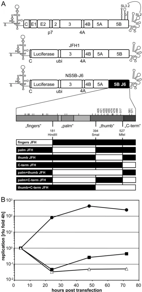
FIG. 1. Structure of monocistronic replicons and importance of the NS5B gene for efficient JFH1 replication. (A) Top, schematic view of an HCV genome and the subgenomic replicons JFH1 and NS5B-J6 used for transient replication assays. Coding sequences are depicted by rectangles, and the NTRs, as well as a critical CRE in the NS5B-coding sequence, are shown by their characteristic stem-loop structures. A kissing-loop inter-action between SL3.2 of this CRE and SL2 within the X tail of the 3⬘NTR is indicated by an arrow. JFH1 and heterologous sequences are indicated by white boxes; J6-derived sequences are depicted in black. This code was kept throughout the whole study. Middle, a schematic blowup of NS5B reveals the NS5B domains fingers, palm, thumb and C terminus. The limits of the four segments that were swapped between JFH1 and J6 are indicated by their respective positions within the coding sequence and the restriction enzymes used for subcloning. Polymorphisms between JFH1 and J6 are indicated by lines, and numbers refer to residues analyzed by site-directed mutagenesis. Bottom, schematic representation of chimeric
NS5B genes analyzed in this study. Luciferase, gene encoding firefly lu-ciferase; ubi, gene encoding human ubiquitin; C, HCV core; 2, 3, 4A, 5A, and 5B, genes encoding HCV nonstructural proteins. (B) Replication efficiencies of subgenomic replicons JFH1 and NS5B-J6 compared to that of a replication deficient mutant. Five micrograms ofin vitrotranscripts corresponding to JFH1 (F), 5BJ6 (f), and JFH1⌬GDD (‚) was trans-fected into Huh7-Lunet cells. Firefly luciferase activity was determined in cell lysates prepared at 4 h, 24 h, 48 h, and 72 h posttransfection. Repli-cation efficiency was determined by normalizing the relative light units (RLU) of the different time points to the respective 4-h value.
on November 7, 2019 by guest
http://jvi.asm.org/
antibiotic instead of ampicillin for BL21(DE3) and carbenicillin and chloram-phenicol were used for Rosetta(DE3). The bacterial cells were grown to an optical density at 600 nm of 0.6, expression was induced by the addition of 1 mM
isopropyl--D-thiogalactopyranoside, and cells were further incubated overnight
for JFH1 or for 4 h for J6 at 23°C with shaking and sedimented for 10 to 15 min
at 6,000⫻g. The pellets from 1-liter (JFH1) or 600-ml (J6) cultures were
resuspended in 40 ml lysis buffer (20 mM Tris-HCl [pH 7.5], 500 mM NaCl, 20
mM imidazole [Sigma-Aldrich], 0.2%n--D-octylglucopyranoside [n-OG], 20%
glycerol, 0.25 mg/ml lysozyme [Sigma-Aldrich], 25 U/ml benzonase [Merck, Darmstadt, Germany], and protease inhibitors [Complete EDTA free; Roche]) or in 20 ml lysis buffer (20 mM Tris-HCl [pH 7.5], 500 mM NaCl, 50 mM imidazole, 1% Triton X-100, 20% glycerol, 0.75 mg/ml lysozyme, 25 U/ml ben-zonase, and protease inhibitors), respectively. Incubation for 2 h with mild shaking at 4°C was followed by five freeze-thaw steps and a centrifugation for 20
min at 10,000⫻gat 4°C. The supernatants for JFH1 and J6 were loaded at 0.5
ml/min on a 1-ml self-packed column (1 ml of Ni-Sepharose; GE Healthcare) and a prepacked HisTrap HP 1-ml GE Healthcare column, respectively. The columns were equilibrated and washed with buffer A (50 mM sodium phosphate
[pH 8.0], 500 mM NaCl, 20 mM imidazole, 0.2%n-OG, 20% glycerol) for JFH1
and with a slightly modified buffer (20 mM Tris-HCl [pH 7.5], 500 mM NaCl, 50 mM imidazole) for J6. The bound proteins were eluted with 20 ml (for JFH1) or 30 ml (for J6) of a linear gradient of buffer A and buffer B (with buffer B containing 250 mM and 500 mM imidazole for JFH1 and J6, respectively) at a
flow rate of 1 ml/min at room temperature. The fractions containing NS5B⌬21
were identified by SDS-PAGE and pooled. The pool was dialyzed against 10 to 20 mM Tris-HCl (pH 7), 350 mM ammonium acetate, 1 mM EDTA, and 4 mM DTT and concentrated on a 30-kDa-cutoff ultrafiltration unit. Purified NS5B was
flash frozen by dripping into liquid nitrogen and kept at⫺80°C until use. Protein
concentrations were determined by OD280with extinction coefficients calculated
from the constructs’ sequences.
RdRp assays.All RdRp assays were done in a reaction buffer containing 20
mM Tris-HCl (pH 7.5), 5 mM MgCl2, 1 mM DTT, 25 mM KCl, 1 mM EDTA,
and 20 U of RNasin (Promega) in a total volume of 25l, and mixtures were
incubated for 1.5 h at room temperature. Standard reactions on homopolymeric
templates included 10Ci of [␣-32P]GTP (Perkin-Elmer), 50M CTP, UTP,
and ATP, 500M GTP, 2g poly(C) RNA template (GE Healthcare), and 1g
of purified polymerase. For quantitative analysis by liquid scintillation counting, samples were precipitated with 10% trichloroacetic acid (TCA) and 0.5% tetra-sodium pyrophosphate, passed through GF-C microfilters (GE Healthcare) washed five times with 1% trichloroacetic acid and 0.1% tetrasodium pyrophos-phate, and air dried. After addition of 4 ml of Ultima Gold (Perkin-Elmer), samples were subjected to liquid scintillation counting. All measurements were done in triplicates. Specific activities on poly(C) templates were expressed in
pmol GTP incorporated perg of NS5B per hour of reaction time and calculated
by determining the fraction of incorporated radioactive GMP, present at 132 nM (ca. 3.3 pmol), relative to the total GTP concentration.
Crystallization.All crystals were obtained by the hanging-drop vapor diffusion method and flash cooled by plunging into liquid nitrogen. New crystallization screens were set up for JFH1_NS5B (4.5 to 11.6 mg/ml) using robotics (Cartesian MicroSys) with the vapor diffusion method from 200-nl sitting drops. These screens produced better-looking and easier-to-handle microcrystals than our previously reported (41) crystals of JFH1_NS5B when mixed with a reservoir solution of 15% polyethylene glycol (PEG) 2000, 0.02 M sodium citrate, and 0.1 M sodium dihydrogen phosphate (pH 6.2) or with a reservoir solution of 12% PEG 4000 and 0.05 M sodium phosphate (pH 6.8). After optimization of the conditions, the crystals with the best diffraction quality were grown from a 1:1 mixture of protein solution (11.7 mg/ml) and reservoir solution (12% PEG 3350,
0.2 M sodium phosphate, pH 6.5) in 2-l hanging drops. Before flash cooling in
liquid nitrogen, the crystals were briefly transferred to a solution containing 12% PEG 3350, 0.2 M sodium phosphate (pH 6.5), and 35% glycerol.
J6_NS5B was concentrated to 2 mg/ml. Initial crystallization screens, set up using robotics (Cartesian MicroSys) with the vapor diffusion method from 200-nl sitting drops, produced small rods when mixed with a reservoir solution of 30% PEG 300 and 0.1 M Tris-HCl, pH 8.5. After optimization of the conditions, the crystals with the best diffraction quality were grown from a 2:2:2 mixture of protein solution (2 mg/ml), dialysis buffer (see “Expression and purification of NS5B” above), and reservoir solution (10% PEG 300, 0.1 M Tris-HCl, pH 8) in
6-l hanging drops. Before being flash cooled in liquid nitrogen, the crystals were
briefly transferred into a solution containing 32.5% PEG 300 and 0.1 M Tris-HCl, pH 8.
Structure determination and refinement.High-resolution X-ray diffraction data were collected at beam line Proxima 1 of the SOLEIL Synchrotron (St. Aubin, Gif-sur-Yvette, France). Diffraction data for JFH1_NS5B were processed
with the XDS package (21) and those for J6_NS5B with programs of the CCP4 suite (4), i.e., MOSFLM (through the imosflm interface) and SCALA. For J6_NS5B, molecular replacement was carried out with MOLREP (45) using Protein Data Bank (PDB) code 3I5K (41) as a search model. A first automatic rebuilding was performed with ARP/wARP (35), and then manual building was continued with COOT (16). Refinement was done first with Refmac5 (34) of the CCP4 suite and then with phenix.refine of the PHENIX suite (2). The high-resolution JFH1_NS5B structure in the new crystal form was determined starting from rigid-body refinement of a JFH1T385A_NS5B point mutant structure that had been previously refined to 1.8-Å resolution (see the supplemental material). This new JFH1_NS5B structure was carefully rebuilt with COOT and refined with phenix.refine. The same test set was used for the high-resolution JFH1_NS5B and JFH1T385A_NS5B data sets.
Objective comparisons of structures.Differences in main-chain conformation between pairs of molecules were objectively assessed using the program ESCET (39). As in our previous work (20, 41), we used significance values (ESCET “lolim” parameter) of 5.0 sigmas to identify broader conformational differences and 2.5 sigmas to take into account all significant conformational differences. When comparing the two high-resolution structures reported here, we also used a lolim value of 2.0. In significantly different structures, all rigid groups larger than 10 residues were sought. Figures were generated with PYMOL (the PyMOL Molecular Graphics System [2002] at http://www.pymol.org).
Infectivity assays.Huh7.5 cells were electroporated within vitrotranscripts of JcR2a or JcR2a chimeras and seeded in six-well plates. Supernatants were
harvested 24 h, 48 h, and 72 h postelectroporation, filtered through 0.45-
m-pore-size filters, and applied to 4⫻105Huh7.5 cells seeded in 24-well plates. At
72 h after infection, cells were lysed in 100l lysis buffer (see “Luciferase assays”
above), and infectivity was determined by measuringRenillaluciferase activity
(r-RLU) for 10 s in a plate luminometer (Mithras LB940; Berthold) after
addi-tion of 400l assay buffer (see “Luciferase assays” above).
Protein structure accession numbers.Coordinates and structure factors for the crystal structures described in Table 2 are available from the Protein Data Bank under accession codes 2XWH (J6_NS5B) and 2XXD (JFH1_NS5B_O).
RESULTS
Mapping of regions within NS5B that are critically involved in JFH1 replication. Our previous analysis showed that the polymerase of HCV isolate JFH1 had a higher efficiency ofde novoRNA synthesisin vitrothan the RdRp of isolate J6. This higher activity correlated with a very closed conformation of the structure of the enzyme compared to a genotype 2a con-sensus structure. To address whether the enzymatic and struc-tural properties of NS5B were indeed underlying the outstand-ing replication of JFH1 in cell culture, we performed a comprehensive analysis of the replication efficiency of repli-cons harboring chimeric NS5B sequences compared to the RdRp activity of the corresponding purified enzymes. Since NS5B was not the only determinant of efficient JFH1 replica-tion (32), we decided to use a subgenomic replicon with a JFH1 backbone as a starting point and replaced only the NS5B-coding sequence of isolate JFH1 with the J6 counterpart (Fig. 1A, NS5B-J6). To mimic the translational properties of a full-length genome most closely, we used a monocistronic architecture and included a firefly luciferase reporter gene to facilitate quantitation of replication (Fig. 1A) (8). The repli-cation efficiency of this RNA can easily be quantified by de-termining firefly luciferase activity in cell lysates. The luciferase activity obtained from the chimeric replicon harboring the J6 NS5B (NS5B-J6) was very low compared to that of the paren-tal JFH1 replicon and only slightly above that of a replication-deficient mutant containing a 10-amino-acid deletion in the active center of the polymerase (⌬GDD) (Fig. 1B). This result was expected from a previous study (32) and confirmed the important role of the NS5B-coding region for efficient JFH1 replication.
on November 7, 2019 by guest
http://jvi.asm.org/
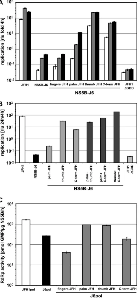
The NS5B-coding regions of JFH1 and J6 differed at 133 positions in the RNA sequence, resulting in 29 amino acid deviations in the protein sequence. In order to identify those positions involved in efficient JFH1 replication, we first re-placed larger regions of NS5B-J6 with the corresponding JFH1 sequences, aiming to rescue replication to JFH1 levels. For this gross mapping analysis we used convenient restriction sites at the approximate borders of the NS5B subdomains (Fig. 1A) and named the chimeras according to the dominant part of the inserted JFH1 domain, i.e., “fingers,” “palm,” “thumb,” and “C-term” (Fig. 1A). Surprisingly, three different JFH1 NS5B segments, palm, thumb, and C-term, increased replication ef-ficiency of NS5B-J6, albeit to various extents (Fig. 2A). The JFH1 palm segment resulted in a 7-fold increase of replication efficiency compared to NS5B-J6, whereas C-term and thumb stimulated replication by ca. 100- and 1,000-fold, respectively, thereby nearly reaching JFH1 levels.
To evaluate the contribution of the increasedde novo initi-ation efficiency of JFH1 RdRp to repliciniti-ation in cell culture, we constructed identical chimeric coding sequences lacking the 21-amino-acid C-terminal membrane insertion sequence (po-sitions 571 to 591) and fused to a hexahistidine tag in a bac-terial expression vector. All chimeric proteins were expressed and purified with similar efficiencies (data not shown) and analyzed for enzymatic activity using a homopolymeric poly(C) template in the absence of a primer (Fig. 2C). These assay conditions allowed the most sensitive and robust quantitation ofde novo-initiated RNA synthesis (41). However, since the readout of this assay includes initiation and elongation, effects on elongation cannot be entirely excluded. JFH1 RdRp exhib-ited a 6-fold-higher RNA synthesis efficiency than J6 under these conditions (Fig. 2C). Replacement of either the palm or thumb segment of J6 by JFH1 increased RdRp activity by ca. 3-fold, whereas the JFH1 fingers segment had a negative effect on enzymatic activity. These results indicated that the contri-butions of the palm and thumb segments to efficient JFH1 replication in cell culture were at least in part due to increased
[image:5.585.46.288.72.587.2]de novo-initiated RdRp activity, as proposed in our previous study (41). The discrepancy between the palm and thumb seg-ments in their ability to stimulate replication in cell culture compared to effects on RdRp activity might be due to other functions of NS5B independent of polymerase activity. In con-trast to the case for the palm and thumb segments, we found no stimulating effect of the C terminus of JFH1 NS5B on RdRp activityin vitro(Fig. 2C), despite the strong effect of this domain on the efficiency of the replicon (Fig. 2A), arguing for a mechanism independent of polymerase activity. The entire C-terminal domain contained only two coding polymorphisms, at positions 561 and 571; the latter was formally lacking in our enzymes due to the deletion of 21 C-terminal amino acids. However, due to a BglII restriction site linking the NS5B-coding sequence to the hexahistidine tag, position 571 by
FIG. 2. Rescue of J6-NS5B by replacement with different JFH1 segments. (A and B) Replication efficiencies of chimeric NS5B-J6 replicons with the indicated replacement of one (A) or two (B) ho-mologous segments of JFH1 NS5B (Fig. 1A).In vitrotranscripts cor-responding to the indicated replicons were transfected into Huh7-Lunet cells. Cell lysates were obtained at different time points after transfection and analyzed for firefly luciferase activity (RLU). The ratios of RLU obtained at 24 h (white bars) 48 h (gray bars), and 72 h (black bars) (A) or 24 h (B) to that at 4 h after transfection are shown. (C) RNA synthesis of purified chimeric RdRps on a homopolymeric template. Standard RdRp assay mixtures using 1g of purified JFH1, J6, and chimeric J6 polymerases as for panel A were incubated for 1.5 h on a poly(C) template with 10Ci [␣-32P]GTP and 500M cold GTP. Incorporation of radioactivity was quantified by TCA precipita-tion and liquid scintillaprecipita-tion counting. After background subtracprecipita-tion,
picomoles of incorporated GMP per microgram of enzyme per hour were calculated based on the ratio of labeled to unlabeled GTP. Lu-ciferase and RdRp assays were performed two to four times indepen-dently for each construct. Data are mean values and standard devia-tions for a representative experiment.
on November 7, 2019 by guest
http://jvi.asm.org/
chance was a serine in all of our polymerase expression con-structs, thereby representing the JFH1 sequence. Therefore, we also mutated position 571 to leucine, which is present in J6. We found no impact of this residue onde novo-initiated RdRp activity in vitro (see Fig. S1 in the supplemental material), ruling out any positive effect of the C-terminal region of JFH1 NS5B on enzymatic activity in our assay system, at least for polymerases lacking the membrane insertion sequence.
Our data implied that the palm and thumb segments of JFH1 NS5B were both enhancing JFH1 replication in cell culture at least in part by increasing RdRp activity, whereas the C-terminal domain was acting by a different mechanism. We aimed to confirm this assumption by combining two of these domains derived from JFH1 in an NS5B-J6 replicon (Fig. 1A and 2B). Interestingly, a combination of the palm and thumb segments of JFH1 did not increase replication efficiency be-yond the level for the thumb segment alone, supporting our hypothesis that both acted by the same mechanism. In contrast, the C terminus of JFH1 additively enhanced the replication efficiency of the chimeras harboring the palm or the thumb segment, for the latter even 2-fold above the level of wild-type JFH1 (Fig. 2B). The additive effect of the C-terminal domain with the palm and thumb segments on replication efficiency in cell culture provided a second piece of evidence arguing for an independent mechanism of action.
In summary, our data showed that the palm and thumb segments of JFH1 increasedde novo-initiated RdRp activity and replication in cell culture of J6 NS5B, suggesting a mech-anistic link as proposed in our previous study (41). The C-terminal region of the NS5B-coding sequence also substan-tially contributed to efficient JFH1 replication in cell culture, but most likely by a mechanism independent of RdRp activity.
A single mutation in the thumb domain mainly contributes to enhanced RNA replication andde novo-initiated RdRp ac-tivity of JFH1 NS5B.Our previous analysis showed that the polymerase of HCV isolate JFH1 had a higher efficiency ofde novo-initiated RNA synthesisin vitrothan the RdRp of isolate J6. This higher activity of the enzyme correlated with a more closed conformation of the JFH1 polymerase structure than of the consensus 2a (Con2a) structure, which was mediated mainly by critical residues in the thumb domain. Therefore, we speculated that the closed conformation of the JFH1 polymer-ase was critical for efficientde novo-initiated RNA replication of this isolate. Since the thumb domain of JFH1 indeed had a major impact on replication efficiency in cell culture and strongly increased de novo initiated-RNA synthesis of the RdRp (Fig. 2A and C), we aimed to prove or disprove our concept by a comprehensive analysis of mutants and chimeras harboring subsegments of the thumb in regard to enzymatic activity and replication efficiency.
The overall pattern was rather complex but revealed inter-esting similarities and discrepancies between replicon and polymerase activities in vitro (Fig. 3 and Table 1). The N-terminal part of the JFH1 thumb segment (positions 394 to 470) strongly increased RdRp activityin vitro, to the level of the entire thumb segment, and replication in cell culture, albeit to a lower extent. The middle part (positions 470 to 494) entirely abrogated both polymerase activity and RNA replica-tion. The C-terminal part (positions 494 to 527) stimulated replicon activity without having an impact on RdRp activityin
vitro. Due to this complex pattern, we decided to analyze the individual polymorphisms in the thumb segment in more de-tail. We exchanged individual amino acids in the NS5B-J6 sequence to check whether they would rescue replication and RdRp activity to the levels conferred by the JFH1 thumb segment. Most single mutations showed no or even a negative phenotype (Fig. 3; Table 1). A421V and S455N affected
nei-FIG. 3. Identification of individual mutations in the thumb that are critically involved in RNA replication andde novo-initiated RNA syn-thesis. (A)In vitrotranscripts corresponding to wild-type, chimeric, or mutant subgenomic replicons were transfected in Huh7-Lunet cells. Firefly luciferase activity was determined as described for Fig. 1B. The ratios of luciferase activity at 24 h (white bars), 48 h (gray bars), and 72 h (black bars) to that at 4 h after transfection are shown. (B)De novo-initiated RdRp activities of purified wild-type, chimeric and mu-tant NS5B⌬C21 proteins expressed inE. coli. Standard RdRp assay mixtures using 1g of purified JFH1pol, J6pol, and chimeric or mutant J6pol variants as for panel A were incubated for 1.5 h on a poly(C) template with 10 Ci [␣-32P]GTP and 500 M cold GTP. Incorporation of radioactivity was quantified by TCA precipitation and liquid scintillation counting. After background subtraction, picomoles of incorporated GMP per microgram of enzyme per hour were calcu-lated based on the ratio of labeled to unlabeled GTP. Numbers refer to amino acid positions of the NS5B protein. Luciferase and RdRp assays were performed two to four times independently for each con-struct. Data are mean values and standard deviations for a represen-tative experiment.
on November 7, 2019 by guest
http://jvi.asm.org/
ther RdRp nor replicon activity. P479H even abrogated RdRp activity of the mutant polymerase as well as replication in cell culture, which probably explains the negative impact of the subsegment from amino acid 470 to 494. A435V and L474M did not impair RdRp activity but resulted in inactive replicons.
Some of these mutations (e.g., A435V and P479H) had even a stronger negative effect on apparent replication than the rep-lication-deficient negative control (⌬GDD), probably due to an impairment of RNA stability. In contrast, A499V and R517K did not increase or only slightly increased RdRp activ-ity but rescued NS5B-J6 replicon replication by a factor of 10-fold each, which corroborates the effects of the subsegment from amino acid 494 to 527. Only two mutations, V405I and A450S, mimicked the phenotype of the entire thumb domain, by stimulatingde novo-initiated RNA synthesis of the RdRp and enhancing replication of the replicon. Most striking was V405I, which rescued RdRp activity to the level of the chimeric polymerase harboring the entire JFH1 thumb domain and in-creased replication efficiency of the NS5B-J6 replicon by more than 100-fold.
Although the pattern of phenotypes exerted by individual mutations in the thumb domain of J6 was complex, we could identify a single polymorphism, V405I, which increased the RdRp activity of J6 polymerase and the replication efficiency of the NS5B-J6 replicon almost to the levels for the entire thumb domain of JFH1.
[image:7.585.43.284.87.350.2]High-resolution crystal structures of J6_NS5B and JFH1 NS5B confirm that extra hydrophobic contacts of residue 405 are JFH1 specific. We previously reported that JFH1_NS5B has a thumb in a slightly but significantly more closed confor-mation than that of the previously published (9) consensus 2a (Con2a) NS5B structures and correlated this fact to the higher efficiency of JFH1 NS5B than of J6 NS5B inde novo RNA synthesis (41). To gain further insight into the direct mecha-nism particularly underlying those polymorphisms stimulating RNA replication and RdRp activity, we required a more con-cise side-by-side structural comparison of JFH1 NS5B with the same construct that we used for the functional analysis. There-fore, we crystallized this J6 NS5B construct and refined its structure to high resolution (Table 2). We also obtained a new crystal form (here designated by the letter “O”) of JFH1 NS5B
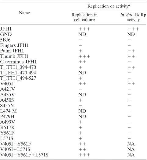
TABLE 1. Schematic comparison of replication efficiency and
in vitroactivity
Name
Replication or activitya
Replication in cell culture
In vitroRdRp activity
JFH1 ⫹⫹⫹ ⫹⫹⫹
GND ND ND
5BJ6 ⫺ ⫺
Fingers JFH1 ⫺ ⫺
Palm JFH1 ⫹ ⫹⫹
Thumb JFH1 ⫹⫹⫹ ⫹⫹
C terminus JFH1 ⫹⫹ ⫺
T_JFH1_394-470 ⫹ ⫹⫹
T_JFH1_470-494 ND ⫺
T_JFH1_494-527 ⫹ ⫺
V405I ⫹⫹ ⫹⫹
A421V ⫺ ⫺
A435V ND ⫺
A450S ⫹ ⫹
S455N ⫺ ⫺
L474 M ND ⫺
P479H ND ⫺
A499V ⫹ ⫺
R517K ⫹ ⫺
Y561F ⫹ ⫺
L571S ⫹ ⫺
V405I⫹Y561F ⫹⫹ NA
V405I⫹L571S ⫹⫹ NA
V405I⫹Y561F⫹L571S ⫹⫹⫹ NA
aFold replication or activity relative to that for J6 was determined. Data are
based on three different experiments. ND, not detectable (inactive); NA, not
analyzed. Replication:⫺, up to 5-fold;⫹, up to 100-fold;⫹⫹, up to 1,000-fold;
⫹⫹⫹, more than 1,000-fold. Activity:⫺, up to 1.2-fold;⫹, up to 2-fold;⫹⫹, up
to 5-fold;⫹⫹⫹, more than 5-fold.
TABLE 2. Data collection and refinement statisticsa
Parameter J6_NS5B JFH1_NS5B
Crystallization conditions (addition for cryogenic cooling)
0.1 M Tris (pH 8), 10% PEG 300 (22.5% PEG 300)
0.2 M sodium phosphate (pH 6.5), 12% PEG 3350 (35% glycerol)
Space group; unit cell parameters (Å, °) P212121;a⫽64.8,b⫽94.3,c⫽113.3,
␣ ⫽90, ⫽90,␥ ⫽90
C2221;a⫽99,b⫽110.4,c⫽115.0,␣ ⫽90,
 ⫽90,␥ ⫽90
Resolution range (Å) 42–1.8 (1.9–1.8) 40–1.9 (1.93–1.88)
No. of reflections 64,666 51,130
Completeness (%) 99.4 (100) 99.5 (95.0)
Rsymb(%) 11.1 (55.7) 11.5 (71.9)
I/sigmaI 7.8 (2.0) 14.1 (2.8)
No. of molecules in asymmetric unit 1 1
Avg multiplicity 3.0 (2.9) 7.3 (7.1)
Rc(%) 16.5 (20.5) 15.7 (17.8)
Rfree
c(%) 20.6 (24.6) 20.4 (22.9)
Root mean square deviation, bond (Å) 0.006 0.006
Root mean square deviation, angle (°) 1.02 0.99
X-ray source Soleil synchrotron, Proxima 1 Soleil synchrotron, Proxima 1
PDB code 2XHW 2XXD
a
Statistics for the highest-resolution shell are given in parentheses.
b
Rsymwas determined by the equationRsym⫽⌺hkl⌺j兩Ihkl.j⫺ 具Ihkl典兩/⌺hkl⌺jIhkl, whereh,k, andlare the unique indices of all reflections measured more than once and
jis the index for symmetry-redundant reflections.
c
RandRfreewere determined by the equationR⫽ ⌺hkl兩兩Fobs兩 ⫺k兩Fcalc兩兩/⌺hkl兩Fobs兩, whereh,k, andlare the indices of the reflections used in refinement (R) or of
5% of the reflections set aside and not used in refinement (Rfree). The same set of reflections was used forRfreein all structures.Fobsare the structure factors deduced
from the measured intensities andFcalcthe structure factors calculated from the model.kis a scale factor to putFcalcon the same scale asFobs.
on November 7, 2019 by guest
http://jvi.asm.org/
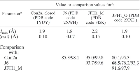
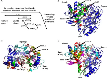
[image:7.585.44.538.500.678.2]that yielded significantly better data (Table 2) than our previ-ously reported crystal form (here designated by the letter “M”). Similarly to as described in our previous report (41), we subjected these structures to an objective comparison (39) with those of both previously published subtype 2a NS5B structures, namely, JFH1_NS5B_M and Con2a NS5B. We found that all four structures are conformationally different from one an-other when using the typical cutoff of 2.5 sigmas for signifi-cantly different positions of the same residues in two structures (Table 3). Due to a small forward rotation, the thumb of JFH1_NS5B_O is actually even more closed than that of JFH1_NS5B_M (Fig. 4A). J6 NS5B, however, falls between Con2a_NS5B and JFH1_NS5B_M with regard to the thumb’s closure (Fig. 4A). The displacements between the thumbs of J6 and JFH1 NS5B are quite small and of the same order of magnitude as those seen for JFH1 NS5B in different crystal packing environments (Fig. 4A). We therefore checked J6_NS5B and JFH1_NS5B_O for the six positions where we previously (41) reported special polymorphisms of JFH1_NS5B to induce specific conformational changes stabi-lizing the closed thumb conformation of JFH1_NS5B_M, par-ticularly extra hydrophobic interactions of I405 and V435. We consistently find that the extra interactions are indeed present in JFH1_NS5B and absent in J6_NS5B, despite the fact that the latter’s thumb is only slightly more open than the former’s. We thus confirm that stabilization of the thumb in a closed conformation is a special feature of JFH1 NS5B.
Delineation of the more pronounced differences between JFH1_NS5B and J6_NS5B highlights a general rearrange-ment of the thumb-fingertip contact and local displacerearrange-ments involving residues 405 and 450.The differences between the four structures in Table 3 are small enough that when only more substantial deviations are considered (cutoff of 5.0 sig-mas), pairs not including JFH1_NS5B_O are conformationally
identical (above the 98% threshold of alpha carbons in iden-tical positions). However, JFH1_NS5B_O is significantly dif-ferent from the other three structures even at this higher-tolerance cutoff. This is due on the one hand to larger differences in conformation (Fig. 4A) and on the other hand to the higher precision of JFH1_NS5B_O compared to JFH1_NS5B_M (Table 3,冓esd冔). This allows us to assess the main conformational differences between JFH1_NS5B and J6_NS5B (Table 3 [underlined value]; Fig. 4B). We find that these larger differences point to a concerted movement of a rigid block (in green in Fig. 4B) comprising the back of the thumb, including helix S, and the distal fingertips, including helix A (we use the nomenclature defined in reference 13). Surprisingly, there are also some significant but very local movements, each involving only a few residues (in red in Fig. 4B). Apart from surface residues involved in crystal contacts (such as at the top of the fingers), two two-residue stretches are thus outlined, right at or close to the two polymorphisms of JFH1_NS5B that mimic the phenotype of the entire thumb domain when introduced into J6 NS5B (Fig. 3): first, residues V405I-406 at the point of contact between the thumb, fingers, and top of the beta flap, and second, the 448-449 (right before A450S) tip of the beta flap where it interacts with the linker.
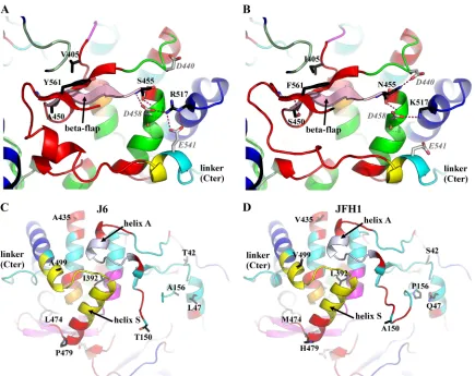
[image:8.585.44.284.89.212.2]A detailed comparison of the high-resolution structures of JFH1_NS5B and J6_NS5B highlights complex, concerted dis-placements of the fingertips, back of the thumb, and C termi-nus. The high precision of JFH1_NS5B_O and J6_NS5B al-lows a more detailed breakdown of rigid blocks to be computed, with a tolerance of 2 sigmas (Table 3 [bold value]; Fig. 4C and D) instead of the typical 2.5. This breakdown in turn allows us to refine and extend our previous findings about the likely role of some key JFH1 polymorphisms (Fig. 5). Thus, the portion of the fingers facing position 405 (in light green in Fig. 5A and B) is slightly more open in this J6/JFH1 compar-ison, as it was in the Con2a/JFH1_M comparison (41). When JFH1 is taken as the reference, the shift of this part of the fingers is less pronounced in J6 than in Con2a due to J6 being in an intermediate conformation between those of Con2a and JFH1 (Fig. 4A). As stated above, our previous report that the JFH1 V405I polymorphism induces a gain of hydrophobic in-teractions buttressing the thumb-fingers interaction in this re-gion is nevertheless confirmed in the comparison with J6. These hydrophobic interactions lead to a distinct displacement of the segment from amino acid 402 to 408 near the beta flap. In the beta flap itself, the strand from amino acid 450 to 455 is highlighted as a distinct rigid body (in light pink in Fig. 4C and D and 5A and B) due to changes in hydrogen bonding at either end (see below for S455N): S450 in JFH1 indeed makes an extra hydrogen bond to I560 of the linker (Fig. 5B) compared to A450 in J6 (Fig. 5A). We previously remarked that, in contrast, there is one fewer hydrogen bond between the linker and flap in JFH1 because of the Y561F polymorphism (41). The case is not quite as clear-cut in the J6/JFH1 comparison, because the extra side chain hydroxyl in Y561 of J6 now makes only a water-mediated hydrogen bond to the top of the beta flap (hence, this hydrogen bond is not depicted in Fig. 5A, where we show only variations in protein-protein hydrogen bonding). However, this extra hydroxyl actually participates in coordinating a network of water molecules (clearly visible at these resolutions) that bridges the gap from the top of the beta
TABLE 3. Pairwise conformational comparisons between J6_NS5B, JFH1_NS5B_ O, and previously published subtype 2a HCV NS5Bs
Parametera
Value or comparison values forb
: Con2a, closed
(PDB code 1YUY)
J6 (PDB code 2XWH)
JFH1_M (PDB code 3I5K)
JFH1_O (PDB code 2XXD)
dmin(Å) 1.9 1.8 2.2 1.9
具esd典(Å) 0.10 0.07 0.15 0.10
Comparison with:
Con2a 85.3/98.1 95.0/99.8 80.1/95.3
J6 93.7/99.6 68.5/76.2/93.3
JFH1_M 91.6/97.9
ad
min, high-resolution limit; 具esd典, mean coordinate uncertainty for alpha
carbons present in all structures.
bThe models are entered in order of increasing closure of the thumb (see text
and Fig. 4A for details). The structures for the consensus 2a NS5B and JFH1_NS5B in crystal form M were previously published. The comparison values are the percentages of residues whose alpha carbons are not in significantly different positions. Two models can be considered to be in identical conforma-tions if this value is larger than 98% (39). The values are given for all pairs of molecules for the two tolerance thresholds (ESCET lolim parameter) of 5.0 (right) and 2.5 (left) sigmas. For the J6/JFH1_O comparison only, the value at 2.0 sigmas (leftmost) is also given and in bold. This value was used for the ESCET detailed breakdown in rigid groups depicted in Fig. 4C and D and 5. The value at 5.0 sigmas (underlined) was used in the cruder breakdown in Fig. 4B, where only more substantial deviations are sought.
on November 7, 2019 by guest
http://jvi.asm.org/
flap to the fingers in J6, a water cap that is starkly reduced above the hydrophobic patch made by Y561F in JFH1. Further up the beta flap, polymorphisms S455N and R517K also con-siderably change the pattern of hydrogen bonding (Fig. 5A and B). We could find no discernible conformational effect of this in our previous work. It is now clear from the new structures that these changes participate in the coordination of several concerted displacements involving regions both at the back and at the front of the thumb. This is accomplished most obviously through two salt bridges of R517 in J6 with E541 of the linker and D458 (Fig. 5A) that cannot both be done by JFH1’s K517 (Fig. 5B). The linker is thus tethered to a rigid block (in cyan in Fig. 4C and 5) going from the linker through the central helix bundle of the thumb to the fingertips (at the base of which are three polymorphisms at positions 42, 47, and 156). Remarkably, this draws a previously unobserved partitioning of the thumb that is completed by two disconnected rigid blocks. First, the second half of the short helix harboring E541
in the linker is associated with the platform at the back of the thumb (in yellow in Fig. 4C and 5C and D) upon which the fingertips rest. This platform reaches to the polymorphism at position 499 and is reoriented by polymorphisms at positions 478 and 479 at the base of helix S. The second disconnected rigid block (in blue-white in Fig. 4C and 5) comprises helix A on the one hand and the connection between helices R and S (which includes the polymorphism at position 474) on the other hand. Residue 474 is buried between this connection and another rigid block (in magenta in Fig. 4C and D and 5) that comprises the 15-16 beta hairpin and extends into the thumb to the polymorphism at position 392. The 15-16 hairpin was pre-viously recognized as a hinge around which the genotype 1b NS5B thumb rotates (1), while residue 392 is one of two JFH1 polymorphisms (with residue 435) that we reported to modify the interaction of helix A with the thumb (41).
[image:9.585.73.509.72.388.2]In summary, the comparison of the JFH1 and J6 NS5B structures shows two distinct features. The first, and simple,
FIG. 4. Comparisons involving the high-resolution structures of subtype 2a NS5B reported in this paper. (A) Schematic of the range of thumb closures seen in various crystal structures of subtype 2a HCV NS5B. Maximum displacements when aligning the structures on the rigid block containing the palm subdomain are at the top of the thumb. These displacements are indicated for relevant pairs of structures. In italics are the structures and displacement values previously reported (9, 41). (B, C, and D) The ESCET program was used to objectively assess rigid groups of NS5B in significantly different positions in J6_NS5B and JFH1_NS5B_O. A ribbon representation of JFH1_O is shown, divided in rigid groups derived from comparison of the two structures. These groups are colored in order of decreasing size: blue, green, and (for panels C and D) cyan, magenta, light green, yellow, blue-white, orange, and light pink. The red zones indicate more flexible regions not assignable to one of the rigid groups. In all three views, helix S of the thumb and helix A of the fingertips are labeled with arrows. (B) Comparison with a tolerance threshold for residues in identical positions (ESCET lolim parameter) of 5.0 sigmas. Positions 405 and 450, at or near two red regions of high local differences between the two structures, are displayed as spheres. (C and D) Comparison with a tolerance threshold of 2.0 sigmas, displayed in two orientations: a “top” orientation allowing convenient visualization of fingertips, beta flap, and linker (labeled) (C) and an orientation as in panel B, displaying the hinges in the base of the thumb at the back of NS5B (D).
on November 7, 2019 by guest
http://jvi.asm.org/
feature is a network of extra interactions in JFH1_NS5B in-volving residues 405 and 450 at the junction between the thumb, fingers, beta flap, and linker. These interactions con-tribute to stabilizing the closed thumb conformation of the JFH1 polymerase, thereby enhancing the de novo initiation efficiency of RNA synthesis. The second, and quite complex, feature is a series of concerted displacements of the fingertips, back of the thumb, and part of the C-terminal segment. Coor-dination is achieved by several channels potentially involving up to 10 JFH1 polymorphisms. One channel is a remodeling of the hydrogen bonding pattern connecting the C terminus to the thumb, central to which is residue 517. For reasons out-lined in Discussion, this channel at least is presumably associ-ated with the transition from initiation of RNA synthesis to elongation. Although residue 561 is in a position to participate in both of these features (Fig. 5A and B), the polymorphism Y561F at this position has no clear structural effect on either.
Two independent mechanisms in the C-terminal segment of NS5B contribute to efficient JFH1 replication.The initial map-ping identified the important contribution of polymerase ac-tivity to efficient JFH1 replication in cell culture, which we
[image:10.585.75.510.66.411.2]could assign primarily to position 405 within the thumb domain (Fig. 3). Next we aimed to determine the critical positions in the C-terminal segment, which also had a significant role in JFH1 replication seemingly independent of the RdRp activity of the isolated enzyme. JFH1 and J6 differed at only two positions in their amino acid sequence in the C-terminal seg-ment (positions 528 to 591) (Fig. 1A) that comprises the linker (positions 530 to 570) and transmembrane helix (positions 571 to 591); however, the situation was complicated by the fact that an important CRE resides in this region, containing three stem-loop structures termed SL3.1, SL3.2, and SL3.3 (54). Therefore, we also included noncoding polymorphisms in our analysis. Recent studies have particularly demonstrated the essential role of SL3.2 in HCV RNA replication, acting via a kissing-loop interaction with a loop-sequence (SL2) in the X-tail region of the 3⬘NTR (18). Interestingly, both variations resulting in a change of the amino acid sequence were located in the SL3.2 sequence: U9348A/Y561F in the loop region, thereby increasing the length of the kissing-loop interaction, and C9378U/L571S at the base of the stem. We again used replicon NS5B-J6 as a starting point and tried to rescue the
FIG. 5. Comparison of polymorphisms in J6_NS5B and JFH1_NS5B. J6_NS5B (left) and JFH1_NS5B (crystal form O, right) are displayed as ribbons and colored as in Fig. 4C and D. Polymorphisms are labeled and displayed in stick representation and colored by atom type (black, carbons; blue, nitrogens; red, oxygens; yellow, sulfurs). Residues making hydrogen bonds to the polymorphisms in either or both structures are similarly displayed but with light gray carbons. For clarity, only side chains are shown unless the main chain is itself involved in the hydrogen bonding pattern. The linker is labeled in all views. (A and B) View highlighting residue 405 and the differential hydrogen bonding pattern of the linker with the thumb and beta-flap. (C and D) View from the back highlighting the hinges at the base of the thumb and in the fingertips.
on November 7, 2019 by guest
http://jvi.asm.org/
replication efficiency of the entire C-terminal segment (C-term JFH) by exchanging J6 sequences with all polymorphisms in individual stem-loops of the CRE (SL3.1 JFH, SL3.2 JFH, and SL3.3 JFH), all noncoding variations (silent JFH), and both amino acid mutations (Y561F⫹L571S) (Fig. 6A). Replicons SL3.2 JFH and Y561F⫹L571S both exhibited replication at the level of C-term JFH1, whereas even the combination of all silent mutations replicated at the level of NS5B-J6. Individual analysis of replicons harboring only mutation L571S or Y561F revealed that both rescued NS5B-J6 to the same extent.
Based on these data, it was not possible to decide whether the change in the nucleotide or amino acid sequence was functionally important. To address this important point, we generated constructs allowing the analysis of the CRE in the absence of protein-coding functions by disturbing the RNA structures in the CRE within NS5B and by insertion of an intact copy of these elements in the variable region of the 3⬘NTR. This approach had already been successful in the case of a genotype 1b replicon (18). We introduced a maximum number of silent nucleotide substitutions in the CRE of the 5B-coding sequence and indeed entirely abrogated replication (JFH1 mut3.1-3) (Fig. 6B and C). Duplication of the CRE in the absence of any changes within the NS5B gene strongly reduced replication efficiency compared to that in wild-type JFH1, probably by disturbing RNA secondary structures at the insertion site in the 3⬘NTR (JFH1 dup3.1-3) (Fig. 6B and C). More importantly, the duplication of the CRE was able to rescue replication of a construct with a scrambled CRE in the NS5B gene (JFH1 ins3.1-3) (Fig. 6B and C), demonstrating that the duplicated CRE was able to replace thecisfunctions of the endogenous stem-loop structures. We now introduced the J6 nucleotide polymorphisms into the duplicated version of the CRE, while retaining the NS5B-coding sequence
unaf-FIG. 6. Functional analysis of mutations in the C-terminal segment of the NS5B-coding region. (A) Replicons harboring the NS5B gene of J6 and different coding and noncoding polymorphisms of JFH1 in the C-terminal segment of NS5B were analyzed for replication efficiency in
cell culture. SL3.1 JFH, SL3.2 JFH, and SL3.3 JFH refer to replace-ments of the individual stem-loops of the CRE in NS5B. Silent JFH and Y561F⫹L571S contain all noncoding or coding polymorphisms of JFH1, respectively. For construct L571S-ACG, the serine was gener-ated by an ACG codon instead of the original UUA codon found in JFH1. Luciferase activity was determined as described for Fig. 1B. The ratios of firefly luciferase activity at 24 h (white bars), 48 h (gray bars), and 72 h (black bars) to that at 4 h after transfection are shown. (B) Schematic presentation of replicon constructs used to evaluate the impact of nucleotide substitution in the NS5B CRE. The 5⬘ and 3⬘NTRs and the three stem-loop structures in NS5B are indicated by their secondary structures. The kissing-loop interaction of NS5B SL3.2 and SL2 of the 3⬘NTR is depicted by arrows. CREs containing accu-mulated silent nucleotide substitutions disrupting the SL structures and the kissing-loop interaction are crossed. Names of the correspond-ing replicons are given on the right of each scheme. The relative positions of mutations U9348A and C9378U in the loop and at the base of SL3.2, respectively, in the duplicated CRE are indicated by dots in JFH1ins3.1. (C) Impact of alterations in the CRE within NS5B on the kissing-loop interaction with the 3⬘NTR. Representative results from transient-replication assays using the basic replicons shown in panel B are shown. (D) Analysis of replication competence by using the JFH1ins3.1-3 replicon with additional mutations (U9348A, C9378U, or U9348A⫹C9378U) in the inserted 5B SL structures. All luciferase activities were evaluated as described for Fig. 1B. The ratios of firefly luciferase activity at 24 h (white bars) 48 h (gray bars), and 72 h (black bars) to that at 4 h after transfection are shown. Data are mean values and standard deviations for a representative experiment.
on November 7, 2019 by guest
http://jvi.asm.org/
[image:11.585.60.265.64.687.2]fected, to focus on the cis functions of these positions. In contrast to the case for our previous gain-of-function experi-ments (rescuing J6 sequences by introducing JFH1 sequences), this experimental system addressed a loss of function by intro-ducing J6 variations in a JFH1 sequence. Interestingly, a change at position 9348 (U9348A, corresponding to position 561 in NS5B), strongly reduced replication efficiency (Fig. 6D). The facts that this nucleotide resided in the loop of SL3.2 and that the mutation decreased the kissing-loop interaction with SL2 in the X tail from 8 to 7 nucleotides clearly argued for a mechanistic role of this polymorphism in the CRE and not in the coding sequence of NS5B. In contrast, a change at position 9378, at the base of the stem of SL3.2, had no impact on replication (Fig. 6D), either individually or in combination with U9348A, suggesting a function at the level of the coding sequence. To further rule out any role of the specific nucleo-tide sequence at this position, we introduced the L571S muta-tion using an AGC codon instead of UUA as found in JFH1. Indeed, both L571S mutants replicated to exactly the same level (Fig. 6A, L571S and L571S-AGC). Therefore, position 571 seems to be an important determinant in the NS5B-coding sequence but not for enzymatic activity itself (Table 1; see Fig. S1 in the supplemental material). However, since this resi-due was directly adjacent to the 6⫻His tag in our NS5B ex-pression constructs and since we did not obtain any structural information beyond position 563, we cannot entirely rule out a potential role of 571S in the RdRp activity of a full-length polymerase.
Taking the findings together, both coding polymorphisms in the C-terminal segment of NS5B were critical for JFH1 repli-cation and rescued NS5B-J6 to the same extent, but by entirely different mechanisms. The polymorphism at position 9478 (en-coding amino acid 561) enhanced replication by increasing the length of the kissing-loop interaction between SL3.2 and SL2. In contrast, position 571 was important in the protein-coding sequence, without affecting RdRp activity in our in vitro de novo-initiated RdRp assay.
Combinations of two substitutions enhance NS5B-J6 repli-cation to levels close to those of JFH1 and rescue generation of infectious virus.We next aimed to rescue NS5B-J6 by com-bining the most promising candidates. After identification of single residues in the NS5B gene contributing to efficient rep-lication of JFH1, it was clear that at least four different mech-anisms were involved: (i) changes in the protein-coding se-quence stimulating RNA replication and de novo-initiated RdRp activity, most likely by favoring a closed conformation of the polymerase (positions 405 and 450) (Fig. 3; Table 1); (ii) deviations enhancing replication of the NS5B-J6 replicons without a detectable impact on RdRp activity in our assay but probably acting on the transition from initiation to elongation (positions 499 and 517) (Fig. 3; Table 1); (iii) position 561, a polymorphism encoded within the CRE, enhancing RNA rep-lication by extending a critical kissing-loop interaction (Fig. 6; Table 1); and (iv) positions increasing replication in cell cul-ture with a yet-undefined mode of action (positions 421 and 571) (Table 1). The additive effects exerted by the thumb and C-terminal segments (Fig. 2B) suggested that the combination of different mechanisms might be more efficient than the ac-cumulation of residues acting by the same mode of action. Therefore, we decided to combine the strongest
representa-tives of each cluster, V405I, L571S, and Y561F, to reach JFH1 replication levels with a minimal number of substitutions in the NS5B gene of J6 (Fig. 7A). Both polymorphisms in the C-terminal segment, Y561F and L571S, individually enhanced the effect of mutation V405I to similar extents, increasing replication of the NS5B-J6 replicon almost to JFH1 levels, albeit with slightly slower kinetics (Fig. 7A; Table 1). However, a combination of all three mutations in NS5B-J6 (V405I, Y561F, and L571S) generated a replicon indistinguishable from that of JFH1.
Since a hallmark of isolate JFH1 replication was the efficient generation of infectious virus, we wanted to evaluate whether the increased replication efficiency of NS5B-J6 produced by the JFH1 polymorphisms would also rescue virus production. Therefore, we introduced the doubly and triply mutated NS5B-J6 gene into an infectious viral genome. A full-length J6 genome seemed not to be a plausible option, since it has been shown that the polymerase, helicase, and 3⬘NTR of JFH1 are required to rescue J6 replication (32). Instead, we chose a monocistronic reporter virus termed JcR-2A (Fig. 7B) harbor-ing a chimeric genome termed Jc1 containharbor-ing the genes for the structural proteins, p7, and the first transmembrane domain of NS2 from isolate J6 and the remaining coding sequence from JFH1 (36). To facilitate quantitation of replication and virus production, the genome was flanked by theRenillaluciferase gene, linked by the 2A autoprotease of FMDV to the core-coding region (Fig. 7B). As for the subgenomic replicon, replication (Fig. 7C) as well as virus production (Fig. 7D) was severely impaired by replacement of the NS5B-coding sequence with the J6 counterpart. By introduction of two mutations in the NS5B-J6 sequence (V405I⫹Y561F or V405I⫹L571S), replica-tion as well as infectious virus producreplica-tion was restored to very close to the efficiency of JcR-2A (Fig. 7C and D). The triple mutant NS5B-J6/V405I⫹Y561F⫹L571S replicated slightly more efficiently than JcR-2A, with identical kinetics and virus titers.
In summary, we identified a series of mutations resulting in enhanced replication efficiency of chimeric replicons harboring the J6 NS5B gene by different mechanisms. Two combinations of double mutations, each including V405I, restored replica-tion capacity and virus producreplica-tion of J6-NS5B almost to JFH1 levels. A J6-NS5B mutant containing a combination of three mutations, V405I, Y561F, and L571S, replicated as efficiently as JFH1.
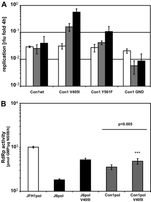
A critical residue in the thumb domain of NS5B enhances RNA replication across genotypes.After the identification of key residues in the NS5B gene important for efficient replica-tion of JFH1, we wanted to evaluate whether the same muta-tions would also enhance replication of a different genotype. We chose Con1, a typical genotype 1b isolate requiring repli-cation-enhancing mutations for efficient replication in cell cul-ture. These replication-enhancing mutations have been iden-tified in persistent selectable replicon cell lines and act by a yet-ill-defined mechanism (10, 24, 26, 27). A downside is that most of them strongly interfered with virus particle production (37). Since the JFH1 isolate replicated with high efficiency and produced infectious virus in cell culture, it appeared to be plausible to apply the same mechanism of replication enhance-ment to genotype 1b isolates. We focused on positions 405 and 561, because those were identical in J6 and Con1, whereas position 571 was divergent in the three isolates and therefore
on November 7, 2019 by guest
http://jvi.asm.org/
was not analyzed further. Interestingly, a valine-to-isoleucine switch at position 405 of NS5B indeed clearly enhanced repli-cation of a Con1 replicon in cell culture by 5- to 10-fold (Fig. 8A). In line with the case for JFH1, we also found a slightly but significantly increased RdRp activity for the purified Con1 V405I enzyme compared to the wild type, arguing for a con-served mechanism of action (Fig. 8B). In contrast, mutation Y561F did not alter Con1 replication, although it should in theory have the same impact on the kissing-loop interaction in Con1 as in JFH1, and a combination of V405I and Y561F also did not result in an increase of replication efficiency compared to that with V405I alone (data not shown). Comparison of the only available Con1 NS5B structure (PDB code 3FQL) with that of JFH1 NS5B shows a displacement of the portion of the fingers facing position 405 (in light green in Fig. 5A and B) very similar to that seen in the J6/JFH1 comparison. This is consis-tent with our proposed mechanism, although we cannot rule
out that this is due in part to this Con1 NS5B being in complex with a nonnucleoside inhibitor of initiation (19). Although the replication levels of the V405I mutant did not reach those previously published for adapted Con1 replicons, this mutation remains a promising candidate for combination with other mutations to generate genotype 1b isolates with high replica-tion levels and the capability of producing infectious virus.
DISCUSSION
The hepatitis C virus genotype 2a isolate JFH1 is currently the only viral strain that replicates efficiently in cell culture and
in vivowithout any need for adaptive mutations. Previous work pointed to an important contribution of the viral NS5B gene (8, 32). This was corroborated functionally by a very highde novoRNA synthesis efficiency of purified JFH1 polymerasein vitrocompared to NS5B from the related isolate J6, which is
FIG. 7. Rescue of J6-NS5B replication and infectious particle production by combinations of mutations. (A) Transient replication of JFH1, NS5B-J6, and replicons with single amino acid substitutions. Huh7-Lunet cells were transfected with 5g ofin vitrotranscripts co