Functional Mapping
Zhigang Yi,a,bZhenghong Yuan,bCharles M. Rice,aand Margaret R. MacDonalda
Laboratory of Virology and Infectious Disease, The Rockefeller University, New York, New York, USA,aand Key Laboratory of Medical Molecular Virology, Shanghai Medical College, Fudan University, Shanghai, Chinab
DNAJC14 is an Hsp40 family member that broadly modulates flavivirus replication. The mechanism by which DNAJC14
stoichi-ometrically participates in flavivirus replication complex (RC) formation is unknown; both reduced and elevated levels result in
replication inhibition. Using yellow fever virus (YFV), we demonstrate that DNAJC14 redistributes and clusters with YFV
non-structural proteins via a transmembrane domain and a newly identified membrane-binding domain (MBD), which both mediate
targeting to detergent-resistant membranes. Furthermore, the RC and DNAJC14 reside as part of a protein interaction network
that remains after 1% Triton solubilization. Mutagenesis studies demonstrate that entry into this protein interaction network
requires the DNAJC14 C-terminal self-interaction domain. Fusion of the DNAJC14 MBD and self-interaction domain with
an-other Hsp40 family protein is sufficient to confer YFV-inhibitory activity. Our findings support a novel model of DNAJC14
ac-tion that includes specific membrane targeting of both DNAJC14 and YFV replicaac-tion proteins, the formaac-tion of protein
interac-tions, and a microdomain-specific chaperone event leading to RC formation. This process alters the properties of the RC
membrane and results in the formation of a protein scaffold that maintains the RC.
T
he family
Flaviviridae
, consisting of the
Flavivirus
,
Pestivirus
,
and
Hepacivirus
genera, causes a significant worldwide disease
burden (
30
). Within the
Flavivirus
genus are numerous
arthro-pod-borne human pathogens (reviewed in references
16
and
17
),
including yellow fever virus (YFV), dengue virus (DENV), West
Nile virus (WNV), Japanese encephalitis virus, and tick-borne
en-cephalitis viruses, each associated with clinically important
dis-eases. Viruses in the
Flavivirus
genus have similar genome
organi-zation and replication strategies (
30
). Translation of the viral
positive-sense genomic RNA produces a polyprotein that is
co-and posttranslationally cleaved by host co-and viral proteases.
Struc-tural proteins C, prM, and E are located in the polyprotein amino
terminus, followed by nonstructural (NS) proteins NS1, NS2A,
NS2B, NS3, NS4A, 2K, NS4B, and NS5. Similar to the expression
of other positive-sense RNA viruses, the expression of NS proteins
induces intracellular membrane modification, resulting in a
virus-specific membrane structure housing the viral replication
com-plex (RC) (
9
,
35
,
54
). Electron microscope tomography studies
have revealed that the flavivirus RC results from invagination of
the endoplasmic reticulum (ER) membrane with the RC interior
connected to the cytoplasm by a pore (
15
,
54
). Virus RC formation
is probably driven by viral NS protein self-interactions (
34
,
41
,
45
)
and also needs concerted viral protein and viral protein-host
fac-tor interactions (
10
,
11
,
37
). In addition, many positive-strand
viruses exploit host lipids to facilitate RC assembly and the specific
lipids potentially provide a scaffold or modify membrane
curva-ture to help generate the RC (
20
,
21
,
33
,
43
,
48
).
DNAJC14 (also designated DRIP78, Jiv, and LIP6) was
identi-fied as interacting with the dopamine D1 receptor and modulates
receptor transport from the ER to the plasma membrane (
2
). It
interacts with the lysosomal trafficking regulator protein that is
implicated in SNARE complex-mediated transport (
50
). It is also
involved in the life cycles of several
Flaviviridae
family members.
The bovine homolog of this factor, Jiv, is essential for polyprotein
cleavage and replication of the pestivirus bovine viral diarrhea
virus (BVDV) and acts as a cofactor for NS2-3 cleavage by the NS2
autoprotease (
27
,
38
,
44
). Like other Hsp40 cochaperones,
DNAJC14 contains a conserved 70-amino-acid J domain that
in-teracts with Hsp70 family members to stimulate ATP hydrolysis
during the chaperone process (
24
,
52
). In the case of BVDV,
bo-vine DNAJC14 or a 90-amino-acid domain downstream of the J
domain, designated Jiv90 and containing putative zinc fingers, can
function to facilitate NS2-3 cleavage (
44
). We identified
DNAJC14 as a broadly acting flavivirus replication modulator that
also affects members of the
Hepacivirus
and
Flavivirus
genera
when overexpressed. Using YFV as a model flavivirus, our initial
mutagenesis studies implicated the J domain and a C-terminal
domain capable of self-interaction, but not the zinc fingers, as
important for YFV inhibition upon DNAJC14 overexpression.
The inhibition occurred after viral genome translation and prior
to genome amplification, suggesting inhibition at the step of RC
formation. DNAJC14 is likely involved in YFV RC assembly in a
stoichiometric manner, since both overexpression and
knock-down of DNAJC14 reduce YFV replication (
56
). We hypothesized
that endogenous DNAJC14, which colocalized with
double-stranded RNA (dsRNA) in YFV-infected cells, was likely involved
in the regulation of RC assembly (
56
).
Despite extensive progress in the characterization of flaviviral
RCs, the mechanism by which viral proteins interact with host
membranes and host factors to assemble the RC is largely
un-known. In this study, we used DNAJC14’s dual role in RC
forma-tion, namely, its requirement for viral replication and its
interfer-ence with the same process when overexpressed, to probe the
events that lead to RC formation. Through mapping of the
do-Received25 April 2012Accepted12 August 2012
Published ahead of print22 August 2012
Address correspondence to Margaret R. MacDonald, [email protected].
Copyright © 2012, American Society for Microbiology. All Rights Reserved.
doi:10.1128/JVI.01022-12
on November 7, 2019 by guest
http://jvi.asm.org/
mains of the DNAJC14 protein that are functional in YFV
repli-cation modulation, we elucidate a model of DNAJC14 action and
propose an RC assembly model involving protein targeting to
de-tergent-resistant membrane (DRM) subdomains within the ER,
followed by DNAJC14-facilitated protein-protein interactions
and RC formation.
MATERIALS AND METHODS
Plasmids.Retroviral vectors pV1, pTrip-EGFP, and pTrip-TagRFP and plasmids FL, CT1, pV1-hDNAJC14-NT5, and pV1-hDNAJC14-NT5CT1 have been described previously (6, 13,22,56,57). Derivatives were generated by standard methods; all PCR-generated sequences were verified by sequencing. The sequences of the primers used are listed inTable 1.
Plasmids pV1-hDNAJC14-NT5.1 (primers, RU-O-16849/RU-O-13945), pV1-hDNAJC14-NT5.2 (primers, RU-O-16850/RU-O-RU-O-16849/RU-O-13945), hDNAJC14-NT5.3 (primers, RU-O-16851/RU-O-13945), hDNAJC14-NT5.4 (primers, RU-O-16852/RU-O-13945), and pV1-hDNAJC14-NT5.5 (primers, RU-O-16853/RU-O-13945) were generated
by PCR amplification usingPfuTurboDNA polymerase (Stratagene) from the hDNAJC14 cDNA plasmid (Open Biosystems), followed by SfiI diges-tion and ligadiges-tion into similarly digested pV1.
Plasmid pTrip-EGFP-hDNAJC14-FL was generated by PCR amplifi-cation from the hDNAJC14 cDNA plasmid by using primers RU-O-14383 and RU-O-14384. After BsrG1 and XhoI digestion, the PCR product was ligated into similarly digested pTrip-EGFP. Plasmids pTrip-EGFP-hDNAJC14-pTM and pTrip-RFP-pTrip-EGFP-hDNAJC14-pTM were generated by PCR amplification with primers RU-O-15362 and RU-O-15363. After BsrG1 and XhoI digestion, the PCR products were ligated into similarly digested pTrip-EGFP or pTrip-TagRFP. These plasmids express hDNAJC14 (or the pTM region of DNAJC14) fused in frame to the C terminus of enhanced green fluorescent protein (EGFP) or red fluores-cent protein (RFP).
Plasmids pTrip-EGFP-hDNAJC14-NT3 (primers, RU-O-16562/RU-O-14384), pTrip-EGFP-hDNAJC14-NT4 (primers, RU-O-16563/RU-O-14384), pTrip-EGFP-hDNAJC14-NT5 (primers, RU-O-15162/RU-O-14384), pTrip-EGFP-hDNAJC14-NT4.1 (primers, RU-O-16543/ 14384), and pTrip-EGFP-hDNAJC14-NT6 (primers, RU-O-TABLE 1Sequences of primers used in this study
Primer Sequence (5=¡3=)
RU-O-13506 TGGCATATTCCAGTCAACCTT
RU-O-13507 GAAGCCCAAGATGGAATCAACT
RU-O-14383 AAATGTACAAGGCCCAGAAGCACCCCGGAG
RU-O-14384 TTTCTCGAGTCAACGTTGGAAGGGCCTCCTC
RU-O-15362 AAATGTACAAGGCAGGCTTTTGGTGGCTGATTG
RU-O-15363 TTTCTCGAGTCAGTCACCTAGTCCCACCAGAAACCG
RU-O-16562 AAATGTACAAGGGAGTGTGGACAGGGCGGT
RU-O-16563 AAATGTACAAGTTTACTCGTTTTCTTAAGCTGCTGGGTGCT
RU-O-15162 AAATGTACAAGCTGGTGGGACTAGGTGACCGG
RU-O-16543 AAATGTACAAGTTTTTGGGCTTTCTACAGTTGGGATGG
RU-O-15159 AAATGTACAAGATGGCTGGGGTTCCTGAGG
RU-O-15971 ATGGACGAGCTGTACAAGATCTATGCCTGCAGGCAA
RU-O-15972 TTGCCTGCAGGCATAGATCTTGTACAGCTCGTCCAT
RU-O-15977 TTTCGAGTTTGGATGGGACTGTTTACTCGTTTTCTT
RU-O-15978 AAGAAAACGAGTAAACAGTCCCATCCAAACTCGAAA
RU-O-15973 ATGGACGAGCTGTACAAGCTGTTTACTCGTTTTCTT
RU-O-15974 AAGAAAACGAGTAAACAGCTTGTACAGCTCGTCCAT
RU-O-16162 GTACAAGCGTTTTCTTAAGCTGCTGGGTGCTTTGCTGCTCCTGGCTCTGGCCCTCTTTTTGGGCTTTCTACAGTTGGGATG
GCGGTTTTGAC
RU-O-16163 TCGAGTCAAAACCGCCATCCCAACTGTAGAAAGCCCAAAAAGAGGGCCAGAGCCAGGAGCAGCAAAGCACCCAGCAGCTT
AAGAAAACGCTT
RU-O-16191 GTACAAGCGTTTTTTGCTGCTCCTGGCTCTGGCCCTCTTTTTGGGCTTTCTACAGTTGGGATGGCGGTTTTGAC
RU-O-16192 TCGAGTCAAAACCGCCATCCCAACTGTAGAAAGCCCAAAAAGAGGGCCAGAGCCAGGAGCAGCAAAAAACGCTT
RU-O-16189 GTACAAGCGTTTTGGTGCTTTGCTGCTCCTGGCTCTGGCCCTCTTTTTGGGCTTTCTACAGTTGGGATGGCGGTTTTGAC
RU-O-16190 TCGAGTCAAAACCGCCATCCCAACTGTAGAAAGCCCAAAAAGAGGGCCAGAGCCAGGAGCAGCAAAGCACCAAAACGCTT
RU-O-17194 AAAGGCCATTACGGCCATGGGTAAAGACTACTACCAGACGTTGG
RU-O-17195 AAAGGCCGAGGCGGCCTCACAGATCCTCTTCTGAGATGAGTTTTTGTTCTATTGGAAGAACCTGCTCAAGTACG
RU-O-16849 AAAGGCCATTACGGCCATGGGCTGGAGGGATAAGGCTACC
RU-O-16957 TAGTCTTTACCCATTAGCTCATCCTCAGGAACCCCAG
RU-O-16958 AGGATGAGCTAATGGGTAAAGACTACTACCAGACGTTGG
RU-O-17195 AAAGGCCGAGGCGGCCTCACAGATCCTCTTCTGAGATGAGTTTTTGTTCTATTGGAAGAACCTGCTCAAGTACG
RU-O-15154 GGTGCCTGGTATTGGAAGAACCTGCTCAAGTACG
RU-O-15155 GCTAAAAGCCCAGGCACCAGAGGGCG
RU-O-15153 AAAGGCCGAGGCGGCCTC
RU-O-13945 AAAGGCCGAGGCGGCCTCACAGATCCTCTTCTGAGATGAGTTTTTGTTCACGTTGGAAGGGCCTCCTCACTTTC
RU-O-16850 AAAGGCCATTACGGCCATGTCTCCAGCCTTGCAGCGTTG
RU-O-16851 AAAGGCCATTACGGCCATGGATAGCAGGCCATGGCAGC
RU-O-16852 AAAGGCCATTACGGCCATGTGGGGCTGGCTGGAGTTG
RU-O-16853 AAAGGCCATTACGGCCATGCCTGAAGAGGAAGTGGCTCGAC
RU-O-16893 AAACTCGAGATGCTGGTGGGACTAGGTGACCGGT
RU-O-16897 TTTGGATCCCGTAGCTCATCCTCAGGAACCCCAG
on November 7, 2019 by guest
http://jvi.asm.org/
[image:2.585.40.555.81.500.2]15159/RU-O-14384) were generated by PCR amplification, BsrG1 and XhoI digestion, and ligation into similarly digested pTrip-EGFP.
Plasmids pTrip-EGFP-hDNAJC14-⌬TM1, pTrip-EGFP-hDNAJC14-⌬TM2, and pTrip-EGFP-hDNAJC14-⌬TM1⫹2 were generated by site-directed mutagenesis with primer pairs O-15971/O-15972, RU-O-15977/RU-O-15978, and RU-O-15973/RU-O-15974, respectively. Plasmid pTrip-EGFP-hDNAJC14-TM3 was generated by directly ligating synthesized oligonucleotides RU-O-16162/RU-O-16163 into BsrG1- and XhoI-digested pTrip-EGFP. Plasmids pTrip-EGFP-hDNAJC14-TM17 (primers, RU-O-16191/RU-O-16192) and pTrip-EGFP-hDNAJC14-TM19 (primers, RU-O-16189/RU-O-16190) were generated by ligating the oligonucleotide pairs indicated into BsrG1- and XhoI-digested pTrip-EGFP.
Plasmid pV1-DNAJB1 (primers, RU-O-17194/RU-O-17195) was generated by PCR amplification from the hDNAJB1 cDNA plasmid (Open Biosystems), followed by SfiI digestion and ligation into similarly digested pV1. To generate DNAJC14-DNAJB1 hybrids, first fragment I (DNAJC14 membrane-binding domain [MBD]) was generated by PCR amplification from the hDNAJC14 cDNA plasmid by using primers RU-O-16849 and RU-O-16957. Fragment II (DNAJB1 with a C-terminal myc tag) was generated by amplification from hDNAJB1 cDNA using primers RU-O-16958 and RU-O-17195. Fragment III (DNAJB1) was generated by amplification from hDNAJB1 cDNA by using primers RU-O-17194 and RU-O-15154. Fragment IV (DNAJC14 C-terminal self-interaction do-main with a myc tag) was generated by amplification from plasmid pV1-hDNAJC14-FL using primers RU-O-15155 and RU-O-15153. Purified fragments I and II were fused by PCR amplification with primer RU-O-16849 and primer RU-O-17195 to generate pV1-MDNAJB1. Fragment III and fragment IV were fused by PCR amplification with primer RU-O-17194 and primer RU-O-15153 to generate pV1-DNAJB1C. Fragment V (DNAJC14 MBD fused to DNAJB1) was amplified with primers RU-O-16849 and RU-O-15154 using pV1-MDNAJB1 as the template. Frag-ments V and IV were fused by PCR with primers O-16849 and RU-O-15153 to generate pV1-MDNAJB1C.
Plasmid pEGFP-N1-MBD was generated by PCR amplification from the hDNAJC14 cDNA plasmid with primers 16893 and RU-O-16897, and after XhoI and BamHI digestion, the PCR product was ligated into similarly digested pEGFP-N1 (Clontech).
Antibodies.YFV NS3/4A, NS4B, and NS5 rabbit polyclonal antisera were previously described (5). NS3/4A and NS4B antisera were used at a 1:10,000 dilution for Western analyses and at 1:1,000 for immunofluores-cence analyses. NS5 antiserum was used at 1:1,000 for Western blot anal-ysis. Rabbit polyclonal anti-GFP antiserum was generated as described previously (7) and used at a 1:20,000 dilution in Western analyses. Mouse monoclonal anti-calnexin antibody (610523; BD Biosciences) was used in Western and immunofluorescence analyses at 1:500 and 1:50 dilutions, respectively. Mouse monoclonal anti-actin (A5441; Sigma) antibodies were used in Western analyses at a 1:5,000 dilution. Rabbit polyclonal anti-DNAJC14 antibody (HPA017653; Sigma) was used in Western and immunofluorescence analyses at 1:2,000 and 1:200 dilutions, respectively. Mouse monoclonal anti-dsRNA J2 antibody (English & Scientific Con-sulting, Bt., Szirak, Hungary) was reconstituted and diluted with an equal volume of glycerol and used at a 1:1,000 dilution in immunofluorescence analyses. Guinea pig polyclonal anti-adipophilin (ADRP) antibody (20R-AP002; Fitzgerald) was used at a 1:500 dilution in immunofluorescence analyses. Rabbit polyclonal anti-calnexin antibody (sc-11397; Santa Cruz Biotechnology, Inc.) was used at a 1:1,000 dilution in Western blot anal-yses. Mouse monoclonal anti-flotillin-1 antibody (610820; BD Biosci-ences) was used at 1:2,000 in Western blot analyses. Mouse monoclonal anti-Hsp70 antibody (SPA-810; Stressgen) was used at a 1:2,000 dilution in Western blot analyses. Mouse monoclonal anti-myc antibody (sc-40; Santa Cruz Biotechnology, Inc.) was used at 1:50 in coimmunoprecipita-tion analyses. Mouse monoclonal anti-GFP antibody (B-2; Santa Cruz Biotechnology, Inc.) was used at 1:50 in coimmunoprecipitation analyses. Alexa Fluor 488 donkey anti-mouse IgG (A-212020; Invitrogen), donkey
anti-guinea pig IgG (706-546-148; Jackson ImmunoResearch), and Alexa Fluor 594 goat anti-rabbit IgG (A-11012; Invitrogen) were used in immu-nofluorescence analyses at a 1:1,000 dilution. Horseradish peroxidase (HRP)-conjugated secondary anti-mouse (115-035-146; Jackson Immu-noResearch) and anti-rabbit (31462; Pierce) IgG antibodies were used at a 1:20,000 dilution. Normal mouse IgG used in immunoprecipitations was from Santa Cruz Biotechnology, Inc. (sc-2025).
Cell lines, viruses, and virus titration.SW13 (human adrenal carci-noma), Huh-7.5 hepatoma (3), and HEK293T cells and the BHK-J line of BHK-21 cells (29) were maintained as described previously (56). SW13 and Huh-7.5 cells were used interchangeably, and assays with the two cell lines gave similar results. YFV (strain 17D) stocks were generated by elec-troporation within vitro-transcribed RNA (26) from plasmid pACNR-YF17D (4), and titers were determined as described previously (56). Len-tiviral stock generation and transduction of cells were performed as described previously (56).
Cell fractionation.SW13 cells were seeded into six-well plates and, after transduction, scraped into 400l hypotonic buffer (5 mM Tris-Cl [pH 7.5], 15 mM KCl, 2.5 mM MgCl2). After 15 min swelling on ice, the
cells were passed 20 times through a 27-gauge needle and then centrifuged (900⫻gfor 5 min) to remove nuclei. One-tenth volume of 5 M NaCl was added to the postnuclear supernatants, and after incubation on ice for 20 min, the membranes were collected by centrifugation at 15,000⫻gfor 20 min. Pellets (membrane fraction, P) were resuspended in 50l sodium dodecyl sulfate (SDS) loading buffer. Proteins in the supernatant (cytosol fraction, S) were concentrated by adding 4 volumes of methanol, centri-fuged (10 min, 12,000⫻g), and resuspended in 50l SDS loading buffer. In situcell permeabilization and solubilization.SW13 cells were washed once with cold buffer C (20 mM HEPES-KOH [pH 7.7], 110 mM potassium acetate, 2 mM magnesium acetate, 1 mM EDTA) and perme-abilized by incubation in room temperature buffer C containing 50g/ml digitonin for 5 min or solubilized by incubation in room temperature buffer C containing 1% Triton X-100. Supernatants were collected if nec-essary. The reaction was stopped by three washes with cold buffer C. The cells were fixed for immunofluorescence analyses, scraped into cold buffer C for proteinase K digestion, or lysed in 2⫻SDS loading buffer (100 mM Tris-Cl [pH 6.8], 20% glycerol, 4% SDS, 3%-mercaptoethanol, 0.02% bromophenol blue).
Immunoprecipitation.To demonstrate the GFP-pTM and NT5CT1 interaction with YFV NS3, SW13 cells were transduced with pTrip-GFP-DNAJC14-pTM or pV1-DNAJC14-NT5CT1 and 2 days later infected with YFV (multiplicity of infection [MOI], 5). After 1.5 days, the cells were scraped into ice-cold phosphate-buffered saline (PBS), solubilized with 300l buffer A (10 mM HEPES [pH 7.5], 150 mM KCl, 3 mM MgCl2, 0.5% NP-40, 1⫻Roche proteinase inhibitor cocktail), and
dis-rupted by being passed through a 27-gauge needle 10 times. After clarifi-cation by centrifugation twice at 1,000⫻gfor 5 min at 4°C, the soluble fraction was incubated overnight at 4°C with anti-myc antibody at a dilu-tion of 1:50. Prewashed protein G-Dynabeads (100.03D; Invitrogen) were then added, and after 2 h of incubation, protein-antibody complexes were captured by a magnet and washed four times with 600l buffer B (10 mM HEPES [pH 7.5], 150 mM KCl, 3 mM MgCl2, 0.5% NP-40). The bound
proteins were eluted by boiling in SDS sample buffer and subjected to Western blot analysis.
To demonstrate the DNAJC14-NS3 interaction afterin situ solubili-zation, SW13 cells were transduced with pV1-DNAJC14-FL and 1.5 days later infected with YFV (MOI, 10). After 4 days, cells were solubilizedin situwith buffer C (see above) supplemented with 1% Triton X-100. After being washed with buffer C, the remaining material was scraped into buffer B (10 mM HEPES [pH 7.5], 150 mM KCl, 3 mM MgCl2, 0.5%
NP-40) and disrupted by being passed through a 27-gauge needle 10 times. After clarification by centrifugation at 15,000⫻gfor 10 min at 4°C, the soluble fraction was incubated overnight at 4°C with myc anti-body at a dilution of 1:50. Prewashed protein G-Dynabeads (100.03D; Invitrogen) were then added, and after 2 h of incubation,
on November 7, 2019 by guest
http://jvi.asm.org/
body complexes were captured by a magnet and washed four times with 600l buffer B. The bound proteins were eluted by boiling in SDS sample buffer and subjected to Western blot analysis.
Proteinase K digestion.Afterin situcell permeabilization or solubili-zation, and being scraped into cold buffer C, the samples were incubated with or without 10g/ml proteinase K at 37°C for 5 min; the reaction was terminated by the addition of phenylmethylsulfonyl fluoride to 1 mM. Proteins were concentrated by trichloroacetic acid (TCA) precipitation by adding an equal volume of 40% TCA. After the precipitates were washed with acetone and solubilized in SDS loading buffer, the samples were boiled for 10 min and used in Western blot analyses.
Membrane flotation.Membrane flotation assays were conducted es-sentially as described previously (47,51). Cells grown in 60-mm dishes were washed, scraped into PBS, and collected by centrifugation (500⫻g, 5 min). After resuspension in 200l hypotonic buffer (10 mM Tris-HCl [pH 7.8], 10 mM NaCl) and swelling (15 min on ice), the cells were disrupted by being passed 25 times through a 27-gauge needle. After cen-trifugation (900⫻gfor 5 min), postnuclear factions were treated with Triton X-100 (final concentration, 1%) on ice for 30 min. The entire 0.2-ml fraction was mixed with 1.6 ml 72% (wt/wt) sucrose in low-salt buffer (LSB; 50 mM Tris-HCl [pH 7.5], 25 mM KCl, 5 mM MgCl2)
over-laid with 2 ml of 55% (wt/wt) sucrose in LSB, followed by 0.6 ml of 10% (wt/wt) sucrose in LSB. For flotation afterin situsolubilization, cells from 100-mm dishes were solubilized with 2.5 ml buffer C containing 1% Tri-ton X-100. Supernatants (S1) were collected. After washing, cells were scraped into 600l buffer C containing 1% Triton X-100, disrupted by needle passage, and clarified by centrifugation at 15,000⫻gfor 5 min. Supernatants (S2) were collected. A 0.2-ml fraction of S1 or S2 was mixed with 1.6 ml 72% (wt/wt) sucrose in low-salt buffer and overlaid with 55% and 10% (wt/wt) sucrose in LSB as described above. The sucrose gradient was centrifuged at 46,000 rpm in a Beckman Coulter MLS-50 Optimax ultracentrifuge rotor for 16 h at 4°C. Fractions (400l) were taken from the top of the gradient, and proteins were precipitated from 300l of each fraction by the addition of 4 volumes of methanol and centrifugation at 12,000⫻gfor 10 min. RNAs were extracted from 100l of each fraction by dilution with 100l H2O, addition of 700l RLT (Qiagen) buffer, and
purification using the RNeasy minikit (Qiagen).
Western blot analysis.Cells were directly lysed with 2⫻SDS loading buffer and boiled for 5 min. Proteins were separated by SDS-polyacryl-amide gel electrophoresis and transferred to a Hybond ECL nitrocellulose membrane (GE Healthcare Life Sciences). The membrane was incubated in blocking buffer (PBS, 0.05% Tween 20, 5% dried milk) for 2 h and then incubated with primary antibody diluted in blocking buffer at 4°C over-night. The membrane was washed three times in PBS supplemented with 0.05% Tween 20 and incubated for 2 h at room temperature with HRP-conjugated secondary antibody. After three washes, the membrane was visualized by ECL Supersignal West Pico or Femto chemiluminescent substrate (Thermo Scientific). Protein bands were quantified by densi-tometry with ImageJ if necessary.
Immunofluorescence and microscopy.Cells were fixed with 4% paraformaldehyde in PBS and permeabilized with 0.2% Triton X-100 or 0.5% Triton X-100 (for dsRNA) in PBS for 5 min at room temperature. Permeabilization was omitted forin situ-solubilized cells. After being washed with PBS, samples were blocked and incubated overnight with primary antibody in PBS containing 3% bovine serum albumin at 4°C. After three washes with PBS, samples were incubated at room tempera-ture for 2 h with an Alex488- or Alex594-conjugated secondary antibody. For confocal microscopy, coverslips were mounted with Mowiol mount-ing medium and observed with a Zeiss LSM510 confocal microscope equipped with a 100⫻1.3 numerical aperture oil immersion objective. Images were captured by using the LSM software and processed by ImageJ. Photoshop was used to adjust the contrast and brightness; all changes for a given channel were applied to the entire image. In some images, Pearson (rp) and Spearman (rs) correlation coefficients were used to calculate colocalization as described previously (14). For
high-resolu-tion microscopy, coverslips were mounted with ProLong Gold antifade reagent (Invitrogen). Samples were initially observed using a Nikon Eclipse TE300 inverted fluorescence microscope. Images were acquired with a Nikon Plan Fluor 20⫻0.45 numerical aperture objective, a Digital Sight DS2MBW camera, and Nikon Elements software. For high-resolu-tion images, the samples were observed with an OMX Blaze 3D-SIM su-perresolution microscope (Applied Precision) equipped with a 100⫻1.40 numerical aperture UPLSAPO oil immersion objective. Images were cap-tured and deconvoluted by using SoftWoRx software and processed by ImageJ. All of the data shown are representative results obtained with cells in at least two independent experiments.
Reverse transcription-quantitative PCR (RT-qPCR).RNAs from each sucrose fraction were purified by using the RNeasy minikit (Qiagen) and eluted in 40l H2O. After heating at 85°C for 10 min, 8l RNA
sample was used for RT with random primers and the Superscript III first-strand synthesis kit (Invitrogen). Each sample was reverse tran-scribed in triplicate. Four microliters of cDNA was used for qPCR with the QuantiTect SYBR green PCR kit (Qiagen) and a LightCycler 480 (Roche) for detection as previously described (56). The primers used for YFV genome amplification (oligonucleotides RU-O-13506 and RU-O-13507, Table 1) were as described previously (28).
Bioinformatics. The transmembrane (TM) prediction program SOSUI (http://bp.nuap.nagoya-u.ac.jp/sosui) was used for membrane he-lix prediction. The program for secondary structure prediction was PSIPRED (http://bioinf.cs.ucl.ac.uk/psipred/).
RESULTS
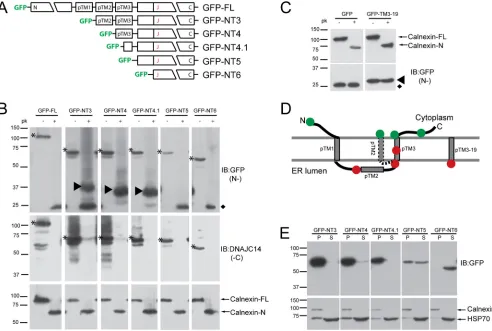
DNAJC14 is a polytopic membrane protein with both N and C
termini in the cytoplasm.
In order to gain insight into its function
in YFV RC assembly, we first undertook a topology study of
DNAJC14. DNAJC14 contains three potential TM domains
(re-ferred to as pTM1, pTM2, and pTM3, with the region designated
the pTM region), although it has been hypothesized to have both
its N and C termini in the cytoplasm (
2
). We used selective
per-meabilization and protection from protease digestion to
deter-mine the orientation of DNAJC14 with respect to the ER
mem-brane. We generated constructs expressing GFP fused to the
amino termini of DNAJC14 N-terminal truncation constructs
containing successively fewer of the putative TMs (
Fig. 1A
). SW13
cells expressing these mutant constructs were treated with
digi-tonin to permeabilize the plasma membrane and left without
fur-ther treatment or treated with proteinase K. Proteinase K
diges-tion efficiency and ER membrane integrity were monitored with
an antibody that recognizes the calnexin N terminus. Upon
pro-teinase K digestion, full-length calnexin was cleaved and the N
terminus within the ER lumen was protected (
Fig. 1B
, bottom
panels), indicating that proteinase K digestion was essentially
complete and that the ER membrane remained intact under our
experimental conditions. We monitored the location of the C
ter-mini by using an antibody that recognizes the DNAJC14 C
termi-nus. Proteinase K digested all of the C termini (
Fig. 1B
, middle
panels), indicating that the DNAJC14 C terminus resides in the
cytoplasm. In the absence of proteinase K digestion,
faster-migrat-ing bands were apparent for the most highly expressed mutant
constructs (GFP-FL, GFP-NT3, and GFP-NT4), most likely
be-cause of nonspecific degradation. A very small fraction of
GFP-NT3 and GFP-NT4 remained resistant to proteinase K digestion.
We monitored the location of the N termini with an antibody to
GFP. GFP is known to be resistant to protease digestion (reviewed
in references
8
and
53
). In cells expressing GFP, which is known to
be present in the cytosol (see
Fig. 5E
), exposure to proteinase K
had no discernible effect on the size or levels of GFP, confirming
on November 7, 2019 by guest
http://jvi.asm.org/
the resistance of the GFP domain to proteinase K digestion
(
Fig. 1C
, left panels). We thus expected that cytosolic exposure of
the GFP-DNAJC14 fusion proteins to proteinase K would result in
release of the protease-resistant GFP domain, while protection
within the ER lumen would result in protected fragments larger
than GFP but smaller than the intact fusion protein. A
protease-resistant GFP domain remaining after digestion was detected for
constructs GFP-FL, GFP-NT3, GFP-NT5, and GFP-NT6,
suggest-ing that the GFP domain was present in the cytoplasm and
sus-ceptible to proteinase K digestion (
Fig. 1B
, top panels).
GFP-con-taining larger protected fragments were observed for GFP-NT3,
GFP-NT4, and GFP-NT4.1. This indicates that the
amino-termi-nal GFP tag for each of these constructs was located in the ER
lumen, even though no signal sequence was present in the amino
termini of the constructs. The ER localization was likely mediated
by pTM3, since fusion of pTM3 (data not shown) or 19 residues
from this region of DNAJC14 to the C terminus of GFP
(GFP-pTM3-19; see schematic in
Fig. 3A
) results in the translocation of
GFP into the ER lumen and protection from proteinase K
diges-tion (
Fig. 1C
, right panels). The apparent molecular masses of the
protected GFP fragments of NT3, NT4, and
GFP-NT4.1 (
⬃
37, 34, and 32 kDa, respectively) are slightly larger than
the predicted molecular masses that would occur upon the
diges-tion of each protein C terminal to pTM3 (
⬃
33, 30, and 28 kDa,
respectively). This could be due to features inherent to this protein
(e.g., charge) or due to the protection of a portion of a
membrane-binding domain (MBD; see below) localized downstream of
pTM3. GFP-NT3 was an interesting case, in that evidence for both
cytosolic localization (GFP-sized band) and luminal location
(larger band) was found. This suggests that in the absence of the N
terminus and pTM1, pTM2 may adopt an alternative TM
topol-ogy that places the GFP moiety in the cytosol. Whether pTM2
inserts itself into the membrane bilayer or whether the pTM
re-gion can take on alternate conformations in the context of
full-length DNAJC14 (DNAJC14-FL) is an interesting area for future
study. Despite this uncertainty, the model of DNAJC14 topology
FIG 1Studies of DNAJC14 topology. (A) Schematic of DNAJC14-FL and pTrip-GFP mutant constructs fused in frame to the C terminus of GFP. The three individual putative TMs (pTMs) are indicated. (B) SW13 cells transduced with the indicated DNAJC14 mutant constructs were permeabilized with 50g/ml digitonin, scraped, and resuspended in buffer for treatment with 10g/ml proteinase K (pk) or no treatment. Proteins were TCA precipitated and analyzed by Western blotting with the antibodies indicated (IB). Top panels assessed N-terminal protection (N-), while the middle panels assessed C-terminal protection (-C). Arrowheads in the top panel indicate protected N-terminal GFP-containing proteins. Asterisks indicate the undigested DNAJC14 mutant constructs. The diamond indicates the protease-resistant GFP domain released after exposure to proteinase K. Calnexin (bottom panels) serves as a control for proteinase K digestion. (C) SW13 cells expressing GFP or GFP with 19 amino acids from pTM3 fused to the C terminus were treated and analyzed as in panel B. The protease-resistant GFP domain released by proteinase K is indicated by the diamond, and the protected larger species containing the TM sequence is indicated by the arrowhead. (D) Summary of proteinase K digestion results. The N-terminal GFP tag location is represented by a circle; green indicates that the GFP was not protected, while red indicates protection from proteinase K digestion. The alternative topology of pTM2 is indicated (dashed line). (E) Subcellular fractionation of DNAJC14 mutant constructs. Cells were disrupted by hypotonic buffer, and postnuclear lysates were separated by centrifugation into a crude membrane fraction (P) and a cytosolic fraction (S), which were analyzed by Western blotting.
on November 7, 2019 by guest
http://jvi.asm.org/
[image:5.585.46.538.67.398.2]shown in
Fig. 1D
is the most consistent with the results; pTM1 and
pTM3 serve as TM domains, and both the N and C termini reside
on the cytoplasmic side of the ER membrane.
To confirm the membrane association of these mutant
con-structs, cells were fractionated by differential centrifugation into
membrane (pellet, P) and cytosolic (soluble, S) fractions.
Cal-nexin served as a membrane protein marker, and Hsp70 served as
a cytosolic protein marker. Mutant constructs containing pTM3
(GFP-NT3, GFP-NT4) or even a portion of pTM3 (GFP-NT4.1)
were almost totally membrane associated (
Fig. 1E
). Similar results
were observed with equivalent C-terminally myc-tagged proteins
(data not shown). These data confirm that pTM3 is indeed a TM
helix. Although a portion of GFP-NT5 resided in the cytosolic
fraction, a similar amount resided in the membrane fraction,
while GFP-NT6 resided totally in the cytosolic fraction (
Fig. 1E
).
Therefore, a region in NT5 not found in NT6 is able to mediate
membrane association by a pTM3-independent mechanism.
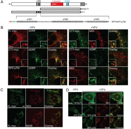
DNAJC14 is redistributed and recruited to YFV NS protein
clustering sites.
Overexpressed DNAJC14 localized both with the
ER and in cytoplasmic structures that we identified as probable
lipid droplets (LD,
Fig. 2B
). DNAJC14-FL and the CT1 mutant
construct lacking the C-terminal self-interaction domain
(sche-FIG 2DNAJC14 is redistributed and recruited to YFV NS protein clustering sites. (A) Schematic of DNAJC14 mutant constructs. DNAJC14-FL and the NT5CT1 and CT1 mutant constructs are in plasmid pV1 and contain a C-terminal myc epitope tag. Putative TM helices (pTM, black), the J domain (red) with a conserved HPD motif, a zinc finger motif (blue) in the JIV90 domain, and the self-interaction domain (gray) are shown. The pTM region was expressed fused to the C terminus of GFP or RFP in a pTrip-GFP (or -RFP) plasmid. The predicted hydrophobic helices are in gray. (B) Huh-7.5 cells were transduced with lentiviruses expressing FL-myc, CT1-myc, RFP-pTM (left panels), or CT1-myc (right panels). Cells were left uninfected or infected with YFV (MOI, 5) and fixed 1 day later and stained with antibodies against myc, the LD marker ADRP, or YFV NS3 as indicated. (C) Huh-7.5 cells expressing FL-myc were infected with YFV (MOI, 10) and fixed 4 days later. Cells were stained with antibody against myc and YFV NS3 or NS4B and examined by confocal microscopy. (D) SW13 cells expressing myc-tagged NT5CT1 or GFP-pTM were left uninfected or infected with YFV (MOI, 5) and fixed 1 day later. The cells were examined by confocal microscopy after staining for myc or NS proteins as indicated. Representative images are shown; similar results were obtained in independent experiments. Bars, 10m.
on November 7, 2019 by guest
http://jvi.asm.org/
[image:6.585.73.510.66.495.2]matic mutant construct representations are shown in
Fig. 2A
)
colocalized with the LD marker adipocyte differentiation-related
protein (ADRP,
Fig. 2B
, left panel, top and middle). The pTM
region likely mediated DNAJC14 colocalization with the LD
marker, since it conferred ADRP colocalization on RFP (RFP-TM,
Fig. 2B
, left panel, bottom), a heterologous protein that is
nor-mally distributed diffusely in the cytoplasm and nucleus (data not
shown). Since endogenous DNAJC14 was not found to localize
with the LD marker at steady state (see below), similar to
caveo-lin-1 (
40
), detection of the LD localization is likely due to the high
level of protein expressed during transduction and might not
re-flect DNAJC14’s normal localization. We then examined the
lo-calization of DNAJC14 in YFV-infected cells (
Fig. 2B
, right
pan-els) by using the noninhibitory CT1 mutant construct. Electron
microscopy of infected cells suggested a large increase in RC
for-mation between 18 and 24 h (data not shown), so we examined the
cells 1 day after infection, when RC formation is likely ongoing.
We found DNAJC14-CT1 and NS3 colocalized in aggregates that
did not appear to be vesicular in nature. As had been previously
observed in DENV-infected cells (
19
), we also noticed reduced LD
staining in YFV-infected cells, with the LD marker ADRP present
in smaller punctate structures (compare
Fig. 2B
, middle panels
with and without YFV). Unlike in uninfected cells, where ADRP
and DNAJC14-CT1 strongly colocalized (
Fig. 2B
, left middle
pan-els), in YFV-infected cells, much of the DNAJC14-CT1 was found
unassociated with the LD marker (
Fig. 2B
, right middle panels),
presumably because of the recruitment of DNAJC14-CT1 to sites
of YFV NS proteins. Consistent with this, YFV NS3 also did not
colocalize with ADRP (
Fig. 2B
, right bottom panels).
Determina-tion that the NS3-DNAJC14-CT1 colocalizaDetermina-tion was reflecting the
biological properties of DNAJC14-FL is complicated by the
inhib-itory activity exerted on YFV by increased levels of DNAJC14. We
previously found that although DNAJC14 overexpression
inhib-ited YFV RNA replication, at late times after infection, the virus
was able to overcome the inhibition (
56
). Huh-7.5 cells expressing
DNAJC14-FL were therefore infected with YFV and fixed late (4
days) after infection. Transduced DNAJC14-FL colocalized
exclu-sively with NS3 and NS4B, which displayed a punctate staining
pattern (
Fig. 2C
). Taken together, overexpressed DNAJC14
re-sides in the ER and LD, but during YFV infection, DNAJC14 has
reduced association with the LD marker ADRP because of
DNAJC14 recruitment to NS protein clustering sites.
We then mapped the minimal elements that mediate
DNAJC14 recruitment to YFV NS protein clustering sites. We
found that mutant construct NT5CT1, lacking both the pTM
re-gion and the C-terminal interaction domain, and GFP fused to the
pTM region (
Fig. 2A
) could colocalize with NS3; moreover,
GFP-pTM colocalization with NS4B was also confirmed (
Fig. 2D
). This
indicated that while the pTM region can confer localization with
NS proteins, another region, present in NT5CT1, is also capable of
this activity. We focused on the pTM element and further deleted
the individual TMs. GFP-
⌬
pTM1, GFP-
⌬
pTM2, and
GFP-⌬
pTM1
⫹
2, each containing pTM3, all colocalized with NS3
(
Fig. 3A
and
B
), suggesting that pTM3 mediates NS3
colocaliza-tion. For unknown reasons, we were unable to stably express
GFP-⌬
pTM3 lacking pTM3. We next focused on pTM3 to define the
minimal sequence necessary for targeting to NS protein clustering
sites. We fused pTM3 to the C terminus of GFP and found that it
redistributed and colocalized with NS3 (
Fig. 3C
). Mutant
con-structs with reduced TM3 lengths as small as 19 or 17 amino acids
(aa) were able to confer NS3 colocalization on GFP (
Fig. 3C
). In
contrast, the ER protein calnexin only partially colocalized with
NS3 (
Fig. 3C
). Given the inability to express GFP-pTM with the
pTM3 sequences deleted, it remains possible that other pTM
se-quences can also mediate localization to NS protein clustering
sites. However, DNAJC14 recruitment to and colocalization with
YFV NS protein clustering sites can be mediated by its third
puta-tive TM with a length as short as 17 aa. We noted that in the
absence of infection, DNAJC14 mutant constructs lacking pTM1
or pTM2 but not both (
⌬
pTM1
⫹
2,
Fig. 3B
; GFP-pTM3,
Fig. 3C
)
were associated with vesicular structures suggestive of LDs (inserts
in
Fig. 3B
, left panels). Clarification of whether pTM1 or pTM2 is
sufficient for LD recruitment requires further investigation.
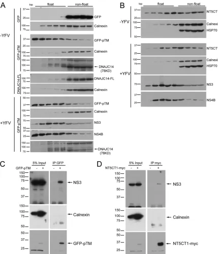
DNAJC14 and YFV NS proteins are targeted to DRMs.
Since
DNAJC14 subdomains mediate recruitment to YFV NS protein
clustering sites and NS proteins of hepatitis C virus (HCV) and
DENV reside on DRMs (
39
,
47
), we wondered if DNAJC14 and
YFV NS proteins were targeted to DRMs. We conducted
mem-brane flotation assays with 1% Triton X-100-treated cell lysates to
evaluate the flotation behavior of GFP fused to the DNAJC14
pTM region (GFP-pTM) and the noninhibitory mutant construct
lacking TMs (NT5CT1) in the absence or presence of YFV
infec-tion. NS3 and NS4B floated to lighter membrane fractions (
Fig.
4A
and
B
). GFP-pTM, in contrast to GFP alone, floated as
effi-ciently as NS4B in the presence or even the absence of YFV
infec-tion (
Fig. 4A
). Furthermore, GFP fused to the third putative TM
(GFP-pTM3) floated as efficiently as GFP-pTM (data not shown).
NT5CT1 (
Fig. 4B
) also floated, albeit less efficiently. Surprisingly,
neither endogenous nor overexpressed DNAJC14 floated to
lighter fractions in the presence or absence of YFV infection (
Fig.
4A
). We believe that this is due to the presence of the C-terminal
self-interaction domain, which mediates interaction, allowing
chaperone activity and the formation of protein interactions that
preclude flotation (see below and Discussion). Taken together, the
results show that two subregions of DNAJC14, pTM3 and
NT5CT1 lacking both pTM and the carboxyl-terminal
self-inter-action domain (
Fig. 2A
), colocalized and cofractionated with YFV
NS proteins on DRMs. We next tested if GFP-pTM and the
NT5CT1 mutant construct physically associate with YFV proteins
in detergent-containing lysis buffer. SW13 cells expressing
GFP-pTM or NT5CT1 were infected with YFV, and cell lysates were
prepared for GFP-pTM or NT5CT1 immunoprecipitation. In
both situations, NS3 was detected in the precipitated complex,
while the ER protein calnexin was not (
Fig. 4C
). We were unable
to assess the coprecipitation of NS4B because of the presence of a
nonspecific band of the same size. These results indicate that the
pTM region of DNAJC14 and the NT5CT1 DNAJC14 mutant
construct each physically associate with YFV NS proteins in a
membrane microdomain.
A newly identified MBD is essential for DNAJC14 function.
As DNAJC14 NT5 is still membrane associated while NT6 is not
(
Fig. 1E
), we proposed that there is another MBD in DNAJC14
following the pTM3 and before the J domain (
Fig. 5A
). To map the
MBD and test whether it is important for antiviral activity, we first
generated truncated forms of the putative MBD (
Fig. 5A
) and
examined the resultant anti-YFV activity and intracellular
distri-bution. There are three predicted
␣
-helices within the putative
MBD. On the basis of these, we generated constructs with
trunca-tions in this region (
Fig. 5A
) and examined their relative
expres-sion levels (
Fig. 5B
) and anti-YFV activities (
Fig. 5C
). The
on November 7, 2019 by guest
http://jvi.asm.org/
sion levels of the mutant proteins were similar, although some
were expressed more robustly than others. This is unlikely to
con-tribute substantially to differences in antiviral efficacy, since our
previous work demonstrated that mutant constructs with very low
expression levels could demonstrate potent antiviral activity (
56
).
NT5.1 and NT5.2, each containing all three putative
␣
-helices,
had anti-YFV activities comparable to that of NT5, causing a 2-log
reduction in YFV titers (
Fig. 5C
). NT5.3 lacking the first putative
␣
-helix (H1) had partially impaired anti-YFV activity and reduced
virus titers by about 1 log. Truncation mutant constructs NT5.4
and NT5.5, each lacking both the first (H1) and second (H2)
pu-tative
␣
-helices, were no longer antiviral (
Fig. 5A
and
C
). These
two mutant constructs also failed to localize in the cytosol and
resided primarily in the nucleus (
Fig. 5D
), which may explain
their failure to inhibit YFV replication. These results indicate that
the first and second putative
␣
-helices in the MBD are required for
DNAJC14’s maximal antiviral function. Next, we fused the
puta-tive MBD to GFP to examine if the MBD could mediate
mem-brane targeting of GFP and recruitment to YFV NS protein
clus-tering sites. Similar to NT5 truncation constructs NT5.1, NT5.2,
and NT5.3, MBD-GFP displayed a cytosolic pattern and also
co-localized with YFV NS4B in YFV-infected cells (
Fig. 5E
). These
data demonstrate that the MBD of DNAJC14 is required for
anti-YFV activity and mediates recruitment to anti-YFV NS protein
cluster-ing sites.
We also verified that the MBD, like NT5CT1, could be targeted
to DRMs. GFP was used as an internal nonfloating control, and
cells were cotransfected with plasmids expressing GFP and
MBD-GFP. Triton-treated cell lysates were subjected to membrane
flo-tation assays, and fractions were examined by Western blotting.
As shown in
Fig. 5F
, flotillin-1, a protein known to float, was
found in the floating fractions, while GFP and calnexin, as
ex-pected, did not float. MBD-GFP displayed an obvious floating
pattern, although it did not float as efficiently as flotillin-1. Thus,
either the DNAJC14 pTM region or MBD can mediate
localiza-tion to DRMs, where YFV NS proteins also reside.
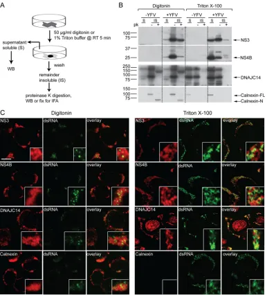
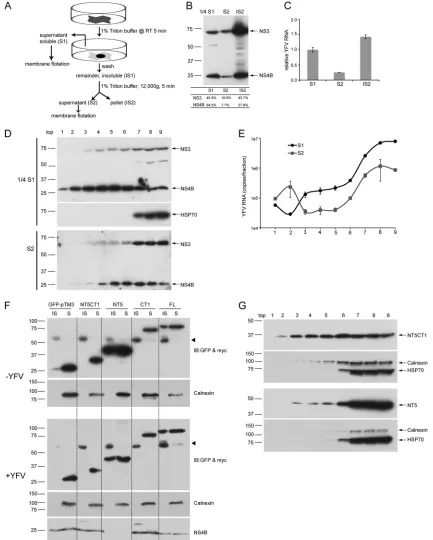
The YFV RC resides on a detergent-insoluble fraction.
Since
DNAJC14 subdomains colocalized and cofractionated with YFV
NS proteins on DRMs, we wondered if DRMs might be the site of
YFV RC assembly. We carried out
in situ
permeabilization with
the mild detergent digitonin, which retains ER membrane
integ-rity, or
in situ
solubilization with 1% Triton X-100, which
pre-sumably disrupts the ER membrane (
Fig. 6A
). We then examined
if viral proteins remained after permeabilization or solubilization
and the remaining viral proteins’ sensitivity to proteinase K
diges-tion, since viral proteins within the RC interior are presumably
FIG 3DNAJC14 pTM3 mediates recruitment to YFV NS protein clustering sites. (A) Schematic of DNAJC14 pTM mutant constructs. Various combinations of the individual TMs (gray) of the pTM region were fused in frame to the C terminus of GFP in the pTrip-GFP plasmid as indicated. (B, C) Huh-7.5 cells transduced to express the indicated DNAJC14 pTM mutant constructs were infected with YFV (MOI, 5) or not infected and fixed 1 day later. Cells were stained with antibodies to NS3 or calnexin as indicated and examined by confocal microscopy for DNAJC14 mutant construct or calnexin (green) and NS3 (red) localization. Representative images are shown. Bars, 10m.
on November 7, 2019 by guest
http://jvi.asm.org/
[image:8.585.100.487.63.394.2]protected from proteinase K digestion (
36
,
42
,
51
). In addition, we
tracked the RC by staining for dsRNA (
15
,
25
,
54
) in the cell
fractions remaining after permeabilization or solubilization.
Upon digitonin permeabilization (
Fig. 6B
, left panels), the
major-ity of NS3, NS4B, calnexin, and DNAJC14 remained with the
in-soluble fraction. The majority of the inin-soluble NS3 was resistant to
protease digestion, whereas only a small part of NS4B was resistant
and almost all of DNAJC14 was sensitive to proteinase K
diges-tion, which indicates that DNAJC14 is unlikely to be incorporated
into the RC interior. Proteinase K digestion was efficient, as
mon-FIG 4DNAJC14 mutant constructs are associated with YFV NS proteins on DRMs. SW13 cells expressing GFP, GFP-pTM, DNAJC14-FL (A), or NT5CT1 (B) were infected with YFV (MOI, 5) or not infected. Cells were harvested 1.5 days postinfection, and 1% Triton X-100-treated lysates were subjected to membrane flotation assay. Fractions were taken from the top to the bottom, and proteins from each fraction were methanol precipitated and analyzed by Western blotting with the antibodies indicated. The identity of the endogenous DNAJC14 protein band was previously verified by small interfering RNA-mediated silencing (56). (C, D) Coimmunoprecipitation of GFP-DNAJC14-pTM (GFP-pTM) and NT5CT1-myc with YFV NS3. SW13 cells left untransduced or transduced to express GFP-pTM (C) or NT5CT1-myc (D) as indicated were challenged with YFV (MOI, 5). Cells were harvested 1.5 days later and immunoprecipitated with anti-GFP or anti-myc antibodies, respectively. Precipitated proteins were subjected to Western blot analysis with antibodies to NS3, calnexin, GFP, or myc, and relevant proteins are indicated. Similar results were obtained in several independent experiments. The values to the left are molecular sizes in kilodaltons.
on November 7, 2019 by guest
http://jvi.asm.org/
[image:9.585.76.505.64.565.2]FIG 5Identification a new MBD in DNAJC14. (A) Schematic of NT5 and truncation mutant constructs. All constructs expressed the MBD region (or truncation constructs thereof) at the amino terminus. MBD truncation mutant constructs contained a myc epitope tag at the carboxyl terminus. (B, C) SW13 cells transduced with V1-NT5 or the indicated truncation mutant constructs were challenged with YFV (MOI, 5). (B) Cells were harvested at 1 day postinfection and analyzed by Western immunoblotting (IB) and (C) virus in the medium was enumerated by plaque assay. Data are the mean titer⫾the standard error of the mean;n⫽3. Abbreviations: pfu, plaque-forming units; V, V1 vector control. (D) The intracellular distribution of mutant constructs was examined by immunostaining with anti-myc antibody (green) and confocal microscopy 1.5 days after transduction. (E) Localization of the DNAJC14 MBD fused to GFP. SW13 cells were transfected to express GFP or MBD-GFP and mock infected or infected with YFV (MOI, 5) 1 day later. After 1 day, the cells were fixed for immunostaining and observed by confocal microscopy. Green (GFP), red (NS4B), and merged channels of a representative image are shown. Bars, 20m. (F) The DNAJC14 MBD confers detergent resistance and membrane flotation on GFP. SW13 cells were cotransfected to express both GFP and MBD-GFP. Postnuclear lysates were treated with 1% Triton X-100 and subjected to membrane flotation; fractions were taken from the top to the bottom. Proteins from each fraction were methanol precipitated and analyzed by Western blotting with the antibodies indicated. The values to the left of panels B and F are molecular sizes in kilodaltons.
on November 7, 2019 by guest
http://jvi.asm.org/
[image:10.585.115.477.69.562.2]itored by the complete loss of full-length calnexin in the
protease-treated samples. Upon
in situ
solubilization with 1% Triton, cell
morphology was lost while the nuclear shape was maintained (see
Fig. 8B
). The ER integral protein calnexin was completely
solubi-lized and was detected only in the supernatant (S) fractions
(
Fig. 6B
, right panels). Although some NS3 was solubilized and
detected in the supernatant, substantial NS3 remained insoluble
(IS). Even after Triton treatment, a small portion of NS3 remained
proteinase K resistant. Compared to NS3, NS4B was solubilized
more efficiently, with a greater portion detected in the
superna-tants (S). The insoluble NS4B was largely protease sensitive.
Sub-stantial endogenous DNAJC14 remained in the IS fraction and
was protease sensitive.
When what remained of the cells after digitonin
permeabiliza-tion or Triton solubilizapermeabiliza-tion was examined by confocal
micros-copy (
Fig. 6C
), dsRNA foci were detected near YFV NS proteins
and DNAJC14 but were not completely colocalized. Although
good colocalization of dsRNA and NS protein has been
demon-strated with DENV (
54
), in some viral studies, such as those with
severe acute respiratory syndrome virus and HCV (
25
,
49
), only
partial colocalization was noted. Failure to completely colocalize
may be explained by differences in the relative antigen abundance
of viral proteins and dsRNA within the RCs and/or by the
partic-ipation of only a small part of the viral proteins in RC function (
42
,
51
). Upon Triton solubilization, calnexin was totally removed.
Although NS protein signals were reduced, in contrast, the dsRNA
FIG 6The YFV RC is on a detergent-insoluble fraction. (A) Schematic of experimental procedures for panels B and C. WB, Western blotting; IFA, immuno-fluorescent staining. (B) Proteinase K digestion of YFV-infected cells. SW13 cells were infected with YFV (MOI, 5) and permeabilizedin situ1 day later with 50 g/ml digitonin or solubilized with 1% Triton X-100. The supernatant (S) was collected for Western blotting. The remaining detergent-insoluble cell material (IS) was then washed, scraped, and treated with 10g/ml proteinase K or not treated. Proteins were TCA precipitated and analyzed by Western blotting with the antibodies indicated. The values to the left are molecular sizes in kilodaltons. (C) Afterin situpermeabilization and washing, cells were fixed and permeabilized with 0.5% Triton for immunofluorescence analyses and examination by confocal microscopy. The results shown are representative of multiple similar experi-ments. Bar, 10m.
on November 7, 2019 by guest
http://jvi.asm.org/
[image:11.585.101.487.65.491.2]signal was enhanced, indicating that dsRNA is tightly associated
with the NS proteins and likely protected within a Triton-sensitive
membrane structure as described previously (
36
,
42
,
51
). After
in
situ
Triton treatment, the dsRNA remains associated with the NS
protein and DNAJC14 network but is more accessible because of
the Triton-mediated membrane disruption. Although Triton
treatment may remove some factors important for RC function
(
55
), these data indicate that the YFV RC, along with DNAJC14,
resides on a Triton-insoluble fraction.
The YFV RC is maintained by a protein-protein interaction
network.
To further address the relationship between YFV RCs
and DRMs and examine if the Triton-insoluble fraction
contain-ing the YFV RC is a DRM, we used progressive solubilization steps
(
Fig. 7A
). We first solubilized YFV-infected cells
in situ
with 1%
Triton as in
Fig. 6
and collected the solubilized supernatant
frac-tion (S1). The remaining insoluble fracfrac-tion (IS1), which contains
YFV RCs, was scraped off and solubilized again in Triton buffer by
being passed through a needle. This physically disrupted sample
was fractionated into solubilized supernatant (S2) and the
insol-uble nuclear pellet (IS2). As shown in
Fig. 7B
, and consistent with
results in
Fig. 6B
, a greater proportion of NS4B than of NS3 was
solubilized with Triton (S1). Substantial amounts of the NS3 and
NS4B proteins (
Fig. 7B
) and the YFV genome (
Fig. 7C
) remained
associated with the insoluble nuclear pellet (IS2) and failed to be
solubilized in the S2 fraction. We subjected S1 and S2 (derived
from the YFV RC-containing insoluble fraction, IS1) to the
mem-brane flotation assay and examined the distribution of NS3, NS4B,
and the viral genome. As shown in
Fig. 7D
, analysis of the S1
fraction revealed that NS4B floated very efficiently, while NS3 also
partially floated, but not as efficiently. In contrast, in the S2
frac-tion, the flotation of NS4B was reduced compared to that of NS4B
in the S1 fraction. NS3 present in S2 was found primarily in the
nonfloating fraction. The YFV genome in both the S1 and S2
frac-tions failed to float (
Fig. 7E
). This, together with the results in
Fig.
6
, demonstrates that the YFV RC components could survive after
Triton solubilization and fail to float. Thus, although the YFV NS
proteins are targeted to DRM domains within the ER and can float
in the membrane flotation assay, the RC components (dsRNA and
NS proteins) are not present in the floating fractions but instead
reside in a Triton-resistant network.
Interestingly, endogenous DNAJC14, like YFV RCs, resides on
a Triton-insoluble but nonfloating fraction in the membrane
flo-tation assay (
Fig. 4A
). This is in contrast to the behavior of the
DNAJC14 pTM or NT5CT1 subdomain, each of which can reside
on floating fractions (
Fig. 4
). To address this apparent
contradic-tion and further define the relacontradic-tionship between Triton solubility
and flotation ability, we analyzed the
in situ
Triton solubility of
DNAJC14 mutant constructs with different flotation behaviors
(
Fig. 7F
). We analyzed GFP-pTM3, NT5CT1, and CT1, each of
which could be recruited to and colocalized with YFV NS protein
clustering sites (
Fig. 3C
and
2D
and
B
, respectively). Of these,
DNAJC14-pTM3 (data not shown) and NT5CT1 (
Fig. 4
) were
able to float. We also included DNAJC14-FL and mutant
con-struct NT5 lacking the pTM region, both of which are capable of
YFV inhibition. Cells were transduced to express the proteins,
infected with YFV or left uninfected, and then
in situ
solubilized
with 1% Triton. Solubilized supernatants (S) and the remaining
insoluble fractions (IS) were collected and analyzed by Western
blotting. In the presence or absence of YFV infection, GFP-pTM3,
NT5CT1, and CT1, each lacking the C-terminal self-interaction
domain, were efficiently solubilized and detected only in the
su-pernatant fraction. In contrast, NT5 and FL (each containing the
C-terminal self-interaction domain) were partially resistant to
Triton solubilization (
Fig. 7F
). Note that NS4B was not detected in
the NT5 sample because of very efficient transduction levels and
the strong inhibitory effect of NT5 on YFV replication. These data
suggest that self-interaction of DNAJC14 is required for resistance
to
in situ
Triton solubilization and suggest that the ability to
par-tition into a Triton-insoluble fraction correlates with
DNAJC14-mediated YFV inhibition. We examined whether the
Triton-in-soluble NT5 mutant construct, in contrast to NT5CT1, resides
predominantly in a nonfloating fraction, similar to endogenous
DNAJC14 (
Fig. 4
). Membrane flotation studies (
Fig. 7G
)
demon-strated that NT5CT1 was able to float while NT5 floated much less
efficiently. Taken together, the YFV RC component resides on a
nonfloating structure or complex that is likely mediated by
pro-tein-protein interactions and this structure is resistant to
in situ
Triton solubilization. Only a portion of the YFV NS proteins
en-ters into the RC. Self-interaction of DNAJC14, which is essential
for its anti-YFV activity, also is required for entering into this
nonfloating structure.
DNAJC14 associates with the YFV RC via direct
protein-pro-tein interaction.
Since both the YFV RC and DNAJC14 reside in a
nonfloating, Triton-insoluble fraction, we wondered if the YFV
RC and DNAJC14 both reside within a protein network and
whether DNAJC14 physically associates with YFV RC
compo-nents. We first examined the viral RC structure and endogenous
DNAJC14 by high-resolution microscopy. After
in situ
Triton
sol-ubilization (
Fig. 8A
and
B
), dsRNAs and NS3 and NS4B appeared
as a clustered network and dsRNAs were attached to the NS3 and
NS4B network (
Fig. 8C
). In YFV-infected cells, DNAJC14 was
redistributed and recruited to a dsRNA-containing network. We
next examined if transduced DNAJC14, like endogenous
DNAJC14, associates with the YFV RC-containing protein
net-work after
in situ
Triton solubilization. Since overexpressed
DNAJC14 inhibits YFV replication, we took advantage of the fact
that, at late time points after infection (
Fig. 2
C), YFV can
over-come DNAJC14-mediated inhibition in
DNAJC14-FL-trans-duced cells (
56
). Colocalization of DNAJC14-FL with NS3, NS4B,
and dsRNA after Triton solubilization was analyzed by confocal
microscopy (
Fig. 8D
). DNAJC14-FL colocalized well with NS3 (
r
p,
0.67;
rs
, 0.75) and NS4B (
rp
, 0.76;
rs
, 0.81). As demonstrated above
for endogenous DNAJC14 (
Fig. 8C
and
6C
), dsRNA foci were also
found in association with overexpressed DNAJC14 (
rp
, 0.51;
rs
,
0.54).
To test for a physical interaction of RC components with
DNAJC14, we performed coimmunoprecipitation using the
RC-containing insoluble fraction. Using
in situ
solubilization,
as outlined in
Fig. 8A
, of YFV-infected cells expressing
DNAJC14-FL-myc, an S2 fraction was prepared and DNAJC14
was immunoprecipitated using anti-myc antibody. YFV NS3
and NS5, but not NS4B, were coprecipitated with
DNAJC14-FL-myc, while flotillin-1 and

-actin were not (
Fig. 8E
),
indi-cating that DNAJC14-FL associates with NS3 and NS5 either
directly or indirectly within a protein-protein interaction
net-work. These results imply a protein scaffold within the YFV RC
and support the presence of a protein-protein interaction
be-tween DNAJC14 and components of the YFV RC.
The DNAJC14 MBD and C-terminal self-interaction domain
can confer activity to a heterologous Hsp40 family member.
On
on November 7, 2019 by guest
http://jvi.asm.org/
FIG 7The YFV RC is maintained by a protein-protein interaction network. (A) Schematic of the experimental procedures for panels B to E. RT, room temperature. (B) Proteins from the supernatants (S1 and S2) were precipitated with methanol, and the pellets from each of the three fractions (S1, S2, and IS2) were solubilized in equal volumes of SDS loading buffer and analyzed by Western blotting with anti-NS3 and -NS4B antisera. Bands were quantified by densitometry, and the percentage of the total NS protein found in each fraction is indicated at the bottom. Note that only one-fourth of the S1 fraction was run on the gel. (C) Distribution of RNA genomes in the fractions. YFV RNA levels were quantified by RT-qPCR and normalized to the level found in S1. (D) YFV-infected SW13 cells were solubilized and fractionated as outlined in panel A. Fractions S1 and S2 were subjected to a membrane flotation assay. Proteins from each fraction were analyzed by Western blotting with the antibodies indicated. (E) YFV RNA levels from each fraction of S1 and S2 after membrane flotation were analyzed by RT-qPCR, and the mean values from triplicate cDNA syntheses are plotted. Error bars indicate standard deviations. (F) SW13 cells expressing the indicated DNAJC14 mutant constructs were infected or not infected with YFV (MOI, 5), and then cells werein situpermeabilized with 1% Triton as described in the legend toFig. 6A. After supernatant (S) collection, the remaining Triton-insoluble material was washed and harvested (IS). Proteins in the supernatants were precipitated with methanol. Proteins from S and IS were solubilized in an equal amount of SDS loading buffer and analyzed by Western blotting with the antibodies indicated. Arrowheads indicated nonspecific bands. (G) SW13 cells expressing DNAJC14 NT5CT1 and NT5 mutant constructs were treated with Triton, and lysates were subjected to a sucrose gradient membrane flotation assay. Proteins from each fraction were analyzed by Western blotting with anti-myc antibodies (NT5 and NT5CT1) or the antibodies indicated. For panels D and E, similar results were obtained in independent experiments. The values to the left of panels B, D, F, and G are molecular sizes in kilodaltons.
on November 7, 2019 by guest
[image:13.585.76.509.34.574.2]FIG 8DNAJC14 is associated with the YFV RC. (A) Schematic of the experimental procedures for panels B and C. coIP, coimmunoprecipitation; IFA, immunofluorescent staining; RT, room temperature. (B, C) SW13 cells were infected with YFV (MOI, 5), and 1 day later they werein situsolubilized with 1% Triton X-100, fixed, and then subjected to immunostaining. In panel B, samples were examined using an inverted fluorescence microscope (TE300; Nikon). In
on November 7, 2019 by guest
http://jvi.asm.org/
[image:14.585.74.509.70.695.2]the basis of our findings described above, we hypothesized that
DNAJC14 first is targeted via its TM or MBD to a unique DRM
microdomain within the ER where YFV NS proteins also reside.
Within this DRM, we hypothesized that DNAJC14 then interacts
with a substrate viral protein(s) and itself via proteprotein
in-teractions and serves to chaperone conformational changes of
vi-ral proteins to allow formation of the RC. In this model,
overex-pression of DNAJC14 would alter the stoichiometry of this
process and disrupt RC formation. Overexpression of DNAJC14
mutant constructs unable to enter into this protein-protein
inter-action network would fail to inhibit YFV replication. This model
predicts that the MBD (or TM) and the C-terminal interaction
domain are key elements of DNAJC14 that play important roles in
its function. To test this hypothesis, we asked whether the MBD
and the C-terminal self-interaction domain could confer antiviral
activity on an unrelated noninhibitory Hsp40 protein. We
se-lected DNAJB1, another Hsp40 family member that has a similar
domain arrangement but lacks an MBD or a membrane-targeting
motif (
23
). We fused the DNAJC14 MBD or the C-terminal
self-interaction domain individually or in combination to DNAJB1
(
Fig. 9A
) and then examined each hybrid protein’s localization
and anti-YFV activity. DNAJB1 was distributed throughout the
cell in both the nucleus and the cytoplasm, and fusion of the
DNAJC14 self-interaction domain (DNAJB1C) did not change
the localization of DNAJB1 (
Fig. 9B
). DNAJB1 with an
N-termi-nal fusion of the DNAJC14 MBD (MDNAJB1), like
DNAJC14-NT5, displayed a cytosolic distribution, although it resided more
peripherally than DNAJC14-NT5. Further fusion of the
DNAJC14 self-interaction domain to MDNAJB1 (MDNAJB1C)
changed its distribution and resulted in a cytoplasmic localization
more similar to that of DNAJC14-NT5 (
Fig. 9B
). When tested for
the ability to inhibit YFV replication, DNAJB1 did not exhibit
significant anti-YFV activity, lowering titers less than 1 log
(
Fig. 9D
). Fusion of either the DNAJC14 self-interaction domain
(DNAJB1C) or the MBD (MDNAJB1) to DNAJB1 did not confer
anti-YFV activity. However, fusion of both the MBD and the
self-interaction domain of DNAJC14 (MDNAJB1C) conferred
signif-icant (
P
⬍
0.05) anti-YFV activity on DNAJB1 (
Fig. 9D
), reducing
panel C, samples were observed by high-resolution microscopy. Uninfected cells stained with anti-DNAJC14 antibody are shown on the left. YFV-infected cells were stained with antibodies to dsRNA and DNAJC14, NS3, or NS4B, as indicated. Enlarged images of the boxed areas are shown below. Scale bars are as indicated. (D, E) SW13 cells were transduced with DNAJC14-FL and infected with YFV (MOI, 10). After 4 days, the cells werein situsolubilized with 1% Triton. In panel D, the cells were fixed and stained with the antibodies indicated and examined by confocal microscopy. Bar, 10m. (E) Afterin situsolubilization, the remaining insoluble cell fractions (IS) were scraped into lysis buffer and disrupted by needle passage. Clarified cell lysates were immunoprecipitated with anti-myc antibody or an IgG control, and immunoprecipitates were analyzed by Western blotting with the antibodies indicated. Asterisks indicate IgG heavy chain. Input, 6.7%. Similar results were obtained in multiple independent experiments. The values to the left are molecular sizes in kilodaltons.
FIG 9The DNAJC14 MBD and self-interaction domain together confer anti-YFV activity on a noninhibitory Hsp40 family member. (A) Schematic of DNAJC14-NT5 and DNAJB1 mutant constructs. The DNAJC14 MBD (green) or self-interaction domain (red) was fused to the amino or carboxyl terminus, respectively, of DNAJB1, either alone or in combination. The chimeric proteins were cloned into the V1 plasmid and contained a C-terminal myc epitope tag. (B) Intracellular distribution of DNAJB1 and mutant constructs. SW13 cells were transduced to express DNAJB1 or the mutant constructs, and after 1.5 days, they were fixed for immunostaining with anti-myc antibody and analysis by confocal microscopy. Bar, 10m. (C) Protein expression of DNAJB1 and mutant constructs. Cells transduced to express the indicated proteins were analyzed by Western blotting with anti-myc antibody. (D) Anti-YFV activity of DNAJB1 and mutant constructs. SW13 cells transduced to express the V1 vector alone (V) or the proteins indicated were infected with YFV (MOI, 5). After 1 day, the virus in the medium was quantified by plaque assay. Data represent the mean titer⫾the standard error of the mean;n⫽3. Asterisks indicate statistically significant differences from titers obtained in cells transduced with the vector alone or between the indicated pairs (*,P⬍0.05; **,P⬍0.01; ns, not significant [unpaired
ttest]). pfu, plaque-forming units.
on November 7, 2019 by guest
http://jvi.asm.org/
[image:15.585.136.456.64.308.2]viral titers by greater than 1 log compared to those obtained with
the vector alone. These data indicate that DNAJC14
MBD-medi-ated membrane targeting and self-interaction are each essential
but not sufficient for YFV-inhibitory activity.
DISCUSSION
We recently identified DNAJC14 as a broadly active modulator of
flavivirus replication. It plays a role after viral genome translation,
likely in YFV RC function (
56
). In the present study, we extended
our investigation to further characterize the individual domains of
DNAJC14 and their roles in YFV RC assembly. We found that
DNAJC14, like the YFV NS proteins, could be targeted to DRMs
within the ER via its TM or MBD (
Fig. 4
). DRMs or lipid rafts,
which are found mainly in the plasma membrane, are minor
frac-tions of cell membranes enriched in sphingolipid and cholesterol
that are preserved after mild detergent (e.g., 1% Triton X-100)
extraction (
31
,
46
). DRMs or lipid rafts provide the platform for<