0022-538X/07/$08.00⫹0 doi:10.1128/JVI.01523-07
Copyright © 2007, American Society for Microbiology. All Rights Reserved.
The Mre11/Rad50/Nbs1 Complex Limits Adeno-Associated Virus
Transduction and Replication
䌤
Rachel A. Schwartz,
1,2Jose Alejandro Palacios,
3Geoffrey D. Cassell,
1† Sarah Adam,
1‡
Mauro Giacca,
3and Matthew D. Weitzman
1*
Laboratory of Genetics, Salk Institute for Biological Studies, La Jolla, California 920371; Biology Graduate Program,
University of California, San Diego, California 920932; and Molecular Medicine Laboratory,
International Center for Genetic Engineering and Biotechnology, Trieste, Italy3
Received 11 July 2007/Accepted 17 September 2007
Adeno-associated virus (AAV) is a parvovirus with a small single-stranded DNA genome that relies on cellular replication machinery together with functions supplied by coinfecting helper viruses. The impact of host factors on AAV infection is not well understood. We explored the connection between AAV helper functions supplied by adenovirus and cellular DNA repair proteins. The adenoviral E1b55K/E4orf6 proteins induce degradation of the cellular Mre11 repair complex (MRN) to promote productive adenovirus infection. These viral proteins also augment recombinant AAV transduction and provide crucial helper functions for wild-type AAV replication. Here, we show that MRN poses a barrier to AAV and that the helper function provided by E1b55K/E4orf6 involves MRN degradation. Using a fluorescent method to visualize the viral genome, we show an effect at the viral DNA level. MRN components accumulate at AAV replication centers and recognize the viral inverted terminal repeats. Together, our data suggest that AAV is targeted by MRN and has evolved to exploit adenoviral proteins that degrade these cellular factors.
Cells encode a vast network of molecules designed to detect and repair all kinds of DNA damage. If this DNA damage response becomes compromised, deleterious mutations can accumulate, resulting in genome instability. Two phosphatidyl-inositol 3-kinase-related kinases, ATM and ATR, are central in transducing the damage response to DNA double-strand breaks and replication fork stresses (1). They phosphorylate a variety of effectors that ultimately coordinate events within the cell to promote DNA repair either by nonhomologous end joining (NHEJ) or by homologous recombination (50). The Mre11 complex (MRN), composed of Mre11, Rad50, and Nbs1 (Xrs2 inSaccharomyces cerevisiae), is a conserved multi-protein complex that is also critical to DNA damage sensing, signaling, and repair (13, 55). It has been shown to be required for ATM and ATR activation after DNA double-strand break induction (9, 26, 28, 57), suggesting that it may have a role in DNA damage sensing. This finding is supported by the rapid relocalization of MRN to sites of DNA damage in cells (4, 26, 34, 36) and its presence at cellular replication forks (33, 37). The importance of MRN functions is highlighted by the lethal-ity of null mutations in mammalian cells, but hypomorphic mutations in the genes encoding Nbs1 and Mre11 have been found in patients with the cancer-prone diseases Nijmegen breakage syndrome (NBS) and ataxia-telangiectasia-like disor-der (A-TLD), respectively (8, 52).
A growing body of literature suggests that viruses interact
with cellular DNA repair factors while establishing productive infections (30, 61). Viral DNA recognized by the DNA damage response can induce signaling cascades and repair functions that interfere with viral replication. For example, infection with an E4-deleted mutant human adenovirus (Ad) results in a DNA damage response and processing of viral genomes, which requires the MRN complex (5, 9, 53). Ad has evolved multiple mechanisms to inactivate MRN, including targeted degrada-tion of these cellular proteins through a proteasome-depen-dent pathway (9, 53).
The adeno-associated virus (AAV) genome is composed primarily of single-stranded DNA with inverted terminal re-peats (ITRs) at either end (31). Wild-type (WT) AAV mini-mally encodes four Rep gene products, involved in viral repli-cation and packaging, and three Cap gene products, which are structural. The ITRs form double-hairpin structures that pro-vide priming for replication and are required for integration and packaging. Recombinant AAV (rAAV) vectors consist of transgenes flanked by the ITRs, replacing the viral Rep and Cap genes. A number of features make rAAV vectors attrac-tive for gene therapy purposes, including ease of manipulation and their ability to transduce nondividing cells (22, 23, 47). No viral genes are expressed from rAAV vectors, and transduction is therefore regulated by host cell factors (22, 23, 47). Efficient AAV replication requires an unrelated helper virus, such as Ad. Previous studies have delineated a minimal set of Ad proteins required for replication that includes E1A, E1b55K, E4orf6, and DNA-binding protein (DBP) (21). E1b55K and E4orf6 act as a complex to enhance rAAV transduction and WT AAV replication by overcoming barriers to second-strand synthesis of the genome (19, 20), although the mechanism is not understood. E1b55K/E4orf6 has also been shown to form a cullin-containing ubiquitin ligase to promote degradation of the Mre11 complex, DNA ligase IV, and p53 during Ad
infec-* Corresponding author. Mailing address: Laboratory of Genetics, Salk Institute for Biological Studies, La Jolla, CA 92037. Phone: (858) 453-4100, ext. 2037. Fax: (858) 558-7454. E-mail: [email protected]. † Present address: Nativis, Inc., 10975 N. Torrey Pines Road, La Jolla, CA 92037.
‡ Present address: Centre de Recherche Pharmapeptides, Univer-site´ de Gene`ve, Geneva, Switzerland.
䌤Published ahead of print on 26 September 2007.
12936
on November 8, 2019 by guest
http://jvi.asm.org/
tion (3, 25, 40, 53, 65). Degradation of these host cell targets prevents apoptosis, DNA damage signaling, and viral genome processing during Ad infection (9, 41, 53).
Published reports have suggested a relationship between AAV, DNA damage, and components of the cellular DNA damage machinery (12, 27, 42, 46). Genotoxic agents have been observed to stimulate rAAV transduction and WT AAV replication (2, 44, 67), although the mechanism(s) remains unclear. A number of studies have focused on the molecular fate of rAAV vector genomes and DNA repair proteins that affect transduction. AAV vectors can be converted into circular and concatemeric forms that persist episomally, and this has been correlated with long-term gene expression (15, 16, 48). ATM and other NHEJ factors appear to have a deleterious effect on rAAV transduction and rAAV genome processing (12, 46, 68). DNA-dependent protein kinase, which is another phosphatidylinositol 3-kinase-related kinase and NHEJ pro-tein, also takes part in rAAV genome processing in vivo (12, 17, 51). UV-inactivated rAAV genomes have been used to study interactions with ATR and downstream effectors (27). Despite increasing research in this area, the connection be-tween these observations remains unclear, and it is not known which specific steps of AAV infection are impacted by these cellular proteins.
Since E1b55K/E4orf6 degrades cellular substrates and pro-vides helper functions for AAV, we wished to determine whether these activities are linked. Here, we show that degra-dation of the Mre11 complex by E1b55K/E4orf6 promotes rAAV transduction, WT AAV replication, and accumulation of high-molecular-weight viral DNA. We propose that MRN is a direct obstacle to the AAV life cycle since transduction and replication are enhanced in mutant or short hairpin RNA (shRNA)-containing cell lines. Furthermore, we demonstrate that MRN localizes to AAV replication centers, suggesting that viral genomes are recognized as DNA damage. Consistent with this observation, purified proteins bind AAV ITRs in vitro, indicating that the ITR may serve as a recognition signal. These results demonstrate that AAV is a target of MRN, and degradation of these cellular factors contributes to the previ-ously described E1b55K/E4orf6-mediated helper function for AAV. Since MRN is required for ATM activity, our data provide biological context for observations that ATM inhibits rAAV transduction. AAV may therefore have evolved to rely on Ad proteins to inactivate the Mre11 complex in order to promote a productive infection.
MATERIALS AND METHODS
Plasmids and transfections.The WT full-length AAV type 2 genome was supplied by the plasmid pNTC244 (11). For production of rAAV vectors, 293T cells were transfected with three plasmids: pXX2, which supplied Rep and Cap proteins (66), pXX6, which contained the Ad helper functions (66), and the vector plasmids pAAV.LacZ or pAAV.GFP, which consist of transgenes under the control of the cytomegalovirus (CMV) promoter cloned between viral ITRs. The transgene expressing the lactose repressor fused to green fluorescent protein (GFP) (GFP-LacR) with a nuclear localization signal was isolated from the plasmid p3⬘ssdEGFP, kindly provided by A. S. Belmont (University of Illinois at Urbana-Champaign), and cloned into the pLPC retrovirus vector under a CMV promoter (49). To make the rAAV.LacO.14 vector, a 292-bp LacO repeat was isolated from pSV2dhfr 8.32 (a plasmid provided by A. S. Belmont that contains 10 kb of 292-bp Lac operator repeats) by digestion with EcoRI and cloned into the EcoRI site of pMCS3⬘(a modified version of the pCMV-MCS plasmid; Stratagene, La Jolla, CA). The 8-mer repeat was then amplified by directional
cloning (43) to obtain a vector containing 14 LacO repeats, for a total of 112 LacR binding sites. The 14 LacO repeats were cloned into the NotI site of pAAV-MCS to generate pAAV.LacO.14. The Ad-DBP and E4orf6 Ad helper genes were expressed under the CMV promoter in expression vectors pcDNA3.1 (Clontech) and pRK5, respectively. Subconfluent monolayers of cells were trans-fected by calcium phosphate precipitation according to standard protocols or with Lipofectamine 2000 (Invitrogen) according to the manufacturer’s recom-mendations.
Cell lines and drug treatments.HeLa, U2OS, and 293 cells were purchased from the American Tissue Culture Collection. W162 cells for the growth of E4-deleted viruses were obtained from G. Ketner (59). Stable cell lines derived from HeLa and U2OS cells that express WT and mutant E1b55K proteins from retrovirus vectors have been described previously (9). NBS cells and comple-mented counterparts were obtained from P. Concannon (10). HCT116 cells stably expressing shRNA against Rad50 were obtained from D. Ferguson and have previously been described (69). A-TLD 3 cells and complemented coun-terparts were also previously described (53). To generate cell lines expressing GFP-LacR, HeLa E1b55K-expressing cells were transduced with retrovirus con-taining GFP-LacR. Puromycin-resistant clones were selected and maintained in media with 1g/ml puromycin. HeLa WT E1b55K and H354 GFP-LacR clones were maintained in puromycin and G418 (400g/ml). All cells were cultured in Dulbecco’s modified Eagle’s medium (DMEM) supplemented with 10 or 20% fetal bovine serum (FBS) and penicillin-streptomycin, with appropriate selection at 37°C in a humidified atmosphere containing 5% CO2. For proteasome
inhib-itor treatment, cells were infected with rAAV.LacZ for 24 h before superin-fection with rAd.E4orf6 or Ad5. At 2 hours postsuperinsuperin-fection, MG132 and epoxomicin (Calbiochem), or dimethyl sulfoxide as a control, were added to a final concentration of 10M and 2M, respectively, for a further 16 h.
Viruses and infections.The E4 mutant virusesdl1004 anddl1016 have previ-ously been described (6, 7) and were obtained from G. Ketner. The rAd rAd.E4orf6, deleted of E1a and E1b and expressing E4orf6 under the CMV promoter, has previously been described (40) and was obtained from P. Branton. Mutant viruses with deletions in E4 were grown on the complementing cell line W162 (59), and all other Ads were propagated on 293 cells. All Ad propagation, purification, and titration have previously been described (9). Propagation and purification of rAAV vectors rAAV.LacZ and rAAV.GFP have previously been described (20, 54). rAAVs with LacO binding sites were generated in 293 cells, using a dual-plasmid cotransfection procedure with pDG as a packaging helper plasmid (kindly provided by J. A. Kleinschmidt, Heidelberg, Germany) as pre-viously described (68). The titers of rAAV.LacO.14 were measured by Southern blotting, using serial dilutions of the input pMCS3⬘lacO.14 plasmid as a standard. All other AAV titers were determined by quantitative PCR (QPCR), using SYBR green I double-stranded DNA binding dye and an ABI Prism 7700 sequence detection system (PE Biosystems). All infections were performed on monolayers of cultured cells in DMEM supplemented with 2% FBS. After 2 h at 37°C, DMEM was supplemented with 10% or 20% FBS. All AAV infections were performed at a multiplicity of infection (MOI) of 1,000 unless otherwise stated.
Antibodies, immunofluorescence, and immunoblotting.The primary viral an-tibodies used in this study were Rep (rabbit polyclonal; gift from J. Trempe), Ad-DBP (mouse monoclonal B-6; gift from A. Levine), E4orf6 (rabbit poly-clonal; gift from P. Branton), and E1b55K (mouse monoclonal 2A6; gift from A. Levine). The commercially available antibodies used in this study were pur-chased from Calbiochem (E1A), Novus (Nbs1), Genetex (Mre11-12D7 and Rad50-13B3), Santa Cruz (Ku86), and American Research Products, Inc. (Rep clone 303.9). Secondary antibodies for immunofluorescence were coupled to Alexa fluorophores (Invitrogen). 4⬘,6⬘-Diamidino-2-phenylindole (DAPI) was used to stain DNA. Cells grown on coverslips in 24-well plates were processed for immunofluorescence for viral replication centers as previously described (9, 53). Images were obtained with a Nikon microscope in conjunction with a charge-coupled-device camera (Cooke Sensicam) in double or triple excitation mode and processed using SlideBook and Adobe Photoshop. Western analysis was performed as previously described (9).
Visualization and quantitation of rAAV.LacO foci. The LacR, GFP-LacR WT E1b55K, and GFP-GFP-LacR H354 HeLa cells were plated on eight-well chamber slides before infections, as described in the figure legends. After infec-tions, cells were fixed with 3.7% paraformaldehyde in phosphate-buffered saline (PBS) and then analyzed by confocal microscopy (Axiovert 100 M; Carl Zeiss, Germany). Three groups, a minimum of 120 cells each, per treatment were selected randomly and acquired (optical slice of 0.9m). The number of cells with clearly visible rAAV.LacO foci and the number of foci per cell were counted manually.
on November 8, 2019 by guest
http://jvi.asm.org/
Reporter assays for transduction.For-galactosidase staining, infected cells were washed with 1⫻PBS before fixation with 2% formaldehyde-0.2% glutar-aldehyde in PBS for 10 min at room temperature. After being washed, cells were stained with 1 mg/ml X-Gal (5-bromo-4-chloro-3-indolyl--D-galactopyranoside) in staining solution (5 mM potassium ferricyanide, 5 mM potassium ferrocya-nide, and 2 mM magnesium chloride in PBS) for 2 to 4 h at 37°C. For quanti-tative-galactosidase assays, cells were lysed with 1⫻lysis buffer (Promega). After one freeze-thaw cycle, lysates were spun down and analyzed for -galac-tosidase activity by using a Tropix Galacto-Light Plus kit (Applied Biosystems) according to the manufacturer’s recommendations.
Hirt extracts, Southern analysis, and QPCR.To analyze viral DNA, HIRT extracts from infected cells were made as previously described (19). For alkaline gel electrophoresis, HIRT DNA was electrophoresed on a 1% agarose gel (with 1 mM EDTA and 50 mM NaCl) equilibrated with alkaline running buffer (50 mM NaOH, 1 mM EDTA). Gels were run overnight at 20 V for⬃18 h before being processed for Southern blotting as previously described (54). Viral DNA was visualized by hybridization with a probe for GFP. For WT AAV, HIRT DNA was run on a neutral 1% agarose gel overnight and processed accordingly with a probe to Rep. Alternatively, HIRT DNA was subjected to QPCR, as described above, with primers to the Rep gene.
Purified proteins and electrophoretic mobility shift assay.Mre11 and Nbs1 proteins were purified as described in reference 38 and were provided by T. T. Paull. Electrophoretic mobility shift assay reaction conditions were adapted from those used for the study in reference 38. Briefly, reactions were performed in 25 mM MOPS (morpholinepropanesulfonic acid; pH 7), 100 mM NaCl, 0.1% Tri-ton X-100, 100 ng/ml bovine serum albumin, 2 mM dithiothreitol, and 100 ng/ml poly(dI-dC). The AAV ITR was digested from pSub201 with Xba1 and PvuII, radioactively 5⬘end labeled with [␥-32P]ATP and T4 polynucleotide kinase, filled
in with Klenow fragment, and gel isolated. To form the secondary structure, the ITR was boiled and snap-cooled before being filled in with Klenow fragment. The blunt ITR was used at a final range of 0.05 to 0.1 pmol. Approximately 150 ng purified Mre11/Nbs1 proteins was added, and the reaction mixtures were
incubated at room temperature for⬃30 min. Competition experiments used the same unlabeled ITR, sonicated salmon sperm DNA, or water, added after 30 min for an additional 15 min at 10⫻concentration. Upon completion, 1l 50% glycerol was added to each reaction mixture before being loaded on a 5% native polyacrylamide gel. Gels were electrophoresed at 4°C and 100 V for 6 h and dried. Analysis was performed with a Molecular Dynamics PhosphorImager.
RESULTS
[image:3.585.99.485.68.329.2]Degradation of MRN enhances rAAV transduction.The Ad E1b55K/E4orf6 protein complex has been shown to enhance rAAV transduction dramatically (19, 20, 24). We wished to determine whether destabilization of a cellular target by E1b55K/E4orf6 is required for this activity. To address this possibility, we first examined the effects of proteasome inhib-itors on rAAV transduction in the presence of E1b55K/E4orf6. Stable HeLa-based cell lines (9), expressing either WT E1b55K or GFP as a control, were infected with an rAAV vector ex-pressing -galactosidase (rAAV.LacZ). E4orf6 was subse-quently provided by a recombinant Ad (rAd.E4orf6) and in-duced proteasome-mediated degradation of cellular substrates in WT E1b55K-expressing cells but not in GFP-expressing cells (9). Proteasome inhibitors were then added approximately 24 h after rAAV application to avoid stimulating transduction through modulation of intracellular trafficking (14, 18). Ex-pression of E4orf6 in the WT E1b55K cell line stimulated rAAV transduction, while proteasome inhibitors abrogated
FIG. 1. Enhanced rAAV transduction correlates with MRN degradation by E1b55K/E4orf6. (A) Protein degradation is required for the enhancement of rAAV transduction by E1b55K/E4orf6. WT E1b55K- or GFP-expressing HeLa cells were infected with rAAV.LacZ 24 h before superinfection with rAd.E4orf6 (MOI of 50). MG132 and epoxomicin, or dimethyl sulfoxide, were added at 2 h postinfection for another 16 h. Cells were stained or assayed for-galactosidase. Quantitation of enzyme activity is shown in the graph below, in which error bars represent standard errors of the means for triplicate samples. inhib., inhibitors. (B) Enhanced transduction correlates with MRN degradation. Stable cell lines expressing GFP, WT E1b55K, or E1b55K mutants were infected with rAAV.LacZ in the presence or absence of rAd.E4orf6 (MOI of 50) for 24 h before-galactosidase analysis as described for panel A. Enhanced rAAV transduction is observed only with E4orf6 in the WT E1b55K and R240A
cell lines, where MRN is degraded.
on November 8, 2019 by guest
http://jvi.asm.org/
this effect (Fig. 1A). These results argue that E1b55K/E4orf6-mediated degradation of a cellular substrate is critical to helper-dependent enhancement of rAAV transduction.
To determine which of the known degradation targets are relevant to augmenting rAAV transduction, we used our pre-viously characterized mutant E1b55K-expressing cell lines (9). The R240A mutant-expressing cell line can degrade the Mre11 complex but not p53 in the presence of E4orf6. The H354 mutant-expressing cell line, however, degrades p53 but not MRN in the presence of E4orf6. We assayed rAAV.LacZ transduction in these mutant cell lines as well as in the WT E1b55K- and GFP-expressing cells (Fig. 1B). In the absence of rAd.E4orf6, similar basal levels of-galactosidase expression were observed in all cell lines. In the presence of rAd.E4orf6, however, we noted a dramatic increase in rAAV transduction only in the cell lines that degrade the Mre11 complex (i.e., in WT E1b55K- and R240A-expressing cells). These experiments utilized our HeLa-based cell lines, but we found that stable E1b55K-expressing U2OS cell lines displayed the same pattern (data not shown). This demonstrates that the effect is not cell type specific and that p53 degradation is not required to aug-ment transduction. To preclude any effects of the recombinant Ad, E4orf6 was transfected into the E1b55K cell lines before rAAV infection, and this yielded similar results (data not shown). Together, these data suggest that degradation of MRN, but not p53, by E1b55K/E4orf6 is required to stimulate rAAV transduction.
rAAV DNA accumulation is enhanced by MRN degradation.
E1b55K/E4orf6 was previously thought to boost rAAV trans-duction by promoting second-strand synthesis through an un-known mechanism (19, 20). We examined this process in the E1b55K mutant cell lines by subjecting rAAV DNA to alkaline gel electrophoresis and Southern blot hybridization (Fig. 2A). In the presence of E4orf6, more double-stranded rAAV DNA accumulated in cells where the Mre11 complex was degraded (Fig. 2A) and transduction was enhanced (only in WT E1b55K and R240A cells). HeLa parental control cells did not display increased second-strand synthesis regardless of E4orf6 expres-sion. Additionally, U2OS-based E1b55K cell lines yielded sim-ilar results (data not shown). These data suggest that MRN degradation is crucial to double-stranded rAAV DNA accu-mulation.
[image:4.585.58.517.68.308.2]In order to examine second-strand synthesis in single cells, we recently developed a system to visualize the conversion of rAAV vector genomes from single-stranded to double-stranded DNA in real time. Our approach combines a lactose repressor-GFP fusion protein (GFP-LacR) and an rAAV ge-nome containing 112 lactose repressor binding sites (rAAV. LacO) (T. Cervelli, J. A. Palacios, L. Zentilin, M. Mano, R. A. Schwartz, M. D. Weitzman, and M. Giacca, submitted for publication). Only when rAAV.LacO becomes double stranded does GFP-LacR bind and form fluorescent nuclear foci. We found that double-stranded rAAV DNA accumulates
FIG. 2. Degradation of MRN correlates with increased rAAV DNA accumulation. (A) WT- or mutant E1b55K-expressing HeLa cells were infected with rAAV.GFP in the presence or absence of rAd.E4orf6 (MOI of 25) for 24 h. Hirt extracted viral DNA was analyzed by alkaline gel electrophoresis and Southern blot hybridization with a GFP probe. The arrowhead indicates a double-stranded form of rAAV DNA in WT E1b55K and R240A cells plus E4orf6, where MRN was degraded. ssDNA (single-stranded DNA) refers to input rAAV DNA. (B) HeLa, WT E1b55K, or H354 cell lines expressing GFP-LacR were infected with rAd.E4orf6 (MOI of 25) for 24 h before superinfection with rAAV.LacO (MOI of 400) for another 16 h. Cells were fixed and analyzed by confocal microscopy for rAAV.LacO foci. Arrowheads show cells with bright foci. (C) Deg-radation of MRN in GFP-LacR cell lines. HeLa-derived cell lines expressing GFP-LacR or GFP-LacR with E1b55K proteins were analyzed by immunoblotting for E1b55K expression and degradation of the Mre11 complex in the presence of E4orf6. (D) Quantitation of rAAV.LacO foci in E1b55K cell lines. For each treatment, the average for three groups of at least 120 cells each is presented. Error bars represent standard errors
of the means.
on November 8, 2019 by guest
http://jvi.asm.org/
in discrete foci inside nuclei under conditions of enhanced transduction (Cervelli et al., submitted).
We engineered HeLa E1b55K cell lines to express GFP-LacR and then infected them with the rAAV.LacO vector in the presence or absence of rAd.E4orf6. All cell lines had sim-ilar, small percentages of rAAV.LacO foci after transduction without E4orf6 (Fig. 2D). However, significantly more cells displayed rAAV.LacO foci when MRN was degraded (Fig. 2B and D), consistent with a greater accumulation of double-stranded rAAV DNA. Degradation was confirmed by immu-noblotting of cell lysates (Fig. 2C). No foci were observed in cells without rAAV.LacO infection (data not shown). Al-though degradation of MRN correlates with the biggest in-crease in double-stranded rAAV DNA in both assays, H354 cells with E4orf6 displayed a small increase in cells with rAAV.LacO foci. This population may not be sufficient for detection by Southern analysis, as darker film exposures did not yield a slower-migrating band (data not shown). Alterna-tively, it could reflect increased annealing of rAAV genomes under that condition which are not detected in the Southern analysis due to the alkaline electrophoresis conditions. Despite this observation, more double-stranded AAV DNA accumu-lates in both assays when MRN is degraded. This demonstrates that the previously reported E1b55K/E4orf6-mediated in-crease in double-stranded AAV DNA may involve disarming these cellular factors.
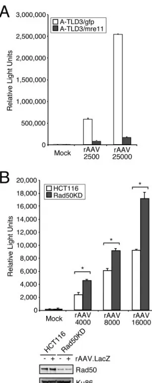
Enhanced rAAV transduction in cells lacking MRN. Our data for E1b55K/E4orf6 suggest that degradation of MRN contributes to increased rAAV transduction. We therefore predicted that rAAV transduction should be greater in cells that do not express functional MRN. To test this, we examined rAAV transduction in cell lines from patients with A-TLD in which Mre11 is mutated (52) versus that in A-TLD cells ex-pressing WT Mre11 (53). We observed higher rAAV transgene expression levels in the mutant cells (Fig. 3A), supporting our hypothesis that the Mre11 complex limits rAAV transduction. This was further evidenced by transduction assays with a cell line stably expressing shRNA against Rad50 (Fig. 3B) (69). The difference in transduction in this latter system is less dra-matic than in the A-TLD cell line, likely because Rad50 was not completely depleted (Fig. 3B) and the remaining MRN proteins are fully functional. The Rad50 knockdown cells, how-ever, still displayed an increase in transduction over different MOIs. Importantly, in both systems, we found that when MRN was compromised, rAAV transduction was enhanced, suggest-ing that this cellular complex poses a barrier to AAV.
WT AAV replication is restricted by MRN.Since E1b55K/ E4orf6 provides a helper function for WT AAV replication, we assessed the contribution of MRN degradation during lytic replication with Ad helper proteins. WT and mutant E1b55K cell lines, or a GFP control cell line, were infected with AAV and an Ad helper virus that cannot limit MRN (dl1016, lacking E1b55K and E4orf3). The accumulation of AAV Rep proteins was assessed by immunoblotting, and replicated viral DNA was measured by QPCR (Fig. 4A). WT Ad was used a helper control, and AAV replication was quantified relative to that of an AAV infection with no helper virus. We consistently ob-served greater Rep expression and viral DNA replication when MRN was degraded (in WT E1b55K- and R240A-expressing cells) than when MRN was present (in H354- and
GFP-ex-pressing cells). DBP levels showed that the cell lines were equally infected withdl1016.
[image:5.585.344.497.65.450.2]To show that the observed differences were not due to vari-ations in helper virus functions, we assayed WT AAV replica-tion after transfecting an AAV infectious plasmid clone, pNTC244 (11), along with Ad helper proteins E4orf6 and DBP, into the E1b55K cell lines. Viral DNA replication was analyzed by Southern blot hybridization (Fig. 4B). Similar to the results obtained above by infection, we observed more AAV replication in the cell lines that degraded the Mre11 complex (WT E1b55K- and R240A-expressing cell lines). To-gether, these assays suggest that degradation of MRN is an
FIG. 3. The Mre11 complex limits efficient rAAV transduction. (A) A-TLD 3 cell lines expressing GFP or WT Mre11 were infected with rAAV.LacZ at the indicated MOIs for 48 h before being pro-cessed for a quantitative-galactosidase assay. Infections were per-formed in triplicate, and error bars represent standard errors of the means (SEM). (B) HCT116 cells or HCT116-derived cells containing shRNA to Rad50 (Rad50KD) were infected in triplicate with rAAV. LacZ at the indicated MOIs and harvested after 48 h for a quantitative -galactosidase assay and Western analysis. Error bars represent SEM. Statistical analysis was performed using an unpaired Studentt test, with asterisks representing aPvalue of⬍0.005.
on November 8, 2019 by guest
http://jvi.asm.org/
important WT AAV helper function provided by E1b55K/ E4orf6.
If the Mre11 complex inhibits WT AAV replication, then more AAV DNA should accumulate in cells lacking MRN. To test this, AAV replication was compared between cell lines harboring hypomorphic mutations in Nbs1 (10) and Mre11 (A-TLD) (52) and WT complemented cells (53). An E4-de-leted Ad that cannot degrade MRN (dl1004) was used as a helper virus in these assays. We found that AAV replication was greater in all the mutant cell lines than in their WT pro-tein-expressing counterparts (Fig. 4C and data not shown), demonstrating that MRN is inhibitory to AAV. Immunoblot-ting showed that levels of viral helper proteins were compara-ble between NBS mutant and WT complemented cell lines (Fig. 4C), suggesting that this does not account for the ob-served differences in AAV replication. As a control, we in-fected both NBS cell lines with WT Ad5, which degrades the remaining MRN members. Similar levels of AAV replication (Fig. 4C and data not shown) were detected in cell lines coin-fected with Ad5. This further indicates that the results ob-tained withdl1004 coinfection are due to the MRN mutations in these cell lines. The use of other mutant Ad helper viruses that do not degrade MRN also yielded similar data (data not
shown), suggesting that the effects on AAV replication are not helper virus specific. These data demonstrate that MRN limits WT AAV replication and suggest that E1b55K/E4orf6 pro-vides a helper function to AAV by degrading the cellular complex.
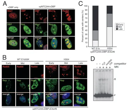
MRN proteins recognize viral replication centers and AAV ITRs.AAV replication centers develop during infection with helper virus (62, 64) or after transfection of the infectious clone pNTC244 with minimal helper virus proteins (54). Since the Mre11 complex localizes to sites of DNA damage and replication within cells (33, 34, 36, 37), we examined MRN localization and its influence on AAV replication compart-ment developcompart-ment by immunofluorescence. HeLa cells were transfected with a plasmid that expresses DBP, with or without pNTC244, and cells were stained for members of the Mre11 complex and viral replication centers (Fig. 5A). In the absence of viral replication centers, DBP and MRN showed a diffuse nuclear staining pattern (Fig. 5A, DBP only). Transfection of pNTC244 along with DBP induced discrete AAV centers in some cells, where DBP and Rep colocalized. We found that MRN accumulated at these viral sites, suggesting that replica-tion compartments may be recognized as DNA damage.
Previous studies (54, 62, 64) have demonstrated that as AAV
FIG. 4. WT AAV replication is restricted by the Mre11 complex. (A) WT- or mutant E1b55K-expressing HeLa cells were infected with WT AAV with or without the Ad mutantdl1016 or Ad5 (MOI of 50) for 24 h before being harvested for immunoblot analysis or Hirt DNA extraction and QPCR of viral DNA. QPCR levels were normalized to those in GFP-expressing cells infected with AAV alone. Viral replication and Rep accumulation were greatest in E1b55K cells where the MRN complex was degraded (Ad5 or WT E1b55K and R240A cells withdl1016). (B) AAV replication in E1b55K cell lines by transfection with helper plasmids. HeLa-derived cell lines expressing GFP or E1b55K proteins were transfected with the AAV plasmid pNTC244 in the presence or absence of plasmids expressing E4orf6 and DBP. Infection with Ad5 (MOI of 50) served as a positive control. Viral DNA was extracted from cells after 48 h and analyzed by Southern blot hybridization with a Rep probe. Rfd (replication form dimer), Rfm (replication form monomer), and ssDNA (single-stranded DNA) refer to replicative forms of AAV. (C) NBS cell lines complemented with empty vector (NBS⫺) or WT Nbs1 (NBS⫹) were infected with WT AAV in the presence or absence of Ad5 (MOI of 50) or the Ad mutantdl1004 (MOI of 50) for 24 h. Cells were processed for viral DNA and immunoblotting. DNA was subject to Southern blot hybridization with a Rep probe. Rfd, Rfm, and ssDNA refer to replicative forms of AAV as in panel B. Lysates were analyzed by immunoblotting for levels of cellular (MRN) and adenoviral (E1b55K, DBP, and E1A) proteins as shown below.
on November 8, 2019 by guest
http://jvi.asm.org/
replication progresses, the Rep pattern changes from small discrete intranuclear foci (early stage) to larger globular struc-tures (middle stage) and eventually to a more diffuse pattern where Rep structures fill much of the nucleoplasm (late stage). To further assess how MRN affects AAV replication compart-ment progression, we compared AAV centers when MRN was present or absent. The AAV infectious clone pNTC244 was cotransfected with DBP and E4orf6 plasmids into either WT E1b55K or H354 cell lines, and the relative number of cells at each replication stage was quantitated (Fig. 5B and C). We found that a larger proportion of late AAV replication centers were present in cells where MRN was degraded (WT E1b55K cells), whereas more early centers were visible in cells express-ing MRN (H354 cells). Additionally, durexpress-ing the course of these experiments, we found that many more cells displayed repli-cation centers when MRN was degraded (WT E1b55K) than
when MRN was present (H354). This observation is consistent with the results shown in Fig. 2B and C, further indicating a barrier in the initiation of viral replication. Rad50 localized to viral centers in all H354 cells, whereas MRN was degraded in 95% of transfected WT E1b55K cells. Together, these exper-iments indicate that MRN may recognize AAV as DNA dam-age, localizing to AAV centers and limiting the efficiency as well as the onset of viral replication.
[image:7.585.82.498.67.427.2]Purified Mre11 complex components have been shown to recognize a variety of DNA structures, including hairpins (13). Since the AAV ITR hairpin structures are genetic elements common between WT AAV and rAAV, we wanted to test whether the complex could recognize AAV ITRs directly. Pu-rified Mre11 and Nbs1 proteins were obtained and tested for association with the ITRs in vitro. The electrophoretic mobility shift assay we employed was previously used to study DNA
FIG. 5. MRN components localize to AAV replication compartments and bind AAV ITRs. (A) MRN localizes to AAV replication centers. HeLa cells were transfected with a DBP expression plasmid with or without the AAV infectious clone pNTC244. Cells were fixed and stained for Rep and DBP to mark AAV centers and for MRN components. (B) AAV replication centers formed by plasmid transfection. Shown are examples of stages of replication centers formed when WT E1b55K or H354 cells were transfected with pNTC244, DBP, and E4orf6 expression plasmids. Cells were stained for Rep and Rad50. Rad50 staining was absent in WT E1b55K cells as it was degraded with E4orf6. (C) Degradation of MRN results in more-advanced AAV replication centers. WT E1b55K- or H354-expressing cell lines were transfected with pNTC244, DBP, and E4orf6 expression plasmids. Rep-positive cells, 200 per treatment, were quantified for replication center stage and expressed as the percentage of cells with centers. (D) Binding of MRN components to the AAV ITR. The AAV ITR was 5⬘end labeled, incubated with purified Mre11/Nbs1 proteins (MN), and electrophoresed as described in Materials and Methods. The positions of bound (B) and free (F) ITRs are indicated. sssDNA, sonicated salmon sperm DNA.
on November 8, 2019 by guest
http://jvi.asm.org/
interactions with purified Mre11 and Nbs1 (38). We found that the mobility of the ITR shifted in the presence of Mre11/Nbs1 and was competed efficiently by an unlabeled AAV ITR but not by nonspecific DNA (Fig. 5D). These data demonstrate that MRN components can bind AAV ITRs directly and sug-gest that ITRs may contribute to MRN recognition in cells.
DISCUSSION
Recent research has established links between DNA viruses and cellular DNA damage response machinery (30, 61). The Mre11 complex is implicated as a major sensor of DNA dam-age in the cell, recognizing structures such as double-strand DNA ends, single-strand DNA, and hairpins (13, 55). Viral genomes contain such structures, and therefore, some viruses have evolved strategies to inactivate MRN. In the case of Ad, E1b55K/E4orf6 degrades MRN to prevent viral genome pro-cessing and damage signaling (9, 53). Given that these viral proteins provide helper functions to promote AAV transduc-tion and replicatransduc-tion, we examined whether the Mre11 complex impedes aspects of the AAV life cycle and whether the E1b55K/E4orf6-mediated helper function involves the degra-dation of these cellular proteins.
Here, we have shown that degradation of the MRN complex by E1b55K/E4orf6 creates a more permissive state for both WT AAV replication and rAAV transduction. The use of the E1b55K mutants and HeLa-based cell lines in these assays rules out a role for p53 in inhibiting AAV, in agreement with a recent report (12). Cyclin A degradation was previously ob-served in an E4orf6-inducible 293-based cell line and was sug-gested to contribute to the enhancement of rAAV transduc-tion (24). While this activity may be relevant to the particular 293-based cell line used in that study, we did not observe cyclin A degradation in the E1b55K-expressing cell lines used in this work (data not shown). E1b55K/E4orf6 was also recently shown to degrade DNA ligase IV, a cellular protein involved in NHEJ (3). However, all the E1b55K mutants utilized here degrade DNA ligase IV in conjunction with E4orf6 (data not shown), suggesting little contribution of this activity toward AAV helper function. Additionally, by using mutant and shRNA-containing cell lines, we directly demonstrated that MRN proteins negatively regulate viral transduction and rep-lication. This further highlights the degradation of MRN as a critical AAV helper function provided by E1b55K/E4orf6. We cannot, however, rule out additional activities of E1b55K/ E4orf6 that also contribute to AAV helper function. This may include the degradation of unknown relevant substrates by the E1b55K/E4orf6 ubiquitin ligase complex or a role in RNA processing and transport (45).
AAV has evolved to exploit helper viruses such as Ad to establish a cellular environment conducive to its replication. Other viruses, such as herpes simplex virus type 1 (HSV-1), also facilitate aspects of the AAV life cycle (60). Minimal HSV-1 helper proteins do not promote robust second-strand synthesis and transduction from rAAV vectors (M. D. Weitz-man, unpublished data), and this may be due to an inability to neutralize MRN. During WT AAV and HSV-1 coinfection, the utilization of the HSV-1 polymerase for AAV replication (58) may bypass the inhibitory effects of MRN on AAV, al-though the HSV-1 polymerase is not absolutely required (60).
Alternatively, since HSV-1 can sequester MRN (29, 56, 63), this may compete the complex away from AAV genomes, al-lowing second-strand synthesis and replication to occur.
We have used Southern blot hybridization, rAAV.LacO fo-cus formation, and the progression of AAV replication centers to demonstrate that the Mre11 complex restricts the accumu-lation of AAV DNA. In localizing to AAV replication centers and rAAV foci (Cervelli et al., submitted), MRN may recog-nize viral genomes as DNA damage. Our in vitro data support a role for the AAV ITRs in this recognition, although we cannot rule out the contribution of other viral structures. The targeting of the AAV ITRs would be consistent with other published data showing the binding and processing of hairpin structures by the Mre11 complex (32, 38, 39). MRN may re-strict AAV DNA accumulation in a similar manner by process-ing or degradprocess-ing viral genomes directly via its nuclease activity (38, 39). Alternatively, the complex may recruit other factors to process AAV DNA or inhibit cellular factors and/or poly-merases required to synthesize viral DNA.
DNA-damaging agents can support limited rAAV transduc-tion and AAV replicatransduc-tion in the absence of helper proteins (2, 44, 67). In these scenarios, cellular sites of DNA damage may compete MRN away from viral genomes (Cervelli et al., sub-mitted). Previous observations have also suggested a role for ATM in limiting rAAV transduction (46, 68). We and others have shown a requirement for the Mre11 complex in ATM activation and signaling (9, 28, 57). Therefore, E1b55K/ E4orf6-mediated degradation of MRN would suppress the ef-fects of ATM on AAV, providing a biological basis for these observations. Although the mechanism by which MRN inhibits AAV is currently unclear, it likely involves ATM and/or ATM-mediated signaling. In support of this, the knockdown of Nbs1 has no additional effect on rAAV transduction in ATM-defi-cient cells (Cervelli et al., submitted).
An expanding body of work has demonstrated that viruses must contend with cellular DNA damage response machinery (30, 61). Vectors based on these viruses will, therefore, likely face similar challenges. Here, we have shown that the Mre11 complex limits rAAV transduction similarly to other proteins involved in DNA double-strand break repair (12, 46, 68). Our observation that Mre11 binds to AAV ITRs is consistent with previous reports showing that vector circularization involves MRN (12) and is decreased by E4orf6 (15). Vector design and structure, however, may alter the recognition and outcome of vector-host interactions (47). For example, a self-complemen-tary AAV vector, which was already double stranded, was recently shown to require certain DNA repair proteins, includ-ing the Mre11 complex, for circularization (12). UV-inacti-vated AAV genomes, which contain cross-linked viral DNA, were recognized by a number of DNA damage response pro-teins in the ATR pathway (27). The outcome of vector-host interactions may also be impacted by the DNA damage re-sponse capacity of certain tissues in vivo, which may be altered after differentiation. It will be interesting to see how DNA damage-signaling pathways are affected in tissues that display efficient rAAV transduction and the differences between the dividing cells used in our studies and the nondividing cells found in vivo. Clearly, further research is needed to clarify the relationship between viral vectors and cellular DNA repair proteins and pathways.
on November 8, 2019 by guest
http://jvi.asm.org/
Our data have demonstrated that the Mre11 complex, a central player in the cellular DNA damage response, poses a barrier to certain aspects of the AAV life cycle. It will be interesting to determine how the MRN complex impacts other AAV serotypes and more distantly related parvoviruses that have different ITR structures. The MRN complex may also play a role in the establishment/reactivation of AAV latency, and its binding to ITRs could contribute to the integration of rAAV vectors at chromosomal breakage sites (35). Our data clarify an important link between cellular DNA damage re-sponse machinery and the AAV helper function provided by the adenoviral proteins E1b55K and E4orf6. These results imply that AAV must exploit helper viruses that modulate the cellular DNA damage response machinery of the host cell to promote a productive infection.
ACKNOWLEDGMENTS
We thank A. Berk, P. Branton, P. Concannon, D. Ferguson, J. Karlseder, G. Ketner, A. Levine, J. Petrini, R. J. Samulski, Y. Shen, J. Trempe, and T. Paull for reagents. We thank C. Hetzer for cloning LacR-GFP into pLPC and D. Lee for technical support. We thank members of the Weitzman lab for critical reading of the manuscript. This work was supported in part by NIH grants AI43341 and CA97093 (to M.D.W.). R.A.S. and G.D.C. were supported in part by NIH training grants to UCSD and the Salk Institute.
REFERENCES
1.Abraham, R. T.2001. Cell cycle checkpoint signaling through the ATM and ATR kinases. Genes Dev.15:2177–2196.
2.Alexander, I. E., D. W. Russell, and A. D. Miller.1994. DNA-damaging agents greatly increase the transduction of nondividing cells by adeno-asso-ciated virus vectors. J. Virol.68:8282–8287.
3.Baker, A., K. J. Rohleder, L. A. Hanakahi, and G. Ketner.2007. Adenovirus E4 34k and E1b 55k oncoproteins target host DNA ligase IV for proteasomal degradation. J. Virol.81:7034–7040.
4.Bekker-Jensen, S., C. Lukas, R. Kitagawa, F. Melander, M. B. Kastan, J. Bartek, and J. Lukas.2006. Spatial organization of the mammalian genome surveillance machinery in response to DNA strand breaks. J. Cell Biol. 173:195–206.
5.Boyer, J., K. Rohleder, and G. Ketner.1999. Adenovirus E4 34k and E4 11k inhibit double strand break repair and are physically associated with the cellular DNA-dependent protein kinase. Virology263:307–312.
6.Bridge, E., and G. Ketner.1990. Interaction of adenoviral E4 and E1b products in late gene expression. Virology174:345–353.
7.Bridge, E., and G. Ketner.1989. Redundant control of adenovirus late gene expression by early region 4. J. Virol.63:631–638.
8.Carney, J. P., R. S. Maser, H. Olivares, E. M. Davis, M. Le Beau, J. R. Yates III, L. Hays, W. F. Morgan, and J. H. Petrini.1998. The hMre11/hRad50 protein complex and Nijmegen breakage syndrome: linkage of double-strand break repair to the cellular DNA damage response. Cell93:477–486. 9.Carson, C. T., R. A. Schwartz, T. H. Stracker, C. E. Lilley, D. V. Lee, and
M. D. Weitzman.2003. The Mre11 complex is required for ATM activation and the G2/M checkpoint. EMBO J.22:6610–6620.
10.Cerosaletti, K. M., A. Desai-Mehta, T. C. Yeo, M. Kraakman-Van Der Zwet, M. Z. Zdzienicka, and P. Concannon.2000. Retroviral expression of the NBS1 gene in cultured Nijmegen breakage syndrome cells restores normal radiation sensitivity and nuclear focus formation. Mutagenesis15:281–286. 11.Chejanovsky, N., and B. J. Carter.1989. Replication of a human parvovirus nonsense mutant in mammalian cells containing an inducible amber sup-pressor. Virology171:239–247.
12.Choi, V. W., D. M. McCarty, and R. J. Samulski.2006. Host cell DNA repair pathways in adeno-associated viral genome processing. J. Virol.80:10346– 10356.
13.D’Amours, D., and S. P. Jackson.2002. The Mre11 complex: at the cross-roads of DNA repair and checkpoint signalling. Nat. Rev. Mol. Cell Biol. 3:317–327.
14.Douar, A. M., K. Poulard, D. Stockholm, and O. Danos.2001. Intracellular trafficking of adeno-associated virus vectors: routing to the late endosomal compartment and proteasome degradation. J. Virol.75:1824–1833. 15.Duan, D., P. Sharma, L. Dudus, Y. Zhang, S. Sanlioglu, Z. Yan, Y. Yue, Y.
Ye, R. Lester, J. Yang, K. J. Fisher, and J. F. Engelhardt.1999. Formation of adeno-associated virus circular genomes is differentially regulated by adenovirus E4 ORF6 and E2a gene expression. J. Virol.73:161–169. 16.Duan, D., P. Sharma, J. Yang, Y. Yue, L. Dudus, Y. Zhang, K. J. Fisher, and
J. F. Engelhardt.1998. Circular intermediates of recombinant adeno-asso-ciated virus have defined structural characteristics responsible for long-term episomal persistence in muscle tissue. J. Virol.72:8568–8577.
17.Duan, D., Y. Yue, and J. F. Engelhardt.2003. Consequences of DNA-dependent protein kinase catalytic subunit deficiency on recombinant adeno-associated virus genome circularization and heterodimerization in muscle tissue. J. Virol.77:4751–4759.
18.Duan, D., Y. Yue, Z. Yan, J. Yang, and J. F. Engelhardt.2000. Endosomal processing limits gene transfer to polarized airway epithelia by adeno-asso-ciated virus. J. Clin. Investig.105:1573–1587.
19.Ferrari, F. K., T. Samulski, T. Shenk, and R. J. Samulski.1996. Second-strand synthesis is a rate-limiting step for efficient transduction by recombi-nant adeno-associated virus vectors. J. Virol.70:3227–3234.
20.Fisher, K. J., G. P. Gao, M. D. Weitzman, R. DeMatteo, J. F. Burda, and J. M. Wilson.1996. Transduction with recombinant adeno-associated virus for gene therapy is limited by leading-strand synthesis. J. Virol.70:520–532. 21.Geoffroy, M. C., and A. Salvetti.2005. Helper functions required for wild type and recombinant adeno-associated virus growth. Curr. Gene Ther. 5:265–271.
22.Goncalves, M. A.2005. Adeno-associated virus: from defective virus to ef-fective vector. Virol. J.2:43.
23.Grieger, J. C., and R. J. Samulski.2005. Adeno-associated virus as a gene therapy vector: vector development, production and clinical applications. Adv. Biochem. Eng. Biotechnol.99:119–145.
24.Grifman, M., N. N. Chen, G. P. Gao, T. Cathomen, J. M. Wilson, and M. D. Weitzman.1999. Overexpression of cyclin A inhibits augmentation of re-combinant adeno-associated virus transduction by the adenovirus E4orf6 protein. J. Virol.73:10010–10019.
25.Harada, J. N., A. Shevchenko, A. Shevchenko, D. C. Pallas, and A. J. Berk. 2002. Analysis of the adenovirus E1B-55K-anchored proteome reveals its link to ubiquitination machinery. J. Virol.76:9194–9206.
26.Jazayeri, A., J. Falck, C. Lukas, J. Bartek, G. C. Smith, J. Lukas, and S. P. Jackson.2006. ATM- and cell cycle-dependent regulation of ATR in re-sponse to DNA double-strand breaks. Nat. Cell Biol.8:37–45.
27.Jurvansuu, J., K. Raj, A. Stasiak, and P. Beard.2005. Viral transport of DNA damage that mimics a stalled replication fork. J. Virol.79:569–580. 28.Lee, J. H., and T. T. Paull.2004. Direct activation of the ATM protein kinase
by the Mre11/Rad50/Nbs1 complex. Science304:93–96.
29.Lilley, C. E., C. T. Carson, A. R. Muotri, F. H. Gage, and M. D. Weitzman. 2005. DNA repair proteins affect the lifecycle of herpes simplex virus 1. Proc. Natl. Acad. Sci. USA102:5844–5849.
30.Lilley, C. E., R. A. Schwartz, and M. D. Weitzman.2007. Using or abusing: viruses and the cellular DNA damage response. Trends Microbiol.15:119– 126.
31.Linden, R. M., and K. I. Berns.2000. Molecular biology of adeno-associated viruses. Contrib. Microbiol.4:68–84.
32.Lobachev, K. S., D. A. Gordenin, and M. A. Resnick.2002. The Mre11 complex is required for repair of hairpin-capped double-strand breaks and prevention of chromosome rearrangements. Cell108:183–193.
33.Maser, R. S., O. K. Mirzoeva, J. Wells, H. Olivares, B. R. Williams, R. A. Zinkel, P. J. Farnham, and J. H. Petrini.2001. Mre11 complex and DNA replication: linkage to E2F and sites of DNA synthesis. Mol. Cell. Biol. 21:6006–6016.
34.Maser, R. S., K. J. Monsen, B. E. Nelms, and J. H. Petrini.1997. hMre11 and hRad50 nuclear foci are induced during the normal cellular response to DNA double-strand breaks. Mol. Cell. Biol.17:6087–6096.
35.Miller, D. G., L. M. Petek, and D. W. Russell.2004. Adeno-associated virus vectors integrate at chromosome breakage sites. Nat. Genet.36:767–773. 36.Mirzoeva, O. K., and J. H. Petrini.2001. DNA damage-dependent nuclear
dynamics of the Mre11 complex. Mol. Cell. Biol.21:281–288.
37.Mirzoeva, O. K., and J. H. Petrini.2003. DNA replication-dependent nu-clear dynamics of the Mre11 complex. Mol. Cancer Res.1:207–218. 38.Paull, T. T., and M. Gellert.1999. Nbs1 potentiates ATP-driven DNA
unwinding and endonuclease cleavage by the Mre11/Rad50 complex. Genes Dev.13:1276–1288.
39.Paull, T. T., and M. Gellert.1998. The 3⬘to 5⬘exonuclease activity of Mre 11 facilitates repair of DNA double-strand breaks. Mol. Cell1:969–979. 40.Querido, E., P. Blanchette, Q. Yan, T. Kamura, M. Morrison, D. Boivin,
W. G. Kaelin, R. C. Conaway, J. W. Conaway, and P. E. Branton.2001. Degradation of p53 by adenovirus E4orf6 and E1B55K proteins occurs via a novel mechanism involving a Cullin-containing complex. Genes Dev. 15: 3104–3117.
41.Querido, E., R. C. Marcellus, A. Lai, R. Charbonneau, J. G. Teodoro, G. Ketner, and P. E. Branton. 1997. Regulation of p53 levels by the E1B 55-kilodalton protein and E4orf6 in adenovirus-infected cells. J. Virol.71: 3788–3798.
42.Raj, K., P. Ogston, and P. Beard.2001. Virus-mediated killing of cells that lack p53 activity. Nature412:914–917.
43.Robinett, C. C., A. Straight, G. Li, C. Willhelm, G. Sudlow, A. Murray, and A. S. Belmont.1996. In vivo localization of DNA sequences and visualization of large-scale chromatin organization using lac operator/repressor recogni-tion. J. Cell Biol.135:1685–1700.
on November 8, 2019 by guest
http://jvi.asm.org/
44.Russell, D. W., I. E. Alexander, and A. D. Miller.1995. DNA synthesis and topoisomerase inhibitors increase transduction by adeno-associated virus vectors. Proc. Natl. Acad. Sci. USA92:5719–5723.
45.Samulski, R. J., and T. Shenk.1988. Adenovirus E1B 55-Mrpolypeptide
facilitates timely cytoplasmic accumulation of adeno-associated virus mRNAs. J. Virol.62:206–210.
46.Sanlioglu, S., P. Benson, and J. F. Engelhardt.2000. Loss of ATM function enhances recombinant adeno-associated virus transduction and integration through pathways similar to UV irradiation. Virology268:68–78. 47.Sanlioglu, S., M. M. Monick, G. Luleci, G. W. Hunninghake, and J. F.
Engelhardt.2001. Rate limiting steps of AAV transduction and implications for human gene therapy. Curr. Gene Ther.1:137–147.
48.Schnepp, B. C., K. R. Clark, D. L. Klemanski, C. A. Pacak, and P. R. Johnson.2003. Genetic fate of recombinant adeno-associated virus vector genomes in muscle. J. Virol.77:3495–3504.
49.Serrano, M., A. W. Lin, M. E. McCurrach, D. Beach, and S. W. Lowe.1997. Oncogenic ras provokes premature cell senescence associated with accumu-lation of p53 and p16INK4a. Cell88:593–602.
50.Shiloh, Y.2003. ATM and related protein kinases: safeguarding genome integrity. Nat. Rev. Cancer3:155–168.
51.Song, S., P. J. Laipis, K. I. Berns, and T. R. Flotte.2001. Effect of DNA-dependent protein kinase on the molecular fate of the rAAV2 genome in skeletal muscle. Proc. Natl. Acad. Sci. USA98:4084–4088.
52.Stewart, G. S., R. S. Maser, T. Stankovic, D. A. Bressan, M. I. Kaplan, N. G. Jaspers, A. Raams, P. J. Byrd, J. H. Petrini, and A. M. Taylor.1999. The DNA double-strand break repair gene hMRE11 is mutated in individuals with an ataxia-telangiectasia-like disorder. Cell99:577–587.
53.Stracker, T. H., C. T. Carson, and M. D. Weitzman. 2002. Adenovirus oncoproteins inactivate the Mre11-Rad50-NBS1 DNA repair complex. Na-ture418:348–352.
54.Stracker, T. H., G. D. Cassell, P. Ward, Y. M. Loo, B. van Breukelen, S. D. Carrington-Lawrence, R. K. Hamatake, P. C. van der Vliet, S. K. Weller, T. Melendy, and M. D. Weitzman.2004. The Rep protein of adeno-associated virus type 2 interacts with single-stranded DNA-binding proteins that en-hance viral replication. J. Virol.78:441–453.
55.Stracker, T. H., J. W. Theunissen, M. Morales, and J. H. Petrini.2004. The Mre11 complex and the metabolism of chromosome breaks: the importance of communicating and holding things together. DNA Repair (Amsterdam) 3:845–854.
56.Taylor, T. J., and D. M. Knipe.2004. Proteomics of herpes simplex virus replication compartments: association of cellular DNA replication, repair, recombination, and chromatin remodeling proteins with ICP8. J. Virol.78: 5856–5866.
57.Uziel, T., Y. Lerenthal, L. Moyal, Y. Andegeko, L. Mittelman, and Y. Shiloh. 2003. Requirement of the MRN complex for ATM activation by DNA damage. EMBO J.22:5612–5621.
58.Ward, P., M. Falkenberg, P. Elias, M. Weitzman, and R. M. Linden.2001. Rep-dependent initiation of adeno-associated virus type 2 DNA replication by a herpes simplex virus type 1 replication complex in a reconstituted system. J. Virol.75:10250–10258.
59.Weinberg, D. H., and G. Ketner.1983. A cell line that supports the growth of a defective early region 4 deletion mutant of human adenovirus type 2. Proc. Natl. Acad. Sci. USA80:5383–5386.
60.Weindler, F. W., and R. Heilbronn.1991. A subset of herpes simplex virus replication genes provides helper functions for productive adeno-associated virus replication. J. Virol.65:2476–2483.
61.Weitzman, M. D., C. T. Carson, R. A. Schwartz, and C. E. Lilley.2004. Interactions of viruses with the cellular DNA repair machinery. DNA Repair (Amsterdam)3:1165–1173.
62.Weitzman, M. D., K. J. Fisher, and J. M. Wilson.1996. Recruitment of wild-type and recombinant adeno-associated virus into adenovirus replica-tion centers. J. Virol.70:1845–1854.
63.Wilkinson, D. E., and S. K. Weller.2004. Recruitment of cellular recombi-nation and repair proteins to sites of herpes simplex virus type 1 DNA replication is dependent on the composition of viral proteins within prerep-licative sites and correlates with the induction of the DNA damage response. J. Virol.78:4783–4796.
64.Wistuba, A., A. Kern, S. Weger, D. Grimm, and J. A. Kleinschmidt.1997. Subcellular compartmentalization of adeno-associated virus type 2 assembly. J. Virol.71:1341–1352.
65.Woo, J. L., and A. J. Berk.2007. Adenovirus ubiquitin-protein ligase stim-ulates viral late mRNA nuclear export. J. Virol.81:575–587.
66.Xiao, X., J. Li, and R. J. Samulski.1998. Production of high-titer recombi-nant adeno-associated virus vectors in the absence of helper adenovirus. J. Virol.72:2224–2232.
67.Yalkinoglu, A. O., R. Heilbronn, A. Burkle, J. R. Schlehofer, and H. zur Hausen.1988. DNA amplification of adeno-associated virus as a response to cellular genotoxic stress. Cancer Res.48:3123–3129.
68.Zentilin, L., A. Marcello, and M. Giacca. 2001. Involvement of cellular double-stranded DNA break binding proteins in processing of the recombi-nant adeno-associated virus genome. J. Virol.75:12279–12287.
69.Zhong, H., A. Bryson, M. Eckersdorff, and D. O. Ferguson.2005. Rad50 depletion impacts upon ATR-dependent DNA damage responses. Hum. Mol. Genet.14:2685–2693.