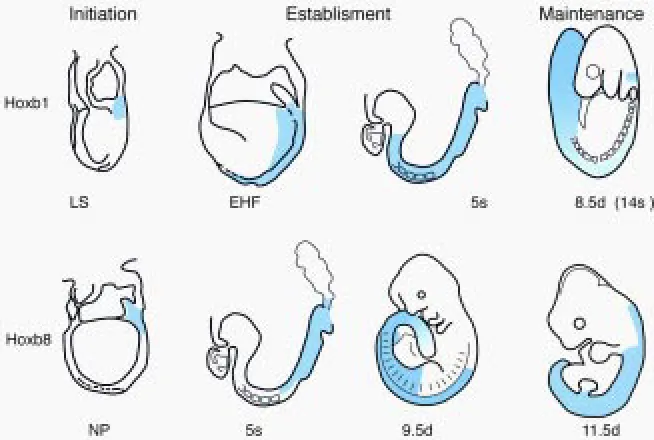
Initiation, establishment and maintenance of Hox gene
expression patterns in the mouse
JACQUELINE DESCHAMPS*, ERIC VAN DEN AKKER, SYLVIE FORLANI,
WIM DE GRAAFF, TONY OOSTERVEEN, BERNARD ROELEN and JEROEN ROELFSEMA
Hubrecht Laboratory, Netherlands Institute for Developmental Biology, Utrecht, The Netherlands
ABSTRACT Spatially and temporally restricted expression of the Hox genes along the main and appendicular axes is essential for correct patterning of vertebrate embryos. In this overview we discuss the latest data that shed light on the mechanisms underlying the generation of the expression domains of the Hox genes. The molecular genetic interactions governing initial transcription of the Hox genes in the posterior part of the primitive streak during mouse and chick gastrulation remain enigmatic. But the recent discovery by Kondo and Duboule (Cell, 97, 1999, 407-417) of a «cluster repressive regulation», will undoubtedly lead to a better understanding of the molecular genetic mechanism underlying colinear and sequential initiation of Hox gene transcription. Recently progress has been booked in characterizing the basal processes driving progression of the Hox expression domains during their establishment. Hox expression is still labile while being established. The transcriptional state of Hox genes in anterior tissues can be reprogrammed under the influence of more posterior locations. Posteriorizing activity may involve RA and FGF signaling. It is only when these interactions and, in some cases at least, regulatory interactions with Hox and cdx gene products occur appropriately, that the Hox expression domains would be correctly established. After the Hox expression domains have been established, regulatory processes involving the products of Polycomb and trithorax- Group genes start operating, perpetuating the transcriptional state of the Hox genes within and outside the expression domains. Whether control at the level of chromatin structure, believed to operate during the late maintenance phase of Hox gene expression, is also involved in regulating concerted initial expression of these genes, is a possibility that has been suggested.
KEY WORDS: Hox genes, antero-posterior patterning, transcriptional regulation
0214-6282/99/$15.00 © UBC Press
Printed in Spain
www.ehu.es/ijdb
*Address for reprints: Hubrecht Laboratory, Netherlands Institute for Developmental Biology, Uppsalalaan 8, 3584 CT Utrecht, The Netherlands. FAX: 31 30 2516464. e-mail: [email protected]
Abbreviations used in this paper: A-P, anterio-posterior; RA, retinoic acid;
RARE, retinoic acid response element; ZPA, zone of polarizing activity; Pc-G, polycomb-Group; trx-Pc-G, trithorax-Group; PRE, polycomb response element.
The Hox genes are key regulators of regional pattern formation along the antero-posterior (A-P) and other embryonic axes. These genes have been involved in transducing positional information to the precursors of embryonic axial and paraxial structures since before the ancestors of insects and vertebrates diverged from each other. In vertebrates as in insects, the precise restricted localiza-tion of each Hox gene product is crucial for correct patterning of tissues surrounding the rostral expression boundary of this gene. The tight transcriptional regulation of the Hox genes is therefore essential to the proper orchestration of embryonic morphogenesis. This overview focuses on particular aspects of the regulatory processes involved in controlling the expression of the Hox genes. The paper deals primarily with the mouse, with some excursions to chick, frog, Amphioxus, nematode and insect em-bryos. Our objective is to discuss a number of current issues regarding the ontogeny of the definitive Hox expression domains
patterns during this phase, such as dynamic forward spreading of the transcript distribution, plasticity of the gene expression state, role of retinoids in establishing the expression domains, regula-tion in the hindbrain versus spinal cord/trunk, regularegula-tion during limb development, and concerted regulatory events within Hox complexes.
I. Initiation of Hox gene transcription
Mouse Hox genes, homologs of Drosophila homeotic genes (HOM/Hox genes) (reviewed by McGinnis and Krumlauf, 1992) start to be expressed in an ordered sequence correlating with the localization of the genes from 3’ to 5’ in the clusters (Dollé et al.,
1989; Izpisua-Belmonte et al., 1991; Gaunt and Strachan, 1996; Van der Hoeven et al., 1996). The earliest (3’) Hox genes start to be expressed in the mesoderm, and more weakly in the primitive ectoderm (epiblast) of the pos-terior primitive streak of mouse and chick em-bryos (Gaunt, 1988; Frohman et al., 1990; Sundin and Eichele, 1992; Gaunt and Strachan, 1994), in late streak stage embryos. Transcrip-tion initiaTranscrip-tion of more 5’ genes occurs in the same region, overlapping the junction between extraembryonic and embryonic ectoderm (Deschamps and Wijgerde, 1993; Gaunt and Strachan, 1994), at progressively later stages but not in the same cells, since gastrulation movements have then brought more lateral epiblast cells into this position, exposing them to the “Hox induction field”. This Hox induction field in the posteriormost part of the embryo, traversed by cells fated to become extraembry-onic mesoderm (Tam and Beddington, 1987; Lawson et al., 1991), must therefore inherently bear a transcription inducing mechanism.
Among the potential Hox inducers present during gastrulation in this embryonic region are fibroblast growth factors and their recep-tors, some of which (i.e. eFGF, Pownall et al., 1996; Isaacs et al., 1998; FGF8, Crossley and Martin, 1995; Meyers et al., 1998; FGFR1, Deng et al., 1994; Yamaguchi et al., 1994) have been proposed to play a role in the control of mesoderm organization and antero-posterior (A-P) patterning. Modulation of Hox gene expression by altered FGF signaling has been observed in Xenopus (Isaacs et al., 1998) and in the mouse (Partanen et al., 1998). Whether initial Hox gene expression is affected in these conditions is not known yet. Evidence has accumulated to strongly sup-port a role of retinoic acid (RA), or a related retinoid, as a factor affecting Hox gene early transcription. RA was shown to turn on the Hox genes according to a colinear sequence from 3’ to 5’ in differentiating human EC cells (Simeone et al., 1990). RA has been shown to be present in the gastrulating chick embryo (stage 4/5, definitive streak stage; Maden et al., 1998). Higher RA levels were measured in the posterior part than in the rest of the embryo including the node, even though the absolute values of RA concentration were rather low. A number of Hox genes were found to carry functional RA response elements (RAREs) (see section II.C.3). Inactivation of the RARE 3’ to Hoxa1 leads to a delay in transcriptional initiation in mutant embryos, pointing to a role of RA signaling in correctly setting the timing of the initial expression of this gene (Dupé et al., 1997). Recent experiments (Niederreither et al., 1999) preventing early RA biosynthesis by inactivating Retinaldehyde Dehydrogenase 2 (Raldh2), suggest that RA is involved in the earliest Hox gene expression, but that it does not totally account on its own for Hox gene initiation. Hoxa1 was expressed at lower levels, and
ingly with a delay in time, in the primitive streak of headfold-stage Raldh2 mutant embryos (Niederreither et al., 1999).
The essential link between the timing of initial Hox gene expres-sion and the position of the genes in their cluster was initially demonstrated by van der Hoeven et al. (1996) who genetically transposed a Hoxd transcription unit to a more 5’ position in its cluster, and observed a delay in the initial expression of the corresponding gene. More recently, Kondo and Duboule (1999) brought the evidence to light that a cis-regulatory element is located at the 5’ side of the Hoxd cluster, some 40 kb from Hoxd13, which is necessary to set up colinear and sequential activation of the Hoxd genes. Deletion of the region containing this remote element leads to a release of a repressive mechanism normally preventing posterior genes to be activated prematurely. This results in the complete loss of colinearity and sequentiality of gene expression. However, the ectopic expression of the prematurely expressed 5’ Hoxd genes in the anterior part of these embryo is transient, probably because the lack of adequate molecular inter-actions in anterior tissues, where 5’ Hoxd genes normally are not expressed, does not allow full establishment and maintenance of gene expression at these anterior levels (Kondo and Duboule, 1999). A mechanism linking the correct sequential timing of initial Hox gene expression and genetically controlled modulation of chromatin structure has been proposed by Kondo et al. (1998) and Kondo and Duboule (1999). Recently, Bel et al. (1999) showed that 5’ Hoxd genes are initially expressed earlier in a null mutant for the Polycomb-Group gene Pc-G M33 than in control embryos (Bel et al., 1999). Furthermore, a given Hox gene was shown to exhibit premature competence to respond to RA activation in M33 null mutant embryos. These data strongly suggest that the Pc-G gene product M33 may play a role in the initiation of Hox transcription and in the time-restricted competence of Hox genes to respond to RA. This will be further discussed in section III.
The mechanism underlying the anterior progression of the initial Hox expressing domains along and lateral to the streak, successively in presumptive extraembryonic, lateral plate and paraxial mesoderm (Fig. 2), is still enigmatic. Early expression is not clonally transmitted (Deschamps and Wijgerde, 1993) but extends in spite of lateral migration of newly formed extraembryonic and embryonic meso-derm leaving the primitive streak (see Fig. 2). Experiments following the expression of Hoxd4 in the chick have shown that rostral spreading of the Hox expression domain along the primitive streak was neither relying on cell movement, nor on the diffusion of inducers from posterior to more anterior positions, at least from the time Hoxd4 was expressed posteriorly (Gaunt and Strachan, 1994).
Tomihara-Newberger et al. (1998) have found that amnionless (amn) is required for proper formation of this middle streak region as the streak extends. Correct functioning of this gene was found to rely on its expression in the visceral endoderm. A role of the endoderm overlying the primitive streak as a guide/instructor in the process of forward spreading of the Hox expression domains from the posterior to the anterior streak at the late streak stage appears as an interesting possibility.
Importantly, recent work by Liu et al. (1999) has made it clear that Wnt signaling is essential for primitive streak and mesoderm formation, and for epiblast differentiation and A-P patterning. The Wnt3 expression pattern and the Wnt3 null phenotype are compat-ible with Wnt3 signaling being involved in a genetic cascade turning on the Hox genes at the late streak stage.
II. Establishment of the Hox domains in axial and
paraxial embryonic tissues and in the limbs
After being initially observed in the posterior primitive streak region, the Hox transcript domains spread anteriorwards in and along the streak, meet and pass the node region and extend further rostrally to reach their anteriormost position (see Fig. 2). Initiation and establishment of the Hox expression domains are therefore a
continuous process, differing essentially by the fact that initiation occurs in cells that will not contribute to the embryo proper (presumptive extraembryonic mesoderm), while subsequent ex-pression involves cells that will contribute descendants in embry-onic tissues. The first embryembry-onic tissue to express a Hox gene following transcriptional initiation is presumptive lateral plate me-soderm, and subsequently paraxial meme-soderm, the cellular precur-sors of which are located at progressively more anterior positions, lateral to the streak, at the late streak stage (Tam and Beddington, 1987) (see Fig. 2). The area lateral to the node containing paraxial mesoderm progenitors also contains neurectoderm progenitors of
hindbrain and spinal cord (Tam and Beddington, 1987; Quinlan et al., 1995; Lawson and Pedersen, 1992 and K. Lawson, personal communication). In this area, inducing signaling events may there-fore operate on precursors of both neural and mesodermal Hox expression domains.
The relationship between initial Hox gene expression in the primitive streak region and gene expression in the definitive expression domains in the three embryonic germ layers is not clearly understood so far. In any case, correction of early spatial and temporal delay in initial expression of the Hoxa1 RARE mutant (Dupé et al., 1997) and of genetically transposed Hoxd11 (van der Hoeven et al., 1996), and lack of maintenance of premature Hoxd gene expression upon deletion of a “locus repressive element” (Kondo and Duboule, 1999) suggests that control mechanisms are superimposed onto the initial Hox gene regulation.
II.A. The rostral Hox expression boundaries are established gradually
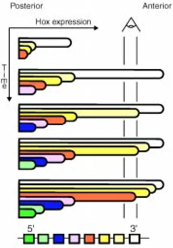
The definitive expression domains of the most 3’ Hox genes along the antero-posterior (A-P) axis of the embryo extend up to rostral limits located at rhombomeric boundaries in the anterior hindbrain in the neurectoderm and rostrally to the first somite in the mesoderm (the expression level decreasing considerably in this germ layer in the case of Hoxb1, Murphy et al., 1989; Wilkinson et al., 1989). More 5’ genes are expressed up to rostral boundaries located at successively more posterior positions. It has become clear that these definitive boundaries are not acquired at once as soon as the expression domains invade embryonic neurectoderm and mesoderm during embryogenesis. Hoxb8, for instance, is and remains expressed from day 8.5 on in a domain extending up to a rostral boundary at the level of somite 11 and its derivatives in the mesoderm, while the expression boundary in the neurectoderm, initially at the axial level of somite 5, shifts from this to a more anterior level in the posterior hindbrain by day 11 (Deschamps and Wijgerde, 1993). Gaunt and Strachan (1994) have clearly shown that the expression domain of chick Hoxd4 in the neurectoderm spreads anteriorwards into initially negative tissue: the expression boundary moves forward from a position located at a level anterior to the node (in the 5-somite embryo) to a position anterior to somite 1, passing a landmark of DiI-stained mesodermal cells at the level of somite 4. Gaunt and colleagues more recently showed that, at least for some Hox genes, Hox expression domains at somite stages also spread forward into initially negative cells within lateral plate mesoderm (Gaunt, 1999) and paraxial mesoderm (Gaunt et al., 1999). Rostral extension of the Hox expression domains in these tissues clearly takes place independently of cell movement, since there is no intersomitic exchange of cells (Stern et al., 1988). Gaunt and colleagues propose a model accounting for these observations, assuming the existence of an early morphogen gradient with a maximum located posteriorly, and a decrease in sensitivity to this morphogen of the genes from 3’ to 5’. For example, the paraxial mesoderm emerging from the streak would carry a morphogen gradient leading to the expression of each gene up to the axial level where the concentration of the morphogen corresponds to its sensitivity, the latter correlating with its position in the cluster (Gaunt and Strachan, 1996; Gaunt et al., 1999). A time-linked mechanism would lead to later induction of each gene at slightly more anterior levels, causing the expression domains to progress further rostrally (Fig. 3). Signaling in the paraxial meso-derm could diffuse into the adjacent neural tube to induce Hox
genes in this tissue. Alternatively, the acquisition of positional values could occur within the neurectoderm as well, provided that it carries an autonomous morphogen gradient in the plane of the neurepithelium. At some point, and by a still unknown mechanism, the definitive expression boundary of a Hox gene would be reached and the Hox expression domains would stop spreading more anteriorly. The transcriptional state of the Hox loci at all A-P positions would then be “frozen”, and further maintained by another molecular mechanism (see section III). A striking feature of mouse and chick Hox expression domains, which is not dealt with in the model, is the difference between the axial position of the rostral expression boundary in neurectoderm and mesoderm. This differ-ence is not likely to be due to a slower anterior spreading of mesoderm cells initiated in the node region: descendants of cells labeled in the node region at a given stage spread to the same anterior level in the ventral neural tube and in the paraxial and lateral mesoderm (K. Lawson, personal communication).
II.B. Plasticity of Hox gene expression state during the establishment phase
Experiments with chick–quail chimeras (Grapin-Botton et al., 1995, 1997) and with chick-mouse chimeras (Itasaki et al., 1996) have brought to light the evidence that posterior signals are able to induce expression of given Hox genes in the neurectoderm where they were not, and would normally never be, expressed. Thus Hox genes in neural tissues are reprogrammed upon transposition of rhombomeric neurepithelium to a more posterior position in the neural tube or upon transposition of posterior somites to more anterior positions until at least the 10-somite stage in the chick. Posteriorly transposed rhombomeres were shown to turn on more
5’ Hox genes under the influence of their landing position, sensing a Hox-inducing posterior signal stronger in this location than at the original site, and the more posterior the position the stronger the signal (Itasaki et al., 1996; Grapin-Botton et al., 1997). These Hox-inducing signals were proposed to be of two types, as shown by additional experiments. On the one hand, transposition of posterior rhombomeric neurepithelium to a more anterior position did not alter its Hox expression status, but gave rise to the induction of more posterior Hox genes in the host epithelium surrounding the graft. “Planar” signals, traveling within the neurepithelium plane, were thus capable of inducing 5’ Hox genes in anterior neural tissue (Grapin-Botton et al., 1997). On the other hand, posterior somites transposed more anteriorly were shown to lead to de novo induc-tion, in the adjacent neural tube, of Hox genes normally corre-sponding to the more posterior position of the explanted somites (Itasaki et al., 1996; Grapin-Botton et al., 1997). Moreover, more posterior somites induced stronger Hox gene expression in the adjacent neural tube, suggesting the existence of a caudo-rostral gradient of inducer activity in the paraxial mesoderm, vertically transmitted to induce novel Hox gene expression in the neurepithelium (Itasaki et al., 1996; Grapin-Botton et al., 1997). In independent experiments, Gould et al. (1998), studied the interac-tions between paraxial mesoderm signaling and Hoxb4 expression in the neurepithelium in explanted mouse tissues in coculture. They obtained results consistent with the reprogramming of Hox gene expression upon tissue transposition in vivo. In addition, they could identify that a RA-mediated process, conserved between mouse and chick embryos, was crucially involved in the signaling event.
It seems therefore clear that the Hox gene expression state can be changed in a particular neural cell provided that this cell is
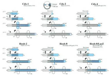
Fig. 4. Schematic representation of the dynamic spreading of cdx and Hox transcripts (in blue) during neural plate/ early headfold (day 7,5 represented) to later stages (day 11.5 represented) of mouse development. For cdx2, the only cdx gene to be expressed before gastru-lation, a sagittal view of an early egg cylinder embryo is depicted, with cdx2 expression in extraembryonic ectoderm (Ext.ect.) all around the boundary with embryonic ectoderm (Emb.ect.). All other schemes show a dorsal view of the stretched embryonic axis. This represen-tation is based on a scheme of cdx1 expression by Meyer and Gruss (1993), and the data are from the work cited and from our own published and unpublished data. Hoxb2 is taken as an example of a 3' Hox gene, Hoxb8 as an example of a more 5' Hox gene, and the particular Hoxb8/lacZ expression is shown because the Hox regulatory region driving lacZ expression contains 4 functional CDX bind-ing sites (see Charité et al., 1998). Note that cdx expression decays from anterior to posterior from early stages on, and that
competent to express Hox genes (this competence slowly de-creasing rostrally to r7) and that its new surrounding corresponds to axial positions posterior enough to induce more 5’ Hox genes (caudally to the r6/r7 border, or adjacent to a somite more posterior than somite 5) (Itasaki et al., 1996; Grapin-Botton et al., 1997). This plasticity of gene expression has been described at developmental stages that are believed to precede the time when the maintenance mode of Hox gene regulation is stabilizing the Hox transcriptional states (day 9.5, according to Yu et al., 1998, looking at mouse Hoxa7, and to Koseki et al., personal communication, looking at mouse Hoxc6).
Considering these experimental data together with the results from Gaunt and colleagues (Gaunt and Strachan, 1994, 1996; Gaunt et al., 1999), it is tempting to speculate that the molecular mechanism responsible for de novo induction of more posterior Hox genes after tissue transposition, is actually the same as the mechanism accounting for rostral extension of the Hox expression domains in the normal situation (see II.A).
II.C. Upstream regulators and effectors in the establishment phase: transduction of positional information to the Hox genes
Signals mediating A-P positional information in the embryonic germ layers have to be transduced to the Hox transcriptional machinery. Several transcription factors have been shown to contribute to controlling the spatially restricted expression of the Hox genes. Some of these factors seem to modulate rostral spreading of Hox gene expression along most of the rostrocaudal embryonic axis while others confine their effect to specific hind-brain compartments (rhombomeres). One can distinguish be-tween these regulatory processes on a time basis as well, i.e. between the early establishment phase of the Hox expression domains from posterior regions to their maximum anterior exten-sion, and the later definition and reinforcement of rhombomeric positional identity. Hox gene expression in the hindbrain seems to be subject to specific, additional regulatory circuits restricting and reinforcing gene expression in very specific territories, individual rhombomeres or subsets of rhombomeres and associated neural crest (see section II.C.3).
II.C.1. Molecular genetic interactions presiding to the establish-ment of the Hox expression domains in spinal cord and trunk mesoderm
II.C.1.a) Trans-acting regulatory factors: the case of caudal The patterning action of Drosophila homeotic genes is prepared by several categories of genes that are partitioning the blastoderm along the A-P axis into progressively smaller compartments: the maternal and the segmentation (gap, pair-rule and segment polar-ity) genes, respectively (reviewed by St Johnston and Nüsslein Volhard, 1992). However, no correspondence between flies and vertebrates has been uncovered at this level, probably reflecting the fact that the strategies of development differ considerably between them. Work of several laboratories, though, seems to involve the homologs of Drosophila caudal, an upstream regulator of regional A-P patterning (MacDonald and Struhl, 1986; Mlodzik et al., 1990), in the control of Hox gene expression in mice, chick and frog embryos. Drosophila maternal caudal, the expression of which is shaped into a posterior to anterior protein gradient by maternal bicoid gene products (Rivera-Pomar et al., 1995), and
later on zygotic caudal, are known to positively regulate posterior gap and pair-rule gene expression (Dearolf et al., 1989; Rivera-Pomar et al., 1995; Schulz and Tautz, 1995), the latter turning on selected Hox genes (Levine and Harding, 1989).This function of caudal, contributing to shape the expression patterns of the Hox genes, was shown to be conserved in the short germ band insect Tribolium (Wolff et al., 1998).
Among the three mouse caudal homologs, cdx2 seems to be expressed the earliest, transcripts being detected in extraembry-onic ectoderm along the junction with adjacent proximal epiblast at the expanded blastocyst stage (Beck et al., 1995). The three mouse cdx genes begin to be expressed in the embryonic part of the conceptus during gastrulation, in domains initially very poste-rior within and along the primitive streak, spreading forward to a rostral limit in neurectoderm and mesoderm which is gradually more anterior for cdx4 (Gamer and Wright, 1993), cdx2 (Beck et al., 1995) and cdx1 (Meyer and Gruss, 1993). Transcription of these three cdx genes therefore generates nested sets of expres-sion domains overlapping posteriorly, together creating a stepwise gradient of cdx gene products with a posterior maximum (see also Marom et al., 1997, describing a similar situation in the chick). In each case, the expression domain in the mesoderm is slightly more posterior than in the neurectoderm. The characteristics of initiation and early forward spreading of cdx gene expression, and the maximal extension of the cdx transcription domains from the caudal part of the embryos up to the preotic sulcus -r2/r3- for cdx1 at the head fold stage (Meyer and Gruss, 1993), are very reminis-cent of the properties of the Hox genes (Fig. 4). Rereminis-cent findings from experiments on Amphioxus are possibly revealing major clues to this resemblance. The Amphioxus cdx gene belongs to the same cluster as other homeobox genes, this cluster most likely being a paralog of the Amphioxus proper Hox cluster, the ParaHox cluster (Brooke et al., 1998). Ancestor Hox and cdx genes were therefore most presumably close relatives of each other. Recent experimental work in Drosophila in fact reinforces the hypothesis of a close functional likeness between Hox and cdx genes, making it clear that caudal (cad), in addition to modulating early segmen-tation, behaves as a canonical Hox gene (Moreno and Morata, 1999). It is expressed exclusively in the most posterior segment of the fly (A10) and its derivatives, the analia; it is required in this segment for normal analia development; it suppresses in A10 the expression of the immediately more anteriorly acting gene, Abdb, and is able to induce ectopic analia development when expressed in ectopic places. Cad behaves as a Hox gene located immediately 5’ to AbdB. Since cad homologs are not physically linked to the Hox genes in any known animal group, cad would be a paralog of a 5’ neighbor of Abdb on the ParaHox cluster, as shown for its Am-phioxus homolog. caudal would therefore have two functions in Drosophila and in short germ band insects: early on, it would regulate segmentation of the blastoderm under syncitial conditions (in the long germ band Drosophila embryos and in the syncitial part of the short germ band embryos); later on, it would pattern the terminal structures, conferring its identity to the most posterior body segment of the fly (Moreno and Morata, 1999). The expres-sion pattern of caudal in this second phase, in short germ band Tribolium embryos which produce abdominal segments in a sec-ondary growth process, is in fact very reminiscent of that seen in vertebrates (Schulz et al., 1998).
muta-tions in mouse cdx1 (Subramanian et al., 1995) and cdx2 (Chawengsaksophak et al., 1997) genes have been shown to lead to anterior homeotic-like transformations in the vertebral column, and Hox gene expression was shown to be altered in cdx1 null mutants (Subramanian et al., 1995). Moreover, overexpression of each of the three cdx genes in the mouse has been shown to be capable of rostrally shifting Hoxb8lacZ transgene activity as well as en-dogenous Hoxb8 expression upon interaction with CDX binding sites in the upstream proxi-mal region of the Hoxb8 gene (Charité et al., 1998), indicating that cdx gene products are capable of directly stimulating Hox gene ex-pression, and that the three cdx genes have overlapping functions when coexpressed (Charité et al., 1998). Independently, Xeno-pus caudal (Isaacs et al., 1998) was shown to control Hox gene expression, and the C. elegans caudal homolog was found to regu-late the early expression of the Hox gene mab-5 in a particular cell (Hunter et al., 1999). The discovery of a late, very conserved function of caudal as a homeotic-like posterior determi-nant makes the interpretation of the cdx/Hox interactions documented in vivo in mouse
expression being usually more posterior than that of the endog-enous gene. This implies either that remote cis-acting sequences are involved in specifying endogenous Hox expression domains, or that a higher level of Hox gene control is operating (i.e. at the level of the whole cluster), or possibly both. In three cases recently, involvement of a remote control element has been invoked to account for Hox gene expression features. Firstly a Hoxd regula-tory element responsible for expression of Hoxd9 to Hoxd13 in overlapping domains in distal limb buds is believed to reside very far from the corresponding transcription unit (van der Hoeven et al., 1996; Hérault et al., 1999). This element would correspond to a unique function of these genes in patterning the autopod (see section II D). Secondly, a regulatory element essential for maintain-ing the anterior expression boundary of Hoxc8 in neural tube and mesoderm has been localized between 11 and 19 kb downstream of Hoxc8 (Bradshaw et al., 1996). Thirdly, a control element located 30 kb downstream of Hoxb8 may contribute, together with other proximal Hoxb8 control elements, to the specification of the defini-tive rostral expression boundary of this Hox gene in neural tube and mesoderm (Valarché et al., 1997). This element is also a candidate for regulating the definitive expression boundary of several neigh-bor Hoxb genes.
Among the mechanisms presumed to account for the balance between the contribution of different enhancers in properly regulat-ing particular Hox genes, selectivity, competition and sharregulat-ing of regulatory elements have been put forward and experimentally documented (Gould et al., 1997; Sharpe et al., 1998). These interactions between the control sequences of neighbor Hox genes would account at least in part for the fact that the genes have remained clustered during evolution. However, Drosophila homeotic genes do not appear to share enhancers (discussed by Duboule, 1998). Loss of selective pressure to maintain integrity of the cluster
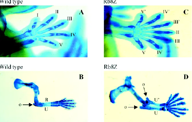
Fig. 5. Alcian blue stained control (wild type) and Hoxb8 overexpressing transgenic (Rb8Z) forelimbs. Control (A) and transgenic (C) forelimbs of day 12.5 embryos show mirror-image duplication of posterior digits and carpal bones of the transgenic limb; Control (B) and transgenic (D) forelimbs of 14.5 day embryos reveal mirror-image duplicated ulna (u) and olecranon process (o) as well as mirror-image duplicated posterior digits in the transgenic limb (see text and Charité et al., 1994).
embryos less straightforward: do the latter reflect the evolutionary conservation of the early function of caudal in insects, regulating early A-P patterning and Hox gene expression (indirectly in the case of insects, and directly in C. elegans and vertebrates), or do they correspond to positive cross-regulatory interactions be-tween related genes? A definitive answer to this question is not available at the moment.
A direct involvement of FGF in the control of Hox gene expres-sion and A-P patterning mediated by a caudal homolog has been reported in Xenopus (Pownall et al., 1996; Isaacs et al., 1998). FGF was shown to be involved in controlling Hox genes also in the mouse (Partanen et al., 1998). The latter authors studied the phenotypes of FGFR1 receptor mutants, and observed homeotic-like transformations in the vertebral column and alterations of Hox gene expression domains. It is possible that the FGF effect on mouse Hox genes operates via the cdx genes as well, although no evidence pointing in this direction has been obtained so far (see Partanen et al., 1998).
II.C.1.b) cis-acting Hox regulatory elements
in this organism may be causally linked to changes in the regulatory interactions to more appropriately fit the strategy of development in this long germ band insect.
II.C.2. RA and the establishment of Hox gene expression domains A possible role of RA in modulating the expression domains of the Hox genes during embryogenesis was suggested by Kessel and Gruss (1991). These authors exposed embryos in utero to exogenous RA, and observed posterior homeotic-like transforma-tions in the vertebral column concomitantly with rostral shifts in Hox expression domains. Depletion of endogenous RA has now con-firmed that this or a related compound plays a crucial role during the establishment of the Hox expression domains: while the initial expression of Hoxa1 in the Retinaldehyde dehydrogenase (Raldh2) null embryos was present at a significantly reduced level, complete loss of Hoxa1 expression was observed in 8.5 and 9.5-day mutant embryos (Niederreither et al., 1999). The significant increase in the level of RA generation at the beginning of somitogenesis in the chick (Maden et al., 1998) might, at least in part, correlate with the role of RA in stimulating Hox gene expression in the rostrally extending axial and paraxial expression domains. Strikingly enough, spinal cord and somites were shown to generate very high levels of RA at early somite-stages, while the CNS anterior to the brain/ spinal cord junction (r1 to r6) did not (Maden et al., 1998). RA is present homogeneously within a large domain with a sharp cutoff at the level of r6/r7, rather than as a gradient with a posterior maximum. In order to account for the expression of the most 3’ Hox genes such as Hoxb1 whose transcription is controlled by a RARE-containing regulatory element, at levels of the hindbrain which do not produce RA (anterior to r7), diffusion of RA from the spinal cord into the presumptive hindbrain in the plane of the neuroepithelium was proposed as a possibility, although no such diffusion could be observed when assaying the presence of RA in the tissue (Maden et al., 1998). Alternatively, because the establishment of the early expression domain of Hoxb4 in the neurectoderm depends on a retinoid-based mechanism in the flanking paraxial mesoderm (Gould et al., 1998), there is a possible role of the RA-producing hindbrain mesenchyme in the induction of 3’ Hox genes in the rostral hindbrain neurepithelium. However, again, no diffusing RA could be visualized in the hindbrain neurectoderm anterior to r7 (Maden et al., 1998). Gould and colleagues elegantly showed that a mesoderm-derived factor and a retinoid signal are necessary for Hoxb4 to be induced in the hindbrain in explant cocultures. The influence of vertical signals from the flanking paraxial mesoderm was shown to induce de novo expression of more 5’ Hox genes in rhombomeres surgically transposed more posteriorly at spinal cord levels in the chick (Itasaki et al., 1996; Grapin-Botton et al., 1997). The Hox genes shown to be induced in posteriorly trans-posed rhombomeres were not only the genes known to be directly RA-sensitive such as paralog groups 1, 2 and 4 (see next section), but all genes belonging to paralogous groups 1 to 8 (Grapin-Botton et al., 1997). Whether the competence to respond to RA is direct or indirect in all cases is not known.
II.C.3. Molecular interactions controlling Hox gene expression in the hindbrain
Several transcription factors have been shown to participate in the regional control of Hox gene expression in the hindbrain. The zinc finger protein Krox-20 directly regulates the expression of
Hoxb2 (Sham et al., 1993; Vesque et al., 1996) and Hoxa2 (Nonchev et al., 1996) in rhombomeres 3 and 5 upon binding to specific sequences in their control regions. Targeted inactivation of Krox20 resulted in the progressive loss of r3 and r5 territories, which then also affected the development of even-numbered rhombomeres (Schneider-Maunoury et al., 1993, 1997). Interest-ingly, recent analysis of Hoxa2-/-/Hoxb2-/- mutants revealed normal generation and maintenance of rhombomeric territories, indicating that the progressive elimination of r3 and r5 in Krox-20-/- embryos must be mediated through different downstream targets (Davenne et al., 1999). The product of the kreisler gene, a basic domain-leucine zipper transcription factor (Cordes and Barsch, 1994), was shown to directly activate Hoxb3 and its paralog Hoxa3 in r5, via conserved binding sites (Manzanares et al., 1997, 1999). The kreisler-dependent activation of Hoxb3 in r5 occurs in combination with an Ets-related transcription factor that binds to a kreisler neighbor site. While a similar Ets-dependent motif is also present in the Hoxa3 enhancer, this site does not appear to be required for Hoxa3 spatially restricted expression (Manzanares et al., 1999). In contrast, kreisler also directs Hoxa3-specific upregulation in r6 through the same high-affinity binding site controlling r5 expres-sion (Manzanares et al., 1999). Altogether these data highlight the common origin of paralogous Hox gene regulatory regions from an ancestral Hox cluster and their subsequent evolutionary diver-gence following Hox cluster duplications, leading to subtle changes in paralogous Hox gene expression domains. Consistent with a role in initiating r5 and r6 segmental identities, kreisler mutants lack r5- and r6-derived structures, including cranial nerves and their associated ganglia (McKay et al., 1994; Manzanares et al., 1997; McKay et al., 1997).
As mentioned above, retinoic acid receptors RARs and RXRs (Kastner et al., 1997) and their ligands are also directly involved in the regulation of Hox genes expressed in the hindbrain. Retinoic Acid Response Elements (RAREs) have been found proximally to promoter regions of Hox gene paralog groups 1 and 4. Hoxb1 is flanked by two distinct RARE containing sequences, a 3’ element shown in transgenic studies to be essential for gene expression in the early neural tube (Marshall et al., 1994), and a 5’ element involved in restricting later expression to r4 (Studer et al., 1994) (see also Ogura and Evans, 1995). Hoxa1 also carries a 3’ RARE containing sequence (Frasch et al., 1995), the inactivation of which has been shown to lead to a delay in the establishment of the anterior expression boundary, resulting in abnormal hindbrain patterning at later developmental stages (Dupé et al., 1997). Double mutant analysis with Hoxb1 3’ RARE, Hoxa1 3’ RARE and Hoxa1 and Hoxb1 null alleles revealed the requirement of Hoxa1, Hoxb1, and endogenous retinoids to initiate Hoxb1 expression, as well as strong genetic interactions between Hoxa1 and Hoxb1 in establishing r4 identity and in patterning r4-derived structures (Gavalas et al., 1998; Studer et al., 1998). Maintenance of Hoxb1 expression in r4 is mediated through an autoregulatory element to which HOXB1 binds together with the product of the pbx gene (homolog of the Drosophila extradenticle gene) as a cofactor (Popperl et al., 1995). Furthermore, Maconochie et al. (1997) reported that cross-regulation by HOXB1 and PBX proteins is also involved in the control of Hoxb2 expression in r4.
gene expression as well (Packer et al., 1998). A RARE containing enhancer is required, together with a mesoderm signal, to induce early expression of Hoxb4 in the neural tube up to an anterior boundary at r6/r7 (Gould et al., 1998) (see section II.B.). It is noteworthy that swapping the Hoxb4 RARE sequence (a DR5-type) with the 3’ RARE of Hoxb1 (a DR2-DR5-type) results in a shift of the rostral expression boundary from r6/r7 to r3/r4, indicating that these sequences bear information necessary for correct position-ing of the anterior boundaries of Hoxb4 and Hoxb1 expression (Gould et al., 1998). Maintenance of Hoxb4 expression in its rostralmost domain at later developmental stages is controlled through a distinct rhombomeric enhancer (Gould et al., 1998). This late enhancer contains a Hox-responsive element which was shown to bind the HOXB4 protein and its paralogs, together with PBX, in a way that appears to be conserved between mammals and Drosophila (Gould et al., 1997). Interestingly, HOXB5 can also bind the Hoxb4 autoregulatory element, resulting in a positive cross-regulation which reinforces Hoxb4 expression in the region of overlap between the Hoxb4 and Hoxb5 expression domains (Gould et al., 1997).
II.D. Control of Hox gene expression in emerging and develop-ing limb buds
The regulation of Hox gene expression is thought to play an important role during two distinct phases of limb development: during the positioning of the limb fields along the A-P axis, and during later control of growth and morphogenesis of the developing limbs (reviewed by Johnson and Tabin, 1997). It is difficult to describe the molecular regulation of the Hox genes during limb outgrowth and patterning without a simultaneous description of the embryological events associated with the (changes in) gene ex-pression features. Early Hox gene exex-pression in the lateral plate mesoderm presumably contributes to specifying the axial position of the future forelimb bud (reviewed by Cohn and Tickle, 1996). Regulation of Hox expression domains in lateral plate mesoderm is believed to have been crucial in the sequential appearance of the two pairs of limbs which are fundamentally non-equivalent (Tabin and Laufer, 1993; Coates and Cohn, 1998), leading to the emer-gence of tetrapods. Control of Hox gene expression in the lateral plate mesoderm is likely to have operated independently from gene control in paraxial mesoderm (Gaunt, 1994; Burke et al., 1995; Cohn et al., 1997; Coates and Cohn, 1998). Control of Hox expression in the presumptive fore- and hindlimb and during limb emergence is likely to be exerted by growth factors and their receptors, important modulators of growth and morphogenetic patterning. FGF10 in particular has been shown to be required for development of both pairs of limbs (Min et al., 1998). The A-P position of the rostral expression boundary of a number of Hoxb genes in lateral plate mesoderm has been proposed to contribute crucial information to positioning the limb field (Rancourt et al., 1995; Cohn and Tickle, 1996; Cohn et al., 1997), and to position the posterior part of the limb field and its signaling center, the Zone of Polarizing Activity (ZPA) (Charité et al., 1994 regarding the mouse; Stratford et al., 1997, and Lu et al., 1997 for chick studies). The molecular-genetic relationship between Hox gene expression and the generation of the ZPA is still enigmatic. The ZPA concept itself has evolved with time to optimally fit the biological observations: it was first defined as the “polarizing activity” present in the posterior limb margin, that could induce development of supernumerary
digits and mirror-image symmetry after being transplanted into the anterior margin of a host chick limb bud (Saunders and Gasseling, 1968); this polarizing activity was found to be present at earlier stages all along the flank mesoderm from the node region up to the limb levels, and to peak at the level of the posterior part of the limb bud at the time limb buds are about to emerge (Hornbruch and Wolpert, 1991). More recently, the ZPA was found to be an organizer, site of very specific gene expression centered on the activity of Sonic hedgehog (Shh), considered to primarily mediate the polarizing activity (Echelard et al., 1993; Krauss et al., 1993; Riddle et al., 1993). The posterior limb margin was also proposed to be an area of stage-dependent differential growth.
A role of early Hoxb gene expression in the lateral plate mesoderm would be to supply positional information specifying and polarizing the limb fields along the A-P axis. Hoxb genes, and Hoxb8 in particular, could possibly confine early asymmetric expression to potential key regulators of Shh such as Gli3 (Schimmang et al., 1992; Masuya et al., 1995) and Alx4 (Qu et al., 1997; Takahashi et al., 1998), resulting in the posterior restriction of the Shh- expressing ZPA. Hoxb8 expression in the lateral plate mesoderm during forelimb bud emergence overlaps the ZPA and precedes Shh expression (Charité et al., 1994). Hoxb8 gain of function mutants exhibit mirror-image duplications of the poste-rior part of the autopod and zeugopod (see Charité et al., 1994). This phenotype is more severe than that of Gli3 (Xt) (Hui and Joyner, 1993), and Alx4 (Lst) (Qu et al., 1997) null mutants, which present mirror-image duplications in the autopod exclusively, suggesting that Hoxb8 is hierarchically higher/operating earlier than the asymmetrically expressed Gli3 and Alx4 genes in the control of limb bud patterning. Moreover, no change in Hoxb8 expression boundary was observed in Gli3 (Xt) (Zúñiga and Zeller, 1999) and Alx4 (Lst), (Chan et al., 1995) polydactyl null mutants.
Re-garding A-P regionalization, the metapterygial axis runs through the stylopod element, through the most posterior element of the zeugopod (ulna and fibula), and through the posterior carpal and tarsal bones (in the arm and leg autopod, respectively), before it bends in the anterior direction, leaving the digits on the postaxial side (see Sordino and Duboule, 1996). Digit morphogenesis there-fore is the result of the extended growth phase in distal limb buds, accounted for by the extended Hoxd13 expression domain driven by the “digit” enhancer (Hérault et al., 1999). Not surprisingly, Hoxd13 null mutants exhibit truncated digits rather than A-P patterning defects (Dollé et al., 1993). The A-P polarity of the early limb bud, foreshadowing the A-P polarity of the complete limb, is unlikely to rely on Shh expression from the beginning on since this gene is only expressed relatively late (in quite developed forelimb buds at about the 30-somite stage in the mouse, Charité et al., 1994). Expression boundaries of early Hoxb genes are candidates to supply positional information polarizing the early limb bud long before Shh is expressed at the posterior margin. Shh would be involved later, both in controlling A-P polarity and growth of the autopod proximo-distally. The phenotype of the Shh null mutant confirms the involvement of this gene in distal growth of the limb buds rather than in the specification of posterior identity exclusively (Chiang et al., 1996).
Expression of Abdb-type Hoxa and Hoxd genes (of paralog groups 9 to 13) in developing limbs is spatially and temporally coupled to limb bud outgrowth (Dollé et al., 1989; Yokouchi et al., 1991). Strikingly, the phenotypes of loss of function mutants in these genes affect limb elements in a way that is colinear with the position of the genes in the cluster (reviewed by Rijli and Chambon, 1997). Recent data reveal an additional level of complexity in the regulation of Hoxd genes in the limbs: Hoxd promoters would differentially integrate the influence of a remote, common 5’ distal “digit enhancer” simultaneously exerted on all promoters, and the influence of a repressor exerted in a colinear way on the different genes (Hérault et al., 1999; Kondo and Duboule, 1999).
II.E. Concerted gene regulation within the Hox complexes
The organization of the Hox genes in clusters has long been proposed to be intrinsically linked to the sequential expression of the genes along the embryonic axis. Colinearity is observed between the order in which the genes reside in their cluster and the rostral extensions of their expression domains (Duboule and Dollé, 1989; Graham et al., 1989). A colinearity between the genes from 3’ to 5’ and their time of first activation during embryogenesis has been described as well (reviewed by Duboule and Morata, 1994). This temporal colinearity rule, was found in short germ band insects (see Kelsh et al., 1994, studying schistocerca) and is thought to have been lost during evolution to the highly specialized Drosophila, where rapid and simultaneous partitioning of the blastoderm into parasegments has abolished temporal sequentiality of events. In the abdominal part of short germ band insect embryos and in the vertebrate embryonic territories patterned by the Hox genes, each Hox gene is assumed to be initially expressed earlier than its 5’ neighbor in the complex (although differences in relative expression levels, and occur-rence of positive autoregulation of certain genes make it hard to document this point in each case). The latter colinearity in fact reflects the chronological progression of embryogenesis, anterior structures (patterned by 3’ Hox genes) being laid down before posterior structures (patterned by more 5’ Hox genes) (reviewed
by Duboule, 1994). A major recent finding (Kondo and Duboule, 1999) suggests that the colinear and sequential initiation of Hoxd gene expression is essentially controlled by a repressive mecha-nism operating via a regulatory element located upstream of the complex. Concerted, directional regulation of the genes within the clusters would thus take place during initiation of gene expres-sion. It would essentially depend on a general repressive mecha-nism, preventing the promoters from becoming active prema-turely, and making them accessible sequentially in time in a way that is not understood so far. Whether this repressive regulation and sequential activation involves the Polycomb and trithorax gene products, known to maintain a repressive or active transcrip-tional state (respectively) by acting at the level of chromatin remodeling (see section III) is not known yet.
Molecular interactions occurring through binding of trans-acting factors to gene-specific enhancers would be a lower operating system, active during the establishment phase of the Hox domains. These interactions are nevertheless essential for the patterns to be maintained at later embryonic stages: after deletion of the “upstream Hoxd repressive element”, genes that were prematurely initiated were ectopically expressed in anterior structures, where the normal interactions with regulators during the establishment phase will not take place, resulting in the absence of maintenance of transcription. The late Hoxd expres-sion patterns in the embryos where the “general repressive element” has been deleted are very similar to that of control embryos.
Enhancers common to several genes would contribute an additional level of integration, either supplementing additional “safety” control, or introducing a regulatory loop to insure specific, locally restricted expression to a subset of the clustered genes, (e.g. the distal limb element in the Hoxd cluster, van den Hoeven et al., 1996; Hérault et al., 1999).
III. Long-term maintenance of the spatially localized
expression of the Hox genes
Hox gene functions in Drosophila are required not only for the early acquisition of positional identity along the A-P axis, but also for the maintenance of this identity after the early Hox inducers, i.e. maternal and segmentation genes, have been turned down. Regu-latory mechanisms allowing maintenance of spatial patterns of gene expression have been conserved between insects and mammals: in both types of organisms the products of two classes of regulator genes come into play to insure cellular memory of the transcriptional states. These genes control the expression of numerous loci, among which the Hox complexes. The Polycomb group (Pc-G) genes maintain the position of the rostral Hox expression boundaries by repressing transcription outside the normal expression domains; the trithorax group (trx-G) genes contribute to maintaining the spatial Hox expression domains by sustaining gene activation within their established transcription domains. These negative and positive “executors” of cellular transcriptional memory have been supposed to exert their action on gene expression by affecting chromatin structure and accessi-bility of cis-control elements to trans-acting factors (reviewed by Paro, 1995; Gould, 1997; Pirrotta, 1997a,b, 1998; Schumacher and Magnuson, 1997).
Hox genes, by some “silencing” mechanism. These Pc-G protein complexes are thought to act upon binding chromatin at Polycomb Response Elements (PREs), found in the regulatory regions of target genes. Recently, a conserved sequence motif was detected between a number of PREs (Mihaly et al., 1998). So far, only one of the known Pc-G proteins binds to DNA in a sequence specific way: the Drosophila Pleiohomeotic (PHO) gene product directly interacts with a PRE from the engrailed gene. Interestingly, the core DNA binding sequence recognized by PHO largely overlaps the conserved motif found in many PREs. It has been proposed that PHO acts to anchor Pc-G protein complexes to DNA (Brown et al., 1998). Nevertheless, the molecular mechanism allowing the Pc-G complexes to affect transcription of the target loci is still not understood. The current hypothesis is that the Pc-G multimeric complexes interact with the regulatory regions of inactive Hox genes and promote a local remodeling of chromatin leading to sustained repression of transcription.
Because Pc-G mutations lead to homeotic-like transformations and ectopic expression of homeotic genes in Drosophila, mutants in these genes could be easily recovered, and some 15 Pc-G genes have been identified and cloned, a number of which have been found to have homologs in mice and humans (reviewed by Gould et al., 1997, and Schumacher and Magnuson, 1997). A number of fly and mammalian Pc-G gene products were found to share a common motif (see Schumacher and Magnuson, 1997), the chromodomain, also found in the heterochromatic protein HP1, and essential for the functional activity of this protein. Others share different essential conserved motifs, some of which involved in protein-protein interactions (specialized zinc finger domains such as the Ring finger motif, the SET domain, also found in a hetero-chromatin protein and in the trithorax protein). The ESC protein and its mouse homolog EED bear a set of WD40 repeats, a motif found in other proteins.
Drosophila Pc-G mutations do not equally affect A-P patterning and lead to derepression of homeotic genes in a varying extent. The homozygous Pc mutant, for example exhibits derepression of all homeotic genes along the entire A-P axis, and all segments are transformed into the eighth Abdominal type (Busturia and Morata, 1988). Most other Pc-G mutants show weaker homeotic pheno-types than Pc, affecting only a few segments. But heterozygous combinations of different Pc-G mutations result in more severe phenotypes, (Jurgens, 1985) suggesting that these genes act synergistically.
In the mouse, Pc-G mutations in most cases cause local derepression of a subset of Hox genes in the region anteriorly flanking the expression domains, giving rise to posterior transfor-mations of the embryonic mesodermal (Akasaka et al., 1996; Van der Lugt et al., 1996; Coré et al., 1997) and neurectodermal (polyhomeotic, Takihara et al., 1997) structures at that level. Recent data reporting the synergistic effect of the mouse bmi1 and M33 (Bel et al., 1998), and bmi1 and mel18 (H. Koseki and A. Berns, personal communication) null mutations document the validity of this feature in mammals as well. The mouse eed Pc-G gene seems to be required for earlier essential embryonic pro-cesses, similarly to its Drosophila counterpart esc, the mutation of which leads to severe phenotypes (Simon et al., 1995). Homozy-gous eed null mutants die early during gastrulation, failing to correctly pattern their mesoderm which is almost integrally di-verted to the posterior extraembryonic region of the conceptus
(Faust et al., 1998). Posterior homeotic-like transformations are observed in the vertebral column of heterozygous eed null mu-tants and homozygous mumu-tants carrying a hypomorphic allele (Schumacher et al., 1996). As expected, an anterior shift of the expression domain of Hoxc8 was observed in heterozygous eed null embryos (J. Wang and T. Magnuson, personal communica-tion).
The fact that mutations in a Pc-G gene like eed (esc) arrest development before the time Hox genes are even starting to be transcribed is very intriguing. The Drosophila homolog of eed, esc is also required early, but at the time Hox silencing is thought to be established, suggesting that it mediates the recruitment of Pc-G proteins in the initial stage of complex formation. Is the mouse gene exerting two consecutive, independent functions during embryo-genesis, and is the regulation of Hox gene expression the latest of those, or does this and possibly other Pc-G gene products exert a general basal function crucially requested at different time points and in different cells during embryogenesis? Frequent association between loss of function of Pc-G genes and impaired lymphoid cell proliferation (reviewed in Schumacher and Magnuson, 1997), and recent work establishing the link between Pc-G gene function and control of cell cycling versus senescence (Jacobs et al., 1999) may well point towards the latter hypothesis. It has become a fact that growth and patterning during vertebrate development are for a large part tightly associated.
Drosophila trithorax -Group (trx-G) genes have been genetically defined by the fact that their mutations suppress the phenotype of Pc-G mutants (Capdevila and Garcia-Bellido, 1981; Ingham, 1983). In addition, mutations in trithorax genes do not affect early expres-sion of the homeotic genes, but result in a late decrease in the expression level with varying degrees of severity depending on the trx mutation and the Hox gene analyzed (reviewed by Paro, 1995; Gould, 1997; Schumacher and Magnuson, 1997). Similarly, the mouse trx homolog Mll has been shown not to affect early Hox gene expression, but to lead to a decrease in the expression of tested Hox genes within their expression domains from day 9.5 on (Yu et al., 1998). According to these data, maintenance of Hoxa7 expression would take place between day 8.5 and day 9.5 post-fertilization. Since there is about a two day-difference between first expression of the most 3’ and the most 5’ genes, a difference between the operation time of the maintenance process is ex-pected as well between 3’ and 5’ genes (see Fig. 1).
trx-G gene osa to regulate transcription of the Antennapedia P2 promoter at specific 5’ sequences (Vazquez et al., 1999). The OSA protein contains a DNA binding motif that may target the BRM complex to some of its regulatory targets (discussed in Vazquez et al., 1999). The mode of action of the product of trithorax itself is not known.
It is becoming clear, in Drosophila, that different trx and Pc-G genes functionally interact, and that the Pc-G and trx-G mediated control of Hox gene expression mutually interfere: PREs bind trx-G gene products as well as Pc-G proteins (Chinwalla et al., 1995). Furthermore, the binding of Pc-G and trx-G proteins to the bithorax complex was shown to be dynamic during early Droso-phila development (Orlando et al., 1998). A major challenge remains to elucidate the molecular interactions leading to specific silencing and positive maintenance of Hox gene expression by the Pc-G and trx-G complexes. It seems clear that Pc-G repression of homeotic genes utilizes a mechanism that is very similar to the silencing mechanism responsible for Position Effect Variegation (PEV). Both Pc-G silencing and PEV involve many proteins, the level of which determines the extent of the silenced region and the degree or stability of silencing (discussed by Pirrotta, 1997a). Moreover, gene silencing in Drosophila has been shown to use a mechanism related to the one used in mammals to prevent expression of an imprinted allele inherited from one of the parents (Lyko et al., 1997).
IV. Discussion: are cluster-involving regulatory
mecha-nisms at the chromatin level modulating both initiation
and maintenance of Hox transcription?
An intriguing question arises from considering the latter sec-tion on the relatively late role of Pc-G and trx-G mediated chroma-tin remodeling in propagachroma-ting the memory of Hox transcriptional states, and the hypothesis mentioned in section I of an involve-ment of a chromatin-mediated control as a mechanism underlying the colinear initiation of the Hox genes in time and space. Van der Hoeven et al. (1996), Kondo et al. (1998) and Kondo and Duboule (1999) propose that the Hox clusters undergo a 3’ to 5’ opening at the level of chromatin structure: progressively, more 5’ genes would become available for transcription as development progresses from the time the earliest Hox gene is initially ex-pressed (day 7.5, later primitive streak stage), till the time the most 5’ gene is turned on (day 9.5, tail bud stage). At a specific stage, later on, which is likely to be reached earlier for the 3’ than for more 5’ Hox genes, “freezing” the transcriptional state of the different genes of the clusters at the different A-P levels, would involve the same or an independent chromatin remodeling mechanism. An involvement of the Pc-G gene products in the initial “opening” of the Hox chromatin has been proposed by Bel et al. (1999) on the basis of their experiments with M33 null mutants. Their hypothesis is that Pc-G gene products control time-restricted initiation of the clustered Hox genes by limiting access of transcription factors, some of which they suggest to be RA mediators (Bel et al., 1999), a model in agreement with the hypothesis of Van der Hoeven et al. (1996), Kondo et al. (1998) and Kondo and Duboule (1999). The seducing aspect of an involvement of chromatin remodeling in the molecular process presiding over the concerted initiation of Hox gene expression is the following: there would be no need for RA (and/or the inducing morphogen) to be distributed in a gradient
if the different RARE elements are “wrapped up” into a higher chromatin structure that would only gradually and sequentially make promoters and regulatory elements accessible to the tran-scription machinery. This would fit with the lack of evidence for any gradient of RA production/distribution in the vertebrate em-bryo, whether it is assayed by tracing RALDH2 protein, visualizing RA produced (Maden et al., 1998), or following RARbeta lacZ expression in embryos (Mendelsohn et al, 1991; Charité et al., 1994).
Summary
Early Hoxb genes might be involved in positioning the limb fields along the rostro-caudal axis, while Abdb-type Hoxa and Hoxd genes modulate later limb growth and morphogenesis along the proximo-distal limb axis.
Acknowledgments
We gratefully thank M. Djabali, P. Dollé, S. Gaunt, H. Koseki, K. Lawson, T. Magnuson, G. Morata and F. Rijli for communicating data prior to publication. We also warmly thank P. Dollé, S. Gaunt, K. Lawson, F. Meijlink, G. Morata, and F. Rijli for reading the manuscript or selected parts of it, and for their helpful comments. We thank F. Vervoordeldonk and J. Heynen for help with the illustrations. We apologise for not citing exhaus-tively the work of all colleagues in this field, for reasons of space limitation. The authors are participants in the Utrecht Research School for Develop-mental Biology, and their work is supported in part by NWO/ALW. S.F. is a grantee from the French Association against Cancer. T.O is supported by NWO/ALW, and J.R. was supported by NWO/SLW.
References
AKASAKA, T., KANNO, M., BALLING, R., MIEZA, M.A., TANIGUCHI, M. and KOSEKI, H. (1996). A role for mel-18, a Polycomb group-related vertebrate gene, during the anteroposterior specification of the axial skeleton. Development 122: 1513-1522.
BECK, F., ERLER, T., RUSSELL, A. and JAMES, R. (1995). Expression of Cdx-2 in the mouse embryo and placenta: Possible role in patterning of the extra-embry-onic membranes. Dev. Dyn. 204: 219-227.
BEL, S., CORÉ, N., DJABALI, M., KIEBOOM, K., VAN DER LUGT, N., ALKEMA, M.J. and VAN LOHUIZEN, M. (1998). Genetic interactions and dosage effects of Polycomb group genes in mice. Development 125: 3543-3551.
BEL, S., CORÉ, N., TERRANOVA, R., GOUDOT, V., BONED, A. and DJABALI, M. (1999). Altered retinoic acid sensitivity and temporal expression of Hox genes in M33-/- mice. Proc. Natl. Acad. Sci. USA. (In press).
BRADSHAW, M.S., SHASHIKANT, G.S., BELTING, H-G., BOLLEKENS, J.A. and RUDDLE, F.H. (1996). A long-range regulatory element of Hoxc8 identified by using the pClasper vector. Dev. Biol. 93: 2426-2430.
BROOKE, N.M., GARCIA-FERNANDEZ, J. and HOLLAND, P.W.H. (1998). The ParaHox gene cluster is an evolutionary sister of the Hox gene cluster. Nature 392: 920-922.
BROWN, J.L., MUCCI, D., WHITELEY, M., DIRKSEN, M. and KASSIS, J.A. (1998). The Drosophila Polycomb Group Gene pleiohomeotic Encodes a DNA Binding Protein with Homology to the Transcription Factor YY1. Mol. Cell 1: 1057-1064.
BURKE, A.C., NELSON, C.E., MORGAN, B.A. and TABIN, C. (1995). Hox genes and the evolution of vertebrate axial morphology. Development 121: 333-346.
BUSTURIA, C.J. and MORATA, G. (1988). Ectopic expression of homeotic genes caused by elimination of the Polycomb gene in Drosophila imaginal epidermis.Development 104: 713-720.
CAPDEVILA, M.P. and GARCIA-BELLIDO, A. (1981), Genes involved in the activa-tion of the bithorax complex of Drosophila. Roux Arch. Dev. Biol. 190: 329-350.
CHAN, D.C., LAUFER, E., TABIN, C. and LEDER, P. (1995). Polydactylous limbs in strong‘s luxoid mice result from ectopic polarizing activity. Development 121: 1971-1978.
CHARITÉ, J., DE GRAAFF, W., CONSTEN, D., REIJNEN, M., KORVING, J. and DESCHAMPS, J. (1998). Transducing positional information to the Hox genes: critical interaction of cdx gene products with position-sensitive regulatory
ele-ments. Development 125: 4349-4358.
CHARITÉ, J., DE GRAAFF, W., SHEN, S.B. and DESCHAMPS, J. (1994). Ectopic expression of Hoxb-8 causes duplication of the zpa in the forelimb and homeotic transformation of axial structures. Cell 78: 589-601.
CHARITÉ, J., DE GRAAFF, W., VOGELS, R., MEIJLINK, F. and DESCHAMPS, J. (1995). Regulation of the Hoxb-8 gene: synergism between multimerized cis-acting elements increases responsiveness to positional information. Dev. Biol. 171: 294-305.
CHAWENGSAKSOPHAK, K., JAMES, R., HAMMOND, V.E., KONTGEN, F. and BECK, F. (1997). Homeosis and intestinal tumours in Cdx2 mutant mice. Nature 386: 84-87.
CHIANG, C., LITINGTUNG, Y., LEE, E., YOUNG, K.E., CORDEN, J.L., WESTPHAL, H. and BEACHY, P.A. (1996). Cyclopia and defective axial patterning in mice lacking Sonic hedgehog gene function. Nature 383: 407-413.
CHINWALLA, V., JANE, E.P. and HARTE, P.J. (1995). The Drosophila trithorax protein binds to specific chromosomal sites and is co-localized with Polycomb at many sites. EMBO J. 14: 2056-2065.
COATES, M.I. and COHN, M.J. (1998). Fins, limbs, and tails: outgrowths and axial patterning in vertebrate evolution. BioEssays 20: 371-381.
COHN, M.J. and TICKLE, C. (1996). Limbs: a model for pattern formation within the vertebrate body plan. Trends. Genet. 12: 253-257.
COHN, M.J., PATEL, K., KRUMLAUF, R., WILKINSON, D.G., CLARKE, J.D.W. and TICKLE, C. (1997). Hox9 genes and vertebrate limb specification. Nature 387: 97-101.
CORDES, S.P. and BARSH, G.S. (1994). The mouse segmentation gene kr encodes a novel basic domain leucine zipper transcription factor. Cell 79: 1025-1034.
CORÉ, N., BEL, S., GAUNT, S.J., AURRAND-LIONS, M., PEARCE, J., FISHER, A. and DJABALI , M. (1997). Altered cellular proliferation and mesoderm patterning in Polycomb-M33-deficient mice. Development 124: 721-729.
CROSSLEY, P.H. and MARTIN, G.R. (1995). The mouse Fgf8 gene encodes a family of polypeptides and is expressed in regions that direct outgrowth and patterning in the developing embryo. Development, 121: 439-451.
DAVENNE, M., MACONOCHIE, M., NEUN, R., PATTYN, A., CHAMBON, P, KRUMLAUF, R and RIJLI, F. (1999) Hoxa2 and Hoxb2 control dorsoventral patterns of neuronal development in the rostral hindbrain. Neuron 22: 677-691.
DEAROLF, C.R., TOPOL, J. and PARKER, C.S. (1989). The caudal gene product is a direct activator of fushi tarazu transcription during Drosophila embryogenesis. Nature 341: 340-343.
DENG, C.X., WYNSHAWBORIS, A., SHEN, M.M., DAUGHERTY, C., ORNITZ, D.M. and LEDER, P. (1994). Murine fgfr1 is required for early postimplantation growth and axial organization. Genes Dev. 8: 3045-3057.
DESCHAMPS, J. and WIJGERDE, M. (1993). Two phases in the establishment of Hox expression domains. Dev. Biol. 156: 473-480.
DOLLÉ, P., DIETRICH, A., LE MEUR, M., SCHIMMANG, T., SCHUHBAUR, B., CHAMBON, P. and DUBOULE, D. (1993). Disruption of the Hoxd13 gene induces locallized heterochrony leading to mice with neotenic limbs. Cell 75: 431-441.
DOLLÉ, P., IZPISUA-BELMONTE, J.C., FALKENSTEIN, H., RENUCCI, A. and DUBOULE, D. (1989). Coordinate expression of the murine Hox-5 complex homeobox-containing genes during limb pattern formation. Nature 342: 767-772.
DUBOULE, D. (1992). The Vertebrate Limb: A Model System to Study the Hox/HOM Gene Network during Development and Evolution. BioEssays 14: 375-384.
DUBOULE, D. (1994). Temporal colinearity and the phylotypic progression: a basis for the stability of a vertebrate Bauplan and the evolution of morphologies through heterochrony. Development (Suppl.): 135-142.
DUBOULE, D. (1998). Vertebrate Hox gene regulation: clustering and/or colinearity. Curr. Opin. Genet. Dev. 8: 381-384.
DUBOULE, D. and DOLLÉ, P. (1989). The structural and functional organization of the murine HOX gene family resembles that of Drosophila homeotic genes. EMBO J. 8: 1497-1505.
DUBOULE, D. and MORATA, G. (1994). Colinearity and functional hierarchy among genes of the homeotic complexes. Trends Genet. 10: 358-364.
DUPÉ, V., DAVENNE, M., BROCARD, J., DOLLÉ, P., MARK, M., DIERICH, A., CHAMBON, P. and RIJLI, F.M. (1997). In vivo functional analysis of the Hoxa-1 3’ retinoic acid response element (3’RARE). Development 124: 399-410.
ECHELARD, Y., EPSTEIN, D.J., StJACQUES, B., SHEN, I., MOHLER, J., McMAHON, J.A. and McMAHON, A.P. (1993). Sonic-hedgehog, a member of a family of putative signaling molecules, is implicated in the regulation of CNS polarity. Cell 75: 1417-1430.
FARKAS, G., SAUSZ, J., GALLONI, M., REUTER, G., GYURKOVICS, H. and KARCH, F. (1994). The trithorax-like gene encodes the Drosophila GAGA factor. Nature 371: 806-808.
FAUST, C., LAWSON, K.A., SCHORK, N.J., THIEL, B. and MAGNUSON, T. (1998). The polycomb-group gene eed is required for normal morphogenetic movements during gastrulation in the mouse embryo. Development 125: 4495-4506.